Preprint
Review

Could Pulmonary Inflammation of COVID-19 ARDS Patients Worsen Due to an Excessive Repetition of Follow up Radiological Studies?

Altmetrics

Downloads

172

Views

323

Comments

0

A peer-reviewed article of this preprint also exists.

  ‡ These authors contributed equally to this work.

This version is not peer-reviewed

Submitted:

09 May 2023

Posted:

11 May 2023

You are already at the latest version

Alerts
Abstract
United Nations Scientific Committee on the Effects of 2006 report was the first document released by an abandoned the classical paradigm that ionizing suppressive, considering the idea that at low doses enhances the appearance of antiinflammatory biomarkers [UNSCEAR 2006]. It considers energetic an immune modulation agent due to the multitude the innate immune system, depending on various age, health status, co-morbidities, genetic background, co-stressors [Lumniczky et al.]. Natural background radiation is the most hazardous public health, followed by medical imaging as a close Naturally occurring radionuclides attach to particulate ionizing radiation after inhalation and deposition in the in this article that exposure to particle radioactivity of inflammation. With that purpose, we have done an on common anti-inflammatory biomarkers between cases on COVID-19 elderly patients, and those found low-intensity natural ionizing radiation in locations with hypothesize that radioactivity increases biomarkers of strategy involved the use of databases from PubMed, (e.g., dose response, hormesis, J-shaped, NLRP3 LNT model, etc.). Extrapolating these effects to artificial ionizing radiation drawn conclusions on the over use of X-ray computed images in elderly ICU admitted patients with pulmonary oxygen species (ROS) generation by this action seems inflammation of leucine-rich protein 3 (NLRP3) inflammasome, waking up an over cytokine production.
Keywords: 
Subject: Medicine and Pharmacology  -   Immunology and Allergy

1. Introduction

Healthy population on Earth locations with larger natural radioactivity levels show increased amounts of autoimmune biomarkers. China, Iran, Brazil, and India are between the countries with higher natural background radioactivity [4]. It was remarkable, during the hardest part of the COVID-19 pandemic, in 2020 Figure 1, that those locations experienced an excess of mortality rates after inflammatory issues related to acute respiratory distress syndrome (ARDS) linked to SARS-CoV-2 [5]. Radiation driven biomarkers were common to those shown by ARDS, and, therefore, a summative effect could be assumed.

2. Ionizing radiation dose response models

Radiation effects on living matter have a variety of consequences on human immunity [6,7]. Diverse self defence paths are found along the whole dose range, from very low to high absorbed radiation levels [8]. According to that publication, dose intervals can be described in four steps: very low dose radiation, ≤ 10 mSv, as the dose to an individual from multiple conventional radiology imaging, not considering either computed tomography or fluoroscopy; low dose radiation dose (LDR), in the closed interval [10, 100] mSv, is to be the dose to an individual from multiple whole-body CT scans; moderate dose radiation, in the closed interval [100, 1000] mSv, is to be the dose to about 100,000 of the recovery operation workers after Chernobyl accident; and high dose radiation (HDR), > 1000 mSv, is to be the dose to individuals after severe radiation accidents or from radiotherapy treatments.
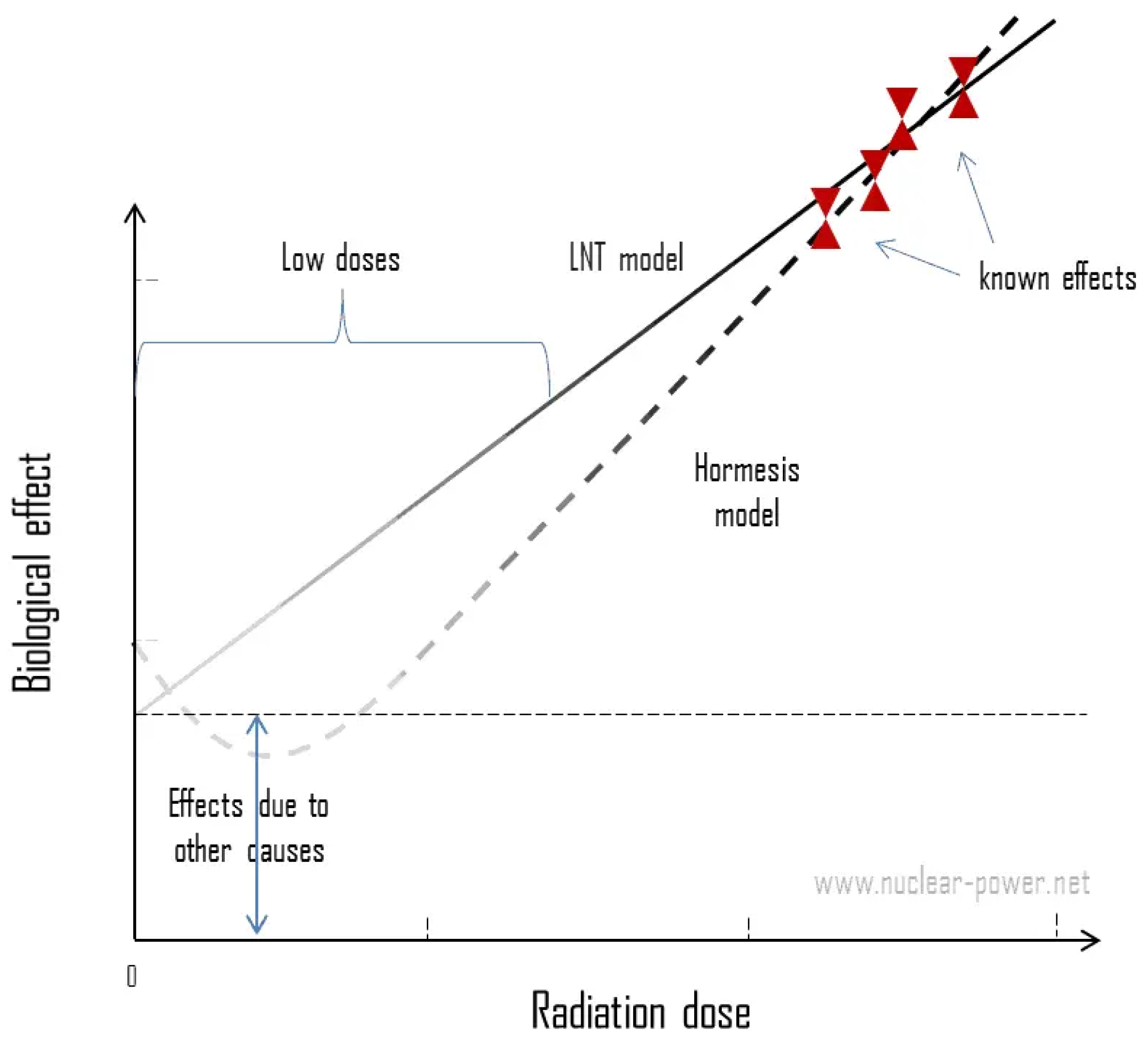
Although Japanese Hiroshima-Nagasaki mathematical radiation effects model for survivors had never fit well enough with the classical Linear Non-Threshold (LNT) hypothesis, where dose response supposed radiation risks as linear to damage (straight line in Figure 2), as it was accepted by the scientific community since 1958 [9]. This attitude was unethically advocated both, by key scientists within radiation genetics, and by editorial practices in four important papers in Science journal, already a well-known and leading international scientific publication at the time. Editorial decisions to circumvent peer review of this work occurred, at least in two cases. One of them, on which a senior editor of the journal was co-author, was alleged to have intentionally falsified the research record [10]. Hence, prominent US scientists suppressed evidence to promote adoption of LNT behaviour, as it was consistently verified an adaptive innate protection promoted by the immune system, where body learned to self-protect when irradiated again after a first low dose exposure [11]. This is call hormesis and it is present with greater evidence in the interval between 10 and 100 mSv in most of the mammals [12]. Delayed temporary adaptive protection involves ROS induced detoxification, deoxyribonucleic acid (DNA) repair enhanced rate, damaged cells removal due to apoptosis followed by normal cell replacement and by cell differentiation and stimulated immune response [13]. Natural shields are developed with a delay of hours and may last for days to months. Hormesis is a biphasic dose response that is generated by almost all biological systems as a result of their interactions with various physical or chemical stimuli. A dose-response relationship is biphasic when low doses are stimulatory and high doses are inhibitory. The low-dose stimulatory response is the Hormetic phase that is often, but not always, associated with beneficial biological effects [14].
Protective gap decreases steadily around 100 to 200 mSv with the presence of M1 type macrophages, and it is not observed anymore above 500 mSv [13]. The activation of the so-called nuclear factor erythroid 2-related transcription factor (Nrf2) is achieved between 500 and 1000 mSv, where anti-inflammatory behaviour is replaced through forcing polarisation shifts from pro-inflammatory macrophage M1 into anti-inflammatory M2 phenotype, rebalancing the immunological redox status [15,16,17]. Nrf2 restores cellular redox, ending ROS proliferation and promoting inflammation resolution. M2 phagocytic capacity is reduced as dose grows towards 1000 mSv, shifting back to M1 and contributing to the increase of apoptotic cells [7].
Therefore, a kind of initial low dose rounded function, up to approximately 100 mSv (see Figure 2) invalidates the LNT linear straight growing line. Hormesis stimulate body defences against LDR danger developing inter-cellular tools to better recover from subsequent X-ray photon hitting and aborting the outdated concept of a linear risk response without protection thresholds. If dose continue to raise before linear behaviour reappears, the number of death cells in the neighbourhood, but outside the radiation field, is increased by the Bystander effects (BE). Bystander dose response function is defined by the linear positive slope rapidly growing up as danger become more important with doses located after the hormesis band, which is closer to the LNT classical model. Because reliable epidemiologic data at low doses are not available, a series of in vitro and in vivo investigations have put into question the validity of the LNT model based on the observations of BE and adaptive response. The BE counts the induction of damage in the cells located in the vicinity of irradiated cells but have not been directly hit themselves. A possible adaptive protection against DNA damage is considered AR or hormesis [18].
A J-shaped dose-response model explains more pronounced non-linearity radiation risks between low and moderate levels [19]. It is preferred to talk about a Hormesis Linear Non-Threshold (H-LNT) behaviour instead of the Linear Non-Threshold scheme (LNT). The LNT is based on the concept that damage to genetic material from ionizing radiation increases in direct proportion to the absorbed dose and that this DNA damage results in causing cancer in proportion to dose. The LNT hypothesis does not consider the complex nonlinear interactions before cancer formation is expressed. The dose-response relationship cannot be based on a simple mathematical equation of the first order, thus, the model should be rejected [18]. Feinendegen suggested, in his dual effect of low-dose ionizing radiation model, that the LNT model appears invalid and should be replaced by a function that includes both linear and nonlinear terms [12]. He summarised the basic assumptions of his model:
-
DNA damage in mammalian cells is proportional to the dose with additional possible Bystander effects.
-
DNA damage comes overwhelmingly from non-radiation sources at background radiation exposure levels.
-
The probability of radiation induced adaptive protection measurably outweighs that of damage from doses well below 200 mSv low-LET (linear energy transfer) radiation.
-
The delayed and temporarily adaptive protection at low doses involves damage prevention, damage repair and immune response.
-
Cell and DNA damage appear increasingly to overrule, negate, or annihilate the more subtle signalling effects seen after low doses, at higher doses in tissue.

3. Inflammation biomarkers due to abnormal levels of radiation

Humans are exposed to small doses of ionizing radiation from natural sources all the time, in particular, cosmic radiation, mainly from the sun, and radon, a radioactive gas that comes from the natural breakdown of uranium in soil, rock, water, and building materials. How much of this background radiation you are exposed to depends on many factors, including altitude and home ventilation. It is estimated a normal average is 3 milliSieverts (mSv) per year but this number may vary upon geographical coordinates.
Cytokines are the most important mediators by which cells of the immune system are increased or decreased due to low dose radiation interaction [20]. IR induces ROS by triggering NLRP3 inflammasome that might change the function and number of immune system cells increasing levels of lymphocytes T and macrophages, given rise to the secretion of several inflammatory intermediators: NF- κ B (Nuclear Factor-Kappa B), IL-1 (Interleukin-1), IL-2 (Interleukin-2), IL-6 (Interleukin-6), IL-8 (Interleukin-8), IL-33 (Interleukin-33), TNF- α (tumor necrosis factor- α ), TGF- β (Tumor Growth Factor- β ), and IFN- γ (Interferon- γ ) [21,22].
Primary radioactive elements causing natural ionizing radiation exposure to humans are potassium (K), uranium (U), thorium ( T h ), and their radioactive decay products, like radium ( R a ) and radon ( R n ). Been deposited in the lungs, 222 R n , a radioactive noble gaseous element part of the 238 U disintegration chain [23], release α radiation after inhalation and is associated with biomarkers of inflammation and endothelial dysfunction [24]. Radon gas tends to accumulate indoors especially in areas with highly permeable soils with a high 226 R a content, resulting in high levels of concentration. Chinese workers exposed to natural background radiation from uranium mines, with high concentrations of indoor 222 R n , showed evident inflammation issues [20]. Two different categories were studied, a control group, underground for < 5 years (cumulative dose < 20 mSv, based on 4mSv per year) and an experimental group, underground for ≥ 5 years (≥ 20mSv). Long-term exposed subjects presented up-regulation of pro-inflammatory cytokines, such as IFN- γ , IL-10, IL-6, and TNF α [20].
Lead (Pb) abundance and toxicity makes it the second most dangerous environmental poison according to the Agency for Toxic Substances and Disease Registry’s Priority Substance List. Although leaded petrol has been removed away from use in many countries, P b compounds are yet being used in aviation fuel [25]. Besides the well-known poisoning by its stable nuclei, there are also unstable radioactive forms suspended in air, like Lead-214 ( 214 P b ) which is a radioactive β emitter. It promotes inflammatory responses producing IL-2, IL-4, IL-8, IL-1 β , IL-6, TNF- α ) IFN- γ , and influencing the immune system cells (T and B lymphocytes, Langerhans cells, macrophages) and the secretion of immunoglobulin A, Immunoglobulin E, Immunoglobulin G, endothelin, and histamine [26,27].
Since the 1960’s tobacco manufacturers have been aware of the α -radioactivity contained in cigarette smoke [28] as several studies links plant fertilisers as the principal source, rich in polyphosphates containing 226 R a , as well as its decay products ( 210 P b and 210 P o ). Polonium-210 ( 210 P o ) may also be generated along the 238 U and 226 R a disintegration chains. Radioactivity in tobacco leaves collected from fifteen different regions in Greece, before the cigarette production, was studied looking for any association between crop root uptake of radionuclides from soil and the effective dose induced to smokers from cigarette tobacco due to the naturally occurring primordial radionuclides ( 226 R a and 210 P b in the uranium series, 228 R a in the thorium series and/or man-made radionuclides, such as 137 C s of Chernobyl origin) [29]. Radiation doses to lung bronchial epithelium from 210 P o inhaled from two packs of cigarettes a day individual smoking is at least seven times that from background sources [30]. Ref. [31] reported that one and a half packs of cigarettes a day deliver 80 mSv per year in areas of bronchial epithelium bifurcation, which is equivalent to the skin dose from 300 chest X-ray plane images per year.
It is well accepted an upper limit of 20 mSv/year as a normal low level exposure to IR in a normal background. However, this threshold is not the same everywhere, depending on the ground composition. There are regions where this background radiation is 10 to 15 times higher than that considered normal. Taleshmahaleh and Chaparsar, two villages in the northern Iran present a higher value, around 260 mSv per year [32]. Some results on blood tests on normal population showed that the total serum antioxidant level in the exposed people was significantly lower than the individuals not exposed to high dose natural IR. These subjects also had higher lymphocyte-induced IL-4 and IL-10 production, and lower IL-2 and IFN- γ production [33].

4. Inflammation biomarkers on COVID-19 cases

SARS-CoV-2 affected patients exhibits a spread spectrum of clinical conditions, ranging from asymptomatic cases to severe pneumonia or even death. Increased levels of proinflammatory cytokines has been observed in the bloodstreamIn of severe COVID-19 cases. NLRP3 inflammasome activation intensely induces cytokine production as an inflammatory response to viral infection, causing what has become known as a cytokine storm [34]. Most infected individuals present with mild flu-like symptoms, while between 5 and 10% are severe cases suffering from a compromised respiratory system and life-threatening pneumonia [35]. In general, severe cases result in acute respiratory distress syndrome (ARDS) and acute lung injury (ALI), which are known to be induced by a storm of inflammatory cytokines, especially IL-1 β , IL-6, and TNF- α [36].
COVID-19 may induce severe lung inflammation leading to acute respiratory distress syndrome (ARDS), respiratory failure, and death despite artificial ventilation. There is an intense inflammatory reaction characterised by mononuclear cells infiltration, fibrin exudates, multinucleated giant cells, and thickened alveoli secondary to proliferating interstitial fibroblasts and type II pneumocyte hyperplasia [37]. Lung hyper-inflammation observed in SARS-CoV-2 patients was also observed in previous pandemics such as SARS-CoV and MERS-CoV [38]. The so-called Macrophage Activation Syndrome (MAS) is the origin of the cytokine storm characterized by elevation of IL-1, TNF- α and IL-6 produced by inflammatory macrophage M1 [39]. When our immune system faces a foreign antigen, i.e. SARS-CoV2, peripheral blood monocytes are recruited into the alveoli where they differentiate into M1 macrophage which produce inflammatory cytokines such as IL-1, IL-6, and IL-18 which in turn attract neutrophils cells into the alveoli to fight the infection, leading to infection clearance through reactive oxygen species (ROS) and phagocytosis [40]. The normal response to an eliminated infection is the reversal polarization to M2 anti inflammation macrophages [41], but in ARDS and some other autoimmune disorders the hyper inflammatory state continues leading not only to lung damage but to multiple normal organs destructions such as kidney failure, cardiac damage and ultimately death. Persistence of inflammatory neutrophils in the alveoli along with increased concentration of ROS and TNF are thought to contribute to lung injury [42].

5. Discussion

Proponents of radiation hormesis typically claim that radio-protective responses in cells and the immune system not only counter the harmful effects of radiation but additionally act to inhibit spontaneous cancer not related to radiation exposure. Radiation hormesis is a dose-response phenomenon and it is one of alternative hypothesis, that low doses of ionizing radiation induces beneficial health effects. According to radiation hormesis, low doses of low LET radiation can stimulate the activation of repair mechanisms, that protect against disease, that are not activated in absence of IR.
Exposure from background sources has not changed since about 1980, but Americans’ total per capita radiation exposure has nearly doubled, and experts believe the main reason is increased use of medical imaging. The proportion of total radiation exposure that comes from medical sources has grown from 15% in the early 1980s to 50% today. CT alone accounts for 24% of all radiation exposure in the United States, according to a report issued in March 2009 by the US National Council on Radiation Protection and Measurements (https://ncrponline.org). In the case of chest CT, the average dose is between the range 4 and 18 mSv, although the actual radiation exposure depends on many things, including the device itself, the duration of the scan, patient size, and the sensitivity of the tissue being targeted [43]. There is direct evidence from epidemiologic studies that the organ doses corresponding to a common CT study (two or three scans, resulting in a dose in the range of 30 to 90 mSv) result in an increased risk of cancer. The evidence is reasonably convincing for adults and very convincing for children [3].
Numbers place X-ray CT monitoring in the dose band ≥ 10 mSv and ≤ 200 mSv, that initiates hard-to-assess inflammatory processes because they are made to old patients with incredibly significant previous inflammation. A retrospective study performed at Tongji Hospital, the largest health center for the treatment of SARS-CoV-2 infected patients in Wuhan, China, showed up that chest CT was used to a wide extent in the follow-up of ordinary COVID-19 cases [44]. Including paediatric cases, 394 patients underwent a total of 1493 exams with a mean time interval between illness onset and discharge of 31.68 ± 8.71 days and a median admitted time of 19 days [44]. The total number of valid CTs and non-value added CTs per patient were related to a high degree to the disease duration [44]. Homayounieh et al. survey [45] remarks that about 30% of the patients (n=225) underwent 2 to 8 chest CT examinations in less than a month [45]. Although it is estimated that no tissue expresses clinically relevant functional deterioration, it has been found that IR produces pro-inflammatory effects even when reduced radiation dose scanning protocols are used [46,47,48].
Low exposures are controversial. It is accepted 100 mSv as a threshold where excess of cancer issues could be potentially observed over the long term [49]. Although pulmonary X-ray CT is a well established technique for diagnosing and managing lung ARDS complications, too much patients were receiving IR doses in the interval between 10 mSv and 200 mSv due to recurrent X-ray exams [50]. There are details not completely understood yet about the effects of ionizing radiation on macrophages [51]. While low doses attenuate inflammation throughout the Abscopal effect, higher levels, like the applied in tumor therapy, increase systemic anti-tumor immune reactions [52]. The Abscopal effect is a phenomenon where the response to radiation is seen in an organ distant to the irradiated area, that is, the responding cells are not juxtaposed with the irradiated cells, like un the Bystander effect.
Despite LDR have been largely related with cancer issues based on the studies over the Japanese survivals affected by the atomic bombs [53], dose response effects are also linked to other secondary effects as inflammation and should be taken in consideration in the LDR Hormetic zone. As the lower dose interval represents a much more relevant exposure scenario for the general population, as it may have broader public health consequences because of the common and frequent indication of X-ray images in general diagnosis [2], this potential danger should limit drastically the number of those type of IR exams if not antioxidant supplements are prescribed along the process [54].

6. Conclusions

Controversy on health effects of IR in the low-range as it has benefits and not risks is served but, currently, there is not valid data supporting the use of LNT in the low-dose range, so dose as a surrogate for risk in X-ray imaging is not appropriate, and therefore, the use of the classical ALARA (As Low As Reasonably Achievable) concept might be considered as obsolete [55]. Concerns have been raised over alleged overuse of CT scanning and inappropriate selection of protocol exams. Although computed tomography is crucial in the follow up of ARDS disease, it cannot be forgotten that it is needed an estimation of the equivalent doses in the organs at risk as it is stated, for instance, in the European Union by the Council Directive 2013/59/Euratom, laying down basic safety standards for protection against the dangers arising from exposure to IR [56].
Extra and additional inflammation throughout X-ray imaging follow-up over elderly population include an excess of lung ionizing radiation shots in a brief time interval of weeks, inappropriate from the radiation protection scope. Obsessive screening will ad up ROS presence to their established inflammatory status while redox products after IR subatomic interactions on tissues will increase free radical basal levels. The interrelationship between the immune system and IR is complex, multi factorial, and dependent on radiation dose and immune cell type. Higher dose radiation levels usually results in immune suppression, while low values modulates a variety of immune responses that have exhibited the properties of immune hormesis [6]. Inflammatory early effects are difficult to isolate and study independently as they are much more subtle and not so obvious functional alterations, and can be easily confused or be hidden by other comorbidities [2].
We hypothesize that the adaptive response given by hormesis due to an excessive number of CT scans in ICU-admitted patients suffering from COVID-19 dramatically adds to the large viral inflammation caused by SARS-CoV-2. It is this enormous lung inflammation that leads to fatal outcomes in ARDS cases. Hormesis protects against cancer but should also be important in autoinflammatory processes. In the absence of data related to LDR and inflammation, we have resorted to the effects that produce the same dose levels but from natural sources to establish analogies between the effects produced but it will required further investigation.

Author Contributions

All authors contributed equally in all the aspects to this work.

Funding

No funding on any of the authors involved is related to this review.

Data Availability Statement

Availability of data and materials: No data or materials have to be declared linked to this work.

Conflicts of Interest

Authors of this manuscript declare no conflicts of interest. They declare no relationships with any companies, whose products or services may be related to the subject matter of the article.

References

  1. UNSCEAR 2006. United Nations Scientific Committee on the Effects of Atomic Radiation Sources, effects, and risks of ionizing radiation. UNSCEAR 2006 Report.
  2. Lumniczky, K.; Impens, N.; Armengol, G.; Candéias, S.; Georgakilas, A.G.; Hornhardt, S.; Martin, O.A.; Rödel, F.; Schaue, D. Low dose ionizing radiation effects on the immune system. Environ Int. 2021, 149, 106212. [Google Scholar] [CrossRef] [PubMed]
  3. Brenner, D.J.; Hall, E.J. Computed tomography—an increasing source of radiation exposure. N Engl J Med. 2007, 357, 2277–2284. [Google Scholar] [CrossRef] [PubMed]
  4. Zoran, M.A.; Savastru, R.S.; Savastru, D.M.; Tautan, M.N. Assessing the relationship between surface levels of PM2.5 and PM10 particulate matter impact on COVID-19 in Milan, Italy. Sci Total Environ. 2020, 738, 139825. [Google Scholar] [CrossRef] [PubMed]
  5. COVID-19 Excess Mortality Collaborators. Estimating excess mortality due to the COVID-19 pandemic: a systematic analysis of COVID-19-related mortality, 2020-21. Lancet 2022, 399, 1513–1536; Erratum in Lancet 2022, 399, 1468. [Google Scholar] [CrossRef]
  6. Cui, J.; Yang, G.; Pan, Z.; Zhao, Y.; Liang, X.; Li, W.; Cai, L. Hormetic Response to Low-Dose Radiation: Focus on the Immune System and Its Clinical Implications. Int J Mol Sci. 2017, 18, 280. [Google Scholar] [CrossRef]
  7. Wang, L.X.; Zhang, S.X.; Wu, H.J.; Rong, X.L.; Guo, J. M2b macrophage polarization and its roles in diseases. J Leukoc Biol. 2019, 106, 345–358. [Google Scholar] [CrossRef]
  8. UNSCEAR 2012. United Nations Scientific Committee on the Effects of Atomic Radiation Sources, effects, and risks of ionizing radiation. UNSCEAR 2012 Report.
  9. Kaminski, C.Y.; Dattoli, M.; Kaminski, J.M. Replacing LNT: The Integrated LNT-Hormesis Model. Dose Response. 2020, 18, 1559325820913788. [Google Scholar] [CrossRef]
  10. Calabrese, E.J.; Giordano, J. LNTgate: How LNT benefited from editorial actions. Chem Biol Interact. 2022, 362, 109979. [Google Scholar] [CrossRef]
  11. Calabrese, E.J. Linear non-threshold (LNT) fails numerous toxicological stress tests: Implications for continued policy use. Chem Biol Interact. 2022, 365, 110064. [Google Scholar] [CrossRef]
  12. Feinendegen, L.E. Evidence for beneficial low level radiation effects and radiation hormesis. Br J Radiol. 2005, 78, 3–7. [Google Scholar] [CrossRef]
  13. Feinendegen, L.E.; Pollycove, M.; Sondhaus, C.A. Responses to low doses of ion- izing radiation in biological systems. Nonlinearity in Biology, Toxicology, and Medicine 2004, 2, 143–171. [Google Scholar] [CrossRef] [PubMed]
  14. Calabrese, E.J.; Giordano, J.J.; Kozumbo, W.J.; Leak, R.K.; Bhatia, T.N. Hormesis mediates dose-sensitive shifts in macrophage activation patterns. Pharmacol Res. 2018, 137, 236–249. [Google Scholar] [CrossRef] [PubMed]
  15. Calabrese, E. The linear Non-Threshold (LNT) dose response model: A comprehensive assessment of its historical and scientific foundations. Chemico-Biological Interactions 2019, 301, 6–25. [Google Scholar] [CrossRef] [PubMed]
  16. Calabrese, E.J.; Kozumbo, W.J.; Kapoor, R.; Dhawan, G.; Lara, P.C.; Giordano, J. Nrf2 activation putatively mediates clinical benefits of low-dose radiotherapy in COVID-19 pneumonia and acute respiratory distress syndrome (ARDS): Novel mechanistic considerations. Radiother Oncol. 2021, 160, 125–131. [Google Scholar] [CrossRef] [PubMed]
  17. McDonald, J.T.; Kim, K.; Norris, A.J.; Vlashi, E.; Phillips, T.M.; Lagadec, C.; Della Donna, L.; Ratikan, J.; Szelag, H.; Hlatky, L.; McBride, W.H. Ionizing radiation activates the Nrf2 antioxidant response. Cancer Res. 2010, 70, 8886–8895. [Google Scholar] [CrossRef] [PubMed]
  18. Kuikka, J.T. Low-dose radiation risk and the linear no-threshold model. Int J Low Radiat 2009, 6, 157–163. [Google Scholar] [CrossRef]
  19. Doss, M. Are We Approaching the End of the Linear Non-Threshold Era? J Nucl Med 2018, 59, 1786–1793. [Google Scholar] [CrossRef]
  20. Li, K.; Chen, Y.; Li, X.; et al. Alteration of Cytokine Profiles in Uranium Miners Exposed to Long-Term Low-dose Ionizing Radiation. Scientific World Journal 2014. [CrossRef]
  21. Hendry, J.H.; Simon, S.L.; Wojcik, A.; et al. Human exposure to high natural background radiation: what can it teach us about radiation risks? J Radiol Prot. 2009, 29, 29–42. [Google Scholar] [CrossRef]
  22. Yahyapour, R.; Amini, P.; Rezapour, S.; Cheki, M.; Rezaeyan, A.; Farhood, B.; Shabeeb, D.; Musa, A.E.; Fallah, H.; Najafi, M. Radiation-induced inflammation and autoimmune diseases. Mil Med Res. 2018, 5, 9. [Google Scholar] [CrossRef]
  23. Analytical Methodology for the Determination of Radium Isotopes in Environmental Samples. IAEA Analytical Quality in Nuclear Applications Series No. 19. IAEA /AQ /19.
  24. Nyhan, M.M.; Ricec, M.; Blomberg, A.; et al. Associations between ambient particle radioactivity and lung function. Environment International 2019, 130. [Google Scholar] [CrossRef] [PubMed]
  25. De Flora, S.; Grassi, C.; Carati, L. Attenuation of influenza-like symptomatology and improvement of cell-mediated immunity with long-term N-acetylcysteine treatment. Eur Respir J. 1997, 10, 1535–1541. [Google Scholar] [CrossRef] [PubMed]
  26. Metryka, E.; Chibowska, K.; Gutowska, I.; et al. Lead (Pb) Exposure Enhances Expression of Factors Associated with Inflammation. Int J Mol Sci. 2018, 19, 1813. [Google Scholar] [CrossRef] [PubMed]
  27. USEPA (United States Environmental Protection Agency). RadNet Monitoring Network. http://www.epa.gov/radnet.
  28. Zagà, V.; Lygidakis, C.; Chaouachi, K.; et al. Polonium and Lung Cancer. J Oncol. 2011. [Google Scholar] [CrossRef]
  29. Papastefanou, C. Radioactivity of Tobacco Leaves and Radiation Dose Induced from Smoking. Int. J. Environ. Res. Public Health 2009, 6, 558–567. [Google Scholar] [CrossRef]
  30. Radford, E.P., Jr.; Hunt, V.R. Polonium- 210: A volatile radioelement in cigarette. Science 1964, 143, 247–249. [Google Scholar] [CrossRef]
  31. Winters, T.H.; Di Franza, J.R. Radioactivity in cigarette smoking. N. Engl. J. Med. 1982, 306, 364–365. [Google Scholar]
  32. Ghiassi-nejad, M.; Mortazavi, S.M.; Cameron, J.R.; Niroomand-rad, A.; Karam, P.A. Very high background radiation areas of Ramsar, Iran: preliminary biological studies. Health Phys. 2002, 82, 87–93. [Google Scholar] [CrossRef]
  33. Attar, M.; Molaie Kondolousy, Y.; Khansari, N. Effect of high dose natural ionizing radiation on the immune system of the exposed residents of Ramsar Town, Iran. Iran J Allergy Asthma Immunol. 2007, 6, 73–78. [Google Scholar]
  34. Zhao, N.; Di, B.; Xu, L.L. The NLRP3 inflammasome and COVID-19: Activation, pathogenesis and therapeutic strategies. Cytokine Growth Factor Rev. 2021, 61, 2–15. [Google Scholar] [CrossRef]
  35. Zhu, N.; Zhang, D.; Wang, W.; Li, X.; Yang, B.; Song, J.; Zhao, X.; Huang, B.; Shi, W.; Lu, R.; Niu, P.; Zhan, F.; Ma, X.; Wang, D.; Xu, W.; Wu, G.; Gao, G.F.; Tan, W. China Novel Coronavirus Investigating and Research Team. A Novel Coronavirus from Patients with Pneumonia in China, 2019. N Engl J Med. 2020, 382, 727–733. [Google Scholar] [CrossRef] [PubMed]
  36. Nieto-Torres, J.L.; Verdiá-Báguena, C.; Jimenez-Guardeño, J.M.; Regla-Nava, J.A.; Castaño-Rodriguez, C.; Fern, ez-Delgado, R. ; Torres, J.; Aguilella, V.M.; Enjuanes, L. Severe acute respiratory syndrome coronavirus E protein transports calcium ions and activates the NLRP3 inflammasome. Virology 2015, 485, 330–339. [Google Scholar] [CrossRef] [PubMed]
  37. Tian, S.; Hu, W.; Niu, L.; Liu, H.; Xu, H.; Xiao, S.Y. Pulmonary Pathology of Early-Phase 2019 Novel Coronavirus (COVID-19) Pneumonia in Two Patients With Lung Cancer. J Thorac Oncol. 2020, 15, 700–704. [Google Scholar] [CrossRef] [PubMed]
  38. Channappanavar, R.; Perlman, S. Pathogenic human coronavirus infections: causes and consequences of cytokine storm and immunopathology. Semin Immunopathol. 2017, 39, 529–539. [Google Scholar] [CrossRef] [PubMed]
  39. McGonagle, D.; Sharif, K.; O’Regan, A.; Bridgewood, C. The Role of Cytokines including Interleukin-6 in COVID-19 induced Pneumonia and Macrophage Activation Syndrome-Like Disease. Autoimmun Rev. 2020, 19, 102537. [Google Scholar] [CrossRef] [PubMed]
  40. Mehta, P.; McAuley, D.F.; Brown, M.; et al. COVID-19: Consider Cytokine Storm Syndromes and Immunosuppression. Lancet 2020, 395, 1033–1034. [Google Scholar] [CrossRef]
  41. Aggarwal, N.R.; King, L.S.; D’Alessio, F.R. Diverse macrophage populations mediate acute lung inflammation and resolution. Am J Physiol Lung Cell Mol Physiol. 2014, 306, L709–L725. [Google Scholar] [CrossRef]
  42. Lu, H.L.; Huang, X.Y.; Luo, Y.F.; Tan, W.P.; Chen, P.F.; Guo, Y.B. Activation of M1 macrophages plays a critical role in the initiation of acute lung injury. Biosci Rep. 2018, 38, BSR20171555. [Google Scholar] [CrossRef]
  43. Mettler, F.A.; et al. Effective Doses in Radiology and Diagnostic Nuclear Medicine: A Catalog. Radiology 2008, 248, 254–263. [Google Scholar] [CrossRef]
  44. Chen, L.; Wang, Q.; Wu, H.; Hu, J.; Zhang, J. Repeat chest ct scans in moderate- to-severe patient’s management during the COVID-19 pandemic observations from a single centre in Wuhan, China. Radiat Prot Dosimetry 2020, 190, 269–275. [Google Scholar] [CrossRef]
  45. Homayounieh, F.; Holmberg, O.; Umairi, R.A.; et al. Variations in CT Utilization, Protocols, and Radiation Doses in COVID-19 Pneumonia: Results from 28 countries in the IAEA study. Radiology 2021, 298, E141–E151. [Google Scholar] [CrossRef] [PubMed]
  46. ICRP 103. Recommendations of the International Commission on Radiological Protection. ICRP Publication 103. Ann. ICRP 37 (2-4). 2007.
  47. Liu, S.Z. Nonlinear Dose-Response Relationship in the Immune System Following Exposure to Ionizing Radiation: Mechanisms and Implications. Nonlinearity Biol Toxicol Med. 2003, 1, 71–92. [Google Scholar] [CrossRef] [PubMed]
  48. Ruhm, W.; Harrison, R.M. High CT doses return to the agenda. Radiat Environ Biophys 2020, 59, 3–7. [Google Scholar] [CrossRef] [PubMed]
  49. Charles, L. Sanders. Radiobiology and Radiation Hormesis. New Evidence and its Implications for Medicine and Society. Springer: 2017. ISBN 978-3-319-56371-8. ISBN 978-3-319-56372-5 (eBook).
  50. Travis, W.D.; Costabel, U.; Hansell, D.M.; et al. An official American Thoracic Society/European Respiratory Society statement: Update of the international multidisciplinary classification of the idiopathic interstitial pneumonias. Am J Respir Crit Care Med 2013, 188, 733–748. [Google Scholar] [CrossRef] [PubMed]
  51. Teresa Pinto, A.; Laranjeiro Pinto, M.; Patrícia Cardoso, A.; Monteiro, C.; Teixeira Pinto, M.; Filipe Maia, A.; Castro, P.; Figueira, R.; Monteiro, A.; Marques, M.; Mareel, M.; Dos Santos, S.G.; Seruca, R.; Adolfo Barbosa, M.; Rocha, S.; José Oliveira, M. Ionizing radiation modulates human macrophages towards a pro-inflammatory phenotype preserving their pro-invasive and pro-angiogenic capacities. Sci Rep 2016, 6, 18765. [Google Scholar] [CrossRef]
  52. Frey, B.; Hehlgans, S.; Rödel, F.; Gaipl, U.S. Modulation of inflammation by low and high doses of ionizing radiation: Implications for benign and malign diseases. Cancer Lett 2015, 368, 230–237. [Google Scholar] [CrossRef]
  53. Lindee, S. Survivors and scientists: Hiroshima, Fukushima, and the Radiation Effects Research Foundation, 1975-2014. Soc Stud Sci. 2016, 46, 184–209. [Google Scholar] [CrossRef]
  54. Calabrese, E.J.; Kozumbo, W.J. The hormetic dose-response mechanism: Nrf2 activation. Pharmacol Res. 2021, 167, 105526. [Google Scholar] [CrossRef]
  55. Oakley, P.A.; Harrison, D.E. Death of the ALARA Radiation Protection Principle as Used in the Medical Sector. Dose Response 2020, 18, 1559325820921641. [Google Scholar] [CrossRef]
  56. EURATOM 2013. Council Directive 2013/59/EURATOM, of , laying down basic safety standards for protection against the dangers arising from exposure to ionizing radiation. Official Journal of the European Union. 5 December.
  57. Backert, S. (Editor). Inflammasome signaling and bacterial infections. Current topics in microbiology and immunology. Springer: 2016; Volume 397, ISBN 978-3-319-41170-5.
  58. Calabrese, E.J.; Selby, P.B. Cover up and cancer risk assessment: Prominent US scientists suppressed evidence to promote adoption of LNT. Environ Res 2022, 210, 112973. [Google Scholar] [CrossRef]
  59. Cuadrado, A.; Pajares, M.; Benito, C.; Jiménez-Villegas, J.; Escoll, M.; Fernández-Ginés, R.; Garcia Yagü, A.J.; Lastra, D.; M. ; a G.; Rojo, A.I.; Dinkova-Kostova, A.T. Can Activation of NRF2 Be a Strategy against COVID-19? Trends Pharmacol Sci. 2020, 41, 598–610. [Google Scholar] [CrossRef] [PubMed]
  60. Dalan, R.; Bornstein, S.R.; El-Armouche, A.; Rodionov, R.N.; Markov, A.; Wielockx, B.; Beuschlein, F.; Boehm, B.O. The ACE-2 in COVID-19: Foe or Friend? Horm Metab Res 2020, 52, 257–263. [Google Scholar] [CrossRef] [PubMed]
  61. Hamarsheh, S.; Zeiser, R. NLRP3 Inflammasome Activation in Cancer: A Double-Edged Sword. Front Immunol 2020, 11, 1444. [Google Scholar] [CrossRef] [PubMed]
  62. Hodgson, A. Natural levels of Polonium-210 in urine. J Radiol Prot 2018, 38, 923–933. [Google Scholar] [CrossRef] [PubMed]
  63. Hoffman, H.M.; Wright, F.A.; Broide, D.H.; W. ; erer, A.A.; Kolodner, R.D. Identification of a locus on chromosome 1q44 for familial cold urticaria. American Journal of Human Genetics 2000, 66, 1693–1698. [Google Scholar] [CrossRef] [PubMed]
  64. Huang, K.J.; Su, I.J.; Theron, M.; et al. An interferon-γ-related cytokine storm in SARS patients. J Med Virol 2005, 75, 185–194. [Google Scholar] [CrossRef]
  65. Ichikado, K.; Muranaka, H.; Gushima, Y.; Kotani, T.; Nader, H.M.; Fujimoto, K.; Johkoh, T.; Iwamoto, N.; Kawamura, K.; Nagano, J.; Fukuda, K.; Hirata, N.; Yoshinaga, T.; Ichiyasu, H.; Tsumura, S.; Kohrogi, H.; Kawaguchi, A.; Yoshioka, M.; Sakuma, T.; Suga, M. Fibroproliferative changes on high-resolution CT in the acute respiratory distress syndrome predict mortality and ventilator dependency: a prospective observational cohort study. BMJ Open 2012, 2, e000545. [Google Scholar] [CrossRef]
  66. Kelley, N.; Jeltema, D.; Duan, Y.; He, Y. The NLRP3 Inflammasome: An Overview of Mechanisms of Activation and Regulation. Int. J. Mol. Sci. 2019, 20, 3328. [Google Scholar] [CrossRef]
  67. ; Lee, C. Managing Radiation Dose from Chest CT in Patients with COVID-19. Radiology 2021, 298, E158–E159. [Google Scholar] [CrossRef]
  68. Lee, E.K.; Kim, J.A.; Park, S.J.; Kim, J.K.; Heo, K.; Yang, K.M.; Son, T.G. Low-dose radiation activates Nrf1/2 through reactive species and the Ca(2+)/ERK1/2 signaling pathway in human skin fibroblast cells. BMB Rep 2013, 46, 258–263. [Google Scholar] [CrossRef]
  69. Lippi, G.; South, A.M.; Henry, B.M. Electrolyte imbalances in patients with severe coronavirus disease 2019 (COVID-19). Ann Clin Biochem 2020, 57, 262–265. [Google Scholar] [CrossRef] [PubMed]
  70. Meister, A. Metabolism and function of glutathione. Dolphin, D.; Poulson, R., Avramovich, O., Eds.; eds. Glutathione: Chemical, biochemical, and medical aspects. Wiley-Interscience: New York. 1989; pp. 367–374. [Google Scholar]
  71. Mense, M.; Stark, G.; Apell, H.J. Effects of Free Radicals on Partial Reactions of the Na,K-ATPase. J. Membrane Biol. 2020, 156, 63–71. [Google Scholar] [CrossRef]
  72. Petruzzi, S.; Musim, B.; Bignami, G. Acute and chronic sulfur dioxide (SO2) exposure: and overview of its effects on humans and laboratory animals. Review. Ann. Ist. Super Sanita. 1994, 30, 151–156. [Google Scholar] [PubMed]
  73. Sheppard, S.C.; Sheppard, M.I. Modeling estimates of the effect of acid rain on background radiation dose. Environ Health Perspect. 1998, 78, 197–205. [Google Scholar] [CrossRef] [PubMed]
  74. Shi, Z.; Puyo, C.A. N-Acetylcysteine to Combat COVID-19: An Evidence Review. Ther Clin Risk Manag. 2020, 16, 1047–1055. [Google Scholar] [CrossRef]
  75. Shprentz, D.S.; Bryner, G.C.; Shprentz, J.S.; Hawkings, D.G. Breath taking: premature mortality due to air pollution in 239 American cities. National Resources Defense Council, Washington, DC. 1996, 99.
  76. Suter, P.M.; Domenighetti, G.; Schaller, M.D.; et al. N-Acetylcysteine enhances recovery from acute lung injury in man: a randomized, double-blind, placebo-controlled clinical study. Chest 1994, 105, 190–194. [Google Scholar] [CrossRef]
  77. Swanson, K.V.; Deng, M.; Ting, J.P. The NLRP3 inflammasome: molecular activation and regulation to therapeutics. Nat Rev Immunol 2019, 19, 477–489. [Google Scholar] [CrossRef]
  78. WHO Regional Office for. Europe, Copenhagen, Denmark; 2000.
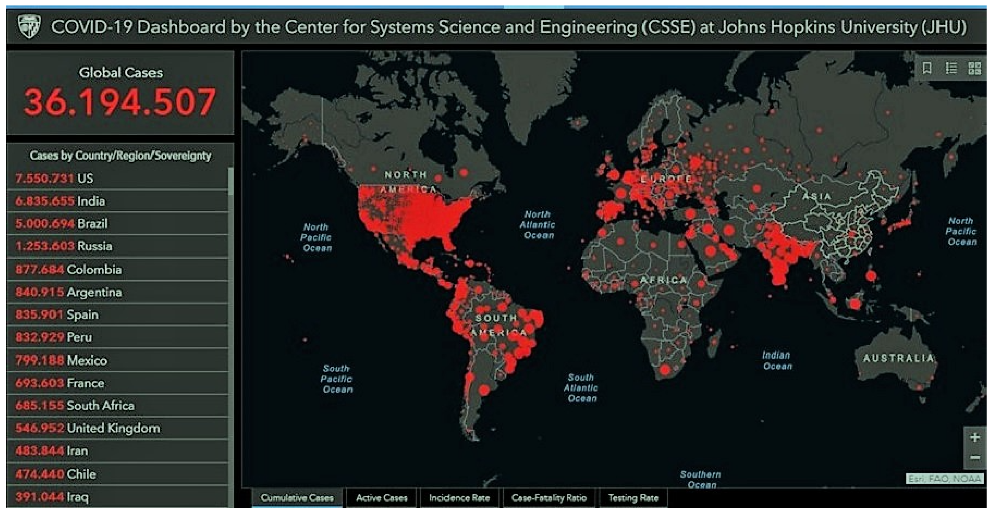
Figure 1. Cumulative confirmed cases last updated 10/8/2020 10:23 a. m. Source: John Hopkins University of Medicine. (https://coronavirus.jhu.edu/map.html). India, Brazil, and Iran are at the top of the ranking.
Preprints 73212 g001
Figure 2. Linear Non-Threshold model (LNT), straight line, versus Hormesis Linear Non-Threshold (H-LNT) behavior, J-shaped line. Source: https://aboutradiation.blogspot.com/2019/02/all-about-radiation-hormesis.html.
Preprints 73212 g002
Disclaimer/Publisher’s Note: The statements, opinions and data contained in all publications are solely those of the individual author(s) and contributor(s) and not of MDPI and/or the editor(s). MDPI and/or the editor(s) disclaim responsibility for any injury to people or property resulting from any ideas, methods, instructions or products referred to in the content.
Copyright: This open access article is published under a Creative Commons CC BY 4.0 license, which permit the free download, distribution, and reuse, provided that the author and preprint are cited in any reuse.
Prerpints.org logo

Preprints.org is a free preprint server supported by MDPI in Basel, Switzerland.

Subscribe

© 2024 MDPI (Basel, Switzerland) unless otherwise stated