Preprint
Hypothesis

Chronic Fatigue Syndrome and Multiple Sclerosis have Reduced Craniospinal Compliance and Dilated Pressurized Bridging Cortical Veins: A Hypothesis Illustrated with Two Case Studies

Altmetrics

Downloads

291

Views

362

Comments

0

A peer-reviewed article of this preprint also exists.

This version is not peer-reviewed

Submitted:

31 May 2023

Posted:

31 May 2023

You are already at the latest version

Alerts
Abstract
Chronic fatigue syndrome (CFS) and multiple sclerosis (MS) share similarities regarding their epidemiology, symptomatology and craniospinal physiology. Indeed, the cardinal feature of CFS, fatigue, is also a major factor in the symptomatology of the majority of MS patients. Recently, we have found that there is a significant reduction in the craniospinal compliance in MS which affects both the stiffness of the walls of the spinal canal and the walls of the cerebral venous system. This change in compliance brings about an alteration in the effectiveness of the pulse wave dampening in the craniospinal system. The result is an impedance mismatch between the cortical veins and their draining sinuses, leading to dilatation of these upstream veins. We deduce this dilatation can only be brought about by an increase in the pressure gradient between the vein lumen and the subarachnoid space (i.e. the transmural pressure gradient). We hypothesise that given the similarities between MS and CFS, a similar mechanism underlies the physiology of CFS. We present two case studies to highlight the expected findings in CFS patients if this hypothesis were proven to be correct.
Keywords: 
Subject: Medicine and Pharmacology  -   Neuroscience and Neurology

Introduction

Chronic fatigue syndrome (CFS), also known as myalgic encephalomyelitis (ME), is a complex disease characterized by a cluster of symptoms including fatigue, malaise, headaches, sleep disturbances, difficulties in concentration, impaired cognitive function and muscle pain [1]. There is no known overarching underlying physiology, blood test or imaging biomarker, so diagnosis rests with clinical criteria. The Canada Consensus Criteria for the diagnosis of CFS/ME lists severe fatigue, post-exertional malaise, pain and neurological and immunological dysfunction as the major criteria for classification [2]. The overall incidence has been estimated to be up to 1.6% of the general population [3]. Multiple sclerosis (MS) is thought to be an autoimmune mediated disruption of the cerebral and spinal white matter [4]. There are several epidemiological similarities between CFS and MS. In CFS the female to male incidence ratio is 3.2:1, with the peak incidence being at 30-39 years [5]. In MS, the ratio of females to males is 3:1 [6] and the mean age at diagnosis is approximately 30 years [7]. The World Health Organisation lists ME/CFS as a post viral neurological disease [8]. In patients with a strong history of a post viral onset of their CFS, one group using antibody responses to two Epstein-Barr virus (EBV) antigens, found an estimated sensitivity of 83% and specificity of 72% of the antigen positivity for the diagnosis of CFS [9]. Similarly, longitudinal analysis of a large number of military recruits showed a 32 fold increase in the prevalence of MS after EBV infection [10]. The similarities extend past the epidemiology into the symptomatology. Patients with ME/CFS and MS both experience severe fatigue, with a worsening of the symptoms with exercise [11]. Fatigue in MS has a prevalence of up to 81%, being more frequent in the progressive forms of disease [12]. With regards to disease progress, both disorders have either a relapsing-remitting or progressive course, with infections worsening the fatigue symptoms [11]. They both have autonomic symptoms, reduced cardiac response to exercise, orthostatic intolerance and postural hypotension [11]. With regards to physiology, little is known with regards to CFS. However, both diseases show decreased cerebral blood flow, atrophy, white matter hyperintensities and increased cerebral lactate [11]. In two studies, a very mild increase in CSF opening pressure was found in CFS approximating 14.5 mmHg [13,14]. This compares to a large study in which the normal CSF pressure at middle age averaged 11.5 mmHg. Similarly, in 32 MS patients the ICP was found to be slightly increased, being approximately 13 mmHg [15]. Rather interestingly, one study suggested Ehlers-Danlos syndrome (a connective tissue disorder) occurred in up to 20% of CFS cases [16]. In a another study, the prevalence of MS was found to be 10 fold greater in Ehlers-Danlos syndrome patients than in the general population [17].
Given the similarities found between CFS and MS, one would be tempted to suggest that CFS is essentially MS without the autoimmune mediated white matter destruction. Recently, we have found the cross-sectional area of the superior sagittal sinus (SSS) to be larger in MS than in controls [18]. This enlargement correlates with disease severity and progression [18]. Modelling indicated the sinuses in MS could be enlarged due to a decrease in the pressure difference between the lumen and the subarachnoid space, an increase in wall thickness or increased wall stiffness [18]. However, only the last two possibilities were feasible [18]. An increase in sinus wall stiffness or thickness would render the craniospinal compartment less compliant than normal. Further study has shown that in MS, the superficial territory cortical veins are 29% larger and the veins of Galen are 25% larger than in the controls [19]. Modelling of these findings indicated that to bring this dilatation about, a significant increase in the bridging vein transmural pressure would be required, estimated to be approximately 6.5 mmHg [19] . Finally, MS patients with significant fatigue have larger cortical veins than those who are not significantly fatigued [19]. These findings lead us to suggest a hypothesis for the underlying physiology of CFS.

Hypothesis

Given the above, the hypothesis tendered is 1) that a reduction in the overall craniospinal compliance in CFS could be related to a stiffening/thickening of the walls of the venous sinuses, leading to dilatation of the sinuses in the majority of cases. This could serve as an imaging marker of the disease and 2) an alteration in the impedance matching at the junction between the bridging cortical veins and the venous sinuses would increase the cortical vein pressure, leading to vein dilatation and an alteration in brain metabolism and the symptoms of the disease.

Illustrating Cases

Given the above hypothesis we wish to discuss two case studies of CFS patients to highlight the imaging findings to be elaborated further.
Case 1 is a 48 year old male who initially complained of a viral throat infection and was noted to be positive for EBV serology. Over the course of 22 months, he developed chronic fatigue and neurocognitive decline with “difficulty thinking and remembering”. He complained of feelings of heaviness and numbness in his limbs with generalised myalgia especially in the neck region. He suffered headaches, dysesthesia and insomnia. He complained of difficulty walking with exercise induced malaise. His symptoms were made worse by inter-current viral infections. An MRI examination with contrast showed no structural brain abnormality. Reconstructions of the 3D post contrast imaging through the venous system showed prominent venous sinuses (see Figure 1A). A cross-sectional image of the superior sagittal sinus 3 cm above the torcular measured 67.6 mm2 (see Figure 1B). The straight sinus measured 28.3 mm2 (see Figure 1C) and the vein of Galen measured 25 mm2 (see Figure 1D). The largest two cortical veins draining into the SSS on each side were measured 1 cm from their junction with the sinus and the average of the four was calculated to be 12 mm2. There was no venous outflow stenosis.
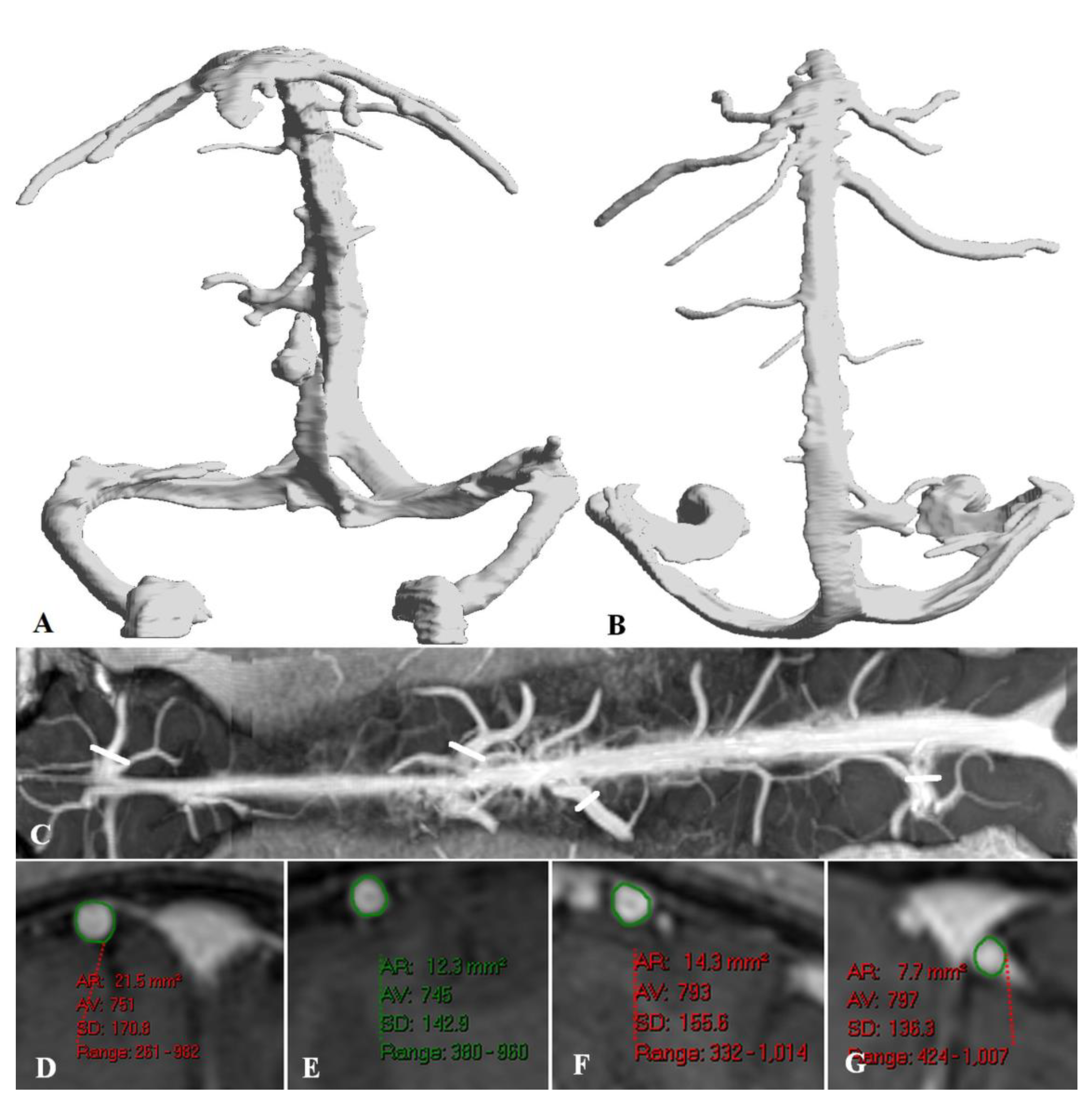
Case 2 is a 20 years old female who also initially developed tonsillitis. In the following 18 months she developed malaise and fatigue made worse by exercise. She complained of chronic headaches and photosensitivity. She suffered from insomnia with pain in the neck muscles and lower limb weakness and limb twitching. An MRI with contrast showed no structural abnormality. Reconstructed images of her cerebral veins showed no outflow stenosis (see Figure 2A-B). Her sagittal sinus measured 62.3 mm2, straight sinus 22.3 mm2 and her vein of Galen measured 41 mm2. A curved reconstruction along the SSS showed prominent cortical veins with no focal stenosis at their junction with the sagittal sinus (see Figure 2C). The two largest cortical veins were measured on each side yielding areas of 21.5 mm2, 12.3 mm2, 14.3 mm2 and 7.7 mm2 (see Figure 2D-G) the average being 14 mm2.
Previously we have measured these veins using an identical technique in several studies. In 50 controls of average age 44.9 ± 10.9 years, the cross-sectional area of the SSS 3cm above the torcular from post contrast 3DT1 images averaged 44.9± 10.9 mm2 and the mid portion of the straight sinus averaged 15.7± 5.4 mm2 [18]. The bridging vein between the deep venous system of the brain and the straight sinus is the vein of Galen. It was found to average 21.9± 4.2 mm2 [19]. The average of the four largest cortical veins draining into the SSS for the controls averaged 5.8± 1.8 mm2 [19]. It can be seen that in case 1 the SSS, straight sinus and cortical vein average are greater than two standard deviations above the mean size for the controls. Similarly in case 2 the vein of Galen and the average of the cortical veins are greater than two standard deviations above the mean size for the controls. These findings would suggest significantly dilated veins.

Discussion

Thus, the illustrative cases indicate there are at least some patents with CFS who have sinus and bridging cortical vein dilatation. We have suggested that the enlargement of the sinuses could render the craniospinal cavity less compliant. The compliance of a structure is a measure of the change in pressure brought about by a change in volume. [20]. The craniospinal cavity is filled by incompliant CSF and the available compliance is provided by the walls of the container i.e. the dura within the spinal canal and the walls of the veins [20]. The compliance of the craniospinal system allows for changes in volume from, for example the arterial pulsation, to be accommodated by expelling CSF into the spinal canal and compressing the terminal portions of the bridging cortical veins in the subarachnoid space to expel blood into the sinuses. This mechanism provides pulse pressure dampening and is known as the windkessel effect [21]. In MS there is evidence of reduced spinal canal compliance. Fjeldstat et al. [22] found patients with MS have lower central arterial compliance than healthy controls, which preferentially affects the CNS vessels. This leads to higher intracranial pulse pressures and an increase in the arterial pulsation volume in systole of 26% [23]. Despite the increased volume to be dissipated, there is a 24% reduction in CSF leaving the foramen magnum in MS [24], which would equate to a 40% reduction in the apparent spinal canal compliance [25]. The systemic arterial tree is also significantly stiffer in CFS than normal [26] suggesting an increase in pulse pressure could also occur in the cranial cavity similar to MS. Is there evidence of reduced spinal canal compliance in CFS? In one study 80% of CFS patients had craniocervical obstructions secondary to osteophytic bars and disc pathology [16]. Obstructing the movement of CSF in this way will reduce the available compliance of the cranial cavity. Three patients with cervical spondylosis and CFS showed a significant improvement in symptoms following surgery to relieve the stenosis [27]. Relieving the stenosis would significantly improve cranial and spinal CSF flow and compliance. In another study in patients with Chiari 1 malformation, 14% had chronic fatigue syndrome and 14% fibromyalgia [28]. The descent of the tonsils in this disorder would reduce craniospinal CSF flow and compliance. Interestingly, MS patients have a higher incidence of cervical spondylosis and develop it at a younger age [29] and there is a higher incidence of MS plaques at the site of spinal canal stenosis than at other levels [30], suggesting another similarity between MS and CFS.
In MS the reduced compliance is also seen as a reduction in the bridging cortical vein dynamic compression. As already discussed, cortical vein compression and the expelling of venous blood into the sinuses in systole is the second part of the windkessel mechanism. A comparison between the changes in the arterial volume over the cardiac cycle compared to the change in the SSS volume (there was an increase in arterial stroke volume of 26 % but a decrease in SSS stroke volume of 31%) indicated there was a 50% reduction in the apparent compliance of the venous system [23]. Intracranial compliance has not been studied extensively in CFS but patients with more severe orthostatic intolerance, showed lower intracranial compliance [31].

Sagittal and straight sinus dilatation

As already discussed, the sinuses are dilated in MS and the illustrative cases indicate the same may be occurring in at least some patients with CFS. The sagittal sinus walls can move and sit between the CSF and venous blood. Modelling using standard engineering equations has indicated that the walls will bow inward due to the pressure of the CSF being 4 mmHg higher than the venous pressure [18]. The amount of this bowing is dependent on the size of the transmural pressure gradient, the thickness of the sinus wall and its intrinsic stiffness [18]. Our modelling in MS showed that the venous transmural pressure was likely to be normal in the majority of MS patients, indicating the walls had to be stiffer and or slightly thicker than normal [18]. In CFS the same three possibilities will also apply if the veins are larger. Higgins et al. have hypothesised that there is a reversal of the transmural pressure gradient in CFS on the basis of a similarity of the disorder to idiopathic intracranial hypertension [32]. Higgins et al. suggest the sinus pressures are elevated but the CSF pressure is moderated by a chronic leakage of CSF [32]. A pressure gradient reversal would also dilate the sinuses, similar to our suggestion that they are stiffer than normal. However, there are some difficulties with the pressure reversal scenario. As already noted, the CSF pressure is elevated by 3 mmHg above normal in CFS, so to reverse the normal transmural pressure would require the venous pressure to rise by at least 7 mmHg above normal to dilate the sinuses. Neither of our illustrative cases had a transverse or sigmoid sinus stenosis to account for this (see Figure 2A, B). Secondly, CSF removal in CFS patients has been shown to improve symptoms in 70% of patients in one study and 85% in another [14,33]. Improvement occurred even in those who had a normal CSF pressure and had their pressure reduced to below normal. How could a reduction in CSF pressure due to a CSF leak be pathological on the one hand and CSF removal at lumbar puncture be therapeutic on the other? Intracranial compliance is governed by the pressure volume curve, which is exponential in the intracranial cavity [20]. Thus, the compliance of the system is low with higher CSF pressures and the intracranial compliance increases as CSF pressure is lowered. Thus, if the intrinsic compliance was very low in CFS, reducing the CSF pressure to below normal could have a therapeutic effect by improving the cranial compliance. Thus, excluding a reversal of the sinus transmural pressure gradient, the only other possibility is thicker or stiffer sinus walls. There is ongoing inflammation in CFS with an elevated C reactive protein [26]. Adams et al. suggested MS was primarily a venous vasculitis [34]. Could long term inflammation of the sinus walls, possibly related to EBV, lead to the compliance changes noted in both diseases?

Bridging vein dilatation

We have suggested that there is a link between a reduction in the intracranial compliance and bridging vein dilatation and pressurisation. As already discussed, an elevation in CSF pressure will take the cranial cavity to a less compliant portion of the pressure volume curve. In an animal modelling study using pigs, a 10 mmHg increase in ICP dilated the cortical veins by 33% in area and a 20% increase in ICP dilated the veins by 57% [35]. Dilatation is dependent on the transmural pressure (the wall thickness and stiffness would not change this quickly) so the venous pressure must have been rising faster than the ICP to accomplish the dilatation. A reduction in intracranial compliance is found in communicating hydrocephalus [36]. In a dog model of naturally occurring hydrocephalus, the ICP was increased by 48% but the cortical vein transmural pressure was increased by 327% compared to controls [37], indicating the pressurisation is also apparent in low compliance states without a grossly elevated ICP. In human subjects, the bridging veins show a mean diameter of 2.04 mm under normal intracranial pressure and increase to 2.65 mm under an increased ICP [38], a 69% increase in cross-sectional area. Finally, in children with hydrocephalus, the superficial territory cortical veins were 22% larger than the controls, with modelling suggesting a significant increase in the superficial vein transmural pressure in childhood hydrocephalus estimated to be approximately 4 mmHg [39]. In MS, the bridging vein dilatation suggested an increase in the transmural pressure estimated to be approximately 6.5 mmHg in the superficial cortical veins overall and those with significant fatigue had greater dilatation in the territory draining the cortical grey matter than those without fatigue [19]. We have previously suggested an impedance mismatch alters the blood flow through the terminal cortical vein segments increasing their pressure with either loss or reversal of the hydraulic engineering effect known as impedance pumping [39]. It is likely that the cortical vein pressure feeds back into the parenchymal venules. In a study comparing the hemodynamics of clinically isolated syndrome (CIS) (the earliest form of MS) and controls, there was a 71% increase in the cerebral blood volume in the average normal appearing white matter and a 59% increase in this metric in the deep grey matter in CIS [40]. One could suggest the venules in MS are just passively enlarged due to atrophy. However, transcranial photoplethysmography following neck compression showed a significant increase in frontal cerebral blood volume in controls but a blunted response in MS. There was no difference between the responses in clinically isolated syndrome vs relapsing remitting patients. The authors suggested the findings indicate the microcirculation appears already congested with less ability for the vessels to dilate [41].

Effect of elevated venous pressure on the brain.

A mouse model of raised venous pressure showed disruption of the blood brain barrier and activation of neuro-inflammatory related gene expression [42]. MR diffusion tensor imaging in CFS showed widespread white matter structural damage [43] suggesting white matter damage is likely in both MS and CFS. A young man with cognitive fatigue and brain fog who had an elevated cerebral venous pressure from a high grade neck vein stenosis found his symptoms resolved with surgery [44]. These findings suggest that there may be an underlying correlation between the increased cortical vein pressure hypothesised and the symptoms CFS patents complain of. Indeed, idiopathic intracranial hypertension, which is known to result from an elevation in venous pressure and CFS, have several clinical findings in common, such as fatigue, widespread pain, paraesthesia, headaches, cognitive dysfunction and brain fog [13]. One CFS patient with borderline elevated ICP underwent venous stenting with improved headache, fatigue, concentration and muscle pain [45]. There is also anecdotal evidence that there may be some improvement in CFS symptoms in those who have had compliance increased from a posterior fossa decompression for concomitant Chiari 1 malformation but there are no published data [46,47].

Funding

This research did not receive any specific grant from funding agencies in the public, commercial, or not-for profit sectors.

Institutional Review Board Statement

The study was approved by the Hospital ethics committee, therefore, the study has been performed in accordance with the ethical standards laid down in the 1964 Declaration of Helsinki. The authorization number 2023/ETH00843 was issued.

Data Availability Statement

All data analysed in this paper are available on reasonable request.

Conflict of Interest

The authors have no conflict of interest.

Abbreviations

3DT1, three dimensional T1 images; CFS, chronic fatigue syndrome; CIS, clinically isolated syndrome; cm, centimetres; CNS, central nervous system; CSF, cerebrospinal fluid; EBV, Epstein-Barr virus; ICP, intracranial pressure; ME, myalgic encephalomyelitis; mm, millimetres; mm2, millimetres squared; mmHg, millimetres of mercury; MRI, magnetic resonance imaging; SSS, superior sagittal sinus.

References

  1. R. Baker and E.J. Shaw, Diagnosis and management of chronic fatigue syndrome or myalgic encephalomyelitis (or encephalopathy): summary of NICE guidance, BMJ. 2007, 335, 446–448. [CrossRef]
  2. B. M. Carruthers, Definitions and aetiology of myalgic encephalomyelitis: how the Canadian consensus clinical definition of myalgic encephalomyelitis works, J Clin Pathol. 2007, 60, 117–119. [CrossRef]
  3. A. R. Valdez, E.E. Hancock, S. Adebayo, et al. Estimating Prevalence, Demographics, and Costs of ME/CFS Using Large Scale Medical Claims Data and Machine Learning, Front Pediatr. 2018, 6, 412. [CrossRef]
  4. H. Lassmann, Multiple sclerosis pathology: evolution of pathogenetic concepts, Brain Pathol. 2005, 15, 217–222. [CrossRef]
  5. I. J. Bakken, K. Tveito, N. Gunnes, et al. Two age peaks in the incidence of chronic fatigue syndrome/myalgic encephalomyelitis: a population-based registry study from Norway 2008-2012, BMC Med. 2014, 12, 167. [CrossRef]
  6. F. Gilli, K.D. DiSano and A.R. Pachner, SeXX Matters in Multiple Sclerosis, Front Neurol. 2020, 11, 616. [CrossRef]
  7. D. S. Reich, C.F. Lucchinetti and P.A. Calabresi, Multiple Sclerosis, N Engl J Med. 2018, 378, 169–180. [CrossRef]
  8. B. M. Carruthers, M.I. van de Sande, K.L. De Meirleir, et al. Myalgic encephalomyelitis: International Consensus Criteria, J Intern Med. 2011, 270, 327–338. [CrossRef]
  9. N. Sepulveda, J. Malato, F. Sotzny, et al. Revisiting IgG Antibody Reactivity to Epstein-Barr Virus in Myalgic Encephalomyelitis/Chronic Fatigue Syndrome and Its Potential Application to Disease Diagnosis, Front Med (Lausanne). 2022, 9, 921101. [CrossRef]
  10. K. Bjornevik, M. Cortese, B.C. Healy, et al. Longitudinal analysis reveals high prevalence of Epstein-Barr virus associated with multiple sclerosis, Science. 2022, 375, 296–301. [CrossRef]
  11. G. Morris and M. Maes, Myalgic encephalomyelitis/chronic fatigue syndrome and encephalomyelitis disseminata/multiple sclerosis show remarkable levels of similarity in phenomenology and neuroimmune characteristics, BMC Med. 2013, 11, 205. [CrossRef]
  12. S. Rooney, L. Wood, F. Moffat and L. Paul, Prevalence of fatigue and its association with clinical features in progressive and non-progressive forms of Multiple Sclerosis, Mult Scler Relat Disord. 2019, 28, 276–282. [CrossRef]
  13. M. Hulens, R. Rasschaert, G. Vansant, I. Stalmans, F. Bruyninckx and W. Dankaerts, The link between idiopathic intracranial hypertension, fibromyalgia, and chronic fatigue syndrome: exploration of a shared pathophysiology, J Pain Res. 2018, 11, 3129–3140. [CrossRef]
  14. N. Higgins, J. Pickard and A. Lever, Lumbar puncture, chronic fatigue syndrome and idiopathic intracranial hypertension: a cross-sectional study, JRSM Short Rep. 2013, 4, 1–7. [CrossRef]
  15. Ragauskas, L. Bartusis, I. Piper, et al. Improved diagnostic value of a TCD-based non-invasive ICP measurement method compared with the sonographic ONSD method for detecting elevated intracranial pressure, Neurol Res. 2014, 36, 607–614. [Google Scholar] [CrossRef]
  16. Bragee, A. Michos, B. Drum, M. Fahlgren, R. Szulkin and B.C. Bertilson, Signs of Intracranial Hypertension, Hypermobility, and Craniocervical Obstructions in Patients With Myalgic Encephalomyelitis/Chronic Fatigue Syndrome, Front Neurol. 2020, 11, 828. [Google Scholar] [CrossRef]
  17. J. Vilisaar, S. Harikrishnan, M. Suri and C.S. Constantinescu, Ehlers-Danlos syndrome and multiple sclerosis: a possible association, Mult Scler. 2008, 14, 567–570. [CrossRef]
  18. G. A. Bateman, J. Lechner-Scott and A.R. Bateman, Modelling of the dilated sagittal sinuses found in multiple sclerosis suggests increased wall stiffness may be a contributing factor, Sci Rep. 2022, 12, 17575. [CrossRef]
  19. G.A. Bateman, A.R. G.A. Bateman, A.R. Bateman and J. Lechner-Scott, Dilatation of the Bridging Cerebral Veins in Multiple Sclerosis Correlates with Fatigue and Suggests an Increase in Pressure, Research Square PREPRINT (Version 1) (2022). [CrossRef]
  20. K. Shapiro, A. Marmarou and K. Shulman, Characterization of clinical CSF dynamics and neural axis compliance using the pressure-volume index: I. The normal pressure-volume index, Ann Neurol. 1980, 7, 508–514. [CrossRef]
  21. M. E. Wagshul, P.K. Eide and J.R. Madsen, The pulsating brain: A review of experimental and clinical studies of intracranial pulsatility, Fluids Barriers CNS. 2011, 8, 5. [CrossRef]
  22. C. Fjeldstad, C. Frederiksen, A.S. Fjeldstad, M. Bemben and G. Pardo, Arterial compliance in multiple sclerosis: a pilot study, Angiology. 2010, 61, 31–36. [CrossRef]
  23. G. A. Bateman, J. Lechner-Scott and R.A. Lea, A comparison between the pathophysiology of multiple sclerosis and normal pressure hydrocephalus: is pulse wave encephalopathy a component of MS?, Fluids Barriers CNS. 2016, 13, 18. [CrossRef]
  24. S. ElSankari, O. Baledent, V. van Pesch, C. Sindic, Q. de Broqueville and T. Duprez, Concomitant analysis of arterial, venous, and CSF flows using phase-contrast MRI: a quantitative comparison between MS patients and healthy controls, J Cereb Blood Flow Metab. 2013, 33, 1314–1321. [CrossRef]
  25. G. A. Bateman, J. Lechner-Scott, A.R. Bateman, J. Attia and R.A. Lea, The Incidence of Transverse Sinus Stenosis in Multiple Sclerosis: Further Evidence of Pulse Wave Encephalopathy, Mult Scler Relat Disord. 2020, 46, 102524. [CrossRef]
  26. V. A. Spence, G. Kennedy, J.J. Belch, A. Hill and F. Khan, Low-grade inflammation and arterial wave reflection in patients with chronic fatigue syndrome, Clin Sci (Lond). 2008, 114, 561–566. [CrossRef]
  27. P. C. Rowe, C.L. Marden, S. Heinlein and C.C. Edwards, 2nd, Improvement of severe myalgic encephalomyelitis/chronic fatigue syndrome symptoms following surgical treatment of cervical spinal stenosis, J Transl Med. 2018, 16, 21. [CrossRef]
  28. J. R. Houston, M.S. Eppelheimer, S.H. Pahlavian, et al. A morphometric assessment of type I Chiari malformation above the McRae line: A retrospective case-control study in 302 adult female subjects, J Neuroradiol. 2018, 45, 23–31. [CrossRef]
  29. V. G. Xydis, A.K. Zikou, V. Kostadima, L.G. Astrakas, P. Kosta and M.I. Argyropoulou, The association between multiple sclerosis and spondylosis: When and why, Eur J Radiol. 2017, 91, 47–51. [CrossRef]
  30. D. Gratch, D. Do, P. Khankhanian, M. Schindler, J.E. Schmitt and J.R. Berger, Impact of cervical stenosis on multiple sclerosis lesion distribution in the spinal cord, Mult Scler Relat Disord. 2020, 45, 102415. [CrossRef]
  31. Finkelmeyer, J. He, L. Maclachlan, A.M. Blamire and J.L. Newton, Intracranial compliance is associated with symptoms of orthostatic intolerance in chronic fatigue syndrome, PLoS One. 2018, 13, e0200068. [Google Scholar] [CrossRef]
  32. J. N.P. Higgins and J.D. Pickard, A paradigm for chronic fatigue syndrome: caught between idiopathic intracranial hypertension and spontaneous intracranial hypotension; caused by cranial venous outflow obstruction, Fatigue. 2021, 9, 139–147. [CrossRef]
  33. M. Hulens, R. Rasschaert, W. Dankaerts, I. Stalmans, G. Vansant and F. Bruyninckx, Spinal fluid evacuation may provide temporary relief for patients with unexplained widespread pain and fibromyalgia, Med Hypotheses. 2018, 118, 55–58. [CrossRef]
  34. C. W. Adams, R.N. Poston, S.J. Buk, Y.S. Sidhu and H. Vipond, Inflammatory vasculitis in multiple sclerosis, J Neurol Sci. 1985, 69, 269–283. [CrossRef]
  35. Y. Yu, J. Chen, Z. Si, et al. The hemodynamic response of the cerebral bridging veins to changes in ICP, Neurocrit Care. 2010, 12, 117–123. [CrossRef]
  36. G. A. Bateman, Magnetic resonance imaging quantification of compliance and collateral flow in late-onset idiopathic aqueductal stenosis: venous pathophysiology revisited, J Neurosurg. 2007, 107, 951–958. [CrossRef]
  37. H. D. Portnoy, C. Branch and M.E. Castro, The relationship of intracranial venous pressure to hydrocephalus, Childs Nerv Syst. 1994, 10, 29–35. [CrossRef]
  38. J. Chen, X.M. Wang, L.M. Luan, et al. Biological characteristics of the cerebral venous system and its hemodynamic response to intracranial hypertension, Chin Med J (Engl). 2012, 125, 1303–1309.
  39. G. A. Bateman, A.R. Bateman and G.M. Subramanian, Dilatation of the bridging cerebral cortical veins in childhood hydrocephalus suggests a malfunction of venous impedance pumping, Sci Rep. 2022, 12, 13045. [CrossRef]
  40. E. Z. Papadaki, V.C. Mastorodemos, E.Z. Amanakis, et al. White matter and deep gray matter hemodynamic changes in multiple sclerosis patients with clinically isolated syndrome, Magn Reson Med. 2012, 68, 1932–1942. [CrossRef]
  41. S. Viola, P. Viola, L. Fiorelli, M.P. Buongarzone and P. Litterio, Transcranial brain photoplethysmography to study the venules of cerebral cortex in patients with multiple sclerosis, Phlebology. 2015, 30, 119–126. [CrossRef]
  42. G. A. Fulop, C. Ahire, T. Csipo, et al. Cerebral venous congestion promotes blood-brain barrier disruption and neuroinflammation, impairing cognitive function in mice, Geroscience. 2019, 41, 575–589. [CrossRef]
  43. K. Thapaliya, S. Marshall-Gradisnik, D. Staines and L. Barnden, Diffusion tensor imaging reveals neuronal microstructural changes in myalgic encephalomyelitis/chronic fatigue syndrome, Eur J Neurosci. 2021, 54, 6214–6228. [CrossRef]
  44. C.T. Primiani, M. C.T. Primiani, M. Lawton, A.E. Hillis and F.K. Hui, Pearls & Oy-sters: Cerebral Venous Congestion Associated With Cognitive Decline Treated by Jugular Release, Neurology (2022). [CrossRef]
  45. N. Higgins, J. Pickard and A. Lever, Borderline Intracranial Hypertension Manifesting as Chronic Fatigue Syndrome Treated by Venous Sinus Stenting, J Neurol Surg Rep. 2015, 76, e244–e247. [CrossRef]
  46. J. S. Cheng, J. Nash and G.A. Meyer, Chiari type I malformation revisited: diagnosis and treatment, Neurologist. 2002, 8, 357–362. [CrossRef]
  47. W. S. Wilke, Can fibromyalgia and chronic fatigue syndrome be cured by surgery?, Cleve Clin J Med. 2001, 68, 277–279. [CrossRef]
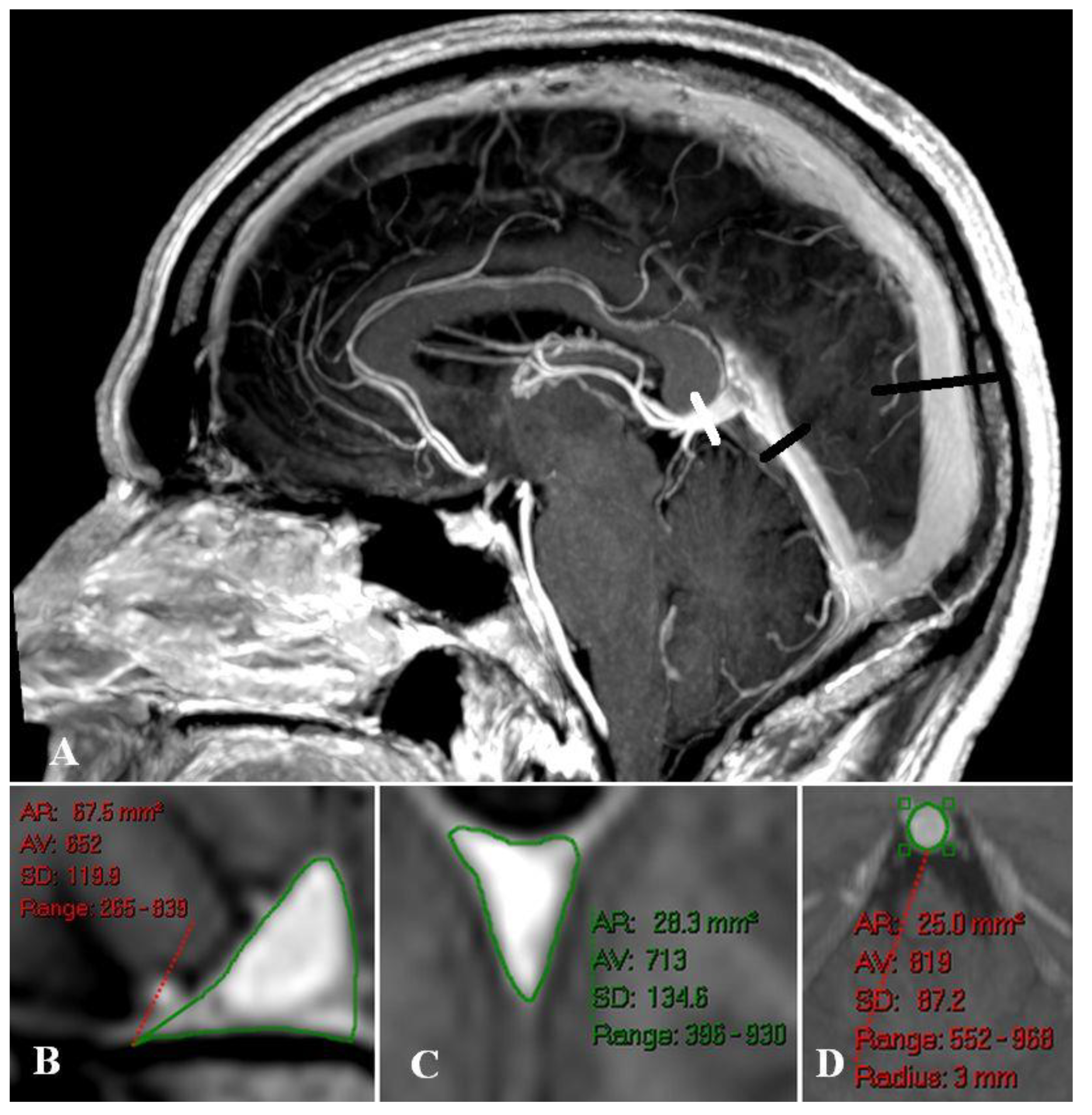
Figure 1. A 48 years old male with CFS. Fig 1A. A sagittal reconstruction of the 3DT1 data showing prominent sagittal and straight sinuses. The long black line indicates the site of SSS measurement, the short black line the straight sinus measurement and the white line the vein of Galen measurement. Fig 1B. A reconstruction showing the SSS to measure 67.5 mm2. Fig 1c. A reconstruction showing the straight sinus to measure 28.3 mm2. Fig 1d. A reconstruction showing the vein of Galen to measure 25.0 mm2.
Preprints 75238 g001
Figure 2. A 20 years old female with CFS. Fig 2A. An anteroposterior surface rendered reconstruction of the venous system taken from the 3DT1 post contrast data. Note no outflow stenosis is evident. Fig 2B. A similar reconstruction to 2A viewed from above. Note the prominent cortical veins with no evidence of obstruction of their junctions with the SSS. Fig 2C. A curved reconstruction of the SSS showing the site of cortical vein measurement. Fig 2D-G reconstructions of the four cortical veins with areas of 21.5, 12.3, 14.3 and 7.7 mm2.
Preprints 75238 g002
Disclaimer/Publisher’s Note: The statements, opinions and data contained in all publications are solely those of the individual author(s) and contributor(s) and not of MDPI and/or the editor(s). MDPI and/or the editor(s) disclaim responsibility for any injury to people or property resulting from any ideas, methods, instructions or products referred to in the content.
Copyright: This open access article is published under a Creative Commons CC BY 4.0 license, which permit the free download, distribution, and reuse, provided that the author and preprint are cited in any reuse.
Prerpints.org logo

Preprints.org is a free preprint server supported by MDPI in Basel, Switzerland.

Subscribe

© 2024 MDPI (Basel, Switzerland) unless otherwise stated