1. Introduction
Blood cells play important roles in the immune response against invading pathogens and in the normal development of metazoan [
1,
2]. In
Drosophila, hematopoiesis occurs in two different waves: in the head mesoderm of early embryos and the lymph glands of larvae [
3,
4,
5]. During this process,
Drosophila prohemocytes terminally differentiate into three types of blood cells: plasmatocytes (phagocytosis), crystal cells (melanization), and lamellocytes (encapsulation) [
2,
5]. Phagocytic plasmatocytes engulf apoptotic bodies and pathogens, such as bacteria and fungi, as the predominant hemocytes [
6,
7]. Lamellocytes, which are rare in healthy conditions, can massively differentiate after infection and form a capsule around foreign pathogens [
2,
8]. Melanization is facilitated by crystal cells that secrete phenol oxidase [
8]. This hemocyte-mediated cellular immune response is involved in the formation of larval melanotic masses.
In
Drosophila, melanotic mass formation, a conspicuous cellular response, can be induced via abnormal immune responses, such as lymph gland overgrowth and massive differentiation of lamellocytes [
2,
9]. The formation of melanotic masses is closely linked to several signaling pathways, including the Toll, Janus kinase/signal transducer and activator of transcription (JAK/STAT),
Drosophila immune deficiency (IMD)/Relish, and Ras/mitogen-activated protein kinase (Ras/MAPK) pathways [
8]. The JAK/STAT signaling pathway is associated with innate immunity and hematopoiesis. According to a model of the JAK/STAT signaling pathway in
Drosophila, binding of the cytokine, Unpaired (Upd), to the Domeless (Dome) receptor induces receptor dimerization and activation of the JAK Hopscotch (Hop in
Drosophila). Activated Hops phosphorylate STAT proteins, allowing them to form dimers and translocate into the nucleus to regulate the expression of target genes, such as the
Suppressor of cytokine signaling at 36E (
Scocs36E) [
10]. Loss of JAK/STAT signaling leads to impaired encapsulation, whereas aberrant activation of the JAK/STAT pathway results in premature lamellocyte differentiation and melanotic mass formation [
10]. Activation of c-Jun N-terminal Kinase (JNK) signaling can affect the JAK/STAT signaling pathway by inducing the expression of
Upds, a ligand of the JAK/STAT pathway [
11]. These Upds can be generated from other larval tissues, including the fat body [
12,
13], and then circulate and trigger activation of the JAK/STAT signaling pathway in the target tissue or cells.
MicroRNAs (miRNAs) are small non-coding RNAs (~21 nucleotides in length) that post-transcriptionally suppress gene expression by binding to the 3′-untranslated region (UTR) of mRNA and inducing RNA degradation and/or translational repression [
14]. Using high-throughput sequencing, a large number of miRNAs have been identified across species, including humans, mice, and flies. According to the miRbase, 469 mature miRNAs have been identified in
Drosophila melanogaster [
15]. Individual miRNAs have been estimated to target approximately 200 mRNA transcripts on average [
16]. Through these complicated regulatory integrations, miRNAs are involved in various biological processes including development, growth, metabolism, and cell death [
16]. In particular, as an miRNA linked to the JAK/STAT signaling pathway,
Drosophila miR-279 is involved in ovarian cell fate and circadian behavior by regulating
stat92E and
upd, respectively [
17,
18]. Additionally, miR-306 and miR-79 enhance the activation of JNK signaling by suppressing RNF146, an E3 ubiquitin ligase [
19]. However, it was revealed roles of only some miRNAs in the signaling pathways controlling various biological processes, and the biological functions of most miRNAs still need to be explored.
The
found-in-neurons (
fne) gene encodes an RNA-binding protein as one of the three paralogs (Rbp9, Fne, and Elav) of the ELAV gene family, and is primarily expressed in neuronal tissues in
Drosophila [
20]. According to previous reports,
fne is associated with several biological processes, such as mushroom body development, male courtship performance, and synaptic plasticity [
20,
21]. In addition, similar to other family proteins, Fne broadly induces 3′-UTR extension in neuronal cells by blocking the use of the proximal polyadenylation site [
22]. The cytoplasmic protein, Fne, undergoes a switch of cellular localization toward the nucleus due to the inclusion of a microexon encoding a nuclear localization signal under Elav-nonfunctional conditions [
22]. However, functional studies on Fne have focused on its role in primarily expressed neuronal cells. As a result, other biological roles of
fne in non-neuronal cells remain unknown.
In the present study, we investigated the biological role of Drosophila miR-274 in larval fat bodies in terms of melanotic mass formation and developmental growth. We found that miR-274 is involved in the activation of the JNK and JAK/STAT signaling pathways, which are closely associated with the observed phenotypic consequences. Furthermore, we revealed that this regulation of miR-274 was mediated by the RNA-binding protein Fne, a biologically relevant target of miR-274. Overall, our findings suggest that miR-274 plays a crucial role in regulating the fne-JNK signaling axis, which in turn affects melanotic mass formation and developmental growth.
3. Results
3.1. miR-274 is associated with melanotic mass formation through the JNK - JAK/STAT signaling pathway axis
To determine the biological roles of miR-274 in the fat bodies of
Drosophila, we overexpressed
miR-274 using
Cg-GAL4, which is activated in fat bodies [
31] (hereafter,
Cg>miR-274) (
Figure 1A). Interestingly,
Cg>miR-274 larvae had varying numbers and sizes of melanotic masses throughout their bodies (
Figure 1B). Ninety-four percent of the
Cg>miR-274 larvae exhibited black masses throughout their bodies, whereas the
Cg/+ control larvae did not exhibit such melanotic masses (
Figure 1C). These black masses persisted in the abdomens of both male and female flies (
Figure 1D). Taken together, these results suggest that miR-274 is involved in the formation of melanotic masses.
We proceeded to elucidate the molecular mechanisms underlying melanotic mass formation. According to previous reports, melanotic mass formation is strongly associated with the JAK/STAT signaling pathway [
10]. Therefore, we sought to determine whether the overexpression of
miR-274 could alter the activity of the JAK/STAT signaling pathway. First, we measured the expression of
socs36E mRNA, a target gene of the JAK/STAT signaling pathway [
10,
32], in the larval fat body of
Cg>miR-274. Indeed,
socs36E mRNA transcripts were significantly upregulated in the fat body of
Cg>miR-274 larvae compared to that in the control larval fat body (
Figure 1E). These results suggest that the overexpression of
miR-274 leads to the activation of the JAK/STAT signaling pathway in the fat body.
Next, we wondered whether miR-274 could affect which step of the JAK/STAT signaling pathway. Thus, we determined the mRNA transcript levels of
upd3, a ligand of the JAK/STAT signaling pathway [
33,
34], in the larval fat body of
Cg>miR-274. Interestingly, we found that the expression level of
upd3 mRNA transcripts was markedly higher than that in the control fat body (
Figure 1F). These data indicate that miR-274 is involved in the upregulation of
upd3, which activates the JAK/STAT signaling pathway.
The expression of the
upd3 cytokine is induced by the JNK signaling pathway [
11]. Thus, to examine whether miR-274 is linked to the activation of the JNK signaling pathway, we determined the level of active phospho-JNK (p-JNK) in the fat body of
Cg>miR-274 larvae. The p-JNK level was found to increase in the larval fat body of
Cg>miR-274 relative to that in the control fat body (
Figure 1G), suggesting that
miR-274 overexpression also activates the JNK signaling pathway.
Activation of the JNK signaling pathway in other tissues can upregulate the expression of
upd3, which triggers a systemic response in hemocytes. This response can increase hemocyte proliferation and lamellocyte differentiation, ultimately resulting in melanotic mass formation [
10,
11]. Therefore, to investigate whether the population of lamellocytes, which are closely associated with melanotic mass, is increased in the hemolymph of
Cg>miR-274 larvae, we determined the level of
Integrin betanu subunit (
Itgbn), a marker gene of lamellocytes [
35] using RT-qPCR. Remarkably, the expression level of
Itgbn was significantly higher in the hemocytes of
Cg>miR-274 larvae than in the control (
Figure 1H), suggesting that lamellocytes were more differentiated in the hemolymph of
Cg>miR-274 larvae. Collectively, our data suggest that miR-274 controls melanotic mass formation by regulating the JNK-JAK/STAT pathway axis.
3.2. Fat body-overexpression of miR-274 leads to growth reduction through defects in the fat body
We additionally investigated the phenotypic consequences observed in the fat bodies of
Cg>miR-274 larvae. Interestingly, the total mass of the fat bodies expressing
miR-274 was markedly reduced compared to that of the control fat bodies (
Figure 2A). Moreover, the size of cells in the fat body was notably reduced in
Cg>miR-274 larvae compared to that in the control larvae (
Figure 2B and 2C). These data indicate that the overexpression of
miR-274 causes a reduction in the tissue growth of the larval fat body, in addition to melanotic mass formation.
As the fat body is a crucial tissue associated with energy metabolism and growth [
36], we continuously monitored the effects of
miR-274 overexpression on developmental growth when
miR-274 was overexpressed in the fat body. Most
Cg>miR-274 larvae developed into the pupal stage; however, more than 50% of these pupae could not eclose into adult flies (
Figure 2D), indicating that miR-274-induced defects in tissue growth of the larval fat body affect normal development.
Interestingly, the growth of
Cg>miR-274 adult flies was reduced relative to that of control flies. To further explore this reduction in growth, we compared the body weights of 3–5-day-old flies between
Cg/+ and
Cg>miR-274. In both males and females, we observed a significant reduction in the body weight of
Cg>miR-274 flies (8.1%-26.5%) compared to
Cg/+ control flies (
Figure 2E). Furthermore, in proportion to body weight, the size of
Cg>miR-274 adult wings remarkably decreased relative to the size of control wings (
Figure 2F and 2G). To determine whether the reduction in whole wing size was due to the size and/or number of wing cells, we analyzed the cell size and number of wings in the indicated genotypes. Both the cell size and number of wings of
Cg>miR-274 flies were lower than those in the control flies (
Figure 2H and 2I). Taken together, these results indicate that miR-274 is involved in growth control through the regulation of total tissue mass in the fat body.
3.3. miR-274 negatively regulates the expression of fne
We wondered how miR-274 controls melanotic mass formation and growth and thus investigated the regulatory mechanism of miR-274. Using the miRNA target prediction tool, TargetScanFly [
9], we first searched for potential target genes that could be regulated by miR-274-5p, the main strand of miR-274. We found 173 transcripts with conserved miR-274-5p binding sites (
Table S2). Among these, based on a screening study of the gene network regulating blood cell homeostasis in
Drosophila [
2], we selected
found in neurons (
fne) that encodes an RNA-binding protein associated with the regulation of mRNA processing, such as splicing and alternative polyadenylation (APA) of mRNA [
22,
37]. According to the prediction using TargetScanFly, miR-274-5p could be bound to two potential sites in the
fne 3′-UTR, one conserved site (miR-274-5p BS1) and one poorly conserved site (miR-274-5p BS2) (
Figure 3A, top).
In
Drosophila,
fne mRNAs are mainly expressed in the nervous system and exists as several isoforms with different lengths of the 3′-UTR [
22]. Thus, we determined whether
fne mRNA is also expressed in the larval fat body, in addition to the nervous system. By semi-RT-qPCR using two different primer sets, we detected
fne mRNA with a short and extended 3′-UTR in the fat body of wandering third-instar larvae and adult heads. Consistent with previous results [
20,
22], the
fne mRNA transcript with an extended 3′-UTR was strongly detected in adult heads and expressed in the fat body, although its expression was lower than that in adult heads (
Figure 3A, bottom). Under Elav-depleted conditions, the nucleus-localized Fne (nFne) bearing the microexon can be induced, which regulates the APA process [
22,
37]. Therefore, we investigated whether
nfne is expressed in the fat body, which is a tissue that does not express Elav. Interestingly, we found that only the
nfne transcript containing the microexon was expressed in the larval fat bodies, whereas both general
fne and
nfne transcripts were expressed at high and low levels, respectively, in adult heads (
Figure 3B). These findings suggest that the APA-related nFne likely functions in the larval fat body.
We next examined whether miR-274 negatively regulates the expression of
fne in larval fat bodies. Accordingly, we determined the levels of
fne mRNA transcripts in the fat bodies of
Cg/+ and
Cg>miR-274 larvae. As expected, the expression of
fne mRNA was significantly reduced in the larval fat bodies overexpressing
miR-274 compared to that in the control fat bodies (
Figure 3C). These data support the hypothesis that miR-274 suppresses
fne mRNA expression in the larval fat bodies.
To clarify the regulatory interaction between miR-274 and
fne mRNA, we performed a luciferase reporter assay. When
miR-274 was overexpressed in S2 cells, the activity of
Renilla luciferase (RL) fused with wildtype
fne 3′-UTR significantly decreased (
Figure 3D and 3E). In contrast, the inhibitory activity of
miR-274 was partially reduced upon co-expression of RL fused with the
fne 3′-UTR containing a mutation in the conserved miR-274-5p binding site (miR-274-5p BS1) (
Figure 3E). Taken together, our results suggest that miR-274-5p directly suppresses
fne expression in
Drosophila.
3.4. fne is involved in melanotic mass formation as the biological target of miR-274
As
fne is suppressed by miR-274 as a direct target in
Drosophila, we sought to determine whether the loss of
fne leads to phenotypic consequences similar to the defects caused by miR-274 driven by
Cg-GAL4. As a result, we knocked down
fne expression in the fat body using
Cg-GAL4 (
Cg>fne-RNAi) (
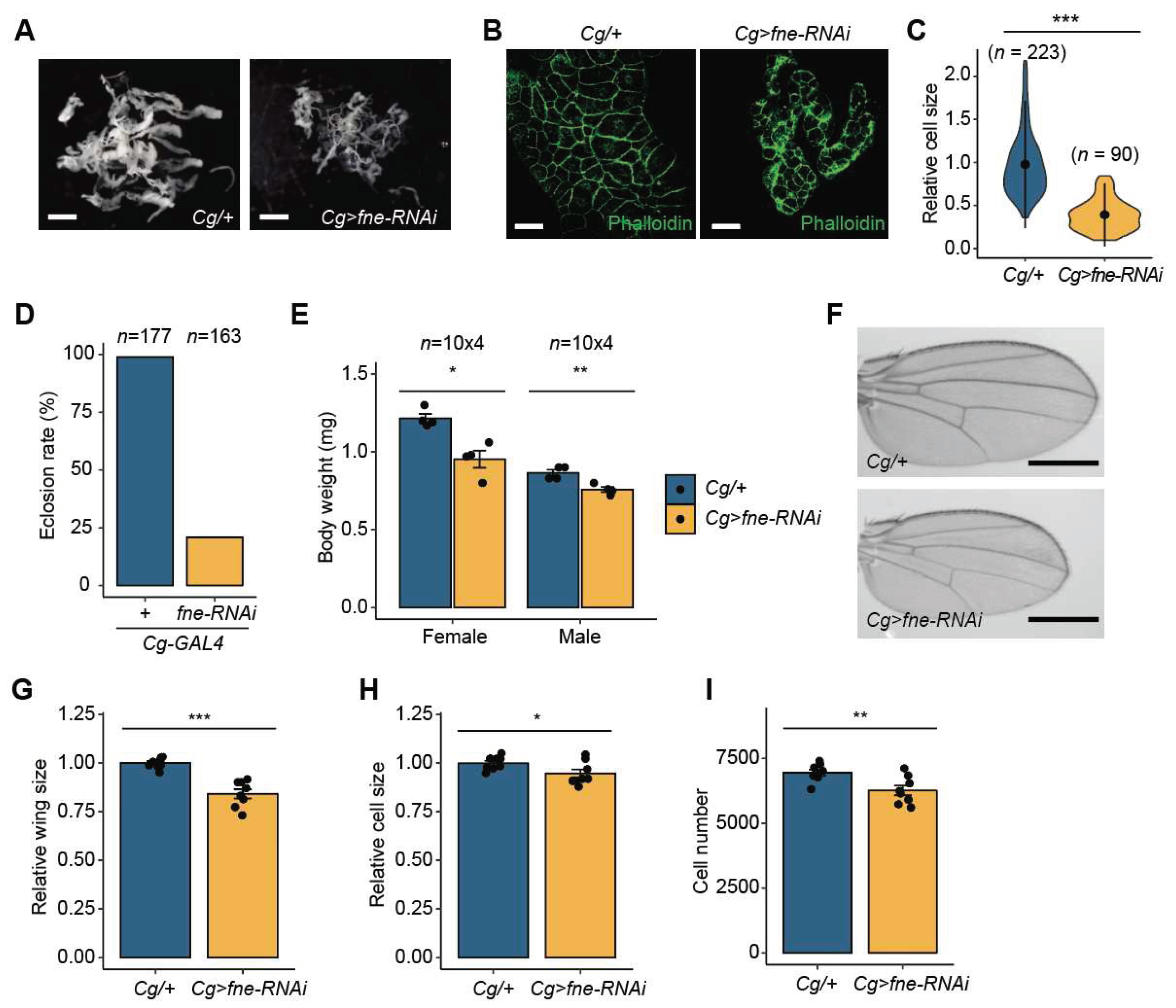
Figure 4A). Consistent with the results for
Cg>miR-274 larvae, we observed that melanotic masses remarkably appeared throughout the body of
Cg> fne-RNAi larvae (
Figure 4B), and 42.3% of the
Cg> fne-RNAi larvae exhibited melanotic mass formation (
Figure 4C). Melanotic masses also persisted in both sexes into adulthood (
Figure 4D). These findings suggest that loss of
fne is associated with melanotic mass formation.
We next examined whether depletion of
fne causes an increase in the activity of the JAK/STAT signaling pathway in the larval fat body, similar to
Cg>miR-274 larvae. The expression level of
socs36E was significantly higher in the fat bodies of
Cg>fne-RNAi larvae than in the control fat bodies (
Figure 4E), indicating that depletion of
fne activates the JAK/STAT signaling pathway. Furthermore, the expression of
upd3 was markedly upregulated in the fat bodies of
Cg>fne-RNAi larvae compared to that in the controls (
Figure 4F). These results suggest that
fne is involved in the JAK/STAT signaling pathway by regulating the expression of
upd3.
Furthermore, we determined whether
fne alters the level of active p-JNK, as observed with
miR-274 overexpression. The level of active p-JNK increased in the fat body of
Cg> fne-RNAi larvae compared to that in the control (
Figure 4G), indicating that
fne depletion activates the JNK signaling pathway.
We investigated whether the reduction in
fne expression leads to increased lamellocyte differentiation. We determined the levels of
Itgbn mRNA in the hemolymph of
Cg>fne-RNAi larvae. Consistent with
Cg>miR-274 larvae,
Itgbn mRNA levels were higher in the hemocytes of
Cg>fne-RNAi larvae than in that of the controls (
Figure 4H). Thus, the results imply that the depletion of
fne causes an increase in the number of lamellocytes. Collectively, our data suggest that
fne, as a biological target of miR-274, is associated with melanotic mass formation through the JNK-JAK/STAT signaling pathway.
3.5. fne depletion leads to growth reduction
We investigated whether
fne is also implicated in developmental growth, similar to miR-274. First, we analyzed the total fat body mass in
Cg>fne-RNAi larvae. Similar to
Cg>miR-274 larvae, the total fat body mass was remarkably reduced in
Cg>fne-RNAi larvae compared to that in control larvae (
Figure 5A). To further examine this phenotype, we compared the sizes of fat body cells between
Cg/+ and
Cg>fne-RNAi larvae after F-actin staining with phalloidin. The cell size of the fat body in
Cg>fne-RNAi larvae was significantly smaller than that in the controls (
Figure 5B and C).
The eclosion rate of
Cg>miR-274 pupae was remarkably reduced compared to that of control pupae (
Figure 5D). To investigate whether defects in the larval fat body of
Cg>miR-274 affected developmental growth, we measured the body weights of
Cg>fne-RNAi flies. Consistent with the reduction in body weight in
Cg>miR-274 flies,
Cg>fne-RNAi flies of both sexes displayed significantly lower body weight than control flies (
Figure 5E). Moreover, we observed a decrease in the whole size of
Cg>fne-RNAi wings relative to control wings in proportion to body weight (
Figure 5F and G). The size and the total number of wing cells in
Cg>fne-RNAi flies were reduced compared to those in control flies (
Figure 5H and I). Together, these observations indicate that
fne, a target of miR-274, also plays a role in developmental growth.
4. Discussion
In Drosophila, the hematopoietic system is involved in important physiological processes, such as immune responses. This system is tightly regulated by various signaling pathways, including the JAK/STAT signaling pathway, under specific conditions driven by external or internal factors. The dysregulation of this process results in an abnormal phenotype. For example, under a condition that induces an increase in lamellocytes, larvae undergo an unusual process, such as melanotic mass formation. However, the underlying mechanisms associated with this process remain unclear.
In this study, we demonstrated that miR-274 regulates the JNK-JAK/STAT signaling axis by targeting
fne, which in turn controls the formation of melanotic mass. However, further studies are needed to address the detailed mechanism by which
fne controls the JNK-JAK/STAT signaling pathways. Based on previous reports [
22], Fne may regulate gene expression through its involvement as an RNA-binding protein in the APA process. In particular, the nucleus-localized nFne is involved in this process under Elav-depleted conditions [
22,
37]. Based on our results, the nFne isoform is likely functional in the APA process in the larval fat body, where Elav is not expressed. Thus, depletion of
fne may induce a shortened 3′-UTR of
fne-target mRNAs, thereby reducing the chance of negative regulation by miRNAs; this is because miRNAs mainly suppress the expression of target genes by binding to the 3′-UTR of mRNAs [
14], which may result in an increase in the expression of genes targeted by Fne. By
targets of
RNA-binding proteins
identified
by
editing (TRIBE) method [
38], target mRNAs directly bound by Fne have been identified in
Drosophila S2 cells [
39]. The identified genes include
Stat92E, a key factor in the JAK/STAT signaling pathway, and positive regulators of JNK signaling, such as
Cell division cycle 42 (
Cdc42),
Rac1, and
cryptocephal (
crc). Thus, changes in the length of the 3′-UTR of these genes involved in the JNK or the JAK/STAT signaling pathway, which are regulated by Fne, may alter their expression level.
Our findings indicate that miR-274 activates JNK signaling by suppressing
fne, which in turn induces the expression of
upd3 in the fat body. Subsequently, Upd3 derived from the fat body may stimulate lamellocyte differentiation, triggering melanotic mass formation. However, miR-274 may regulate the JNK-JAK/STAT signaling pathway in blood cells in addition to the fat body. The
Cg-GAL4 driver primarily activates the fat body but also drives gene expression in blood cells. [
31]. Thus, interactions between the regulatory networks of the fat body and hemocytes may influence melanotic mass formation.
In addition to the observed melanotic mass formation, we noted a significant reduction in tissue mass and activation of the JNK signaling pathway in the fat body when
miR-274 and
fne were overexpressed and depleted, respectively. JNK signaling is well known to play a pro-apoptotic role in cell death and negatively regulates insulin/IGF-like signaling in both mammals and
Drosophila [
40,
41]. The activation of JNK signaling has been demonstrated to cause defects in the normal growth and development of the wing and eye in
Drosophila [
42,
43]. These findings support our observation that miR-274 and
fne controls developmental growth through JNK signaling.
According to previous reports, the expression of
pre-miR-274 is higher in the glia than in other tissues during the larval stage of
Drosophila. However, mature-miR-274 is released as an exosome and broadly distributed to other cells, including synaptic boutons, muscle cells, and tracheal cells [
44]. This finding implies that glia-derived exosomal mature-miR-274 could circulate in the larval hemolymph and affect the expression of its target genes in several target tissues, such as hemocytes and fat bodies, along with miR-274 transcribed in the tissue itself. The biological target gene of miR-274,
fne, is primarily expressed in neuronal tissue in
Drosophila [
20]. The expression of both
miR-274 and
fne in the same tissues indicates that they are likely to maintain a regulatory network, similar to our findings in fat bodies. Based on our previous results, the expression of
miR-274 is upregulated during the larval-to-pupal transition [
26], which suggests that changes in
miR-274 expression may alter the composition of the extended 3′-UTR of neuronal-specific genes by regulating the expression of
fne during this developmental stage. Thus, future studies should investigate the perturbation of APA in neuron-specific genes during metamorphosis.
We observed a reduction in both fne mRNA and reporter activity with the fne 3′-UTR when miR-274 was overexpressed. miR-274-5p was found to negatively regulate the expression of fne at the transcript level by binding to at least one conserved site (miR-274-5p BS1) in the 3′-UTR of fne mRNA. However, the luciferase reporter activity suppressed by miR-274 was found to be partially restored when the region corresponding to the miR-274-5p seed was mutated in the miR-274-5p BS1. This result implies that other binding sites may exist for miR-274-5p in the fne 3′-UTR. The poorly conserved binding site (miR-274-5p BS2) in the fne 3′-UTR could be a potential regulatory binding site for miR-274-5p. Future studies could examine whether miR-274-5p targets miR-274-5p BS2 in the fne 3′-UTR.
A relatively stronger formation of melanotic masses was observed in larvae overexpressing miR-274 than in fne-depleted larvae. Most Cg>miR-274 larvae exhibited a melanotic mass, whereas less than 50% of Cg>fne-RNAi larvae had black dots. The melanotic masses in the individual Cg>miR-274 larvae were numerous and larger than those in the Cg>fne-RNAi larvae. Such small differences between the lines linked in the regulatory network may arise from other genes affected by alterations in miR-274 or fne expression. TargetScanFly identified 173 potential miR-274-5p targets. Some of these targets might directly or indirectly affect the JNK and/or JAK/STAT signaling pathways. For example, depletion of S-adenosylmethionine Synthetase (Sam-S) and slimfast (slif), which are potential targets of miR-274-5p, resulted in an increase in the expression of upd3 and socs36E (data not shown). Collectively, our findings provide insights into the potential molecular mechanisms underlying these phenomena. Further studies should explore these mechanisms in detail to provide a more comprehensive understanding of the physiological processes.
Author Contributions
Conceptualization, H.K.K. and D.-H.L.; Methodology, H.K.K. and D.-H.L.; Software, H.K.K. and D.-H.L.; Validation, H.K.K. and D.-H.L.; Formal Analysis, H.K.K. and D.-H.L.; Investigation, H.K.K., C.J.K., D.J., and D.-H.L.; Data Curation, H.K.K. and D.-H.L.; Writing – Original Draft Preparation, H.K.K. and D.-H.L.; Writing – Review & Editing, D.-H.L.; Visualization, D.-H.L.; Supervision, D.-H.L.; Project Administration, D.-H.L.; Funding Acquisition, D.-H.L. All authors have read and agreed to the published version of the manuscript.
Figure 1.
miR-274 is involved in the formation of melanotic mass. (A) Overexpression of miR-274-5p in the fat body of Cg>miR-274 larvae. U6 snRNA was used as a control for normalization. (B) Wandering third-instar larvae of the indicated genotypes exhibiting melanotic masses. Melanotic masses are marked as arrows. The dashed box image is magnified. (C) Quantitative data showing the percentage of wandering third-instar larvae with melanotic masses. n is the total number of analyzed larvae. (D) Adult flies exhibiting melanotic masses in the indicated genotypes and sexes (M, male; F, female). Melanotic masses are marked as arrows. The dashed box images are magnified. (E) Expression level of socs36E mRNA in the fat body of Cg>miR-274 larvae. rp49 served as a control for normalization. (F) Expression level of upd3 mRNA in the fat body of Cg>miR-274 larvae. (G) Protein level of p-JNK in the larval fat body of Cg>miR-274. Representative band image (left) and quantitative bar graph (right) are shown. β-Tubulin served as a loading control. The bar plot is shown as the mean with individual values from two independent experiments. (H) Expression level of Itgbn mRNA in the hemocytes of Cg>miR-274 larvae. The error bars on the bar plots (A, E, F, and H) indicate the standard error of the mean (SEM). *P < 0.05, **P < 0.01, and ***P < 0.001 compared with the control, as assessed by Student’s t-test.
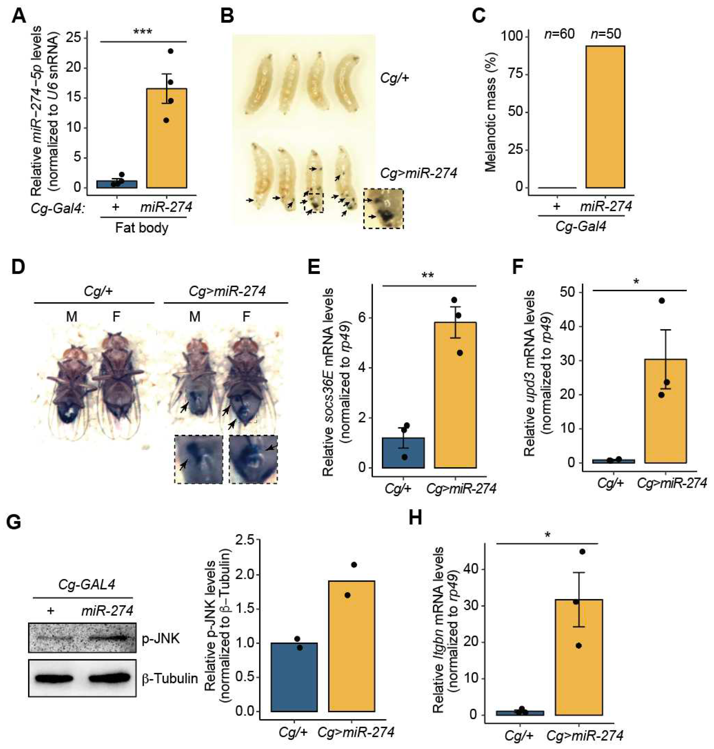
Figure 2.
Overexpression of miR-274 in the fat body reduces developmental growth. (A) Whole fat bodies of wandering third-instar larvae of the indicated genotypes (n = 5). Scale bar, 1 mm. (B) Representative phalloidin staining images of the larval fat body (phalloidin, red; DAPI, blue). Scale bar, 50 µm. (C) Relative size of the fat body cells in Cg>miR-274 larvae. The quantitative violin plot is shown as the mean ± standard deviation (SD). n is the total number of analyzed fat body cells. (D) Eclosion rate from pupae to adult flies in each genotype. n is the total number of analyzed pupae. (E) Body weight of Cg>miR-274 flies. n is the total number of analyzed flies. (F) Representative wing images of female flies of the indicated genotypes. Scale bar, 0.5 mm. (G) Relative comparison of wing size between Cg/+ and Cg>miR-274 flies (n = 10 per each genotype). (H, I) Relative cell size (H) and total cell number (I) of adult wings analyzed in the G panel. All bar plots (E, G, H, and I) are shown as the mean ± SEM. *P < 0.05 and ***P < 0.001 compared with the control, as assessed by Student’s t-test.
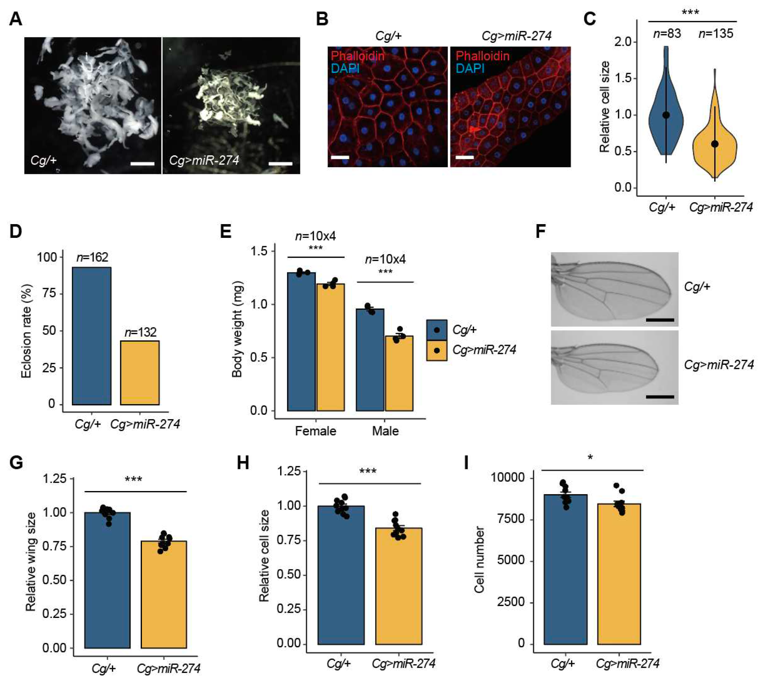
Figure 3.
miR-274 suppresses fne expression. (A) Expression of the fne mRNA transcripts with the short or extended 3′-UTR. Isoforms of fne mRNA transcripts with different lengths of 3′-UTR, each primer binding site (1F/1R and 2F/2R, red box), and two miR-274-5p binding site (BS1 and BS2, blue box) are shown (top). Semi-RT-qPCR at two different sites in the fne 3′-UTR in the larval fat bodies and adult heads (bottom). (B) Expression of nuclear fne (nfne) transcript containing the microexon (red box) in the larval fat bodies. Schematic of the fne microexon (top) and the expression of two fne isoforms in the larval fat bodies and adult heads (bottom). Blue and red boxes indicate exons, and gray lines indicate introns. The primer binding sites for RT-qPCR are marked as arrows. (C) Downregulation of the fne mRNA level in the fat body of Cg>miR-274 larvae. rp49 served as a control for normalization. (D) Overexpression of miR-274-5p in S2 cells. U6 snRNA was used as a control. (E) Relative activity of the Renilla-luciferase (RL) fused with either the wildtype (WT) or mutated (MT) fne 3′-UTR. The sequences of WT and MT fne 3′-UTR and miR-274-5p are shown (top). The mutated sequences are highlighted in bold red. The RL activity was normalized to the firefly luciferase (FL) activity (bottom). All bar plots are shown as the mean ± SEM. *P < 0.05, **P < 0.01, and ***P < 0.001 compared with the control, as assessed by Student’s t-test (C and D) or ANOVA with a supplementary Dunnett’s test (E).
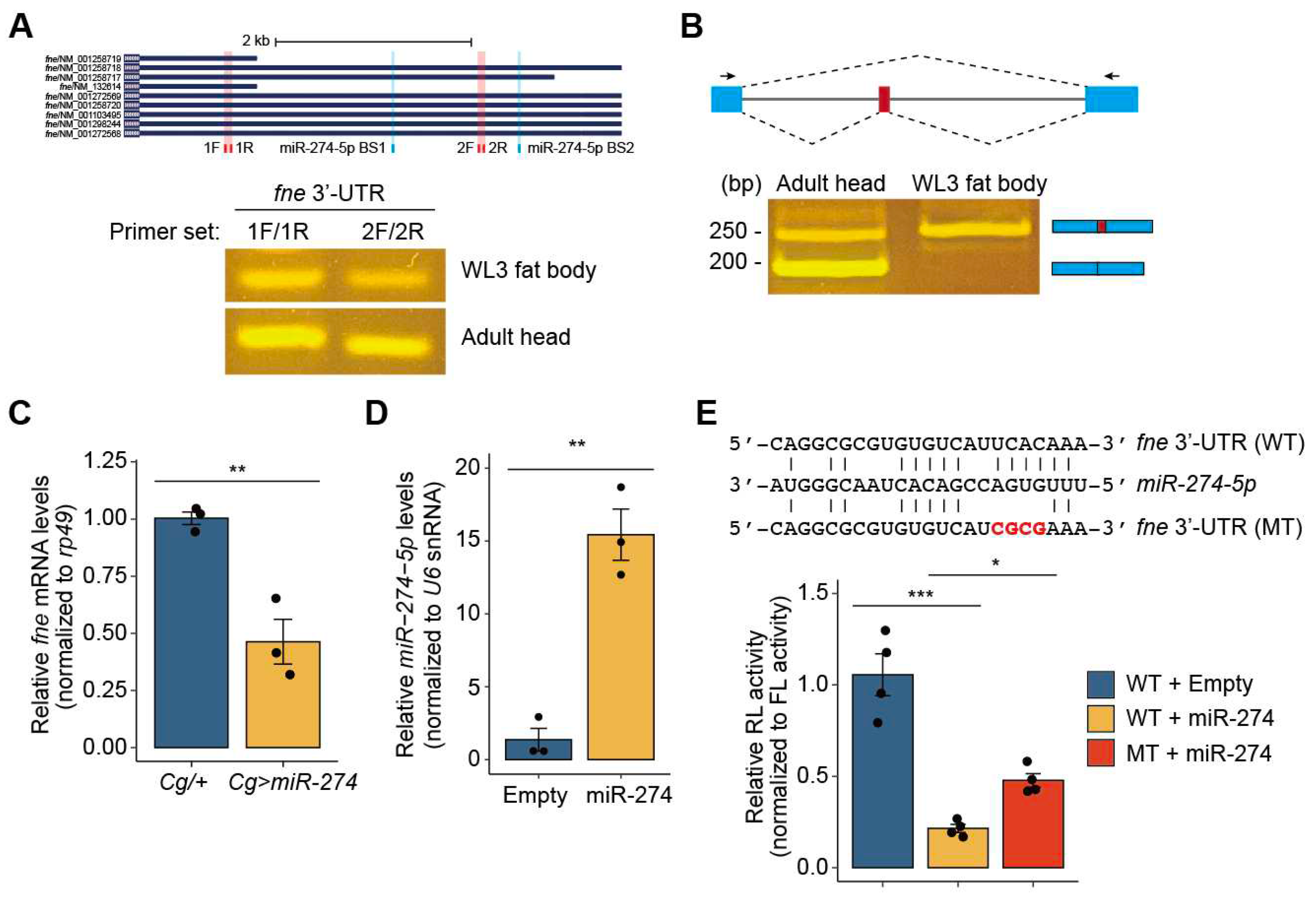
Figure 4.
Depletion of fne results in melanotic mass formation. (A) Knockdown of fne in the fat body using RNAiTRiP line driven by Cg-GAL4. rp49 served as a control for normalization. (B) Melanotic mass formation in Cg>fne-RNAi larvae. Melanotic masses are marked as arrows. The dashed box image is magnified. (C) Percentage of Cg>fne-RNAi larvae exhibiting melanotic masses. n is the total number of analyzed larvae. (D) Maintenance of melanotic mass in Cg>fne-RNAi adults. Melanotic masses are marked as arrows. The dashed box images are magnified. (E) Expression level of upd3 mRNA in the fat body of Cg>fne-RNAi larvae. (F) Expression level of socs36E mRNA in the fat body of Cg>fne-RNAi larvae. (G) Protein level of p-JNK in the larval fat body of Cg>fne-RNAi. Representative band image (left) and quantitative bar graph (right) are shown. The bar plot is shown as the mean with individual value. β-Tubulin served as a loading control. (H) Expression level of Itgbn mRNA in the hemocytes of Cg>fne-RNAi. The error bars on the bar plots (A, E, F, and H) indicate SEM. *P < 0.05 and **P < 0.01 compared with the control, as assessed by Student’s t-test.
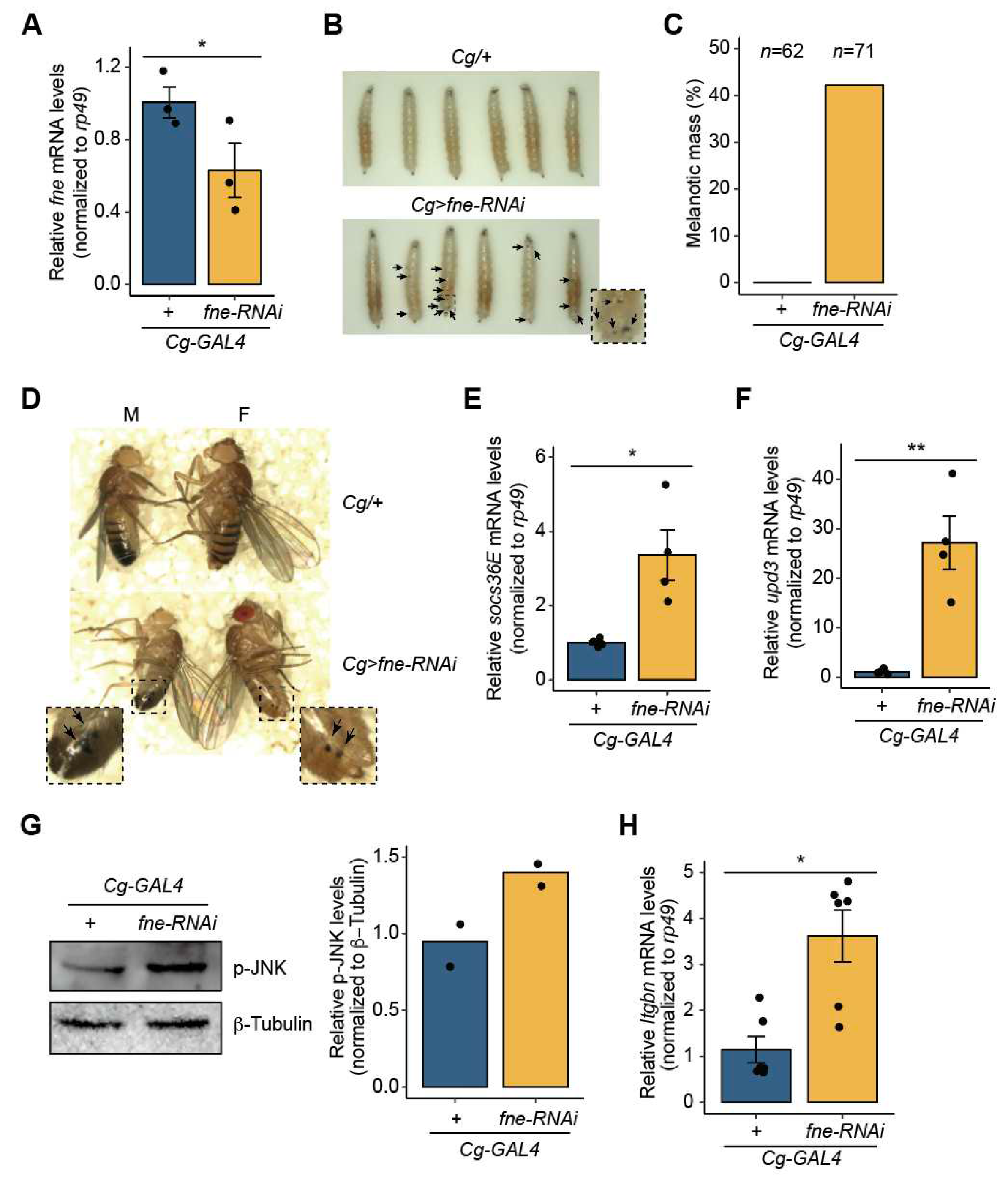
Figure 5.
Knockdown of fne causes growth reduction. (A) Whole fat bodies of wandering third-instar larvae of the indicated genotypes (n = 5). Scale bar, 1 mm. (B) Representative phalloidin staining images of the larval fat body (phalloidin, green). Scale bar, 50 µm. (C) Relative size of the fat body cells in Cg>fne-RNAi larvae. The quantitative violin plot is shown as the mean ± standard deviation (SD). n is the total number of analyzed fat body cells. (D) Reduced eclosion rate of Cg>fne-RNAi pupae. n is the total number of analyzed pupae. (E) Body weight of Cg>fne-RNAi flies. n is the total number of analyzed flies. (F) Representative wing images of male flies of the indicated genotypes. Scale bar, 0.5 mm. (G) Relative comparison of wing size between Cg/+ and Cg>fne-RNAi flies (n = 10 per each genotype). (H, I) Relative cell size (H) and total cell number (I) of adult wings analyzed in the G panel. All bar plots (E, G, H, and I) are shown as the mean ± SEM. *P < 0.05, **P < 0.01, and ***P < 0.001 compared with the control, as assessed by Student’s t-test.
