Preprint
Article

Does Accelerometry at the Centre of Mass Accurately Predict the Gait Energy Expenditure in Patients with Hemiparesis?

Altmetrics

Downloads

74

Views

20

Comments

0

A peer-reviewed article of this preprint also exists.

Submitted:

29 June 2023

Posted:

10 July 2023

You are already at the latest version

Alerts
Abstract
Background: The aim of this study was to compare energy expenditure (EE) predicted by accelerometery (EEAcc) with indirect calorimetry (EEMETA) in individuals with hemiparesis. Methods: Twenty-four participants (12 with stroke and 12 healthy controls) performed a six-minute walk test (6MWT) during which EEMETA was measured using a portable indirect calorimetry system and EEACC was calculated using Bouten’s equation (1993) with data from a 3-axis accelerometer positioned between L3 and L4. Results: Median EEMETA was 9.85 [8.18;11.89] W·kg-1 in the stroke group, and 5.0 [4.56;5.46] W·kg-1 in the control group. Median EEACC was 8.57 [7.86;11.24] W·kg-1 in the control group and 8.2 [7.05;9.56] W·kg-1 in the stroke group. EEACC and EEMETA were not significantly correlated in either the control (p=0.8) or the stroke groups (p=0.06). The Bland-Altman method showed a mean difference of 1.77±3.65 W·kg-1 between EEACC and EEMETA in the stroke group and -2.08±1.59 W·kg-1 in the controls. Conclusions: The accuracy of the predicted EE, based on the accelerometer and the equations proposed by Bouten et al, is low in individuals with hemiparesis and impaired gait. This combination (sensor and Bouten's equation) is not yet suitable for use as a stand-alone measure in clinical practice for the evaluation of hemiparetic patients.
Keywords: 
Subject: Biology and Life Sciences  -   Life Sciences

1. Introduction

Physical activity measurements have been used to indirectly quantify energy expenditure in individuals with various pathologies for several years [1,2,3,4]. Connected devices such as watches, bracelets or smartphone applications, which are designed to increase the activity levels of the general public, have become popular among clinicians due to their ease of use and their low cost. Such devices have thus been integrated into clinical practice and research to indirectly quantify energy expenditure [5]. Studies comparing results from off-the-shelf connected devices with specialised, equivalent medical devices or indirect calorimetry (which is the gold standard) have found that they accurately record data like the number of steps, distance covered and reliably estimate energy expenditure in healthy subject [6,7].
Increasing the level of physical activity for people with a chronic pathology, such as stroke, has been shown to reduce their co-morbidities [8,9]. The evaluation of the impact of stroke treatments would be improved if clinicians could reliably and easily measure the amount of activity performed by their patients [10]. Study have shown that patient with stroke are more inactive than healthy age-matched controls [11]. Research has also shown that energy expenditure is doubled in patients with stroke due the sequalae (mainly weakness and spasticity) of their hemiparesis [12].
Feedback on patients’ activity levels would not only inform healthcare providers, it might also motivate individuals with stroke to perform regular physical activity, and is therefore recommended by the HAS (Haute Autorité de Santé) [13]. The accessibility of new technologies and connected devices that are easily integrated into peoples’ daily lives and which allow activity to be tracked, such as smartphone applications and smart watches, have simplified the collection of detailed data relating to physical activity levels out with the hospital setting [14]. Nevertheless, several studies have indicated that inter-device reliability can be poor due to factors like the device’s position on the body, the recording method used, and the equations used to process the data, all of which result in either an over- or under-estimation of energy expenditure [15]. As a result, the use of connected devices is currently a less reliable measurement technique than indirect calorimetry [16].
Therefore, despite the promise of such devices, the clinical interest in them and the work on their development, there is currently no consensus for their use in individuals with chronic diseases and significant gait asymmetry. Optimal sensor types and positions for the accurate evaluation of physical activity levels and energy expenditure have yet to be identified. One method frequently reported in the literature [17,18,19,20] is Bouten’s method [21]. This method has been validated in healthy individuals although not in people with gait disorders [22]. Bouten’s method uses a regression equation to calculate the integral of signal data recorded by an accelerometer, positioned between L3 and L4 (so as to be close to the person’s centre of mass) in three planes of space (x, y, and z) in order to estimate energy expenditure during gait.
Moreover, following a cardiovascular accident (stroke), we often observe motor impairment caused by either a hemorrhage (hemorrhagic stroke) or a blocked artery (ischemic stroke) in the motor cortex. Neuromuscular disorders result from that, causing locomotor impairments. In terms of spatiotemporal parameters of gait cycle, reduced the speed, cadence, and stride length are observed [23,24]. At the joint kinematic level, we can observe disturbance in flexion [25]. At the hip level, there can be limitations in knee elevation due to impaired flexion and/or hip extension [26]. This can lead to difficulties in overcoming obstacles. In terms of the knee joint, during the stance phase, hyperextension and a deficit in flexion during the swing phase can be observed [27]. These issues can be explained, on one hand, by the overactivity of the triceps surae, resulting in knee extension and plantar flexion disturbance, and on the other hand, possibly by the overactivation of the rectus femoris. Finally, at the ankle level, there is often hyperactivity of the plantar flexors and weakness of the dorsiflexors. These impairments can lead to foot drop [28]. The aforementioned impairments result in a significant increase in energy cost during walking [24]. This means that the patient will expend more energy per unit of distance compared to someone without pathology [29]. The need to evaluate the effects of therapies on these gait disorders is essential. Consequently, the evaluation of the energy cost of walking, or more simply of energy expenditure, is relevant to support clinicians in the overall evaluation of the effects of the therapies chosen. In fact, the connected objects allowing this indirect measurement have a preponderant place in the evaluation of the impact of therapeutics on the autonomy of walking. So, a question arises: are connected tools using the Bouten’s method sufficiently accurate to estimate energy expenditure in patients who have had a stroke?
The aim of this study, therefore, was to compare the accuracy of energy expenditure values calculated using Bouten’s regression equation method [21] with those obtained from the gold standard method of indirect calorimetry. This work would help to validate the use of Bouten’s method as a simple way to assess people who have stroke-related hemiparesis and impaired gait. Data were compared for both methods from two groups of subjects, n=12 individuals with stroke and impaired gait, and n=12 healthy controls during a 6-minute walk test (6MWT).

2. Materials and Methods

2.1. Participants

Participants with stroke or impaired gait were recruited either during a routine follow-up medical consultation, or while they were hospitalised in rehabilitation. Inclusion criteria were: aged over 18 years, able to walk without assistance or assistive devices, able to carry out the 6MWT according to the recommendations and without any known cardiovascular contraindications [22]. Their main sequelae are locomotor disorders due to hemiparesis. The twelve participants with stroke included 10 males and 2 females; their median age was 50.5 years [interquartile range (IQR) 41.25;53.25]; median height: 175 [170.0;177.0]; median weight: 73kg [62.0;83.0]).
A group of twelve control subjects (8 males and 4 females) were also recruited. Their results were important not only as they allowed a comparison between the two experimental groups, but also because their data ensured that any effects noted were not an artefact of the experimental set-up used in this study, since Bouten’s method has been validated in healthy individuals [22]. Inclusion criteria for the control group were: aged over 18 years and with no known neuromuscular pathologies. Their median age was 29 years [IQR 24.0;33.7], median height =177cm [169.8;177.0] and median weight: 69kg [60.0;75.5].
The study was granted ethical approval, all participants provided informed consent for participation and the study was carried out according to the Helsinki declaration.
Study design
All participants performed the 6MWT, as recommended by American Thoracic Society [30], as quickly as possible along a 30m-long corridor that was marked every 2 metres. The distance covered was measured at the end of the test. Participants wore a portable gas exchanger (K4b², COSMED, Rome, Italy) and a three-axis accelerometer (EQO2, Equivital, Cambridge, UK). We choose the 6 Minute Walk Test (and its performance criteria including a walk that covers the greatest distance despite the difference in walking speed) by its common use for functional or cardio-respiratory evaluations.

2.2. Procedures

2.2.1. Indirect Calorimetry

Analysis of the gas expired from each respiratory cycle provided the reference measurement of energy expenditure (EEMETA). The system was calibrated in the corridor where the test was performed according to standard procedures.
EEMETA was calculated when V ˙ O 2 kinetics reached a stable state. The V ˙ O 2 values (Kcal·min-1) were initially smoothed using a 3-point moving average, then the last 150 seconds of each 6MWT were averaged. The EEMETA was then converted to W·kg-1.

2.2.2. Accelerometery

A lightweight (38g), compact (78x53x10mm), three-axis accelerometer (250 Hz) was used to capture the acceleration of the centre of mass in the three planes of space. It was positioned between the third and fourth lumbar vertebrae using a custom-made support. This has previously been recommended for the optimal estimation of energy expenditure [17,31,32]. A connected chest-strap monitor (EQO2, Equivital, Cambridge, UK) was used to measure heart rate during the 6MWT.

2.2.3. Estimation of Energy Expenditure (EEACC)

Bouten’s method was used to estimate energy expenditure [21]. The raw signals were initially filtered using a Butterworth filter (4th order with a 20 Hz cut-off frequency). The absolute values of the signal obtained in the three axes (IAAtot) were then calculated in 30-second periods then summed for the duration of the test [21]. The following equation was used to obtain predicted EEACC (W·kg-1):
E E A C C = 0,104 + 0,23 × I A A t o t

2.3. Statistical Analysis

The results for the descriptive and interferential statistics were described by the median, and the first and last quartiles (Q1 and Q3). The level of significance was set at p ≤0.05. The normality of the distribution was verified using a Kolmogorov-Smirnov test.
The results from Bouten’s method and indirect calorimetry we’re not normally distributed therefore we chose to use a Mann-Whitney test to compared using a Mann-Whitney test (non-homogenous sample with independent samples). The relative agreement between the EEMETA and EEACC values of the groups was compared using Spearman rank correlations. Absolute agreement was calculated using the Bland-Altman method (± limits of agreement set at 95%) [33].

3. Results

The variables measured during the 6MWT are presented in Table 1. There were significant differences between the groups for V ˙ O 2 values and distance walked (highest in the control group), but no difference in heart rate.
We observe a significant difference in measurement between median EEMETA, for patient with stroke and control respectively. Median EEMETA was 9.85 [8.18;11.89] W·kg-1 in the stroke group, and 5.0 [4.56;5.46] W·kg-1 in the control group (p<0.0001). For the accelerometric method, median EEACC was not significantly different. EEACC was 8.2 [7.05;9.56] W·kg-1 in the stroke group and 8.57 [7.86;11.24] W·kg-1 in the control group (p=0.11) (Table 2).
EEACC and EEMETA were not significantly correlated in either the control (Spearman’s r=0.086: p=079) or the stroke groups (Spearman’s r=0.56: p=0.06) (Table 3).
The Bland-Altman analysis showed large differences between EEMETA and EEACC measurements in the stroke group with a mean over-estimation of EEACC of 1.16 ± 3.70 W·kg-1 (p=0.3) relative to EEMETA (Figure 1A). In the healthy group, EEACC was under-estimated by a mean of -2.43 ± 1.45 W·kg-1 (Figure 1B).

4. Discussion

The purpose of this study was to compare the accuracy of energy expenditure values calculated using accelerometry signal, via Bouten’s regression equation method, with those obtained from the oxygen uptake of in-direct calorimetry. The results of this study showed differences between energy expenditure (EE) during a 6MWT calculated by indirect calorimetry (EEMETA) and estimated using Bouten’s method (EEACC) in both healthy volunteers (control) and individuals with stroke. The use of Bouten’s regression equation led to a 17% under-estimation in the control group and a 49% over-estimation in the calculated energy expenditure in comparison to the gold standard indirect calorimetry results in the stroke group (i.e., EEACC>EEMETA).
The first interesting result (Table 2) shows that when EE is calculated using indirect calorimetry, there is a significant difference between the control group and patients with stroke. This difference is consistent with the study by Slawinski, which shows that strokes have a lower EE because their walking speed is significantly lower than that of healthy subjects. In this study, these authors also found that the addition of obstacles during a gait test did not affect V ˙ O 2 in patients with stroke [24] as they were already at their V ˙ O 2 peak and could not increase their O2 consumption further because of their limited gait. Our EEMETA results agree with those from that study, namely that the EEMETA value of the stroke group participants was half that of the control EEMETA. This difference was mainly due to the difference in the distances covered during performance of the 6MWT: the stroke group covered an average of 339 m, whereas the controls covered, on average, 696 m. Collectively these results suggest that the reduction in distance covered by patients with stroke is related to an increase in extraneous movements required for movement control and balance in these patients.
The second interesting result concerns the comparison between strokes and controls in terms of EE estimated using accelerometery and the Bouten’s method. In fact, there is no longer difference in EEACC. In other words, EEACC is the same for both, strokes and controls. These results confirm the previous hypothesis regarding extraneous movements associated with the locomotion of patients with stroke. In the stroke group, the over-estimation of EEACC by Bouten’s method (compared to gold standard method) was likely due to the individuals’ abnormal segmental kinematics. An increase in vertical oscillations of the pelvis is a common gait anomaly following stroke [27]; it is related to various kinematic anomalies such as knee hyperextension (genu recurvatum) or a stiff gait (lack of knee flexion during swing) [34,35]. The position of the accelerometer just above the pelvis (between L3 and L4) meant that all compensatory movements performed by the subjects as a result of motor and sensory impairments were also recorded. The use of the integral of the unit vector of the accelerometer (IAAtot) to calculate EE in Bouten’s method then amplified the EEACC value. More the accelerometer moves due of the compensating movements, higher is the amplitude of the accelerometer signals and bigger is the IAAtot.
The third surprising (table 3 and figure 1) results was to find that the control group results contrasted with those described by Bouten et al.[22] The original paper had reported a mean over-estimation of EEACC of 15% in a group of 11 young healthy adults walking at different speeds. However, for a gait speed of 7 km.h-1, EEACC was overestimated by 8%. By contrast, in the present study, at almost the same gait speed (6.97 ± 0.79 Km.h-1), Bouten’s method actually under-estimated energy expenditure in the control group by 17%. This contradiction has been observed elsewhere: other studies have also reported both over- and under-estimations of EE when using accelerometery and comparing the results to indirect calorimetry in healthy subjects [36]. Indeed, two studies that used accelerometer device reported opposing results: Bai et al. (2016) found an over-estimation [37] while Imboden et al. (2018) found an under-estimation [38]. These variations were likely due to differences in the tasks (gait speed, cadence etc.). There is currently no consensus regarding the level of acceptable errors or whether they relate to under or over-estimations of EE. For strokes, EEACC over-estimate EE of 1.16 ± 3.70 W·kg-1. These results confirm the variability of accelerometric measurements when used to estimate energy expenditure. This measurement variability likely explains the lack of correlation observed between the two measurement methods.
The present results associated with those of previous studies shows that there is currently no consensus regarding the level of acceptable errors or whether they relate to under or over-estimations of EE. The variety of EEACC results obtained by different research groups suggests that it is important to be aware of the limitations in the use of accelerometers. We recommend that in order to take advantage of the convenience of accelerometer measurements, healthcare practitioners should produce their own reference data within their own setting and in patients with different pathologies using both indirect calorimetry and accelerometery in order to make informed interpretations of the accelerometery data.
The present study was different to other studies of EEACC with regards to two methodological aspects: (1) the choice of accelerometer signal processing method and (2) the positioning of the sensor. In terms of the first point, signal processing by the root mean square has been largely replaced by count per minute [39]. Nevertheless, there is currently no accepted consensus in the literature regarding threshold values for activity detection. This may, at least in part, be due to inter-individual variations caused by variables such as age or existing medical conditions. For example, it has been reported that it was difficult to calculate EEACC in older patients when using gait thresholds taken from younger adults: older people had a naturally wider range of inappropriate movements compared to younger adults which led to unreliable detection of EE in the older population [16].
With regards to sensor position, Compagnat et al. (2018)[40] found a mean difference in predicted energy expenditure between 3 and 58% in patients with hemiparesis when the sensor was positioned on the wrist rather than the pelvis. However, Bouten et al. (1997) recommends positioning the sensor between L3 and L4 [17] in order to quantify movement of the centre of mass, and this position is used in many studies [18,20,24,35,36]. We think that it seems more logical to place the sensor around the pelvis if the aim is to record compensatory gait movements, and to objectify the patient's progress during rehabilitation. Finally, recent works[41] demonstrated that the choice of the oxygen cost prediction equation ;can greatly improve the estimation of stokes daily energy expenditure.
The main limitation of this study was the inclusion of patients with diverse gait patterns. Unfortunately, there were too few patients with each type of gait pattern to determine the effects of different compensatory movements and to refine the prediction equation accordingly. On the other hand, our sample was small and did not walk at the same speed. We can also observe a mismatch between gender and age.

5. Conclusions

In conclusion, therefore, results from this study suggest that the 1993 Bouten method [21] does have potential to be of considerable practical value for quantifying rehabilitation-induced changes in gait (better gait is and less compensatory movement acting on the IAAtot). In addition to being it being cheaper and more accessible than indirect calorimetry, Bouten’s method to assess energy expenditure using an accelerometer also accounts for compensatory lower limb movements that occur as part of pathological gait. However, we should also stress that this method is, at present, unvalidated in the wider research community and not always predictable in terms of how EEACC results vary in comparison to EEMETA, even from the same populations. Therefore, this tool is not yet suitable for use as a stand-alone measurement in routine practice for the assessment for any patient with stroke-related hemiparesis and impaired gait. Further investigations are required to ensure that necessary corrective coefficients are known for different patient groups, and pathologies, in order to ensure the accuracy and reliability and reproducibility of EEACC values for the different combinations of patient demographics and pathologies.

Author Contributions

conceptualization, L.B., D.D., and J.S; methodology, L.B., and D.D.; software, L.B., and D.D.; formal analysis, D.D., and J.S.; investigation, L.B.; resources, D.D.; data curation, , L.B., and D.D.; writing—original draft preparation, L.B.; writing—review and editing,. L.B., D.D., and J.S; visualization, L.B., D.D., and J.S; supervision, D.D, and J.S. All authors have read and agreed to the published version of the manuscript.

Funding

not applicable

Institutional Review Board Statement

The study was conducted in accordance with the Declaration of Helsinki – NCT01807247; update 2015-01-04.

Informed Consent Statement

Informed consent was obtained from all subjects involved in the study.

Data Availability Statement

Not applicable.

Acknowledgments

We gratefully thank all the participants involved in this study.

Conflicts of Interest

The authors declare no conflict of interest.

References

  1. Ezeugwu, V.E.; Manns, P.J. Sleep Duration, Sedentary Behavior, Physical Activity, and Quality of Life after Inpatient Stroke Rehabilitation. J. Stroke Cerebrovasc. Dis. 2017, 26, 2004–2012. [Google Scholar] [CrossRef] [PubMed]
  2. Motl, R.W.; Sandroff, B.M.; Pilutti, L.A.; Klaren, R.E.; Baynard, T.; Fernhall, B. Physical Activity, Sedentary Behavior, and Aerobic Capacity in Persons with Multiple Sclerosis. J. Neurol. Sci. 2017, 372, 342–346. [Google Scholar] [CrossRef] [PubMed]
  3. Dalgas, U.; Stenager, E.; Ingemann-Hansen, T. Review: Multiple Sclerosis and Physical Exercise: Recommendations for the Application of Resistance-, Endurance- and Combined Training. Mult Scler 2008, 14, 35–53. [Google Scholar] [CrossRef] [PubMed]
  4. Hansen, B.H.; Kolle, E.; Dyrstad, S.M.; Holme, I.; Anderssen, S.A. Accelerometer-Determined Physical Activity in Adults and Older People. Med. Sci. Sports Exerc. 2012, 44, 266–272. [Google Scholar] [CrossRef]
  5. Jo, A.; Coronel, B.D.; Coakes, C.E.; Mainous, A.G. Is There a Benefit to Patients Using Wearable Devices Such as Fitbit or Health Apps on Mobiles? A Systematic Review. Am. J. Med. 2019, 132, 1394–1400.e1. [Google Scholar] [CrossRef]
  6. Stookey, A.D.; McCusker, M.G.; Sorkin, J.D.; Katzel, L.I.; Shaughnessy, M.; Macko, R.F.; Ivey, F.M. Test-Retest Reliability of Portable Metabolic Monitoring after Disabling Stroke. Neurorehabil. Neural Repair 2013, 27, 872–877. [Google Scholar] [CrossRef]
  7. Evenson, K.R.; Goto, M.M.; Furberg, R.D. Systematic Review of the Validity and Reliability of Consumer-Wearable Activity Trackers. Int. J. Behav. Nutr. Phys. Act. 2015, 12. [Google Scholar] [CrossRef] [PubMed]
  8. Kuwashiro, T.; Sugimori, H.; Ago, T.; Kamouchi, M.; Kitazono, T. Risk Factors Predisposing to Stroke Recurrence within One Year of Non-Cardioembolic Stroke Onset: The Fukuoka Stroke Registry. Cerebrovasc. Dis. 2012, 33, 141–149. [Google Scholar] [CrossRef]
  9. Smith, A.C.; Saunders, D.H.; Mead, G. Cardiorespiratory Fitness after Stroke: A Systematic Review. Int. J. Stroke 2012, 7, 499–510. [Google Scholar] [CrossRef]
  10. Lynch, E.A.; Jones, T.M.; Simpson, D.B.; Fini, N.A.; Kuys, S.S.; Borschmann, K.; Kramer, S.; Johnson, L.; Callisaya, M.L.; Mahendran, N.; et al. Activity Monitors for Increasing Physical Activity in Adult Stroke Survivors. Cochrane Database Syst. Rev. 2018, 109, 103392. [Google Scholar] [CrossRef]
  11. English, C.; Healy, G.N.; Coates, A.; Lewis, L.K.; Olds, T.; Bernhardt, J. Sitting Time and Physical Activity after Stroke: Physical Ability Is Only Part of the Story. Top. Stroke Rehabil. 2016, 23, 36–42. [Google Scholar] [CrossRef] [PubMed]
  12. Waters, R.L.; Mulroy, S. The Energy Expenditure of Normal and Pathologic Gait. Gait Posture 1999, 9, 207–231. [Google Scholar] [CrossRef] [PubMed]
  13. Haute Autorité de Santé Prescription d’activité Physique et Sportive Accidents Vasculaires Cérébraux. 2018, 1–10.
  14. Dobkin, B.H. Wearable Motion Sensors to Continuously Measure Real-World Physical Activities. Curr. Opin. Neurol. 2013, 26, 602–608. [Google Scholar] [CrossRef]
  15. Lyden, K.; Kozey, S.L.; Staudenmeyer, J.W. Energy Expenditure and MET Prediction Equations. 2012, 111, 187–201. [CrossRef]
  16. Hall, K.S.; Howe, C.A.; Rana, S.R.; Martin, C.L.; Morey, M.C. METs and Accelerometry of Walking in Older Adults. Med. Sci. Sport. Exerc. 2013, 45, 574–582. [Google Scholar] [CrossRef]
  17. Bouten, C.V.C.; Sauren, A.A.H.J.; Verduin, M.; Janssen, J.D. Effects of Placement and Orientation of Body-Fixed Accelerometers on the Assessment of Energy Expenditure during Walking. Med. Biol. Eng. Comput. 1997, 35, 50–56. [Google Scholar] [CrossRef] [PubMed]
  18. Caron, N.; Peyrot, N.; Caderby, T.; Verkindt, C.; Dalleau, G. Accelerometry-Based Method for Assessing Energy Expenditure in Patients with Diabetes during Walking. J. Hum. Nutr. Diet. 2019, 32, 531–534. [Google Scholar] [CrossRef]
  19. Levine, J.; Melanson, E.L.; Westerterp, K.R.; Hill, J.O. Tracmor System for Measuring Walking Energy Expenditure. Eur. J. Clin. Nutr. 2003, 57, 1176–1180. [Google Scholar] [CrossRef]
  20. Valenti, G.; Bonomi, A.G.; Westerterp, K.R. Body Acceleration as Indicator for Walking Economy in an Ageing Population. PLoS One 2015, 10, 1–10. [Google Scholar] [CrossRef] [PubMed]
  21. Bouten, C.V.C.; Westerterp, K.R.; Verduin, M.; Janssen, J.D. A Triaxial Accelerometer for the Assessment of Daily Physical Activity in Relation to Energy Expenditure. Eng. Med. Biol. Soc. 1993. Proc. 15th Annu. Int. Conf. IEEE 1993, 985–986. [Google Scholar]
  22. Bouten CV, Westerterp KR, Verduin M, J. J. Assessment of Energy Expenditure for Physical Activity Using a Triaxial Accelerometer. 1994.
  23. Chen, G.; Patten, C.; Kothari, D.H.; Zajac, F.E. Gait Differences between Individuals with Post-Stroke Hemiparesis and Non-Disabled Controls at Matched Speeds. Gait Posture 2005, 22, 51–56. [Google Scholar] [CrossRef] [PubMed]
  24. Slawinski, J.; Pradon, D.; Bensmail, D.; Roche, N.; Zory, R. Energy Cost of Obstacle Crossing in Stroke Patients. Am. J. Phys. Med. Rehabil. 2014, 1–7. [Google Scholar] [CrossRef] [PubMed]
  25. Kerrigan, D.C.; Bang, M.S.; Burke, D.T. An Algorithm to Assess Stiff-Legged Gait in Traumatic Brain Injury. J. Head Trauma Rehabil. 1999, 14, 136–145. [Google Scholar] [CrossRef] [PubMed]
  26. Olney, S.J.; Griffin, M.P.; Monga, T.N.; McBride, I.D. Work and Power in Gait of Stroke Patients. Arch. Phys. Med. Rehabil. 1991, 72, 309–314. [Google Scholar]
  27. Olney, S.J.; Richards, C. Hemiparetic Gait Following Stroke. Part 1: Characteristics. Gait Posture 1996, 4, 136–148. [Google Scholar] [CrossRef]
  28. Waters, R L; Frazier, J; Garland, D E; Jordan, C; Perry, J. Electromyographic Gait Analysis before and after Operative Treatment for Hemiplegic Equinus and Equinovarus Deformity. J. Bone Jt. Surg. 1982, 64, 284–288. [Google Scholar] [CrossRef]
  29. Olney, S.J.; Monga, T.N.; Costigan, P.A. Mechanical Energy of Walking of Stroke Patients. Arch. Phys. Med. Rehabil. 1986, 67, 92–98. [Google Scholar] [CrossRef]
  30. Crapo, R.; Casaburi, R.; Coates, A.; Enright, P.; MacIntre, N.; McKay, R.; Johnson, D.; Wanger, J.; Zeballos, R.; Bittner, V.; et al. American Thoracic Society ATS Statement : Guidelines for the Six-Minute Walk Test. Am J Respir Crit Care Med 2002, 166, 111–117. [Google Scholar] [CrossRef]
  31. Auvinet, B.; Gloria, E.; Renault, G.; Barrey, E. Runner’s Stride Analysis: Comparison of Kinematic and Kinetic Analyses under Field Conditions. Sci. Sports 2002, 17, 92–94. [Google Scholar] [CrossRef]
  32. Demonceau, M.; Donneau, A.F.; Croisier, J.L.; Skawiniak, E.; Boutaayamou, M.; Maquet, D.; Garraux, G. Contribution of a Trunk Accelerometer System to the Characterization of Gait in Patients with Mild-to-Moderate Parkinson’s Disease. IEEE J. Biomed. Heal. Informatics 2015, 19, 1803–1808. [Google Scholar] [CrossRef]
  33. Bland, J.M.; Altman, D.G. Statistical Methods for Assessing Agreement between Two Methods of Clinical Measurement. Lancet (London, England) 1986, 1, 307–310. [Google Scholar] [CrossRef]
  34. Robertson, J.V.G.; Pradon, D.; Bensmail, D.; Fermanian, C.; Bussel, B.; Roche, N. Relevance of Botulinum Toxin Injection and Nerve Block of Rectus Femoris to Kinematic and Functional Parameters of Stiff Knee Gait in Hemiplegic Adults. Gait Posture 2009, 29, 108–112. [Google Scholar] [CrossRef] [PubMed]
  35. Gross, R.; Delporte, L.; Arsenault, L.; Revol, P.; Lefevre, M.; Clevenot, D.; Boisson, D.; Mertens, P.; Rossetti, Y.; Luauté, J. Does the Rectus Femoris Nerve Block Improve Knee Recurvatum in Adult Stroke Patients? A Kinematic and Electromyographic Study. Gait Posture 2014, 39, 761–766. [Google Scholar] [CrossRef] [PubMed]
  36. Price, K.; Bird, S.R.; Lythgo, N.; Raj, I.S.; Wong, J.Y.L.; Lynch, C. Validation of the Fitbit One, Garmin Vivofit and Jawbone UP Activity Tracker in Estimation of Energy Expenditure during Treadmill Walking and Running. J. Med. Eng. Technol. 2017, 41, 208–215. [Google Scholar] [CrossRef]
  37. Bai, Y.; Welk, G.J.; Nam, Y.H.; Lee, J.A.; Lee, J.M.; Kim, Y.; Meier, N.F.; Dixon, P.M. Comparison of Consumer and Research Monitors under Semistructured Settings. Med. Sci. Sports Exerc. 2016, 48, 151–158. [Google Scholar] [CrossRef] [PubMed]
  38. Imboden, M.T.; Nelson, M.B.; Kaminsky, L.A.; Montoye, A.H. Comparison of Four Fitbit and Jawbone Activity Monitors with a Research-Grade ActiGraph Accelerometer for Estimating Physical Activity and Energy Expenditure. Br. J. Sports Med. 2018, 52, 844–850. [Google Scholar] [CrossRef]
  39. Crouter, S.E.; Churilla, J.R.; Bassett, D.R. Estimating Energy Expenditure Using Accelerometers. Eur. J. Appl. Physiol. 2006, 98, 601–612. [Google Scholar] [CrossRef]
  40. Compagnat, M.; Mandigout, S.; Chaparro, D.; Daviet, J.C.; Salle, J.Y. Validity of the Actigraph GT3x and Influence of the Sensor Positioning for the Assessment of Active Energy Expenditure during Four Activities of Daily Living in Stroke Subjects. Clin. Rehabil. 2018, 32, 1696–1704. [Google Scholar] [CrossRef]
  41. Compagnat, M.; Salle, J.Y.; Vinti, M.; Joste, R.; Daviet, J.C. The Best Choice of Oxygen Cost Prediction Equation for Computing Post-Stroke Walking Energy Expenditure Using an Accelerometer. Neurorehabil. Neural Repair 2022, 36, 298–305. [Google Scholar] [CrossRef]
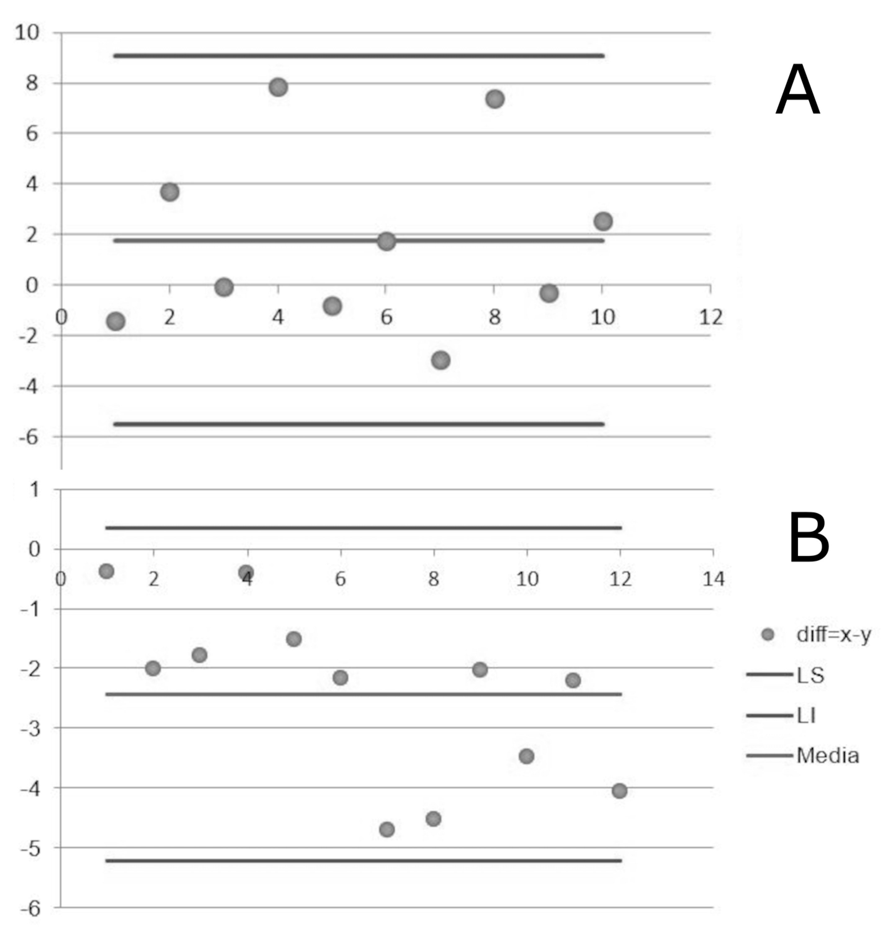
Figure 1. Bland-Altman stroke (A) and with healthy participant. UL (B) between EEMETA and EEACC.
Preprints 78111 g001
Table 1. Variables measured during 6MWT.
Control group Patient with stroke Mann-Whitney
Médian Q1 Q3 Médian Q1 Q3
HR (bpm) 140.0 98.1 143.7 116.0 90.1 126.5 p=0.08
V ˙ O 2 (mL.min-1.kg-1) 28.65 23.35 33.83 13.55 12.63 15.8 p=0.0001
Distance (m) 686.5 660.0 729.7 341.0 310.0 442.0 p=0.0001
Table 2. Comparison of EE measurements between methods (EEACC and EEMETA).
Median Q1 Q3 Mann-Whitney
EEMETA(W·kg-1) Control group 9,85 8,18 11,89 p<0,0001*
Patient with stroke 5,0 4,56 5,46
EEAcc(W·kg-1) Control group 8,57 7,86 11,24 p=0,11
Patient with stroke 8,2 7,05 9,56
Table 3. Correlation between energy expenditure measured using K4b² and the accelerometric method.
Median Q1 Q3 Correlation
coefficient
Control group EEMETA
(W·kg-1)
9.85 8.18 11.89 r=0.09 ; p=0.79
EEAcc(W·kg-1) 8.57 7.86 11.24
Patient with stroke EEMETA
(W·kg-1)
5.0 4.56 5.46 r=0.56 ; p=0.06
EEAcc
(W·kg-1)
8.2 7.05 9.56
Disclaimer/Publisher’s Note: The statements, opinions and data contained in all publications are solely those of the individual author(s) and contributor(s) and not of MDPI and/or the editor(s). MDPI and/or the editor(s) disclaim responsibility for any injury to people or property resulting from any ideas, methods, instructions or products referred to in the content.
Copyright: This open access article is published under a Creative Commons CC BY 4.0 license, which permit the free download, distribution, and reuse, provided that the author and preprint are cited in any reuse.
Prerpints.org logo

Preprints.org is a free preprint server supported by MDPI in Basel, Switzerland.

Subscribe

© 2024 MDPI (Basel, Switzerland) unless otherwise stated