Preprint
Article

Mental Stress Classification based on Selected EEG Channels Using Correlation Coefficient of Hjorth Parameters

Altmetrics

Downloads

182

Views

91

Comments

0

A peer-reviewed article of this preprint also exists.

Submitted:

10 July 2023

Posted:

12 July 2023

You are already at the latest version

Alerts
Abstract
Electroencephalography (EEG) signals provide valuable insights into various activities of the human brain, including the detection of mental stress, which is a complex physiological and psychological response. However, the challenge lies in identifying mental stress accurately while mitigating the limitations associated with a large number of EEG channels. This includes issues such as computational complexity, the risk of overfitting, and the increased setup time for electrode placement, which can be cumbersome for real-life applications. Therefore, it is crucial to develop EEG channel selection algorithms that enable the creation of a wearable device capable of assessing mental stress in real-life scenarios. This study introduces a novel channel selection method aimed at identifying highly accurate channels for detecting mental stress. Our approach, known as the Correlation Coefficient of Hjorth Parameters (CCHP), assesses the correlation between activity, mobility, and complexity in the time domain to nominate the most relevant channels. By selecting channels that exhibit high correlation with the stress task while being uncorrelated with each other, CCHP significantly reduces the number of EEG channels required, without compromising accuracy or performance. To evaluate the effectiveness of CCHP, we conducted experiments using the DEAP public dataset. Comparing our results with other recent algorithms that utilize the full set of EEG channels, CCHP achieved a superior classification accuracy of 81.56% using only eight EEG channels. Furthermore, CCHP outperformed existing channel selection methods by an impressive 8%. These findings strongly indicate that the CCHP algorithm shows great promise in the design of a wearable application for mental stress detection, utilizing a minimal number of EEG channels.
Keywords: 
Subject: Engineering  -   Bioengineering

1. Introduction

According to the latest neurosciences, the human brain is considered the main target organ of mental stress due to its responsibility to distinguish between situational circumstances (stressful/threatening or normal situations) [1]. To study the changes in brain activities during stress conditions, several non-invasive neuroimaging modalities have been used, such as functional magnetic resonance imaging (fMRI), positron emission tomography (PET), magnetoencephalography (MEG), electroencephalography (EEG), and functional near-infrared spectroscopy (fNIRS) [2,3]. EEG is a commonly preferred modality for assessing brain functionalities due to its non-invasive nature, high temporal resolution, ease of setup, commercial availability, and comparative low-cost [4]. Accordingly, researchers use EEG in various domains that involve neural engineering, neurosciences, and biomedical sciences (e.g., brain-computer interfaces, BCI) [5,6]. EEG signal plays a crucial role in several EEG-based research and application areas such as clinical applications for epilepsy [7], depression [8,9], effective monitoring of emotion [10], mental stress [11,12,13], and sinogram application [14]. Multiple EEG channels are often used for brain signal acquisition from multiple locations on the scalp to offer a high temporal and spatial resolution. However, reducing the number of channels in the signal processing setup is necessary since the setup procedure with a high number of channels is time-consuming and results in subject discomfort. Furthermore, it increases the system’s computational complexity, which is required to be low in specific applications [15,16]. Therefore, channel selection methods play a vital role in the reduction of complexity and high dimensionality of the feature vector space to improve overall performance. This increases the chances of building commercial wearable devices to provide better diagnosis and accurate treatment for mental stress [17,18]. The common approach of the channel selection method is based on neuroscience skills where data from each region of the brain is highly correlated to some specific tasks. For example, the prefrontal region of the brain is highly associated with cognitive processing such as emotions, thoughts, and actions [2]. Meanwhile, central lobes relate to motor imagery tasks in BCI systems [15]. Consequently, several EEG channel selection methods based on specific tasks have been proposed [19,20] such as Sequential floating forward selection (SFFS) in BCI [21], normalized mutual information selection (NMIS), and minimum redundancy maximum relevance (mRMR) [22] in emotion recognition, spatiotemporal-filtering-based feature selection [20] in BCI, and harmony search algorithm for alcoholism detection [23], etc. Their findings revealed that they can reduce channels and maintain the classification performance of the given task. Yet, current channel selection methods suffer from poor performance and/or lack a neurophysiological basis [24]. Apart from that, these methods may demonstrate limitations in terms of eliminating irrelevant channels or reducing redundant channels [22]. It is known that the execution of a single task by a participant will trigger functionality changes in different brain regions [15]. It could be said that employing the entire channels of EEG not only increases the complexity of the system but also introduces noise which may result in lowering the classification performance. Thus, finding an optimal channel selection method is needed to reduce computational complexity and minimize the occurrence of the over-fitting problem, which may be caused by the issue known as the "curse of dimensionality", in which the error increases as the number of features increases[22]. In addition to the above, the channel selection methods rely on feature extraction methods that extract temporal, spectral, or spatial EEG patterns of signal processing. The feature extraction approaches have been used effectively in improving the EEG classification performance. However, each EEG channel may contain more than one feature which results in the sharp increase of feature vector space in multi EEG channels. Therefore, current research approaches employed feature selection methods to find the optimal number of features without reducing the EEG channels. Applying feature selection only in multi-EEG channels can be useful in the laboratory because it provides high accuracy due to high spatial resolution. On the other hand, it is not practically effective in home-based applications or daily usage due to the long setup time for electrodes’ placements which increases computational complexity and affects the comfort level of the user wearing the device. As a result, several methods for obtaining the relevant channels to the source localized of intended tasks were proposed. The approach for selecting EEG channels could be seen as a feature selection problem. However, The major difference is that channel selection evaluates all features from one channel as a single entity [22]. In terms of EEG features utilized in channel selection methods, Wang [22] adopted EEG spectrogram representations of short-time Fourier transform (STFT) for each channel by treating the data as time-frequency images passed to SVM for emotion classification. Meanwhile, Yongkoo [15] and Jing [24] employed raw EEG signals of each channel with correlation coefficient methods in motor imagery (MI) tasks. In [22], a channel selection method was proposed to select a relevant subset of EEG channels using normalized mutual information (NMI). The method achieved 74.41% and 73.64% accuracy for emotion classification of valence and arousal respectively with only 8 channels selected. Another proposed method by [25] used the ReliefF algorithm to find the subset channels corresponding to mental fatigue classification using multi-domain features, he succeeded to reduce the number of channels from 16 channels to 8 optimal channels with acceptable accuracy. The EEG signal is non-stationary but has an event-dependent property for the given task. Therefore, it is important to analyze the changes in signal with time. Time-frequency feature extraction methods are preferred because they keep the information of both time and frequency. However, time-frequency features such as STFT have high computational complexity while redundant frequency information remains to be solved in real-time STFT applications [26]. The Hjorth parameter proposed in [26] is considered to be a superior alternative for the STFT due to its high ability to extract important information in both the temporal and frequency domains via a simple computing process. In 1970, Hjorth [27] introduced a set of three time-domain parameters to quantify the EEG signal. The Hjorth parameters referred to the normalized slope descriptors due to their ability to be explained by means of first and second derivatives. The first parameter is a mean power value that represents the signal’s activity. The second parameter, called mobility which is the approximation of the mean frequency. The third parameter is called complexity which estimates the signal’s bandwidth. Hjorth parameters are computed using variance, thus it has a low computing cost in comparison to other methods [28]. According to Hjorth, this approach establishes a link between a physical time-domain interpretation and the more traditional frequency-domain description. Additionally, the time-domain context of the Hjorth representation could be advantageous for scenarios requiring continuous EEG analysis for real-life applications. Several studies have successfully employed Hjorth parameters to extract information from various bio-signals, including detection of the heart rate from the electrocardiogram (ECG) signal, classification of lung sound, classification of Electromyogram (EMG) signal, diagnosis of hyperactivity (ADHD), epilepsy, and emotion [29,30]. Besides, Safi et.al [30] reported that EEG Hjorth features improved the detection rate of Alzheimer’s disease. Recently, different extensions of correlation-based channel selection of common spatial pattern (CSP) methods were proposed. The correlation-based channel selection regularized CSP (CCS-RCSP) methods were proposed to find the optimal channels related to motor imagery (MI) tasks using correlation coefficient [24]. The CCS-RCSP is trained to select the channels that are highly correlated to the MI task. Another extension called filter-bank CSP (FBCSP) was proposed by Park [15] for MI task classification. Additionally, cross-correlation-based discriminant criterion (XCDC) was proposed by [31] to find the optimal subset channels that are capable of discriminating MI tasks. Another extension of CSP was proposed by [32] to select internal features and channels based on the difference and the ratio of average L1-Norm for CSP (DRL1 CSP). However, the results of these approaches still provide many channels for the classification task and are specific to the MI task. Additionally, most of the current channel selection studies tend to find significant channels of each individual using dependent and/or independent tests. However, the brain activities of stress tasks vary among individuals which makes it hard to find the common significant channels among subjects. Therefore, finding common significant channels could help in the development of real-life applications for stress recognition. To address the above-mentioned points, our key contributions in this work are as follows:
  • Proposing an alternative approach based on the correlation coefficient of Hjorth parameters aimed to select general optimal channels among subjects while preserving the classification accuracy.
  • Proposing a new methodology to extract important features from the general optimal channels.
  • Validating and comparing the effectiveness of the proposed method with the state-of-the-art channel selection methods.
The rest of this paper is structured as follows. In Section 2, the methods and material including details of the dataset and data annotations are described. Section 3 describes the main proposed method for channel selection. Section 4 shows the feature set extracted from general optimal channels. Section 5 provides the ML algorithm, the parameters used, and the evaluation matrix. The results of the proposed method and comparison with existing methods are discussed in Section 6. The detailed discussion of this work follows in Section 7 and the conclusion is given in Section 8.

2. Methods and Material

2.1. EEG DATASET

The Dataset for Emotion Analysis using Physiological Signals (DEAP) is a public EEG dataset for emotion recognition [33]. The DEAP comprises data collected at 512 Hz sampling frequency from 40 physiological channels (32 EEG channels and 8 other physiological channels). In total, 32 healthy subjects participated (50% are males and 50% are females). The EEG data were collected while participants were watching a selective music video (40 videos/trials, every trial is one minute long) of the emotion wheel. All participants performed self-assessment manikin (SAM) [34] of their arousal levels, like/dislike, valence, and dominance on a scale from 1 to 9. Overall, there are 40 trials (each trial is 63 s long including 3 s pretrial) for each subject.
The DEAP Authors have offered preprocessed EEG data. The original EEG data was down-sampled to 128 Hz. The band-pass filtered from 4.0–45.0 Hz was applied to omit the noise caused by 50/60 Hz of line-power and the low frequencies < 4 Hz artifacts caused by blinking eyes. Also, the artifacts caused by EOG were removed. In this paper, the preprocessed EEG data provided by DEAP were used for the mental stress classification task.

2.2. EEG Data Annotation

The data from 32 EEG channels were annotated based on the online self-assessment rating SAM scale provided by DEAP for valence and arousal. Therefore, for this paper, the online self-assessment rating was considered to identify calm and stress tasks for each participant by Equations (1), which was derived from [35,36].
s t r e s s = ( v a l e n c e < 3 ) ( a r o u s a l > 5 ) , c a l m = ( 4 < v a l e n c e < 6 ) ( a r o u s a l < 4 )
Valence refers to the pleasantness of the stimulus on a scale of negative to positive, while arousal refers to the intensity level of emotion induced by the stimuli and scaling between calm (or low) to excited (or high). A calm state is considered when arousal is low and valence is high. Meanwhile, the stress state is obtained from the low valence and high arousal. When the criteria obtained from Equations (1) and (2) were applied to each subject data, seven subjects (with participant IDs: 3, 6, 7, 9, 17, 23, 30) were removed since their data did not contain both stress and calm states. Thus, the rest of the analysis continued with the remaining data of 25 participants.

3. Hjorth multi-Correlation coefficient

Figure 1 represents the general flow of channel selection-based correlation to find an optimal EEG channel. In general, the proposed method selects channels that are highly correlated to class tasks and uncorrelated with one another across trials. The reduction of channels is based on the hypothesis that some EEG channels related to EEG Mental Stress Task (MST) contain a similar feature across all experiment trials for the subject performing the same tasks while the rest of the channels contribute less because they are unrelated to MST. Therefore, the following steps were taken to precisely establish a correlation-based channel selection method. First, three features of Time Domain Hjorth Parameters (TDHPs) namely activity, mobility, and complexity were extracted from each EEG channel since they have an advantage of the quantitative evaluation of EEG signal in the time domain. Table 1 shows the details of TDHPs and the equations. The correlation of these channels was conducted based on the statistical measurement of the TDHPs feature set. Second, after extracting the TDHPs feature vectors for each channel, we used feature-wise Z-score normalization by subtracting each sample value from its feature-wise mean and dividing the result by the associated standard deviation. Lastly, we computed the correlation coefficient (CC) method based on the feature extracted for channel-channel based correlations and channel-class based correlations. The below sections describe the details of the CC method.

3.1. Correlation coefficient Measures

Pearson’s correlation uses similarity measurement to find the strength of a linear association between any pair of channels or features in a one-dimensional space. For a given N channel, we can have N(N-1)/2 possible pairs for calculating correlations. The pairs of values are considered highly correlated if the correlation coefficient is near or equal to ± 1 and uncorrelated if the correlation coefficient is 0 or less than the threshold value (i.e., 0.5). The best way to find an optimal projection of the selected channel is to maximize the separation between the two classes. For example, assume that there are two classes of observations (s, c ∈ (stress, calm)). In a one-dimensional feature space, the separation between two classes is defined using the correlation coefficient: Let TDHP ∈ A, C, M identify the features of the activity, complexity, and mobility and corresponding to x∈(s,c) for classes (stress and calm). The channel-channel based correlation is computed as the equation below:
P x ( S , K ) = 1 l x i = 1 l x c o v ( A i S , A i K ) + c o v ( M i S , M i K ) + c o v ( C i S , C i K ) A i S ˜ A i K ˜ + C i S ˜ C i K ˜ + M i S ˜ M i K ˜
where x s , c represents the classes of stress and calm, l x represents the total number of trials of the given class, (S,K) represent the pair channel index, A i s ˜ A i c ˜ , M i s ˜ M i c ˜ , C i s ˜ C i c ˜ are the standard deviations of TDHPs (activity, complexity, and mobility), and c o v ( A i S , A i K ) is covariance of T i S , T i K where T=A,M or C,and can be calculated using :
c o v ( T i s , T i k ) = 1 N 1 i = 1 N ( T i s T i s ¯ ) ( T i k T i k ¯ )
T i s ¯ , T i k ¯ represent the mean of the sample variables T i s and T i s respectively. Then we computed the main of the two pair channels as follows:
C h S , K ¯ = P s ( S , K ) + P c ( S , K ) 2
where P s ( S , K ) and P c ( S , K ) are the average correlation of the pair of channels of two classes (s and c) and C h S , K ¯ is the main of two channels. After getting the correlation of channel-channel based, we computed class-channel based correlation using the equation below of (channel-class correlation):
p ( s , c ) = i = 1 N c o v ( T i s , T i c ) T i s ˜ + T i c ˜
where T T D H P s represents TDHPs’ Activity, i for an index of the channel, s, c for classes(stress and calm), and c o v ( A j s , A j c ) is the covariance of T j s , T j c . The average of two class correlations of single-channel was calculated as:
C h ¯ q = T T D H P s p ( s , c ) , T D H P s = A , M , C .
The F score was used to estimate the discrimination power of the group of TDHPs features as the correlation feature selection depends on a single feature [37]. The use of the evaluation function is to precisely find the channel subsets that are highly correlated with the class and uncorrelated with each other. Irrelevant channels with low-class correlation will be omitted. The activation function can be expressed as follows:
E j = K C h q ¯ K + K ( K + 1 ) K C h ¯ ( s , k )
where E j is the significant channels evaluated per independent subject, k is the number of channels, C h q ¯ is the mean channel-class correlation with ( C h S ) and the C h c h ¯ is the average channel-channel based of inter-correlation.
For general optimal channels among subjects, we counted the frequency of occurrence of each significant channel E j of subjects as the following equation:
U ( j , p t ) = U ( j , p t + 1 ) , i f U j = E ( j , k ) U ( j , p t ) , i f U j E ( j , k )
where U ( j , p t ) is the overall unique significant channels among all subjects and p t represents the total unique occurrences of each channel. Then, we ranked them from high to low occurrences and applied a threshold to select the most common channels among subjects that appeared in the significant channel sets.
G j , o p t i m a l = { U j U ( j , p t ) | p t f t h r }
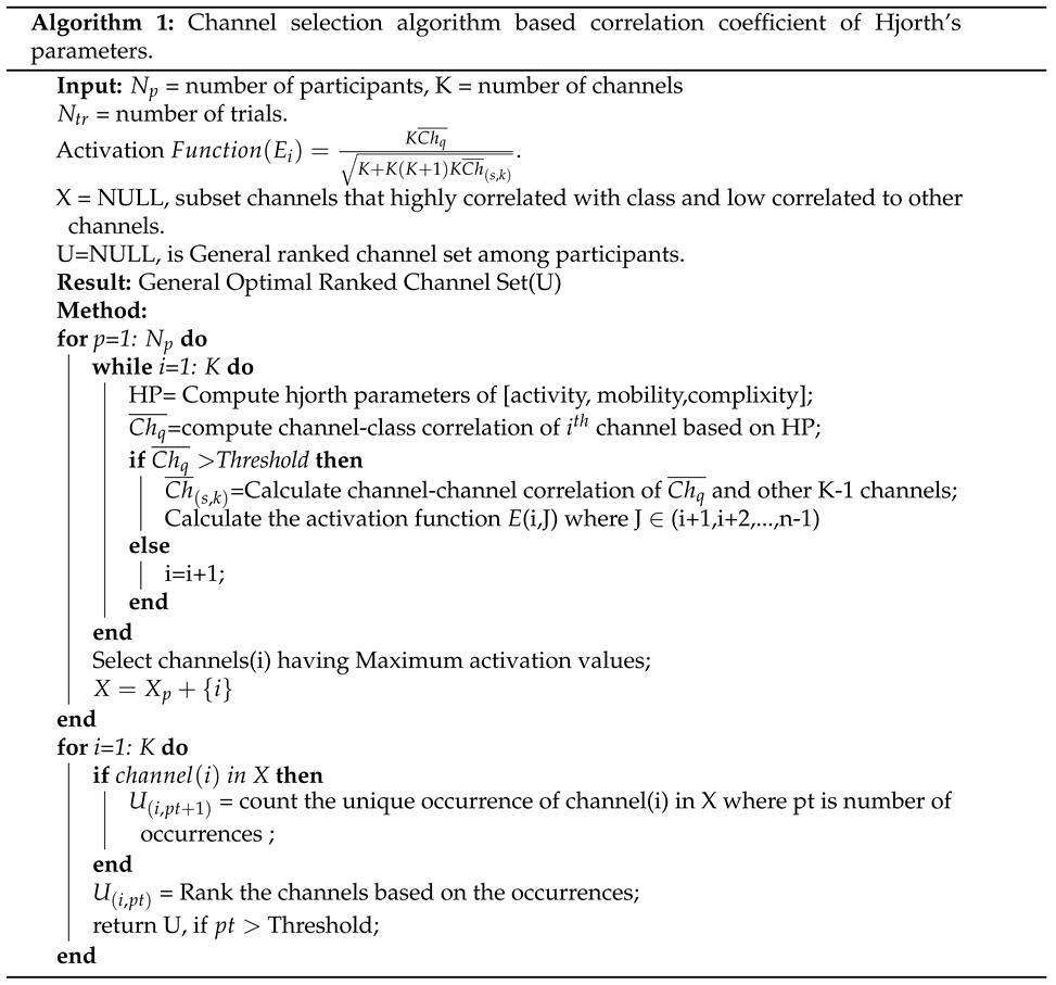
G j , o p t i m a l is the general unique significant channels that exist as significant channels on most independent subjects based on the threshold p t represents the total number of occurrences of each channel, f t h r is the threshold, and U ( j , p t ) represents the matrix of each unique channel with its repeated number of occurrences. The Channel Selection Based Correlation Coefficient of Hjorth Parameters is given by Algorithm 1. These general optimal channels are used for the rest of this paper.
Preprints 79095 i001

4. Feature Extraction

The preprocessed DEAP EEG signals of each participant consist of 40 trials where each trial has 7680 samples (60 seconds long). In the study of Shon [36], each trial was divided into 16 parts, resulting in 480 samples (4 seconds long) per part. This results in a total of 640 segments per subject (40 trials*16 segments) which was used in this study. Then, we computed the EEG feature extraction of time, frequency, and time-frequency domains from the segmented trial having a time window size of 4s of the selected general optimal channels proposed by our model. The selected time window size is supported by previous studies which found that the window size between 3 and 12s to be effective when classifying individuals’ mental status using EEG signals [44,45]. Besides, the number of data points within the 4s is appropriate to show the stationary of EEG signals and thus confirms the reliability for achieving channel selection [46,47]. Table 1 shows the features’ descriptions, mathematical equations, and the count of each feature per channel used in this study. Previous studies have utilized several time-domain features for EEG mental stress and emotion classifications [3,36,45]. In this study, we proposed to extract multi-domain features, from time domain namely; line length, peak-to-peak amplitude, kurtosis, skewness, and Hjorth parameters (activity, mobility, and complexity) of the signal. Meanwhile, five features from the frequency domain were extracted based on Relative Powers [41] of theta θ (4-8Hz), low alpha α (8-12Hz), high alpha α (12-15Hz), low beta β (15-20Hz), and high beta β (20-30Hz). Additionally, eight features from the time-frequency domain were extracted with six features from the energy of wavelet decomposition coefficient (db4, 6 levels) [11,42], and the spectral entropy of PSD-Welch [43] and Katz Fraction Dimension [38]. A total of 20 features that are mathematically defined in Table 1 were used as a feature set for the optimally selected channels.

5. Classification

Both general optimal selected channels of CCHP and all EEG channels were tested to distinguish between mental stress and calm emotional state using two classifiers. In the studies of Hasan and Kim [35,36], KNN was employed to classify mental stress and calm state for the DEAP dataset and was shown to provide high performance. However, according to the report of Alex [48], the most common classifier technique applied in EEG signals is SVM. In addition, we used seven classifiers for EEG analysis of mental stress in our recent work [49], and found that SVM outperformed other classifiers. Therefore, in this study, we employed both SVM and KNN to evaluate the proposed method and determine their performance with a minimum number of channels. In this study, SVM and KNN were implemented in Python to classify data into two classes (stress and calm). Table 2 shows the parameter values assigned to each classifier. In each classifier, an independent subject test with 10-fold cross-validations was performed.
A total of 20 features from multi-domain were extracted from each EEG channel as shown in Table 1 to form a large feature vector. Then, the features of selected EEG channels in each subject were randomly split into 10 equal subsets based on the 10-fold. Each time, one of the unique subsets was treated as the test set and the other 9 subsets were put together to form a training set. We applied the following metrics for evaluating the performance of the classifiers: precision, recall, and accuracy. Precision is the ratio of correctly predicted positive cases to the total predicted positive cases. A recall is defined as the ratio of correctly predicted positive cases to all observations in the actual class while accuracy is defined as the percentage of correct predictions for the test data. The mathematical formulas of precision, recall, and accuracy are presented in Equations 10-12:
P r e c i s i o n = T p T p + F p
R e c a l l = T p T p + F n
A c c u r a c y = T p + T n T p + T n + F p + F n
where Tp represents the total samples of true positive, Fn represents the false negative, Tn represents the true negative, and Fp is for the false positive.

6. Result Analysis and Classification

6.1. Analysis of Channel Selection
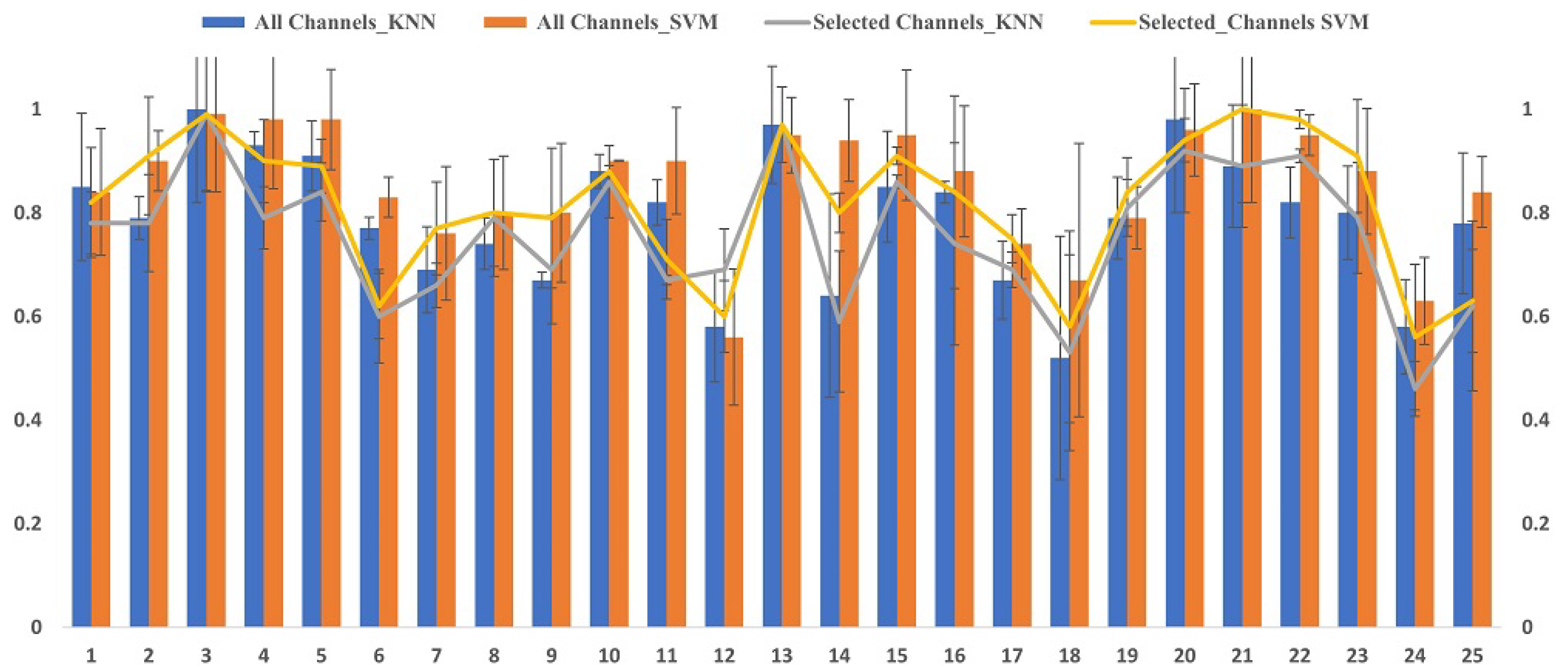
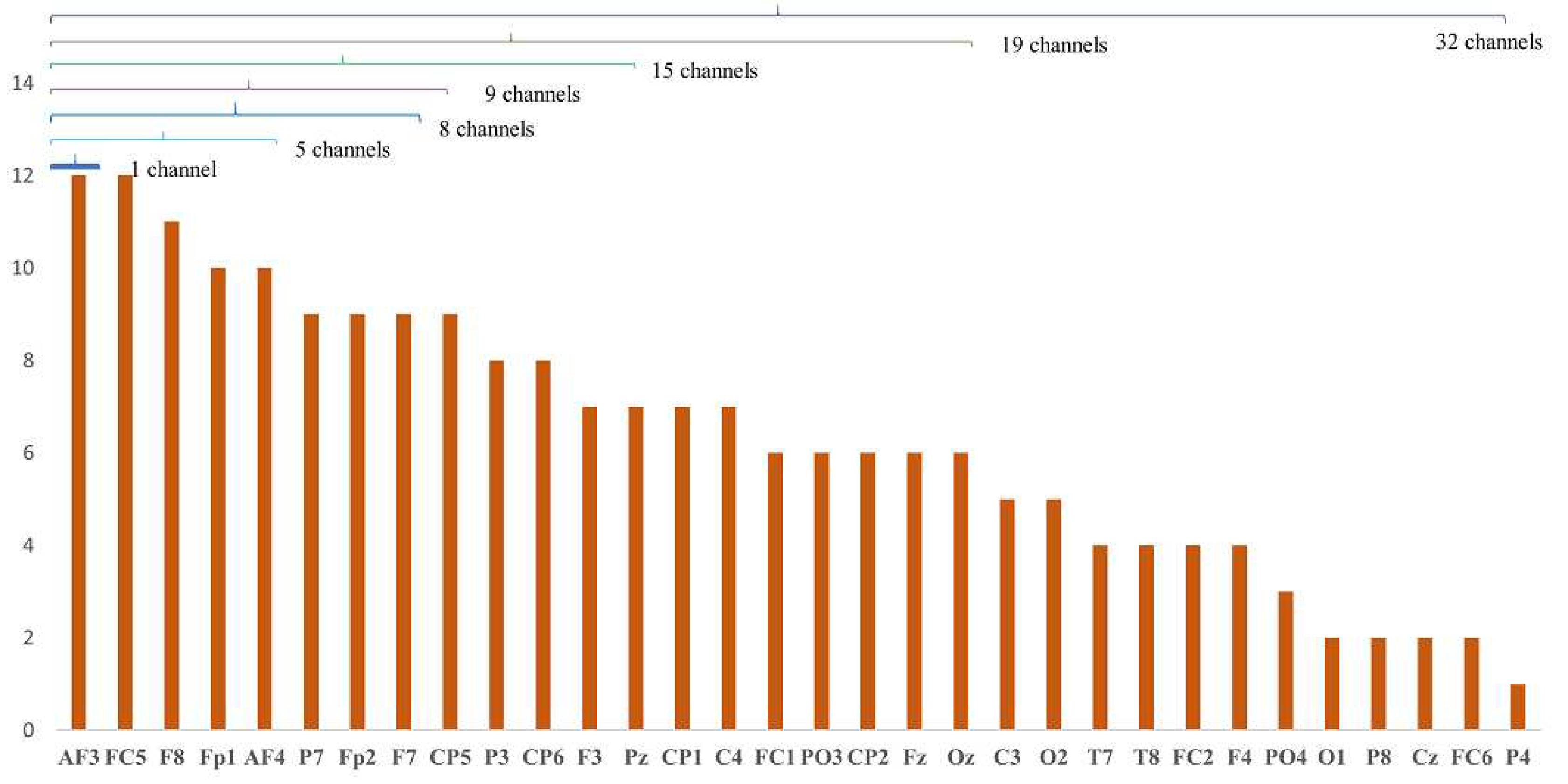
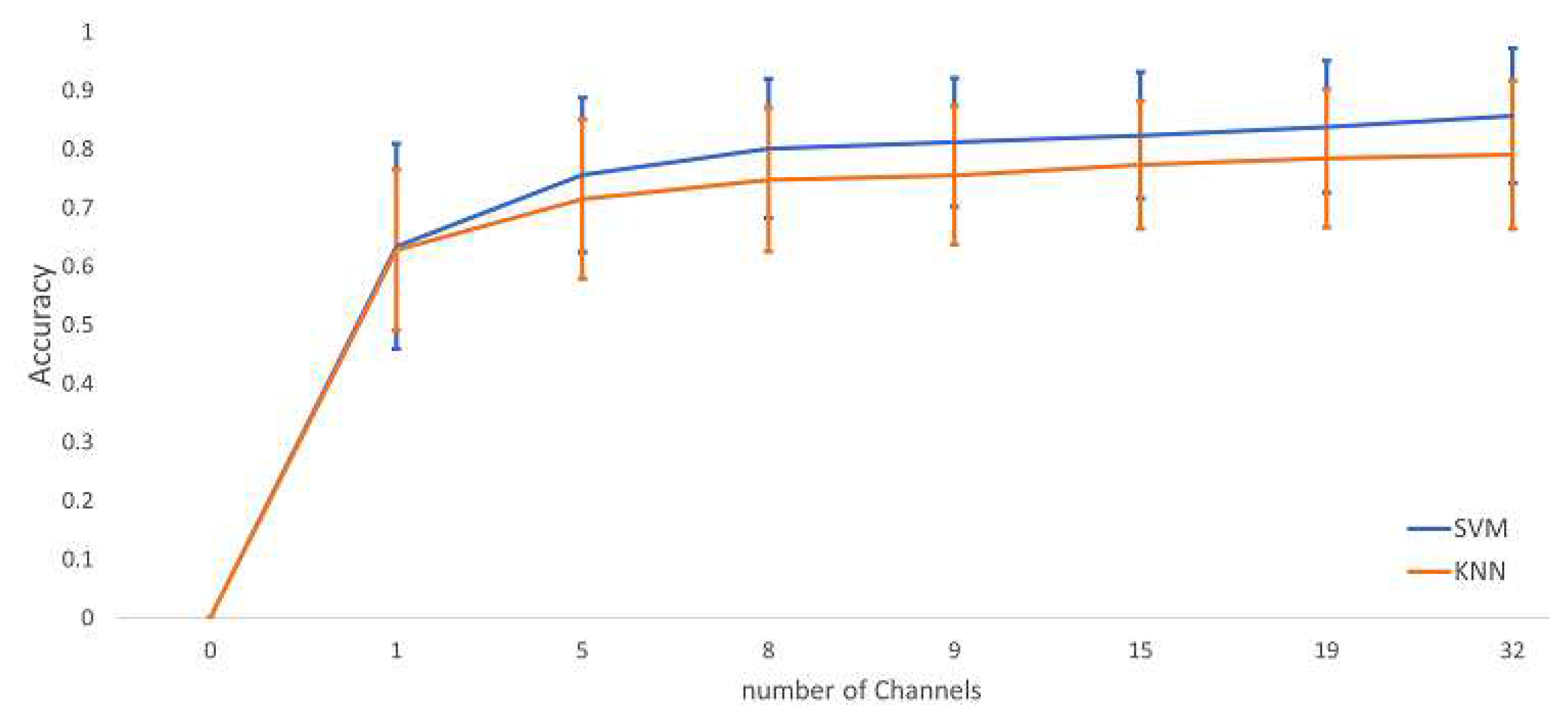
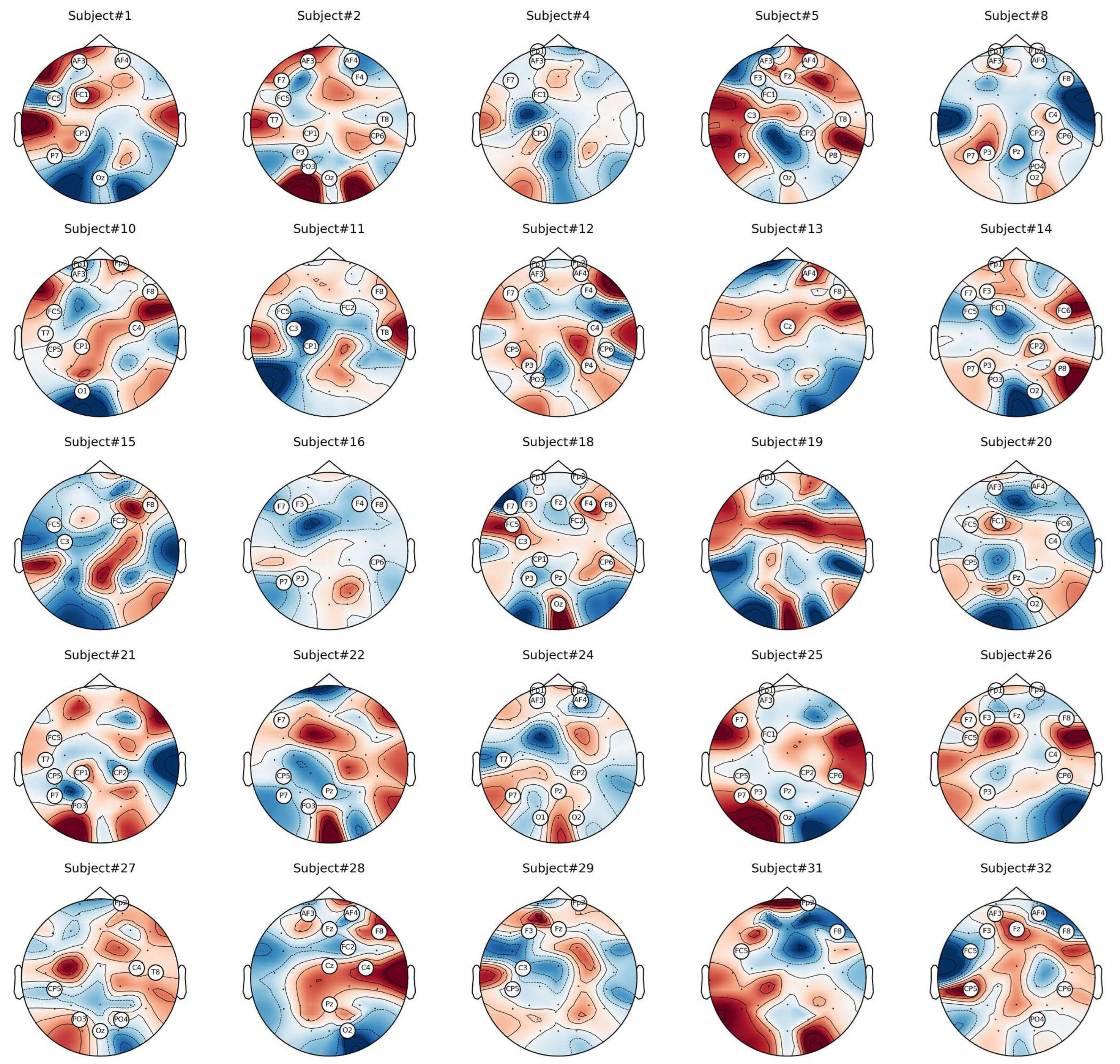
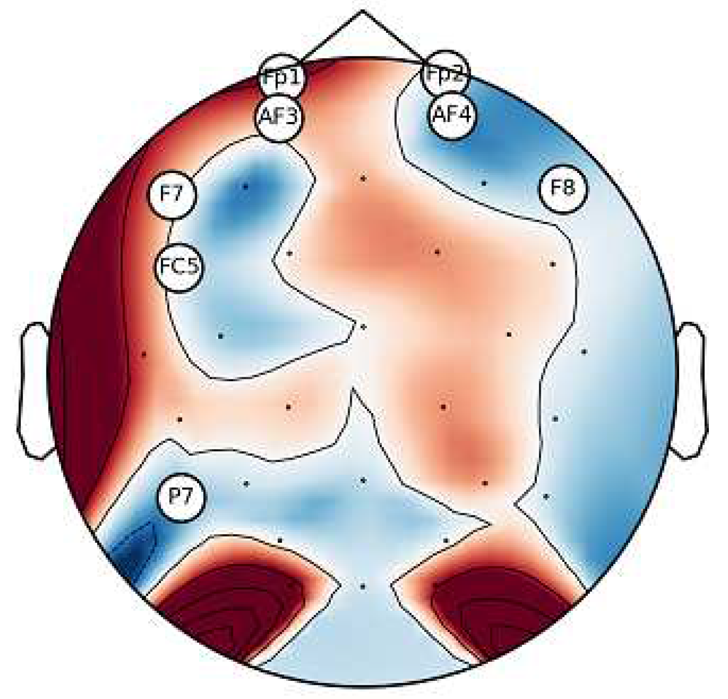
The proposed method of CCHP is based on the filter approach which considers an information-based or statistical criterion to give feedback to the searching algorithms without the classifier’s involvement. The EEG channels were reduced sharply based on the activation function. In particular, for each subject, the proposed CCHP select EEG channels that are highly correlated to mental state class, and less correlated to other channels within the same class. Selecting relevant channels that are highly correlated to class (stress/calm) increased performance accuracy. Similarly, the redundant channels were removed by obtaining the low channel-channel-based correlation from the same class. As a result, each subject presented some important channels that best discriminate mental stress tasks as shown in Figure 4. To find a common channel among all subjects, we have ranked the significant channels based on the occurrence of mode frequency of significant channels as shown in Figure 2, where the high occurrence channels ranked first and so on. To find which set of channels gives an acceptable accuracy, we have compared different sets of ranked channels, 1, 5, 8, 9, 15, 19, and 32 EEG channels, and we found that only 8 EEG channels can significantly classify the mental stress state without affecting much on the performance as shown in Figure 3. Finally, Figure 6 illustrates the selected eight general optimal channels among all subjects that were ranked based on the occurrence of the best subject-independent channels among all subjects.
Figure 4 shows the mean accuracies of identifying mental stress and calm using a different number of ranked optimal channels. The result of different numbers of ranked EEG channels showed that when using full EEG channels (32 channels) to classify mental stress tasks, it yielded an accuracy of 85% compared to 8 channels with an accuracy of 80%. However, with only one channel of the highest-ranked general channels, namely CH3, the accuracy reached (63.4±17.5)% while the ranks of 8, 9, 15, and 19 number of channels obtained the mean accuracy range of 80–84%. The graph shows a slight increase in accuracy when using full EEG channels due to the high spatial resolution that covers different brain regions. However, with only 8 channels, we can obtain an acceptable accuracy that can help in recognizing the stress state with an optimal number of channels. Based on the ranked optimal channel results shown in Figure 4, we have identified 8 channels of [’AF3’, ’FC5’, ’F8’, ’Fp1’, ’AF4’, ’P7’, ’Fp2’, ’F7’] as a general optimal channel to classify mental stress. We further conducted a statistical analysis using a two-sample t-test to confirm the optimum of the selected channels. We found no significant improvements (p>0.05) in the classification accuracy after the selected 8 channels. Figure 6 shows the location of the general optimal EEG channels on the scalp that were ranked based on the occurrence of best subject-independent channels and used for the rest of the paper to recognize the stress/calm mental states of each subject.
Figure 2. Accuracies and standard deviation for the 10-fold cross-validation per independent subject for 32 channels Vs 8 channels. The bars represent the full EEG channels, while the lines represent the selected significant EEG channels.
Preprints 79095 g002
Figure 3. A rank of the common important EEG channels among all subjects.
Preprints 79095 g003
Figure 4. A comparison of mean accuracies based on the number of the most common EEG channels among subjects.
Preprints 79095 g004
Figure 5. A topographic map of the significant eeg channels of mental stress per individual.
Preprints 79095 g005
Figure 6. General optimal channels among all subjects that best discriminate mental stress. A circle around the name of each channel represent the significant channels, while the dot symbols are not significant.
Preprints 79095 g006

6.2. Classification Results

We evaluated the proposed channel selection method of CCHP using KNN and SVM. The mean classification accuracy, recall, and precision of each participant were tested in two methods of all channels and with proposed general optimal selected channels as summarized in Table 3. The classification performance was tested on the two classes (binary classification) of stress and calm states on the EEG data of the DEAP dataset. The classification accuracy of our model using full channels achieved 85.68% and 79.04% of both SVM and KNN respectively which outperform the other stress detection models on the same DEAP dataset. Comparatively, the proposed model with 8 channels achieved an average classification accuracy of 81.56% and 75.68% using SVM and KNN respectively. This verifies that an increased number of EEG channels leads to a slight increase in accuracy performance.

6.3. Performance Comparison of Mental Stress with Existing Methods In DEAP Dataset

To validate the proposed method, we applied the same process methodology to the existing methods. Then, we compared the proposed method with the existing methods listed in [22,36] of min-Redundancy-Max- Relevance (mRMR), Short Time Fourier Transform with Mutual Information (STFT+MI), and Genetic algorithm (GA). Table 4 summarizes the comparison results of these methods taking into account the results of three important parameters: number of selected channels, classification accuracy, and execution time. We further conducted a statistical analysis using the Friedman test between the methods in Table 4 and we found there is no significance in terms of classification accuracy with Fr=4.1667 and p-value = 0.244. This indicates that the proposed CS confirms the reliability of the proposed method. However, in terms of the number of channels selected, CCHPs obtain fewer optimal channels that are most related to mental stress tasks. The results show that the proposed method yielded the best result in selecting an optimal number of channels within the shortest time compared to the rest of the methods, with 8 channels and 340 ms execution time. Additionally, for classification performance, the proposed method achieved higher than mRMR and was slightly lower than STFT-MI and GA. This was due to the higher number of channels in GA and STFT-MI. A comparison of different channel selection methods revealed that a minimum number of EEG channels not only reduced the complexity of feature dimensional space but also preserved the accuracy and reduced the time needed to set up the channels on the scalp. Furthermore, the proposed model results were compared with other related works using EEG signals of the DEAP dataset to recognize mental stress as shown in Table 5. One can notice that the proposed GOC and the design paradigm outperformed related works in terms of stress/calm classification and a minimum number of channels used, with 81.65% accuracy obtained by 8 channels compared to 73.38% highest accuracy achieved by the study of Hassan [35].

7. Discussion

This study aims to find the most sensitive areas of the brain for detecting mental stress states using EEG. This could help in the development of a highly accurate wearable device for detecting mental stress in real-time. For this purpose, a proposed channel selection method using the correlation coefficient of Hjorth parameters was designed to find the most common EEG channels among subjects that can detect mental stress and calm states. Figure 5 shows the topographic maps of significant channels per individual obtained by the proposed method. Since EEG data is non-stationary, we noticed that the brain activation of each individual to mental stress’ stimuli was different. This finding broadly supports other studies in terms of different individual responses to mental stress tasks [50]. Therefore, the results of the proposed method selected EEG channels of [’ AF3’, ’FC5’, ’F8’, ’Fp1’, ’AF4’, ’P7’, ’Fp2’, ’F7’] that were found in most of the subjects as general optimal channels (GOC) to discriminate mental stress. These common EEG channels mostly cover the frontal lobe of the scalp in both hemispheres, and their distribution is shown in Figure 6. This is inline with previous stress EEG and fNIRS studies that highlight the frontal area of the brain as the most sensitive region to stress exposure [51,52,53].
In this paper, we compared the results of proposed GOC with full EEG channels as shown in Table 3 and found a slight increase in accuracy when using full channels compared to GOC with an average accuracy of 85% and 80% respectively. These results are in line with those of previous studies, which found that full EEG channels could increase the accuracy compared to optimally selected channels due to the high spatial resolution provided when using full channels. On the other hand, using full channels may not be applicable for home-based applications due to computational complexity, long setup time for EEG electrodes’ placements, and high cost.
The work presented here provides one of the first investigations into ranking the common important channels for mental stress recognition as shown in Figure 3, where the channels were ranked based on GOC weight among all subjects.
To validate the proposed method, we compared it with existing methods. The proposed GOC in this research provided a promising result that considered computational complexity using execution time, the number of selected channels, and classification performance as shown in Table 4. The results indicate that the proposed method can obtain the common important channels for real-time EEG stress detection and ensure relatively high accuracy with only 8 channels.
Furthermore, the results of the proposed method were based on EEG data from the DEAP dataset and compared with other related works that used the same dataset for stress recognition as shown in Table 5. The two studies proposed by Shon [36] and Hasan [35] utilized EEG data from the DEAP dataset to distinguish stress and calm states by employing the 32 EEG channels with feature selection methods. The accuracy results obtained were 71.76% and 73.38%, respectively. Thus, the results of our proposed CCHP model outperformed the other models for mental stress classification.
Although our proposed method was informative for selecting the commonly related channels for mental stress classification, it had a few limitations. First, we applied our method with the time-domain data, future studies should consider features from other domains. Second, the dimensionality of the feature vector is still high for real-time application, combining our method with other feature selection methods such as Particle swarm optimization (PSO) [54], BAT algorithm [55], Genetic Algorithm(GA) [36], whale optimization algorithm (WOA)[56], and other heuristic optimization methods can reduce the occurrence of the “curse of dimensionality” and increase the classification performance. Third, even though we have achieved high accuracy using the selected channels and SVM with default parameters, however, optimization of SVM parameters is not covered and is worth exploring in future studies for better results, also other approaches such as deep learning with the selected channels should be considered in the future [57]. Finally, we investigated our features within the cortical activation domain, exploring other types of features such as functional connectivity network patterns with graph theory analysis or fusion of them should be considered to improve the performance of stress detection as suggested in [47,58,59].

8. Conclusion

In this study, we attempted to recognize mental stress and calm states using an optimal number of EEG channels. We proposed a CCHP method to find the common optimal channels among subjects that could help in the development of real-life applications for stress assessment. The study found that the frontal area of the brain is the most sensitive to mental stress. The results of CCHP ranked the most common channels among subjects, where 8 channels were selected as general optimal channels to distinguish mental stress among subjects. Then, to extract useful information from the 8 channels, we utilized features from the time domain, frequency domain, and time-frequency domain. Additionally, we employed machine learning algorithms, SVM, and KNN to train and evaluate the proposed model. The proposed method was then compared with the existing methods and related works and showed its superior performance. The results showed that the proposed method could distinguish mental stress with a minimum number of channels (8 channels) and achieved high accuracy of 81.56% using SVM. In summary, we present a novel model to rank the most common EEG channels that could detect mental stress. This could be useful in developing a portable device for detecting mental stress in real-time applications. This helps clinicians to track subtle changes in brain activities in real time and provides historical data for better treatment.

Acknowledgments

The authors would like to thank the support received in part by Taylor’s University and in part of the support by the Journal of Brainsciences.

References

  1. Dedovic, K.; Renwick, R.; Mahani, N.K.; Engert, V.; Lupien, S.J.; Pruessner, J.C. The Montreal Imaging Stress Task: Using functional imaging to investigate the effects of perceiving and processing psychosocial stress in the human brain. Journal of Psychiatry and Neuroscience 2005, 30, 319. [Google Scholar] [PubMed]
  2. Al-Shargie, F.; Tang, T.B.; Kiguchi, M. Stress Assessment Based on Decision Fusion of EEG and fNIRS Signals. IEEE Access 2017, 5, 19889–19896. [Google Scholar] [CrossRef]
  3. Halim, Z.; Rehan, M. On identification of driving-induced stress using electroencephalogram signals: A framework based on wearable safety-critical scheme and machine learning. Information Fusion 2020, 53, 66–79. [Google Scholar] [CrossRef]
  4. Aspiotis, V.; Miltiadous, A.; Kalafatakis, K.; Tzimourta, K.D.; Giannakeas, N.; Tsipouras, M.G.; Peschos, D.; Glavas, E.; Tzallas, A.T. Assessing Electroencephalography as a Stress Indicator: A VR High-Altitude Scenario Monitored through EEG and ECG. Sensors 2022, 22. [Google Scholar] [CrossRef]
  5. Peksa, J.; Mamchur, D. State-of-the-Art on Brain-Computer Interface Technology. Sensors 2023, 23. [Google Scholar] [CrossRef]
  6. Sharif, M.S.; Raj Theeng Tamang, M.; Fu, C.H.Y.; Baker, A.; Alzahrani, A.I.; Alalwan, N. An Innovative Random-Forest-Based Model to Assess the Health Impacts of Regular Commuting Using Non-Invasive Wearable Sensors. Sensors 2023, 23. [Google Scholar] [CrossRef]
  7. Xin, Q.; Hu, S.; Liu, S.; Zhao, L.; Zhang, Y.D. An Attention-Based Wavelet Convolution Neural Network for Epilepsy EEG Classification. IEEE Transactions on Neural Systems and Rehabilitation Engineering 2022, 30, 957–966. [Google Scholar] [CrossRef] [PubMed]
  8. Hashempour, S.; Boostani, R.; Mohammadi, M.; Sanei, S. Continuous Scoring of Depression From EEG Signals via a Hybrid of Convolutional Neural Networks. IEEE Transactions on Neural Systems and Rehabilitation Engineering 2022, 30, 176–183. [Google Scholar] [CrossRef]
  9. Wu, W.; Zhang, Y.; Jiang, J.; Lucas, M.V.; Fonzo, G.A.; Rolle, C.E.; Cooper, C.; Chin-Fatt, C.; Krepel, N.; Cornelssen, C.A.; et al. . An electroencephalographic signature predicts antidepressant response in major depression. Nature Biotechnology 2020, 38, 439–447. [Google Scholar] [CrossRef]
  10. Song, Y.; Yin, Y.; Xu, P. A Customized ECA-CRNN Model for Emotion Recognition Based on EEG Signals. Electronics 2023, 12. [Google Scholar] [CrossRef]
  11. Al-Shargie, F.; Tang, T.B.; Badruddin, N.; Kiguchi, M. Towards multilevel mental stress assessment using SVM with ECOC: an EEG approach. Medical & biological engineering & computing 2018, 56, 125–136. [Google Scholar]
  12. Hag, A.; Handayani, D.; Pillai, T.; Mantoro, T.; Kit, M.H.; Al-Shargie, F. A wearable single EEG channel analysis for mental stress state detection. 2021 IEEE 7th International Conference on Computing, Engineering and Design (ICCED), 2021, pp. 1–6. [CrossRef]
  13. Hag, A.; Handayani, D.; Altalhi, M.; Pillai, T.; Mantoro, T.; Kit, M.H.; Al-Shargie, F. Enhancing EEG-Based Mental Stress State Recognition Using an Improved Hybrid Feature Selection Algorithm. Sensors 2021, 21. [Google Scholar] [CrossRef]
  14. Yu, Y.; Liu, Y.; Yin, E.; Jiang, J.; Zhou, Z.; Hu, D. An Asynchronous Hybrid Spelling Approach Based on EEG–EOG Signals for Chinese Character Input. IEEE Transactions on Neural Systems and Rehabilitation Engineering 2019, 27, 1292–1302. [Google Scholar] [CrossRef]
  15. Park, Y.; Chung, W. Optimal Channel Selection Using Correlation Coefficient for CSP Based EEG Classification. IEEE Access 2020, 8, 111514–111521. [Google Scholar] [CrossRef]
  16. Narayanan, A.M.; Patrinos, P.; Bertrand, A. Optimal Versus Approximate Channel Selection Methods for EEG Decoding With Application to Topology-Constrained Neuro-Sensor Networks. IEEE Transactions on Neural Systems and Rehabilitation Engineering 2021, 29, 92–102. [Google Scholar] [CrossRef] [PubMed]
  17. Gaur, P.; McCreadie, K.; Pachori, R.B.; Wang, H.; Prasad, G. An automatic subject specific channel selection method for enhancing motor imagery classification in EEG-BCI using correlation. Biomedical Signal Processing and Control 2021, 68, 102574. [Google Scholar] [CrossRef]
  18. Shen, J.; Zhang, X.; Huang, X.; Wu, M.; Gao, J.; Lu, D.; Ding, Z.; Hu, B. An Optimal Channel Selection for EEG-Based Depression Detection via Kernel-Target Alignment. IEEE Journal of Biomedical and Health Informatics 2021, 25, 2545–2556. [Google Scholar] [CrossRef]
  19. Yin, X.; Meng, M.; She, Q.; Gao, Y.; Luo, Z. Optimal channel-based sparse time-frequency blocks common spatial pattern feature extraction method for motor imagery classification. Mathematical Biosciences and Engineering 2021, 18, 4247–4263. [Google Scholar] [CrossRef] [PubMed]
  20. Qi, F.; Wu, W.; Yu, Z.L.; Gu, Z.; Wen, Z.; Yu, T.; Li, Y. Spatiotemporal-Filtering-Based Channel Selection for Single-Trial EEG Classification. IEEE Transactions on Cybernetics 2021, 51, 558–567. [Google Scholar] [CrossRef]
  21. Qiu, Z.; Jin, J.; Lam, H.K.; Zhang, Y.; Wang, X.; Cichocki, A. Improved SFFS method for channel selection in motor imagery based BCI. Neurocomputing 2016, 207, 519–527. [Google Scholar] [CrossRef]
  22. Wang, Z.M.Z.M.; Hu, S.Y.S.Y.; Song, H. Channel Selection Method for EEG Emotion Recognition Using Normalized Mutual Information. IEEE Access 2019, 7, 143303–143311. [Google Scholar] [CrossRef]
  23. Bavkar, S.; Iyer, B.; Deosarkar, S. Optimal EEG channels selection for alcoholism screening using EMD domain statistical features and harmony search algorithm. Biocybernetics and Biomedical Engineering 2021, 41, 83–96. [Google Scholar]
  24. Jin, J.; Miao, Y.; Daly, I.; Zuo, C.; Hu, D.; Cichocki, A. Correlation-based channel selection and regularized feature optimization for MI-based BCI. Neural Networks 2019, 118, 262–270. [Google Scholar] [CrossRef] [PubMed]
  25. Liu, Q.; Liu, Y.; Chen, K.; Wang, L.; Li, Z.; Ai, Q.; Ma, L. Research on Channel Selection and Multi-Feature Fusion of EEG Signals for Mental Fatigue Detection. Entropy 2021, 23, 457. [Google Scholar]
  26. Oh, S.H.; Lee, Y.R.; Kim, H.N. A Novel EEG Feature Extraction Method Using Hjorth Parameter. International Journal of Electronics and Electrical Engineering 2014, 2, 106–110. [Google Scholar] [CrossRef]
  27. Hjorth, B. EEG analysis based on time domain properties. Electroencephalography and Clinical Neurophysiology 1970, 29, 306–310. [Google Scholar] [CrossRef] [PubMed]
  28. Leite, J.P.R. Heartbeat classification with low computational cost using Hjorth parameters. IET Signal Processing 2018, 12, 431–438. [Google Scholar]
  29. Rahman, M.M.; Sarkar, A.K.; Hossain, M.A.; Hossain, M.S.; Islam, M.R.; Hossain, M.B.; Quinn, J.M.; Moni, M.A. Recognition of human emotions using EEG signals: A review. Computers in Biology and Medicine 2021, 136, 104696. [Google Scholar] [CrossRef]
  30. Safi, M.S.; Safi, S.M.M. Early detection of Alzheimer’s disease from EEG signals using Hjorth parameters. Biomedical Signal Processing and Control 2021, 65, 102338. [Google Scholar] [CrossRef]
  31. Yu, J.; Yu, Z.L. Cross-correlation based discriminant criterion for channel selection in motor imagery BCI systems. Journal of Neural Engineering 2021. [Google Scholar] [CrossRef]
  32. Jin, J.; Xiao, R.; Daly, I.; Miao, Y.; Wang, X.; Cichocki, A. Internal Feature Selection Method of CSP Based on L1-Norm and Dempster-Shafer Theory. IEEE Transactions on Neural Networks and Learning Systems, 2020; 1–12. [Google Scholar] [CrossRef]
  33. Koelstra, S.; Mühl, C.; Soleymani, M.; Lee, J.S.; Yazdani, A.; Ebrahimi, T.; Pun, T.; Nijholt, A.; Patras, I. DEAP: A database for emotion analysis; Using physiological signals. IEEE Transactions on Affective Computing 2012, 3. [Google Scholar] [CrossRef]
  34. Morris, J.D. OBSERVATIONS: SAM: The Self-Assessment Manikin - An Efficient Cross-Cultural Measurement of Emotional Response. Journal of Advertising Research 1995, 35. [Google Scholar]
  35. Hasan, M.J.; Kim, J.M. A hybrid feature pool-based emotional stress state detection algorithm using EEG signals. Brain sciences 2019, 9, 376. [Google Scholar]
  36. Shon, D.; Im, K.; Park, J.H.; Lim, D.S.; Jang, B.; Kim, J.M. Emotional Stress State Detection Using Genetic Algorithm-Based Feature Selection on EEG Signals. International journal of environmental research and public health 2018, 15. [Google Scholar] [CrossRef]
  37. Yang, Y.; Kyrgyzov, O.; Wiart, J.; Bloch, I. Subject-specific channel selection for classification of motor imagery electroencephalographic data. ICASSP, IEEE International Conference on Acoustics, Speech and Signal Processing - Proceedings, 2013; 1277–1280. [Google Scholar] [CrossRef]
  38. Esteller, R.; Vachtsevanos, G.; Echauz, J.; Litt, B. A Comparison of waveform fractal dimension algorithms. IEEE Transactions on Circuits and Systems I: Fundamental Theory and Applications 2001, 48, 177–183. [Google Scholar] [CrossRef]
  39. Boonyakitanont, P.; Lek-uthai, A.; Chomtho, K.; Songsiri, J. A review of feature extraction and performance evaluation in epileptic seizure detection using EEG. Biomedical Signal Processing and Control 2020, 57, 101702. [Google Scholar] [CrossRef]
  40. Cai, H.; Han, J.; Chen, Y.; Sha, X.; Wang, Z.; Hu, B.; Yang, J.; Feng, L.; Ding, Z.; Chen, Y.; others. A pervasive approach to EEG-based depression detection. Complexity 2018, 2018. [Google Scholar] [CrossRef]
  41. Subhani, A.R.; Mumtaz, W.; Naufal, M.; Mohamed, B.I.N.; Kamel, N.; Malik, A.S. Machine Learning Framework for the Detection of Mental Stress at Multiple Levels. IEEE Access 2017, 5, 13545–13556. [Google Scholar] [CrossRef]
  42. Al-shargie, F.; Tang, T.B.; Badruddin, N.; Dass, S.C.; Kiguchi, M. Mental stress assessment based on feature level fusion of fNIRS and EEG signals. 2016 6th International Conference on Intelligent and Advanced Systems (ICIAS), 2016, pp. 1–5. [CrossRef]
  43. Tian, Y.; Zhang, H.; Xu, W.; Zhang, H.; Yang, L.; Zheng, S.; Shi, Y. Spectral entropy can predict changes of working memory performance reduced by short-time training in the delayed-match-to-sample task. Frontiers in Human Neuroscience 2017, 11, 1–12. [Google Scholar] [CrossRef]
  44. Candra, H.; Yuwono, M.; Chai, R.; Handojoseno, A.; Elamvazuthi, I.; Nguyen, H.T.; Su, S. Investigation of window size in classification of EEG-emotion signal with wavelet entropy and support vector machine. Proceedings of the Annual International Conference of the IEEE Engineering in Medicine and Biology Society, EMBS, 2015; 2015-Novem, 7250–7253. [Google Scholar] [CrossRef]
  45. Jebelli, H.; Hwang, S.; Lee, S.H. EEG-based workers’ stress recognition at construction sites. Automation in Construction 2018, 93, 315–324. [Google Scholar] [CrossRef]
  46. Fraschini, M.; Demuru, M.; Crobe, A.; Marrosu, F.; Stam, C.J.; Hillebrand, A. The effect of epoch length on estimated EEG functional connectivity and brain network organisation. Journal of Neural Engineering 2016, 13, 036015. [Google Scholar] [CrossRef]
  47. Al-Shargie, F.; Tariq, U.; Alex, M.; Mir, H.; Al-Nashash, H. Emotion Recognition Based on Fusion of Local Cortical Activations and Dynamic Functional Networks Connectivity: An EEG Study. IEEE Access 2019, 7, 143550–143562. [Google Scholar] [CrossRef]
  48. Alex, M.; Tariq, U.; Al-Shargie, F.; Mir, H.S.; Al Nashash, H. Discrimination of genuine and acted emotional expressions using EEG signal and machine learning. IEEE Access 2020, 8, 191080–191089. [Google Scholar] [CrossRef]
  49. Hag, A.; Handayani, D.; Pillai, T.; Mantoro, T.; Kit, M.H.; Al-Shargie, F. EEG Mental Stress Assessment Using Hybrid Multi-Domain Feature Sets of Functional Connectivity Network and Time-Frequency Features. Sensors 2021, 21. [Google Scholar] [CrossRef]
  50. Al-Shargie, F.; Tang, T.B.; Kiguchi, M. Assessment of mental stress effects on prefrontal cortical activities using canonical correlation analysis: an fNIRS-EEG study. Biomedical Optics Express 2017, 8, 2583. [Google Scholar] [CrossRef]
  51. Katmah, R.; Al-Shargie, F.; Tariq, U.; Babiloni, F.; Al-Mughairbi, F.; Al-Nashash, H. A Review on Mental Stress Assessment Methods Using EEG Signals. Sensors 2021, 21. [Google Scholar] [CrossRef]
  52. Al-Shargie, F.; Tang, T.B.; Badruddin, N.; Kiguchi, M. Simultaneous measurement of EEG-fNIRS in classifying and localizing brain activation to mental stress. IEEE 2015 International Conference on Signal and Image Processing Applications, ICSIPA 2015 - Proceedings, 2016, pp. 282–286. [CrossRef]
  53. Al-shargie, F.; Tang, T.B.; Kiguchi, M. Mental stress grading based on fNIRS signals. 2016 38th Annual International Conference of the IEEE Engineering in Medicine and Biology Society (EMBC), 2016, pp. 5140–5143. [CrossRef]
  54. Li, Z.; Qiu, L.; Li, R.; He, Z.; Xiao, J.; Liang, Y.; Wang, F.; Pan, J. Enhancing BCI-based emotion recognition using an improved particle swarm optimization for feature selection. Sensors (Switzerland) 2020, 20, 1–16. [Google Scholar] [CrossRef]
  55. Wang, Z.; Zhang, Z.; Wang, W. Emotion recognition based on framework of BADEBA-SVM. Mathematical Problems in Engineering 2019, 2019, 9. [Google Scholar] [CrossRef]
  56. Hussien, A.G.; Oliva, D.; Houssein, E.H.; Juan, A.A.; Yu, X. Binary whale optimization algorithm for dimensionality reduction. Mathematics 2020, 8, 1–24. [Google Scholar] [CrossRef]
  57. Liu, J.; Wu, G.; Luo, Y.; Qiu, S.; Yang, S.; Li, W.; Bi, Y. EEG-Based Emotion Classification Using a Deep Neural Network and Sparse Autoencoder. Frontiers in Systems Neuroscience 2020, 14, 43. [Google Scholar] [CrossRef]
  58. Al-Shargie, F.M.; Hassanin, O.; Tariq, U.; Al-Nashash, H. EEG-Based Semantic Vigilance Level Classification Using Directed Connectivity Patterns and Graph Theory Analysis. IEEE Access 2020, 8, 115941–115956. [Google Scholar] [CrossRef]
  59. Al-Shargie, F.; Tariq, U.; Hassanin, O.; Mir, H.; Babiloni, F.; Al-Nashash, H. Brain Connectivity Analysis Under Semantic Vigilance and Enhanced Mental States. Brain Sciences 2019, 9. [Google Scholar] [CrossRef] [PubMed]
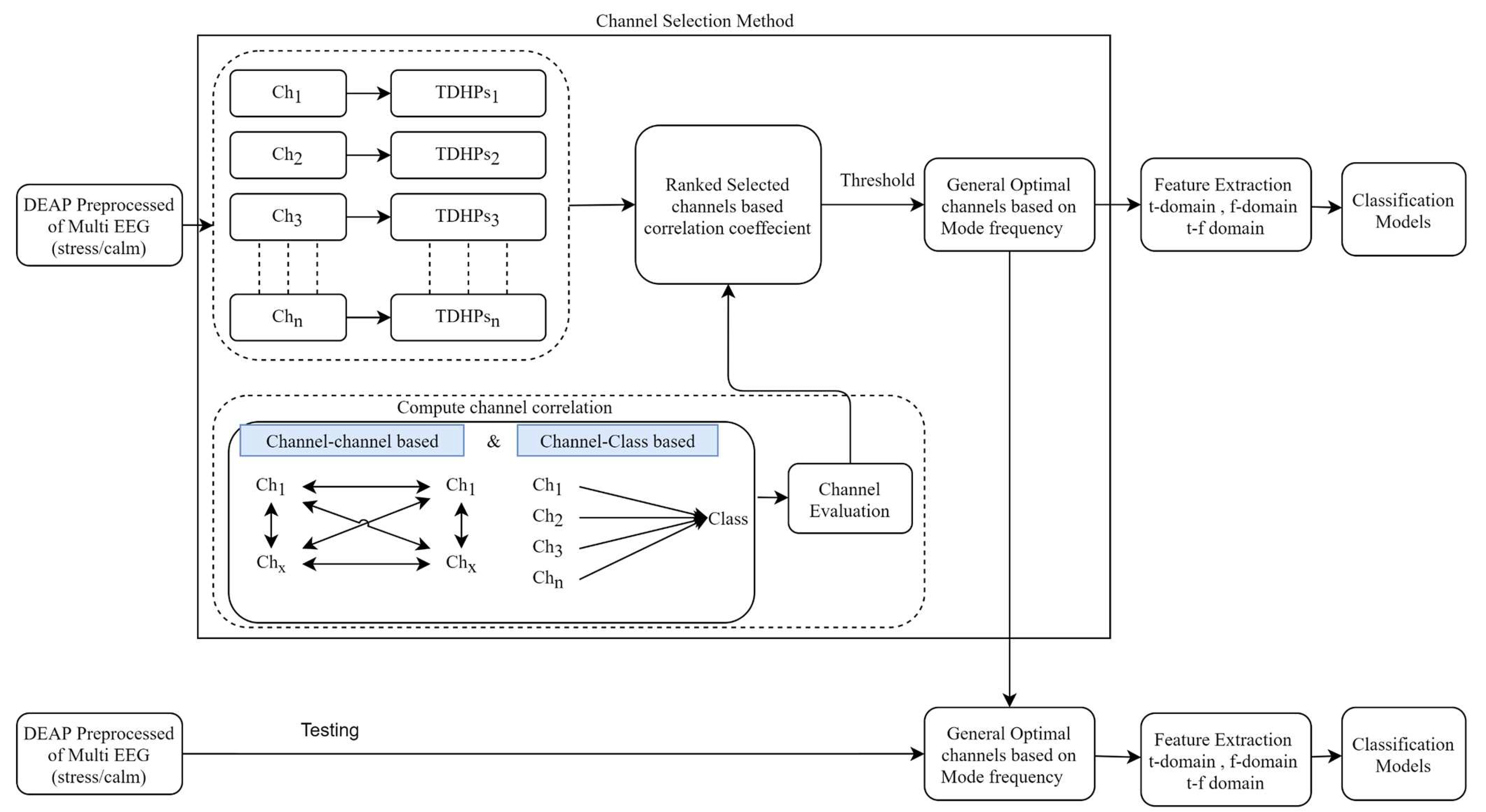
Figure 1. Flow chart of the proposed methodology for EEG common channel selection related to mental stress recognition.
Preprints 79095 g001
Table 1. A summary of the feature extraction methods employed in this study.
Domain Features Equations Description #no. features
Line Length [38] [39] L ( n ) = i = 1 N 1 x [ i ] x [ i 1 ] Called curve length, is the total vertical length of the signal 1
Time Kurtosis [40] K u r t o s i s = 1 T t = 1 T ( x ( t ) μ ) 4 σ 4 Shows the sharpness of EEG signals’ peak 1
Peak to peak amplitude P T P = p k h i g h p k l o w Time of EEG signal peaks between the various windows 1
Skewness [40] S k e w n e s s = 1 T t = 1 T ( x ( t ) μ ) 3 σ 3 A asymmetry of an EEG signal 1
Hjorth Parameters [36,40] A c t i v i t y = v a r ( x ( t ) ) A variance of the time function. 1
M o b i l i t y = v a r ( d y ( t ) d t ) A c t i v i t y ( y ( t ) ) A mean frequency or the proportion of standard deviation of the power spectrum. 1
C o m p l e x i t y = M o b i l i t y ( d y ( t ) d t ) M o b i l i t y ( y ( t ) ) Indicates how the shape of a signal is similar to a pure sine wave. 1
Frequency Relative Power [41] of:
theta (4-8Hz)
alpha(8-12Hz)
sigma(12-15Hz)
low beta(15-20Hz)
high beta (20-30Hz)
R P = p o w e r ( s e l e c t e d _ b a n d ) p o w e r ( t o t a l _ b a n d s ) * 100 Average absolute power of the given band interval. 5
Time-Freqeucny Energy of Wavelet
decomposition coefficients
(db4, 6 level) [11,42].
E n e r g y ( E ) = t = 1 n x t 2 Measure the square sum of wavelet coefficients of each db level 6
Spectral Entropy
(PSD,Welch) [43]
S E = K f = 4 F = 45 P S D ¯ ( F ) *
l o g ( P S D ¯ ( F ) ) , k = 1 .
Measure the distribution of signal power over frequency. 1
katz Fractal Dimension [38] D = l o g 10 ( n ) l o g 10 ( d L ) + l o g 10 ( n ) Compute the maximum distance between the first point and any other point of the Signal’ time window. 1
Table 2. Default parameters for classification techniques.
No. Classifier Default Value
1 SVM C=1.0, Kernal = Radial Basis Function (RBF), 1.0 E 3
2 KNN K=10, distance function= euclidean distance
Table 3. A summary comparison of classification performance for mental stress detection.
  All Channels+KNN   All Channels+ SVM   Proposed Channels+KNN Proposed Channels+SVM
Participant Id precision recall accuracy precision recall accuracy precision recall accuracy precision recall accuracy
1 0.92 0.70 0.85 0.91 0.69 0.84 0.71 0.62 0.78 0.86 0.65 0.82
2 0.82 0.80 0.79 0.90 0.90 0.90 0.84 0.80 0.78 0.91 0.91 0.91
4 1.00 1.00 1.00 0.99 0.99 0.99 0.99 0.99 0.99 0.99 0.99 0.99
5 0.94 0.93 0.93 0.98 0.98 0.98 0.80 0.79 0.79 0.90 0.90 0.90
8 0.91 0.92 0.91 0.98 0.97 0.98 0.83 0.84 0.84 0.92 0.86 0.89
10 0.79 0.77 0.77 0.83 0.83 0.83 0.60 0.60 0.60 0.62 0.62 0.62
11 0.69 0.71 0.69 0.75 0.71 0.76 0.67 0.68 0.66 0.76 0.71 0.77
12 0.67 0.65 0.74 0.81 0.64 0.80 0.74 0.70 0.79 0.76 0.68 0.80
13 0.55 0.55 0.67 0.78 0.58 0.80 0.55 0.54 0.69 0.76 0.57 0.79
14 0.79 0.73 0.88 0.95 0.67 0.90 0.74 0.76 0.86 0.83 0.65 0.88
15 0.84 0.82 0.82 0.90 0.90 0.90 0.69 0.67 0.67 0.72 0.71 0.71
16 0.54 0.53 0.58 0.53 0.52 0.56 0.70 0.66 0.69 0.58 0.57 0.60
18 0.98 0.94 0.97 0.97 0.91 0.95 0.98 0.94 0.97 0.98 0.94 0.97
19 0.65 0.56 0.64 0.95 0.92 0.94 0.56 0.56 0.59 0.83 0.76 0.80
20 0.84 0.84 0.85 0.96 0.94 0.95 0.85 0.86 0.86 0.93 0.88 0.91
21 0.83 0.82 0.84 0.92 0.84 0.88 0.73 0.71 0.74 0.88 0.80 0.84
22 0.62 0.60 0.67 0.73 0.66 0.74 0.66 0.66 0.69 0.74 0.69 0.75
24 0.32 0.38 0.52 0.35 0.48 0.67 0.32 0.38 0.53 0.33 0.42 0.58
25 0.83 0.73 0.79 0.80 0.74 0.79 0.80 0.79 0.81 0.83 0.81 0.84
26 0.98 0.97 0.98 0.97 0.94 0.96 0.90 0.92 0.92 0.96 0.91 0.94
27 0.45 0.50 0.89 1.00 1.00 1.00 0.45 0.50 0.89 1.00 1.00 1.00
28 0.81 0.84 0.82 0.96 0.92 0.95 0.90 0.88 0.91 0.98 0.97 0.98
29 0.86 0.71 0.80 0.92 0.81 0.88 0.82 0.70 0.79 0.94 0.86 0.91
31 0.59 0.58 0.58 0.63 0.63 0.63 0.46 0.46 0.46 0.56 0.56 0.56
32 0.85 0.71 0.78 0.89 0.80 0.84 0.57 0.54 0.62 0.60 0.58 0.63
Average 0.76 0.73 0.79 0.85 0.79 0.85 0.71 0.70 0.75 0.80 0.76 0.81
Table 4. Performance comparison of the proposed model with other popular existing methods.
Method No. Channels Channel Subsets Classifier Accuracy Execution Time
mRMR 11 ’C4’, ’FC2’, ’CP6’, ’Cz’, ’T8’, ’F4’, ’F8’, ’P4’, ’Fz’, ’FC6’, ’Pz’ SVM 0.80±0.12 1.42 s 
KNN 0.74±0.12
STFT+MI 15 ’AF3’,’F7’,’FC5’,’P3’,’P7’,’Pz’,’O2’,’P4’,’FC6’,’Fp2’,’FC1’,’CP2’,’C4’,’F4’,’Fz’   SVM 0.82±0.11 4.46s
KNN 0.74±0.12
GA 13 ’O2’, ’O1’, ’PO3’, ’AF3’, ’P4’, ’P8’, ’F8’, ’P7’, ’C4’, ’CP5’, ’Pz’, ’FC5’, ’Fp2’   SVM 0.82±0.12 1h 3min 34s
KNN 0.76±0.13
Proposed 8 ’AF3’, ’FC5’, ’F8’, ’Fp1’, ’AF4’, ’P7’, ’Fp2’, ’F7’ SVM 0.81±0.11 0.34s
KNN 0.75±0.12
Table 5. Performance comparison of stress detection with related work using EEG signals in the DEAP dataset.
Author Method Number of EEG Channels Dataset Accuracy / Class
Shon [36] Genetic Algorithm-Based Feature Selection 32 DEAP 71.76% (Stress/Calm)
Hasan [35] Boruta-based k-NN feature selection 32 DEAP 73.38% (Stress/Calm)
Proposed Full Channels SET+SVM 32 DEAP 85.68% (Stress/Calm)
CCHP+SVM 8 DEAP 81.56% (Stress/Calm)
Disclaimer/Publisher’s Note: The statements, opinions and data contained in all publications are solely those of the individual author(s) and contributor(s) and not of MDPI and/or the editor(s). MDPI and/or the editor(s) disclaim responsibility for any injury to people or property resulting from any ideas, methods, instructions or products referred to in the content.
Copyright: This open access article is published under a Creative Commons CC BY 4.0 license, which permit the free download, distribution, and reuse, provided that the author and preprint are cited in any reuse.
Prerpints.org logo

Preprints.org is a free preprint server supported by MDPI in Basel, Switzerland.

Subscribe

© 2024 MDPI (Basel, Switzerland) unless otherwise stated