Submitted:
03 August 2023
Posted:
03 August 2023
You are already at the latest version
Abstract
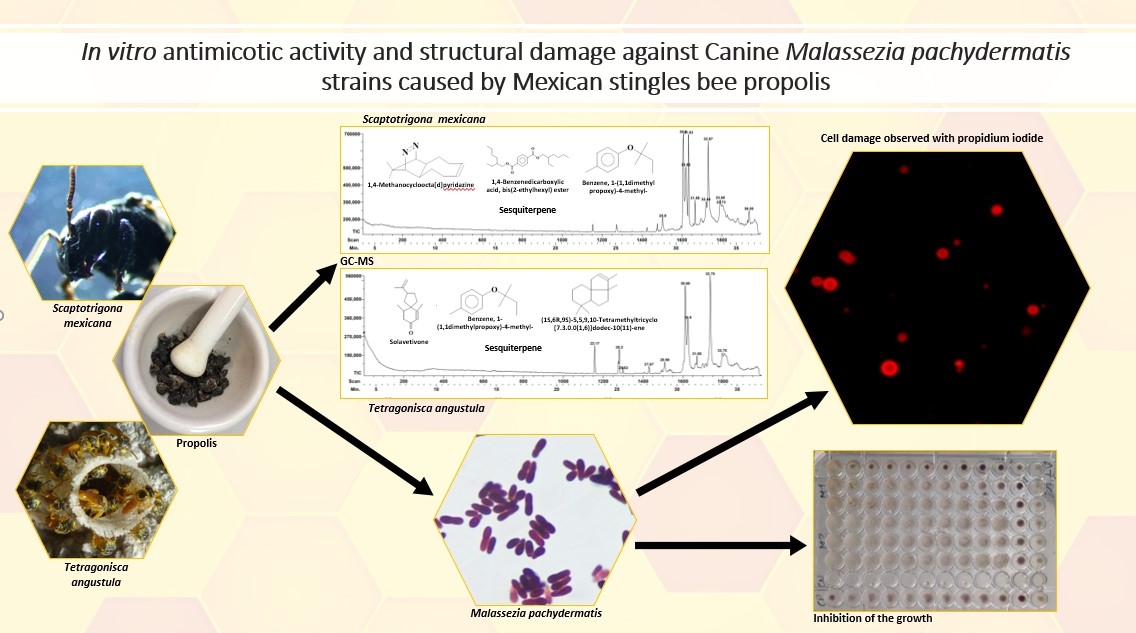
This work demonstrates the antimycotic activity and structural damage of stingless bee propolis against Malassezia pachydermatis,causative agent of canine otitis.Two propolis ethanolic extracts(EEP) derived from the stingless bees, Scaptotrigona mexicana and Tetragonisca angustula were tested against three clinical strains of Malassezia pachydermatis and one of reference (ATCC 14522). Each ethanolic extract of propolis was analyzed by Gas Chromatography – Mass Spectrometry(GC-MS). Antimycotic activity was evaluated by plate microdilution. To evaluate the induced changes in yeasts, by fluorescence microscopy, stains were performed with chalcofluor white and propidium iodide. For both propolis, sesquiterpenes were the main components determined by GC-MS. Minimum inhibitory concentration (MIC) of 32 mg/mL and a minimum fungicidal concentration of 64 mg/Ml were determined in both extracts. EPP of Scaptotrigona mexicana and Tetragonisca angustula caused significant damage to yeast morphology. Propidium iodide penetration was observed, indicating damage to the yeast and with calcofluor-white stain, only morphology deformation was observed..The antimycotic activity and structural damage of propolis from Scaptotrigona mexicana and Tetragonisca angustula against Malassezia pachydermtais was demonstrated .This probably being the first scientific report that demonstrates structural damage in Malassezia pachydermatis of Mexican stingless bee propolis
Keywords:
Mexican stingless bee
; propolis
; antimicotic activity
; structural damage
; Malassezia pachydermatis
1. Introduction
Propolis is a natural resinous substance produced by bees from substances collected from the vegetation in which antifungal, antibacterial, antiviral and antiparasitic activities have been recognized, showing variation in their biological activity depending on their geographical origin [1,2,3].
In Latin America, there is a great variety of ecosystems each with a very diverse vegetation from which native bees’ extract propolis, which in turn results in great medicinal richness. In general, to be able to identify the origin from which bees extracted the material with which they elaborate their propolis, is not as simple as it is to do so with honey or pollen, and for this reason it has been proposed that chemotaxonomic studies should be undertaken or the behavior of each bee species in each region to be observed [4,5,6,7]. Since ancient times, products elaborated by stingless bees from the species Scaptotrigona Mexicana and Tetragonisca angustula have been used, both species being highly employed in central American beekeeping, however, very little information exists in the currently available literature concerning the scientific evidence demonstrating the medicinal efficacy of their products. Also, propolis from the Apis mellifera species presents a fungicidal effect against several fungi species [8,9,10]. Moreover, its applications can prove beneficial in veterinary medicine, as in the case of canines. Propolis coming from Apis mellifera has been used as a prophylactic agent against gastrointestinal and respiratory diseases, mycoses, as well as a wound healing agent, and its therapeutic use has spread to many areas [11,12], one example being that of canine external otitis, which is the inflammation of the external auditory canal and represents between 5 to 20 % of consultation reasons. The main causative agent of canine external otitis is the yeast Malasezzia pachydermatis, which is part of the normal microbiota of the external auditory canal in dogs [13,14,15,16]. Propolis possess an alternative for the treatment of canine otitis, instead of common antifungals, for patients with a high incidence of relapse, because of its antifungal, anti-inflammatory and wound healing properties; however, up to this time the only propolis that has been evaluated is that obtained from the bee Apis mellifera [17]. To undertake studies regarding the potential antimycotic properties of propolis from native bees can provide the scientific ground for its use as an alternative treatment against canine otitis.
2. Materials and Methods
2.1. Ethanolic Extracts Propolis (EEP)
Two propolis samples from stingless bees were obtained. One from, found in the municipality of Yecuatla, Veracruz, geolocation at 19°51 north and 96°46 west, at an altitude of 432 m.a.s.l. The other one from Tetragonisca angustula, found in the municipality of Chalchihuitán, Chiapas, latitude of 16°57 north, longitude of 92°37 west and an altitude of 1461 m.a.s.l. Collected material was evaluated for its physical properties according to Mexican regulation regarding color, odor, taste, and consistency [18]. 30 g of propolis from Sm and 12 g from Ta were weighted and any present impurities were eliminated. Thereafter 100 mL of 70 % ethanol were added to each sample and the obtained mixture was subjected to ultrasonic extraction (Branson, CPX1800H, Danbury, EE. UU.). Each sample was then vacuum filtered and, finally, the obtained filtrates were concentrated by rotovapor (Science MED, SM100-PRO, Finlandia) and left to dry by vacuum pump. Then, both dried extracts were placed in light resistant containers and refrigerated at 4 °C until they were needed [19].
2.2. Gas Chromatographic-Mass Spectrometry (GC-MS)
Chromatographic analysis of ethanolic extracts was performed using a gas chromatograph (6850) coupled to a mass spectrometer (7890 model, JEOL MC-GC-Mate II, Japan). A HP-5MS (30 m × 0.32 mm) capillary column and a film thickness of 0.25µm were used. Helium gas was used as the carrier gas. Elected injection method was split mode with an injection volume of 1 µL. Separation conditions were as follows: 70°C at the beginning for two minutes followed by two ramp increments. The first one of 20°C per minute until a temperature of 230°C was reached; the second one of 8°C per minute until a temperature of 290°C was reached, keeping this temperature for a period of 5 minutes. Total analysis time was of 21.25 minutes. The detected mass range was of 35 m/z to 750 m/z, the sample was subjected to electron impact ionization at 70 eV, with the ionization source reaching a temperature of 230 °C. Compound identification was carried out by comparison with the library database from the equipment [20].
2.3. Evaluation of antimycotic activity:
2.3.1. Inoculum preparation
Three different M. pachydermatis strains were used, one of reference ATCC 14522 and two obtained from clinical isolates from dogs. All three strains were identified by proper biochemical testing [21]. Microorganisms were provided by the Laboratorio del Servicio de Análisis de Propóleos (LASAP), a branch of the Multidisciplinary Research Unit of FES Cuautitlán, UNAM. To activate M. pachydermatis strains, each yeast was seeded in modified Dixon Agar (mDA). Each was seeded in a different petri dish and then incubated for 72 hours at 28 °C. Thereafter, samples were reseeded in other mDA-containing plates and were incubated for 48 hours at 28°C in order to rule out strain contamination [22,23]-
2.3.2. Determination of Minimum Inhibitory Concentration and minimum fungicidal concentration
Broth microdilution was carried out by determination of the minimum inhibitory concentration (MIC) and the minimum fungicidal concentration (MFC). For this purpose, the EEP were subjected to serial dilutions from 32 to 0.0312 mg/mL in Sabouraud Dextrose broth (SDA) supplemented with glucose (Bioxon, México). SDA broth containing the microorganism was used as the positive witness, while SDA broth without the microorganism was used as the negative witness and were incubated for 48 hours at 28°C. To detect the respiratory activity of the microorganisms, a solution of 0.08 % oxidated tetrazolium salt (TTC) was used, which generates a red pigment (formazan) in the presence of the microorganism. The afore mentioned procedure was performed in the following manner: 50 µL of TCC were added to each inoculated well, plates were mixed by plate agitator and incubated at 33 °C for 30 minutes. By the end of the 30 minutes, it was observed that an insoluble, red-colored precipitate had formed, representing the MIC. When there was no growth, the solution stayed clear-colored, indicating the MIC or MFC. To confirm these results, it was determined whether the observed effect was fungistatic or fungicidal by taking a sample of the culture with an inoculation loop and seeding the material in a petri dish containing SDA agar supplemented with glucose, followed by an inoculation period of 72 hours at 33 °C. It was considered that plates exhibiting growth was indicative of a fungistatic effect, while plates lacking growth of the microorganisms was indicative of a fungicidal effect [24].
2.4. Structural damage
To evaluate the structural changes induced by the EEP on Malazassia pachidermatis fluorescence microscopy was used. In this experiment, the reference strain and one clinical strain were used. A concentration of 64 mg/mL of each EEP of Scaptotrigona mexicana and Tetragonisca angustula were added to each strain. Incubated at 28 ° C for 48 hrs. When the incubation ended, the yeast was stained with calcofluor-white (M2R 1g/L,Sigma) and propidium iodide (2.4 mmol, Sigma), as a negative control, a culture without EEP was used. Preparations were viewed on a microscopy Zeiss Axioscop 40, coupled to an Evolution VF Cooled Color camera from Media Cibernetics. All experiments were performed in triplicate [25,26].
3. Results
3.1. Gas Chromatographic-Mass Spectrometry (GC-MS)
Results from each propolis ample analyzed by coupled gas chromatography- mass spectrometry (GC-MS) are shown in Table 2 and Table 3, as well as Figure 1 and Figure 2, these show the main identified compounds with an accurate identification (> 90%) as related to the equipment’s database. For both propolis simples, the main constituent compounds were sesquiterpenes

Table 1.
Constituents of Mexican Scaptotrigona mexicana propolis characterized by CG-EM.
| Bak | Time Retention (TR) |
Composite proposed by the database |
Chemical classification |
Biological activity | Reference |
|---|---|---|---|---|---|
| 2 | 30.6 | 1,4-Methanecycloocta[d]piri-dazine, 1,4,4a,5,6,9,10,10a-octahydro-11,11-dimethyl-, (1.alpha.,4.alpha.,4a.alfa. ,10a.alfa.) - | Pyridazine (heterocyclic compound) | Antioxidant | [27] |
| 5 | 31.58 | Fanersol Isomer a | Sesquiterpene | Antimicrobian | [28] |
| 6 | 32.48 | Ethanone, 1-(1,3a,4,5,6,7-hexahydro-4-hydroxy-3,8-dimethyl-5-azulenyl)- | Ketone Sesquiterpene |
Antimicrobian | [29] |
| 8 | 33.68 | 2H-1-Benzoxacyclohexadecin-16(18aH)-one,3,4,5,6,7,8,9,10,11,12,13,14-dodecahydro-18,18a-dihdroxy-2-methyl | Macrocycle | Activity Not reported | [30,31] |
| 9 | 33.7 | Furan-2,5-dicarbaldhyde 2,5-Furandicarboxaldehydo | Heterocyclic compound with aldehyde groups | Antioxidant Antimicrobian |
[32,33] |
3.2. Evaluation of the antimycotic activity
It was found that all tested Malassezia pachydematis strains were susceptible to all evaluated propolis extracts in the present study, the value of the Minimum Inhibitory Concentration was of 32 mg/mL and that of the Minimum Fungicidal Concentration was of 64 mg/mL (Table 4).
3.3. Structural damage
Figure 3 and Figure 4 show the effect of EEP on the structure of M. pachydermatis (ATCC 14522) and clinical strain. Both EEPs penetrated the yeasts.
The lack of fluorescence in the stain of calcofluor-white in some preparations indicates a probable destruction of the yeast.Propidium iodide was detected in all treated samples, which indicates alteration of the wall and membrane, observing a greater amount of altered yeasts in the reference strain treated with the EEP of Scaptotrigona mexicana.
4. Discussion
Many of the identified metabolites of propolis stingless bees have been reported to exhibit a myriad of biological activities, including antimicrobial, anti-inflammatory, cytotoxic, antioxidant, hepatoprotective, antiulcer, among others [39,40,41,42]. Analyzed propolis extracts exhibit chemical compositions with significant differences among them, this being consistent with other studies which have found that the quantity and diversity of compounds found in propolis is high [43,44], in comparison with the obtained results from the present study for Scaptotrigona mexicana and Tetragonisca angustula as well as other species such as Melipona beecheii and others worldwide [2].It is important to consider that floral diversity, collection time of the year and bee species are all determinant factors for the final composition of each propolis. Compounds belonging to the sesquiterpenes were primarily detected, these have been attributed with antimicrobial activity [34,45,46,47]
Regarding Scaptotrigona mexicana, it is important to mention that a bibliographic research of furan-2,5-dicarbaldehyde, which is a heterocyclic compound with aldehyde groups, was unsuccessful using that exact denomination, however we did find mention of a similar compound, 2-acetil-5-metilfurane, reported to exhibit antimicrobial activity against Escherichia coli, Candida albicans and Staphylococcus aureus [33].
For Tetragonisca angustula, the antibacterial and antifungal activity of solavetivone has been reported [36,37]; on the other hand, concerning l-(1S,6R,9S)-5,5,9,10-tetramethyltricyclo [7.3.0.0(1,6)]dodec-10(11)-ene, there in no specific information regarding its activity; nonetheless, antifungal and antibacterial activities have been reported for a similar compound, namely 3,3,7,7-tetramethyl-5-(2-methyl-1-propenyl)-tricyclo [4.1.0.0(2,4)]heptane [37].
Antimycotic potential of propolis extracts, mainly from Apis mellifera, has been demonstrated against Candida albicans. Also, fungicidal and fungistatic properties of green and red propolis extracts from Brazil against other fungi genera such as Saccharomyces [48] have been observed; inhibition and morphologic alterations of Cyptococcus neoformans [49].
None the less, information regarding the evaluation of the antimycotic activity of propolis coming from stingless bees mainly against Candida albicans has been studied for the following species: Lestrimellata spp., Melipona favora orlinge, Melipona marginata, Melipona quadrifasciata, Melipona scutellaris, Nannotrigona testaceicornis, Plebeia droriana, Plebeia remota, Scaptotrigona bipunctata, Tetragona clavipes [7] and Tetragonisca angustula and that of propolis from Tetragonisca fiebrigi against Candida glabrata [50] .In the case of propolis coming from the Malaysia-stingless bee Trigona thoracica, it has been demonstrated that it acts against Cryptococcus neoformans [51]. Furthermore, an Indonesian propolis from the species Tetragonula sp. has been evaluated as a possible therapeutic agent for the treatment of vaginal candidiasis [52].
Regarding Malassezia pachydermatis, there are studies evaluating the activity of propolis from Apis mellifera, but not from native bees, against this yeast. In a recent study a correlation was established between the antifungal activity of EPP from Brazilian green and red propolis against M. pachydermatis, with a MIC between 4 y 8 mg/mL and a MFC of 8 and 16 mg/mL. It was determined that as the total content of phenols and flavonoids increases, propolis exhibits a better biological activity, proposing that the action mechanism of EEP is due to the rupture of the cellular wall, given the fact that during the investigation it was observed that some azole- resistant Malassezia pachydermatis strains were inhibited by the EEP. However, they also extern concern because it is still unclear if high EPP concentrations could induce cytotoxicity. Therefore, there remains work to be done in order to accurately identify the active principles of propolis, as well as their action mechanisms. Currently the are two theories aiming to explain the antifungal activity of propolis, the first one proposing that it elicits cellular wall lysis and the other proposing that it damages the cellular membrane by inhibiting ergosterol synthesis [53]. Likewise, the efficacy of an Argentinian propolis against M. pachydermatis was evaluated by different in vitro techniques, which concluded that the yeast was vulnerable to all employed propolis concentrations, with a MIC of 0.30 mg/mL, however, they were not able to determine the MFC [17]. The efficacy of a 2.5 % EEP against 48 clinical strains of Malazessia pachydermatis isolated from dogs diagnosed with external otitis was also proven, as it was found that all of them were susceptible to it [54]. Antimycotic activity against clinical isolates from dogs with external otitis has also been observed in a EEP from Rio Grande do Sul, Brazil, with a MIC of 2.6 mg/mL and a MFC of 5.3 mg/mL [55]. In contrast, our results for both propolis of the evaluated bee species regarding MIC and MFC was of 32 mg/mL and 64 mg/ml respectively, both concentrations being significantly higher than those reported in studies of propolis coming from Apis mellifera.
According to the images obtained, the EEP proved to be able to penetrate the membrane cellular, causing severe damage and eventually death of yeasts with structural and functional damage by membrane disruption. Calcofluor white has a high affinity for fungal wall components [56,57]. The alteration observed with this stain was mainly deformity of the morphology and in some cases, we have the hypothesis that the complete destruction of the cell wall caused the yeasts not to be observed, which would be a possible effect by the sesquipertenes present in the EEP as previously described [58]. It would be advisable to perform computational chemistry studies to specifically establish the damage of these compounds in the yeast wall.
On the other hand, propidium iodide binds to nucleic acids and increases its red color when there is damage to the cell membranes. which indicates severe cell damage and death, which was seen in the reference strains and the clinic treated with both EEP, which demonstrates the effectiveness of this type of propolis. This effect with propidium iodide has been observed in other fungi such as Fusarium species and Colletotrichum fructicola, [59] when naturally occurring compounds have been evaluated against their growth [60].
These stains have been used to detect cellular damage using Apis mellifera propolis against other yeast of medical importance such as Candida albicans [61,62,63] as well as the bacterium Staphylococcus aureus [64].
Therefore, it is shown that propolis of the Mexican stinglees bee Scaptrigona mexicana and Tetragonisca angustula have antimycotic effect and cause structural damage on Malazessia pachidermatis, which supports its possible use for therapeutic purposes in the treatment.. The findings and their implications should be discussed in the broadest context possible. Future research directions may also be highlighted.
5. Conclusions
In conclusion, the present study has shown the antifungal properties and damage structural of two propolis ethanolic extracts from two stingless bee species (Scaptotrigona mexicana and Tetragonisca angustula) proceeding from two Mexican municipalities (Yecuatla, Veracruz and Chalchihuitán, Chiapas) against different Malazessia pachydermatis strains, one of reference (ATCC14522) and three clinical isolates, being highly likely that for both propolis. Sesquiterpenes, along with other compounds, are possibly the reason the reason for the antimycotic activity of both extracts. It is important to mention that, to our knowledge, the present work is the first to demonstrate the damage structural and antifungal effect of Mexican stingless bee propolis against this Malazessia pachydermatis. None the less, further research must be undertaken in order to provide more solid scientific basis for the future employment of propolis as an alternative treatment for canine external otitis.
Supplementary Materials
Table 1. Constituents of Scaptotrigona mexicana propolis characterized by CG-EM, Figure 1. Gas chromatogram corresponding to the propolis of Scaptotrigona mexicana and the main compounds detected. Figure 2. Gas chromatogram corresponding to the propolis of Tetragonisca angustula and the main compounds detected. Table 2. Main constituents of propolis of Tetragonisca angustula characterized by CG-EM. Figure 2. Gas chromatogram corresponding to the propolis of Tetragonisca angustula and the main compounds detected. Table 3. Minimum inhibitory concentration (MIC) and minimum fungicidal concentration (FMC) values of stingless bee propolis extracts from two regions of the Mexican Republic on the reference strain M. pachydermatis ATCC 14522 and strains isolated from clinical samples. Figure 3. Effect of EEP of Scaptotrigona mexican and Tetragonisca angustula on reference strain Malassezia pachidermatis were obtained by fluorescence microscopy and dyed with calcofluor-white and propidium iodide. Cultures were exposed to EEP at a concentration of 64 mg/ml for 48 hr at 28 °C. Propidium iodide penetration was observed, indicating damage to the yeast. With calcofluor-white stain, only morphology deformation.(40x magnification).Figure 4. Effect of EEP of Scaptotrigona mexican and Tetragonisca angustula on clinical strain of Malassezia pachidermatis were obtained by fluorescence microscopy and dyed with calcofluor-white and propidium iodide . Cultures were exposed to EEP at a concentration of 64 mg/ml for 48 hr at 28 °C. Propidium iodide penetration was observed, indicating damage to the yeast; with calcofluor-white stain, alteration of morphology was observed. A greater damage was observed since it was not detected the presence with the calcofluor- white of the yeast and only the red of the propidium iodide was detected with Ethanolic Extract of Tetragonisca angustla Propolis. .(40x magnification).
Author Contributions
Evaluation antimicotic activity, D.B.F E.; Analysis of propolis samples ,B.R.P; Collection of propolis in the meliponaries,FHG ; Clinicals and reference strains, and conservation of microorganisms, NTB; Gas Chromatographic-Mass Spectrometry,J.P.F..; Performance of chemical determinations , J.G.P.C. ; Fluorescence microscopy and revision manuscript, C.G.G.T.; Funding acquisition and revision manuscript TACS .All authors have read and agreed to the published version of the manuscript
Funding
The authors acknowledge and appreciate the support provided by the research projects: UNAM-DGAPA, PAPIIT IN223719, IN215723. FESC. Research Chairs CI 2237, CI 2222, CI 2267, and the Bee Team from the South Border College (ECOSUR).
Informed Consent Statement
No applicable
Data Availability Statement
No applicable
Acknowledgments
The authors would like to thank to Lázaro Arroyo and Yliana Delfin Fuentes for his support in propolis sample recollection from stingless bee families and to Francisco Rodolfo González Diaz for his technical support
Conflicts of Interest
The authors declare no conflict of interest
References
- Bakchiche, B.; Temizer, I.K.; Aytaç,G. A.; Çelemli, G.; Yegin, C.B.; Ghareeb, M.A.; Chemical Composition and Biological Activities of Honeybee Products From Algeria. J. Appl. Biotechnol. Rep. 2020, 7, 93–103. [Google Scholar]
- Bankova, V.; Popova, M. Propolis of Stingless Bees: A Promising Source of Biologically Active Compounds. Pharmacogn Rev. 2007, 1, 88–92. [Google Scholar]
- Ramon, S.J.; Peraz, L.E.; Rodríguez, B.R.; Yam, P.A.; Madera, S.T.; Ortiz, VE. Partial characterization of ethanolic extract of Melipona beecheii propolis and in vitro evaluation of its antifungal activity. Rev. Bras. Farmacogn. 2019, 29, 319–324. [Google Scholar] [CrossRef]
- Arnold N, Zepeda R, Vázquez M, Aldasoro, M. Las abejas sin aguijón y su cultivo en Oaxaca, México: Con catálogo de especies. 1a ed; ECOSUR-CONABIO, San Cristóbal de Las Casas. México, 2018, 10-58.
- Ayala, R. Revisión de las abejas sin aguijón de México (Hymenoptera: Apidae: Meliponini). Folia Entomol. Mex. Mex. 1999, 106–123. [Google Scholar]
- Pardini, R.; da Rocha, P.L.B.; El-Hani, C.; Pardini, F. Desafíos y Oportunidades para Cerrar la Brecha entre Investigación e Implementación en Ciencias y Gestión Ecológicas en Brasil. Biología de la conservación: Voces de los trópicos. PH Raven, NS Sodhi & L. Gibson : John Wiley & Sons, Ltd., Reino Unido, 2013, 75-85.
- Hurtado, M.M.; Jara, L.; May, I.J.; Quezada, E.G.; Ruiz, C.P.; De la Rúa, P. A geometric morphometric and microsatellite analyses of Scaptotrigona mexicana and S. pectoralis (Apidae: Meliponini) sheds light on the biodiversity of Mesoamerican stingless bees. J. Insect Conserv. 2016, 20, 753–763. [Google Scholar]
- Mateo, A.J.; Rodríguez, P.B.; González, G.S.; Cruz, T.A. Structural damage in Cryptococcus neoformans caused by a Mexican propolis. Nova Scientia. 2020, 12, 1–17. [Google Scholar]
- Quintero, M.M.L.; Londoño, O.A.; Manzano, G.P.; López, M.R.; Soto, Z.C.I.; Carrillo, M.L.; Penieres, C.J.G.; García, T.C. G,.;Cruz-Sánchez, T.A. Effect of Mexican propolis extracts from Apis mellifera on Candida albicans in vitro growth. Rev Iberoam Micol. 2008, 25, 22–26. [Google Scholar] [CrossRef]
- Rodríguez, P.B.; Canales, M.M.; Penieres, C.J.; Cruz, S.T. Chemical composition, antioxidant properties and antimicrobial activity of Mexican propolis. Acta Universitaria. 2019, 30, 1–29. [Google Scholar] [CrossRef]
- Flores, I.S.; Moreno, M.; Londoño, A. ; Cruz. TA. Use of mexican propolis for the tropical treatment of dermatomycosis in horses. Open J. Vet. Med. 2016, 6, 1–8. [Google Scholar]
- Tovar, N.; García,L. ; Cruz TA. Propolis in dogs: Clinical experiences and perspectives (a brief review). Open J. Vet. Med. 2016, 5, 11–17. [Google Scholar]
- Girãoa, M.R.N.; Brilhanteab, RA.; Cordeirob, A.J.; Monteiroc, J.J.C.; Sidrimb,M. F.G.; Rochaab, P.RS. Malassezia pachydermatis isolated from normal and diseased external ear canals in dogs: A comparative analysis. Vet. J. 2006, 172, 544–548. [Google Scholar]
- Lozina, L.; Boehringer, S.; D’Aquino, M.; Acosta, O. Eficacia del propóleos Sobre Malassezia pachydermatis. Correlación de Diferentes Técnicas in vitro. Acta Farm. Bonaerense. 2006, 25, 560–563. [Google Scholar]
- Cabañes, F. J. Diagnosis of Malassezia pachidermatitis and otitis in dogs and cats, is it just a matter of counting? Rev Iberoam Micol. 2021, 38, 3–4. [Google Scholar] [CrossRef]
- Deegan, K.R.; Fonseca, M.S.; Oliveira, D.C.P.; Santos,L. M.; Fernández, C.C.; Hanna,S.A.; Machado, B.A.S.; Umsa-Guez, M.A.;Meyer,; Portela,R.W. Susceptibility of Malassezia pachydermatis clinical isolates to allopatic antifungals and Brazilian red, green, brown propolis extracts. Front. Vet. Sci. 2019, 6, 1–12. [Google Scholar] [CrossRef]
- Lozina, L.A.; Peichoto, M.E.; Boehringer, S.I.; Koscinczuk, P.; Granero, G.E.; Acosta, O.C. Efficacy of Argentine propolis formulation for tropical treatment of canine otitis externa. Arq. Bras. Med. Vet. Zootec. 2010, 62, 1359–1366. [Google Scholar] [CrossRef]
- Diario Oficial de la Federación. Norma Oficial Mexicana NOM-003-AG/GAN-2017: Propóleos, producción y especificaciones para su Procesamiento. 2017. Available online: https://www.dof.gob.mx/nota_detalle.php?codigo=5500103&fecha=06/10/2017#gsc.tab=0.
- Dos Santos, L.; Hochheim, S.; Boeder, A.M.; Kroger, A.; Tomazzoli, M.M.; Dal Pai,R. ; Maraschin, M.; Guedes, A.; de Cordova, C.M.M.; Chemical characterization, antioxidant, cytotoxic and antibacterial activity of propolis extracts and isolated compounds from the Brazilian stingless bees Melipona quadrifasciata and Tetragonisca angustula. J. Apic. Res. 2017, 56, 543–558. [Google Scholar] [CrossRef]
- Rivera; Y. C.R., Terrazas, L.I.; Jiménez, E.; Campos, J.E.; Flores, O.C.M.; Hernández, L.B.; Cruz, S.T.; Garrido, F.G.I.; Rodríguez-Monroy, M.A.;Canales M.M. Anti-Candida Activity of Bursera morelensis Ramirez Essential Oil and Two Compounds, α-Pinene and γ-Terpinene-An in vitro study. Molecules. 2017, 22, 1–13. [Google Scholar]
- Kaneko, T.; Makimura, K.; Abe, M.; Shiota, R.; Nakamura, Y.; Kano, R.; Hasegawa, A.; Sugita, T.; Shibuya, S.; Watanabe, S.; Yamaguchi, H.; Abe, S.; Okamura, N. Revised Culture-Based System for Identification of Malassezia Species. J. Clin. Microbiol. 2007, 45, 3737–3742. [Google Scholar] [CrossRef]
- Aspiroz, C.; Gilaberte, Y.; Rezusta, A.; Boekhout, T. ; Rubio. M.C. Gentamycin inhibits the growth of Malassezia pachydermatis in culture. Rev. Iberoam. Micol. 2010, 27, 20–21. [Google Scholar]
- Kaneko, T.; Makimura, K.; Abe, M.; Shiota, R.; Nakamura, Y.; Kano, R.; Hasegawa, A.; Sugita, T.; Shibuya, S.; Watanabe, S.; Yamaguchi, H.; Abe, S.; Okamura, N. Revised Culture-Based System for Identification of Malassezia Species. J. Clin. Microbiol. 2007, 45, 3737–3742. [Google Scholar] [CrossRef]
- Balouiri, M.; Sadiki, M.; Ibnsouda, S.K. Methods for in vitro evaluating antimicrobial activity: A review. J. Pharm. Anal. 2016, 6, 71–79. [Google Scholar] [CrossRef] [PubMed]
- K. Sánchez, A.K.;Fernández M.R.F.; Moreno M. M. ;Villegas A.L.; Meneses G.F.; Arenas G.R. Sensitivity and specificity of mycological direct examination with calcofluor hite for the diagnosis of onychomycosis. Med. Cutan. Iber. Lat. Am. 2013, 41, 261–266. [Google Scholar]
- Phillips, A.J. ; Ian Sudbery,I. ;Ramsdale,M. Apoptosis induced by environmental stresses and amphotericin B in Candida albicans. PNAS. 2003, 100, 14327–14332. [Google Scholar] [PubMed]
- Urbizu, G.A.L.; Castillo, R.O.; Martínez, A.G.C.; Torres,C. J.A. Natural variability of essential oil and antioxidants in the medicinal plant Turnera diffusa. Asian Pac. J. Trop. Biomed. 2017, 10, 121–125. [Google Scholar]
- Devequi-Nunes, D.; Machado, B.A.S.; Barreto, G.A.; Silva, J.R.; da Silva, D.F.; a Rocha, J. .LC.; Neves,B.H.; Borges, M.M.;Umzza, G.M.A. Chemical characterization and biological activity of six different extracts of propolis through conventional methods and supercritical extraction. PLoS ONE. 2018, 3, 1–20. [Google Scholar]
- Fatnassi, S.; Zarrouk, H.; Chatti, S. Chemical composition and antimicrobial activity of volatile fraction of the peel of Maclura pomifera fruit growing in Tunisia. J. Soc. Chim. Tunis. 2011, 13, 1–6. [Google Scholar]
- Delmondes, G.A.; Santiago, L.I.C.; Días, D.Q.; Cunha, G.L.D.; Araújo, I.M.; Barbosa, R.; Coutinho, H.D.M.; Felipe, CF.B.; Barbosa, F.J.M.; Lima, N.T.R.; De Menezes, I.R.A.; Kerntopf, M.R. Pharmacological Applications of Farnesol (C15H26O): A patent review. Expert Opin Ther Pat. 2020, 30, 227–234. [Google Scholar] [CrossRef]
- Chen, C.; Change, H.; Kirk, K. Betulachrysoquinone hemiketal: A p-benzoquinone hemiketal macrocyclic compound produced by Phanerochaete chrysospor. Phytochemistry. 1977, 16, 1983–1985. [Google Scholar] [CrossRef]
- Muriira, K.G. : Nyagah, M.N.E.; Machocho, A.K.; Nyawira, W.L., Mungiria, J.N.M. Chemical Composition and in vitro Antioxidant Activities of Ocimum americanum. Adv. Anal. Chem. 2015, 5, 42–49. [Google Scholar]
- Otieno, A.J. Antimicrobial activity and phytochemical profiles of Warburgia ugandensis sprague (canellaceae) extracts from different populations across the Kenyan Rift Valley a Thesis Submitted in Partial Fulfillment of the Requirements for the Award of the Degree of Master of Science (Biotechnology) in the School of Pure and Applied Sciences of Kenyatta University. Kenia 2016.
- Peng, D.; Yu, Z.X.; Wang, C.H.; Gong,B. ; Liu,Y.Y.; Wei, J.H. Chemical Constituents and Anti-Inflammatory Effect of Incense Smoke from Agarwood Determined by GC-MS. Int. J. Anal. Chem. 2020, 4575030, 1–19. [Google Scholar]
- Satyala, P.; Maharjanb, S.; Setzera,W. N. Volatile Constituents from the Leaves, Fruits (Berries), Stems and Roots of Solanum xanthocarpum from Nepal. Nat. Prod. Commun. 2015, 10, 361–364. [Google Scholar] [CrossRef]
- Takahashi, S.; Yeo, Y.; Zhao, Y.; O’Maille, P.E.; Greenhagen, B.T.; Noel, J.P.; Coates, M.R.; Chappell, J. Functional Characterization of Premnaspirodiene Oxygenase, a Cytochrome P450 Catalyzing Regio- and Stereo-specific Hydroxylations of Diverse Sesquiterpene Substrates. Int. J. Biol. Chem. 2007, 282, 31744–31754. [Google Scholar] [CrossRef]
- Bruzua, V.H.; Henríquez, G.W.; Crescente, O; Lanza, J. G. Aceite esencial de Wedelia calycina (Asteraceae): Composición química, actividad antibacteriana y antifúngica. Saber. 2015, 27, 87–93. [Google Scholar]
- Mohd, B.A.A.; Zin, M.; Annisava, A.R.N.; Nafi, N.N.E.; Mohd, K.S. Phytochemical screening and antioxidant properties of stingless bee Geniotrigona thoracica propolis. Mal. J. Fund. Appl. Sci. 2019, 15, 330–335. [Google Scholar] [CrossRef]
- Surek, M.; Fachi. , M.M.;De Fátima, C.A.; De Oliveira, F.F.; Pontarolo, R.; Crisma,A.R.; De Souza, M.W.; Felipe, K.B. Chemical composition, cytotoxicity, and antibacterial activity of propolis from Africanized honeybees and three different Meliponini species. J. Ethnopharmacol. 2021, 269, 1–14. [Google Scholar] [CrossRef]
- Torres, A.R.; Sandjo, L.P.; Friedemann, M.T.; Tomazzoli, M.M.; Maraschin, M.; Mello, C.F.; Santos, A.R.S. Chemical characterization, antioxidant and antimicrobial activity of propolis obtained from Melipona quadrifasciata quadrifasciata and Tetragonisca angustula stingless bees. Braz. J. Med. Biol. Res. 2018, 51, 1–10. [Google Scholar] [CrossRef] [PubMed]
- Yam, P.A.; Santana, H.A.A.; Yah, P.N.; Ramón, S.J.M.; Cáceres, M.R.; Borges, A.R.L.; Ortiz, VE. Pentacyclic triterpenes and other constituents in propolis extract from Melipona beecheii collected in Yucatan, Mexico. Rev. Bras. Farmacogn. 2019, 29, 358–363. [Google Scholar]
- Grajales, C.J.; Elías, C.J.; Lozano, G.E.; Albores, F.V.; López, G.A. Actividad antimicrobiana de propóleos de abejas sin aguijón en combinación con ajo, Allium sativum (Amaryllidaceae). Rev. Biol. Trop. 2021, 69, 23–35. [Google Scholar]
- Bakchiche B, Temizer IK, Aytaç GA, Çelemli G, Yegin CB, Ghareeb MA. Chemical Composition and Biological Activities of Honeybee Products from Algeria. J. Appl. Biotechnol. Rep. 2020, 7, 93–103. [Google Scholar]
- Ramon, S.J.; Peraz, L.E.; Rodríguez, B.R.; Yam, P.A.; Madera, S.T.; Ortiz, VE. Partial characterization of ethanolic extract of Melipona beecheii propolis and in vitro evaluation of its antifungal activity. Rev. Bras. Farmacogn. 2019, 29, 319–324. [Google Scholar] [CrossRef]
- Peng, D.; Yu,Z. X.;, Wang, C.H.; Gong,B.;Liu,Y.Y.; Wei, J.H. Chemical Constituents and Anti-Inflammatory Effect of Incense Smoke from Agarwood Determined by GC-MS. Int. J. Anal. Chem. 2020, 4575030, 1–19. [Google Scholar]
- Sarvesan, R.; Eganathan, P.; Saranya, J.; Sujanapa, P. Chemical composition and antibacterial activity of leaf essential oil of Eugenia cotinifolia ssp. Codyensis (Munro ex wight) Ashton. Int. J. Pharm. Sci. 2015, 9, 3981–3985. [Google Scholar]
- Xie, C.; Wang, S. , Cao, M.Y.; Xiong, W.;Wu, L. (E)-9-Octadecenoic Acid Ethyl Ester Derived from Lotus Seedpod Ameliorates Inflammatory Responses by Regulating MAPKs and NF-κB Signalling Pathways in LPS-Induced RAW264.7 Macrophages. Evid. Based Complementary Altern. Med. 2022, 2022, 6731360. [Google Scholar] [CrossRef]
- Lotti, C.; Castro, G.M.M.; Sá, LFR. Inhibition of Saccharomyces cerevisiae Pdr5p by a natural compound extracted from Brazilian Red Propolis. Braz. J. Pharmacogn. 2011, 21, 21,901–907. [Google Scholar] [CrossRef]
- Hee, Y.C. In vitro evaluation of the antifungal activity of propolis extract on Cryptococcus neoformans and Candida albicans. Mycobiology. 2002, 30, 93–95. [Google Scholar]
- Campos, J.F.; Dos Santos, U.P.; Da Rocha, P.; Dos S Damião, M.J.; Balestieri, J.B.; Cardoso, C.A.; Paredes-Gamero, E.J.; Estevinho, L.M.; De Picoli,Souza K. ; Dos Santos E.L. Antimicrobial, Antioxidant, Anti-Inflammatory, and Cytotoxic Activities of Propolis from the Stingless Bee Tetragonisca fiebrigi (Jataí). Evid. Based Complementary Altern. Med. 2015, 2015, 296186. [Google Scholar]
- Shehu, A.; Ismail, S.; Rohin, M.A.K.; Harun, A.; Aziz, A.; Haque, M. Antifungal Properties of Malaysian Tualang Honey and Stingless Bee Propolis against Candida albicans and Cryptococcus neoformans. Appl. Pharm. Sci. 2016, 6, 44–50. [Google Scholar] [CrossRef]
- Farida. S.; Sahlan, M.; Rohmatin, E.; Adawiyah R. The beneficial effect of Indonesian propolis wax from Tetragonula sp. as a therapy in limited vaginal candidiasis patients. Saudi J. Biol. Sci. 2020, 27, 142–146. [Google Scholar]
- Deegan KR, Fonseca MS, Oliveira DCP, Santos LM, Fernández CC, Hanna SA, Machado, BAS, Umsa-Guez MA, Meyer R, Portela RW. Susceptibility of Malassezia pachydermatis clinical isolates to allopatic antifungals and Brazilian red, green, brown propolis extracts. Front. Vet. Sci. 2019, 6, 1–12. [Google Scholar]
- Lozina LA, Peichoto ME, Boehringer SI, Koscinczuk P, Granero GE, Acosta OC. Efficacy of Argentine propolis formulation for tropical treatment of canine otitis externa. Arq. Bras. Med.Vet. Zootec. 2010, 62, 1359–1366. [Google Scholar] [CrossRef]
- Cardoso, R.L.; Maboni, F; Machado, G; Alves S. H.; de Vargas A.C. Antimicrobial activity of propolis extract against Staphylococcus coagulase positive and Malassezia pachydermatis of canine otitis. Vet. Microbiol. 2010, 142, 432–434. [Google Scholar] [CrossRef] [PubMed]
- Hageage, G.J.; Harrington, B.J. Use of calcofluor white in clinical mycology. Lab. Med. 1984, 15, 109–112. [Google Scholar] [CrossRef]
- Ram, A.F.J.; Klis, F.M. Identification of fungal cell wall mutants using susceptibility assays based on Calcofluor white and Congo red. Nat. Protoc. 2006, 1, 2253–2256. [Google Scholar] [CrossRef]
- Mihai, A.L.; Popa, M.E. In vitro activity of natural antimicrobial compounds against Aspergillus strains. Agric. Agric. Sci. Proc. 2015, 6, 585–592. [Google Scholar] [CrossRef]
- Pei, S.; Liu, R.; Gao, H.; Chen, H.; Wu, W.; Fang, X.; Han, Y. Inhibitory effect and possible mechanism of carvacrol against Colletotrichum fructicola. Postharvest Biol. Technol. 2020, 163, 111126. [Google Scholar] [CrossRef]
- Medina-Romero, Y.M.; Hernandez-Hernandez, A.B.; Rodriguez-Monroy, M.A.; Canales-Martínez, M.M. Essential oils of Bursera morelensis and Lippia graveolens for the development of a new biopesticides in postharvest control. Sci. Rep. 2021, 11, 20135. [Google Scholar] [CrossRef]
- de Castro, P.A.; Bom, V.L.; Brown, N.A.; de Almeida, R.S.; Ramalho, L.N.; Savoldi, M.; Goldman, M.H.; Berretta, A.A.; Goldman, G.H. ; Identification of the cell targets important for propolis-induced cell death in Candida albicans. Fungal Genet. Biol. 2023, 60, 74–86. [Google Scholar] [CrossRef]
- Adomavičiūtė,E. ; Pupkevičiūtė,S.; Juškaitė, V.; Žilius,M.; Stanys,S.; Pavilonis, A.; Briedis, Vitalis. Formation and Investigation of Electrospun PLA Materials with Propolis Extracts and Silver Nanoparticles for Biomedical Applications. J. Nanomaterials. 2017, 8612819. [Google Scholar]
- Alfarrayeh, I.; Pollák, E.; Czéh, Á.; Vida, A.; Das, S.; Papp, G. Antifungal and Anti-Biofilm Effects of Caffeic Acid Phenethyl Ester on Different Candida Species. Antibiotics. 2021, 10, 1359. [Google Scholar] [CrossRef]
- Grecka, K.; Xiong, Z.R.; Chen, H.; Pełka, K.; Worobo, R.W.; Szweda, P. Effect of Ethanol Extracts of Propolis (EEPs) against Staphylococcal Biofilm—Microscopic Studies. Pathogens. 2020, 9, 646. [Google Scholar] [CrossRef] [PubMed]
Figure 1.
Gas chromatogram corresponding to the propolis of Scaptotrigona mexicana and the main compounds detected.
Figure 1.
Gas chromatogram corresponding to the propolis of Scaptotrigona mexicana and the main compounds detected.
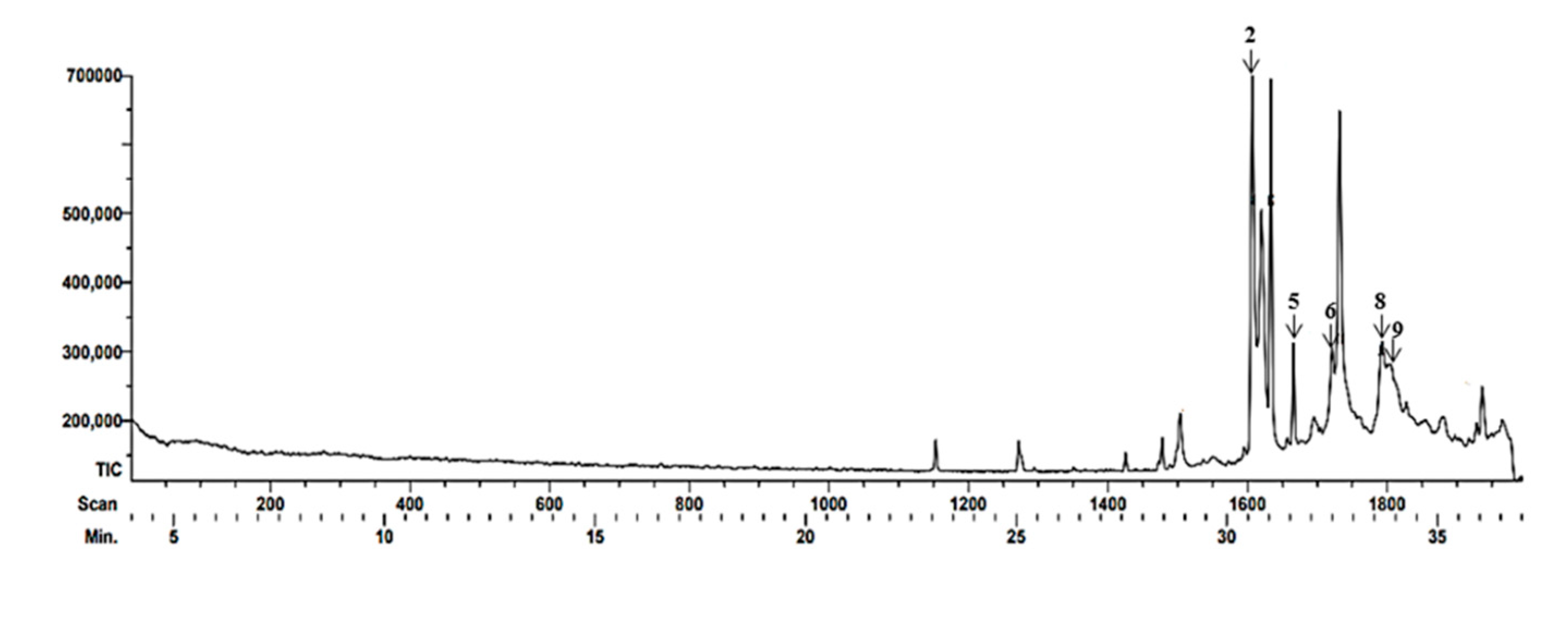
Figure 2.
Gas chromatogram corresponding to the propolis of Tetragonisca angustula and the main compounds detected.
Figure 2.
Gas chromatogram corresponding to the propolis of Tetragonisca angustula and the main compounds detected.
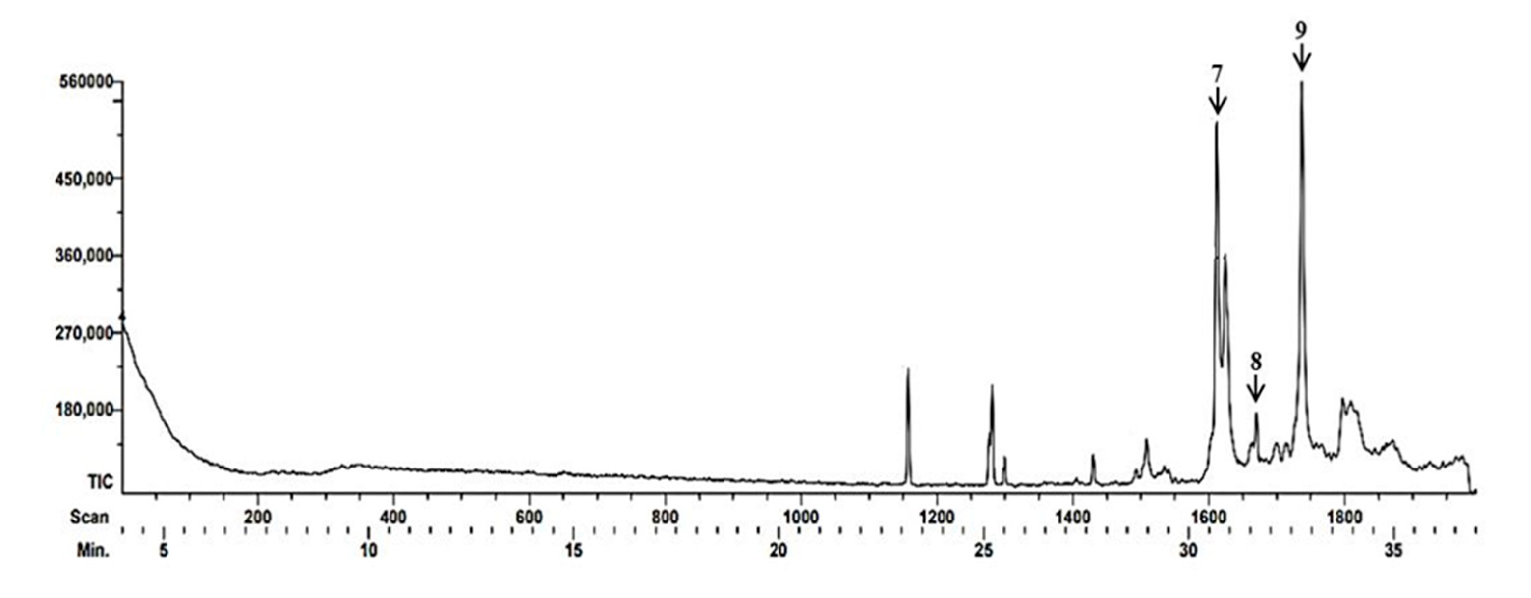
Figure 3.
Effect of EEP of Scaptotrigona mexican and Tetragonisca angustula on reference strain Malassezia pachydermatis ATCC 14522 were obtained by fluorescence microscopy and dyed with calcofluor-white and propidium iodide. Cultures were exposed to EEP at a concentration of 64 mg/ml for 48 h at 28 °C. Propidium iodide penetration was observed, indicating damage to the yeast. With calcofluor-white stain, only morphology deformation. (40x magnification).
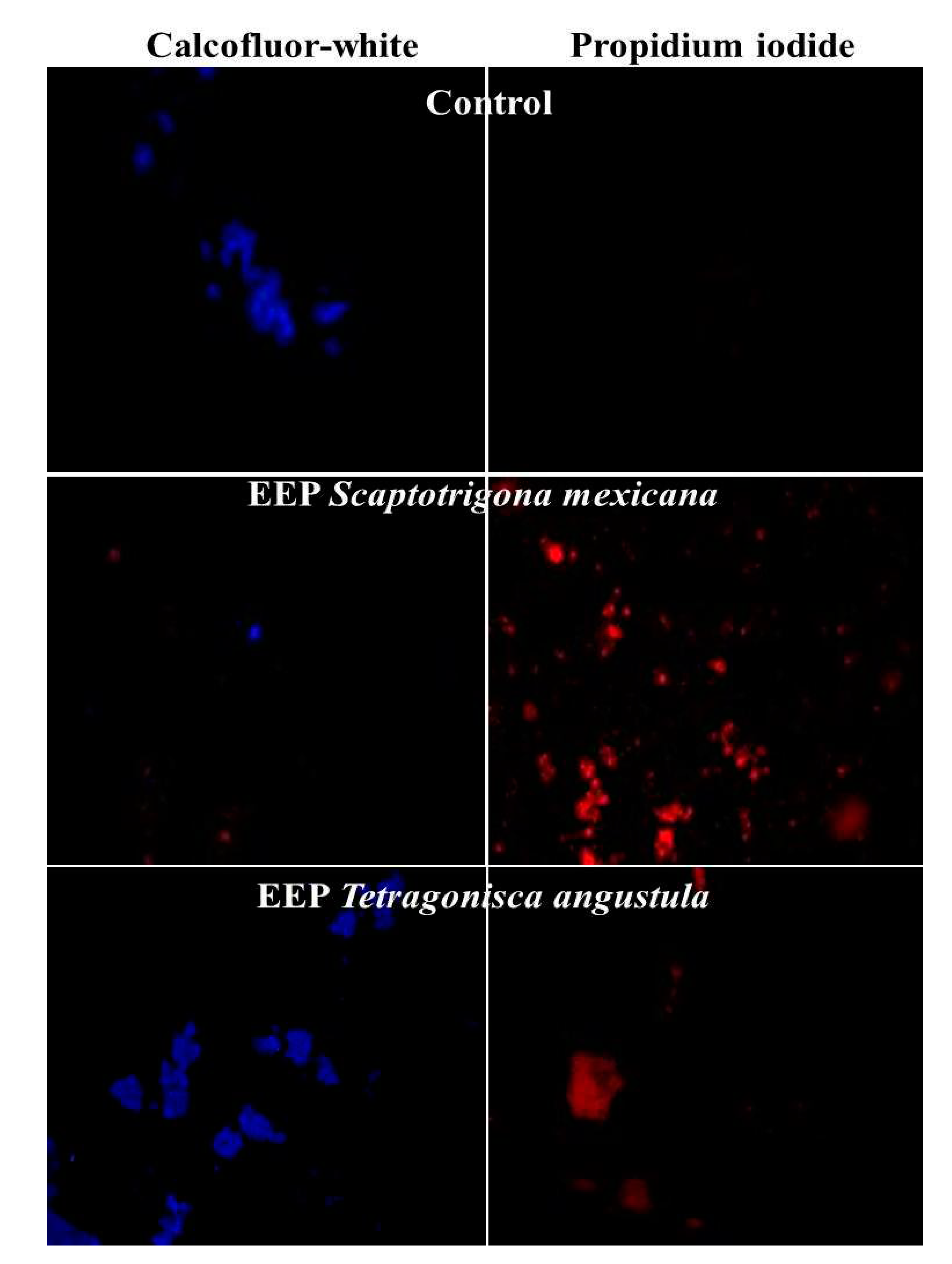
Figure 4.
Effect of EEP of Scaptotrigona mexicana and Tetragonisca angustula on the clinical strain oof Malassezia pachydermatis were obtained by fluorescence microscopy and dyed with calcofluor-white and propidium iodide. Cultures were exposed to EEP at a concentration of 64 mg/ml for 48 hr at 28 °C. Propidium iodide penetration was observed, indicating damage to the yeast. With calcofluor-white stain, alteration of morphology is observed. A greater damage was observed since it was not detected the presence with the calcofluor- white of the yeast and only the red of the propidium iodide was detected with Ethanolic Extract of Tetragonisca angustula Propolis.(40x magnification).
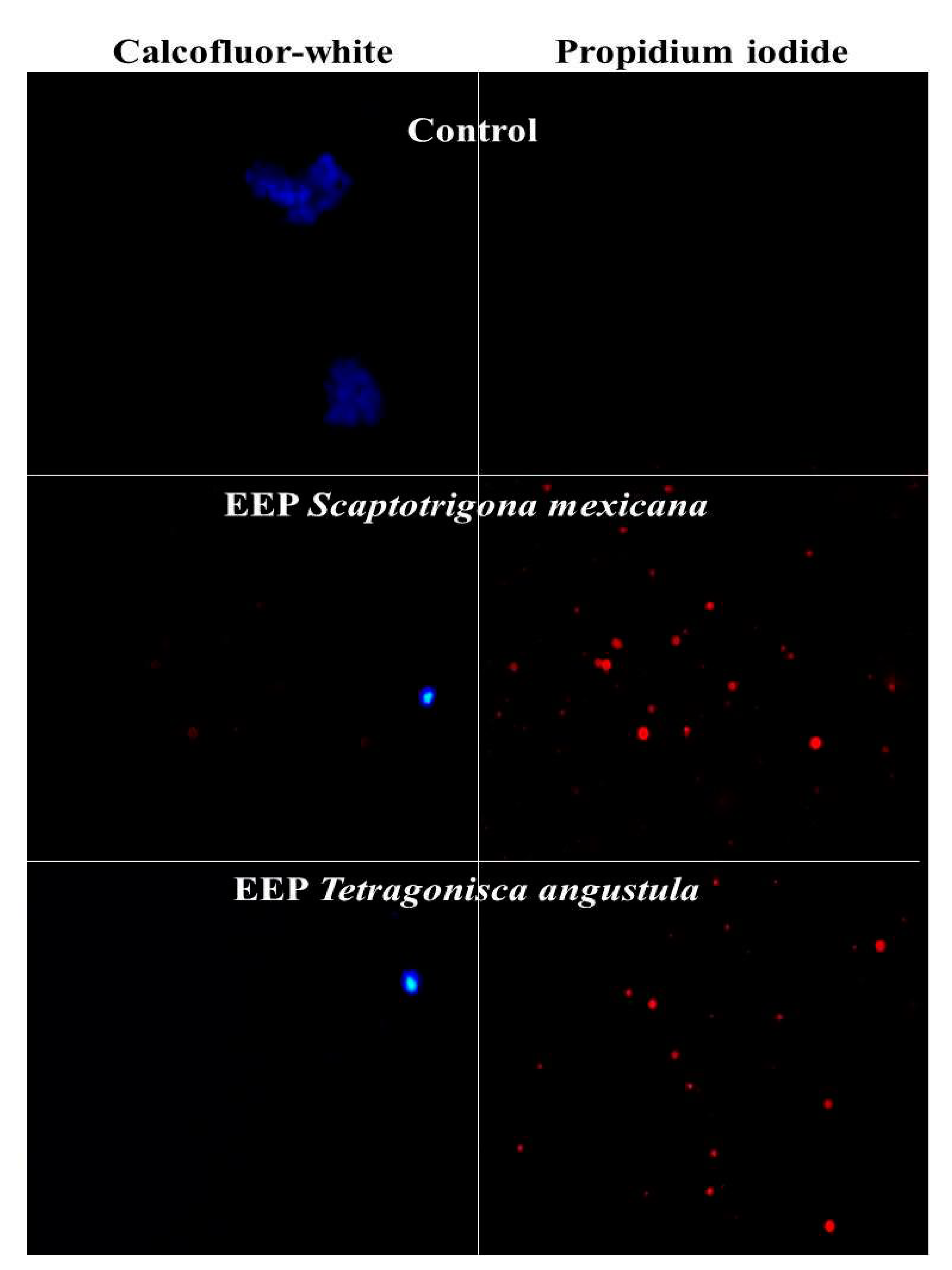
Table 2.
Main constituents of propolis of Tetragonisca angustula characterized by CG-EM.
| Beak | Time Retention (TR) |
Composite proposed by the database |
Chemical classification |
Biological activity |
Reference |
|---|---|---|---|---|---|
| 7 | 30.68 |
Solavetivona | Sesquiterpenoid and a cyclic ketone | Antifungal anti-inflammatory |
[34,35,36] |
| 8 | 31.65 | 2-Methyl-3-(3-methyl-but-2-enyl)-2-(4-methyl-pent-3-enyl)-oxethane |
Heterocycle compound | Not found Information | |
| 9 | 32.75 | (1S,6R,9S)-5,5,9,10-Tetramethyltricycle [7.3.0.0(1,6)]dodec-10(11)-ene | Sesquiterpene | Antibacterian antifúngal |
[37] |
Table 3.
Minimum inhibitory concentration (MIC) and minimum fungicidal concentration (FMC) values of stingless bee propolis extracts from two regions of the Mexican Republic on the reference strain M. pachydermatis ATCC 14522 and strains isolated from clinical samples.
Table 3.
Minimum inhibitory concentration (MIC) and minimum fungicidal concentration (FMC) values of stingless bee propolis extracts from two regions of the Mexican Republic on the reference strain M. pachydermatis ATCC 14522 and strains isolated from clinical samples.
| Origen | MIC (mg/ml) M. pachydermatis ATCC 14522 | MFC(mg/ml) M. pachydermatis ATCC 14522 |
Media CMI (mg/ml ) in isolation Clinical (n = 3) |
Media CMF (mg/ml) in isolation Clinical (n = 3) |
Number of Isolates Clinical Inhibited (n = 3) |
|
|---|---|---|---|---|---|---|
| Scaptotrigona mexicana | Municipio de Yecuatla Veracruz |
32 | 64 | 32 | 64 | 3 |
| Tetragonisca angustula | Municipio de Chalchihuitan Chiapas |
32 | 64 | 32 | 64 | 3 |
Disclaimer/Publisher’s Note: The statements, opinions and data contained in all publications are solely those of the individual author(s) and contributor(s) and not of MDPI and/or the editor(s). MDPI and/or the editor(s) disclaim responsibility for any injury to people or property resulting from any ideas, methods, instructions or products referred to in the content. |
© 2023 by the authors. Licensee MDPI, Basel, Switzerland. This article is an open access article distributed under the terms and conditions of the Creative Commons Attribution (CC BY) license (http://creativecommons.org/licenses/by/4.0/).
Copyright: This open access article is published under a Creative Commons CC BY 4.0 license, which permit the free download, distribution, and reuse, provided that the author and preprint are cited in any reuse.


