Preprint
Article

Polyphenols Analysis by LC-MS-ESI and Potent Antioxidant, Anti-Inflammatory, Antimicrobial Activities of Jatropha multifida L. Extracts Used in Benin Pharmacopoeia

Altmetrics

Downloads

207

Views

50

Comments

0

A peer-reviewed article of this preprint also exists.

Submitted:

26 July 2023

Posted:

04 August 2023

You are already at the latest version

Alerts
Abstract
Jatropha multifida L. is a plant belong to Euphorbiaceae family used in Benin pharmacopoeia because of its medicinal properties. The objective of this work is to appreciate the therapeutic power of this plant through the evaluation of some of its biological activities. After the preliminary phytochemistry, the polyphenols and flavonoids were quantified and identified by the LC-MS-ESI. The antimicrobial power of the extracts was investigated by agar medium diffusion. The antioxidant power of the extracts was evaluated by the reduction of the DPPH radical, the ABTS radical cation, the feric ion (FRAP) and the lipid peroxidation (LPO). Anti-inflammatory activity was assessed by inhibition of protein (albumin) denaturation. Indeed, different phenolic and flavonoid compounds namely, 2-Hydroxybenzoic acid, o-Coumaroylquinic acid, Apigenin-apiosyl-glucoside, Luteolin-galactoside, Luteolin-glucoside, Luteolin-rhamnoside, Quercetin-glucoside, Quercetin-arabinoside, Dicaffeoyquinic acid, Kaempferol-rhamnoside were identify. The J. mutifida extracts has a bactericidal effect with reference strains of wich CMBs vary from 22.67 mg/ml (S. aureus, S. enteridis, L. monocytogenes and C. albicans) to 47.61 mg/ml (E. coli) and with the meat isolated strains (S. equorum, S. saprophiticus, S. haemoliticus, S. cohnii and S. lentus). Ethanolic extract show the highest DPPH radical scavenging activity (IC50 = 0.72±0.03 mg/ml) while methanolic extract showed the highest ferric ion reduction (46.23±1.10 µgEAA/g). Contrary to the FRAP method, the reducing power with ABTS method of water-ethanolic extract was greater (0.49±0.11 mol ET/g). The same extract show the highest albumin denaturation inhibition power (97.31±0.35%) at 1000µg/ml. J. multifida extracts are rich in bioactive compound with good biological activity
Keywords: 
Subject: Biology and Life Sciences  -   Life Sciences

1. Introduction

The recurring threat of antibiotic resistance is a global health challenge in particular its presence in foodborne pathogens [1]. Indeed, food and its production can be a vector of antibiotic resistant bacteria and antibiotic resistance genes for humans, which has an impact on public health. Antibiotic-resistant bacteria are thought to be responsible for 700,000 deaths each year worldwide [2]. Apart from infectious diseases, inflammaging and oxidative stress are associated with several diseases such as cardiovascular disease, chronic obstructive pulmonary disease, arthritis, neurodegenerative diseases, cancer, diabetes, and more others [3]. But the anti-inflammatories currently in use are mostly non-steroidal and have enough adverse effects on the body. Apart from the contradicting effect of non-steroidal anti-inflammatory drugs used in the treatment of certain diseases such as cancer leaving uncertain the decision on their use for the treatment of cancers, NSAIDs are associated with other serious non-cancerous complications, such as myocardial infarction, gastrointestinal bleeding and renal failure [4].
Due to the problem of microorganisms resistance against antibiotics, and the increased of metabolic diseases linked to oxydatif stress and inflammation, a look at medicinal plants would be a good alternative because of their richness in natural compounds. Apart from primary metabolites use for their development and growth, plants also produce secondary metabolites which are multifunctional compounds which for the most part are involved in plant defense and communication with the environment [5]. The secondary metabolites generally described in plants according to their biosynthetic pathways are classified into five major molecular families, in particular phenolics, terpenes, steroids, alkaloids and flavanoids. Its various classes of molecules are known for their defense properties against pathogens, pests and herbivores, environmental stresses and the mediation of interactions between organisms, but also for their roles in modulating plant microbiomes [6]. Plants secondary metabolite are known for various biological activities, including antibacterial, antifungal activity, antioxidant and antiinflammatory activities.
The Benin republic presents, in its forest ecosystems and agro-ecosystems, over 162 forest plant species used for domestic food, commercial, with social meaning, religious or cultural [7]. A part of such uses, these palnts are also frequently used in local medicine. One of them is Jatropha multifida, belonging to Euphorbiaceae family. J. multifida is a shrub that is very often found in the several regions of the world (Asia and Africa) [8]. Popularly known in Benin as “wouèkè”, it is recognized for several properties including its purgative effects, against fever, infections of the skin and those of food origin, wound healing property. This study aimed to appreciate the therapeutic power of this plant collected in Benin through the evaluation of some of its biological activities

2. Materials and Methods

2.1. Chemicals and Microorganisms Culture Medium

The extraction solvents, and some chemical products such as PBS, Na2CO3, AlCl3, Folin-Ciocalteu (RE00180250), ferric chloride FeCl3, potassium acetate, quercetin, gallic acid, vanillin, sulfuric acid, hydrochloric acid HCl, sodium phosphate, ammonium molybdate, DPPH (2,2-diphenyl-1-picrylhydrazyl), were obtained from Sigma-Aldrich, St. Louis, MO, USA. HPLC solvents were purchased from Merck (Germany). Direct-Q UV system was used to purified water by Millipore (USA). The standard compounds for HPLC such as gallic, chlorogenic acid (purity 99% HPLC) and rutin (purity 99% HPLC) were purchased from Sigma (USA). Nutrient broth, Baird-Parker agar, TBX agar, XLD agar, Palcam agar and Muller Hinton agar were from Oxoid Ltd., Basingstoke, Hampshire, England. All the chemical and reagents were of analytical grade

2.2. Plant Material

The aerial organs (leaves) of J. multida used were collected from Aglogbè (06°28’58,6’’ N, 002°40’41,6’’ E), which is located in the department of Oueme, Benin. A voucher specimens No. AAC81108/HNB was deposited at the Benin national herbarium, University of Abomey-Calavi, Cotonou, Benin. The leaves were air-dried at the temperature between 23 ± 2°C for 14 days before powdered using grinder Retsch type SM 2000/1430/Upm/Smf, Haan, Germany.

2.3. Preliminary Phytochemical Profiling

The phytochemical profiling of J. multifida leave powder was perfomed in oder to determine the major group of secondary metabolites (nitrogenous, polyphenolic and terpenic compound, glycosides) using the previous method decribed by Houghton and Raman [9].

2.4. Preparation of Plants Extracts

Five solvents (Ethanol, Methanol, water-ethanol 30:70 (v/v), acetone and dichloromethane) were used to prepared J. multifida leaves extracts. Briefly, 1 g of plant material (leaves powder) was subjected to ultrasonication (35 Hz) in 100 mL of each solvent at room temperature for 2 h. Whatman N° 1 paper was used to collect the filtrate from each mixture which was then evaporated using an rotary evaporator (Heidolph Laborota 400 Rotovap, Schwabach, Germany) before oven dried at 40 °C.

2.5. Total Polyphenol Content Determination

Folin–Ciocalteu method [10] was used to determined J. multifida leaves extracts total polyphenol content. In a 96-well microplate, a mixture consisting of 25 µL of the Folin–Ciocalteu reagent and 10 µL of each extract was prepared. To this mixture were added 25 µL of 20% sodium carbonate solution after 5 min of incubation. This mixture was supplemented with 140 µL of ultrapure water. A blank was prepared under the same conditions, replacing Folin's reagent with ultrapure water. Gallic acid solution (500-0.97 µg/mL) was used as a reference, and the results are given in µg equivalents gallic acid per mg of sample. After 30 min, the absorbance values of the samples were read at 760 nm using a multiwell plate reader (Tecan Pro 200, Tecan Trading AG, Männedorf, Switzerland)

2.6. Total Flavonoid Content Determination

Aluminum chloride method previously described was used to quantified the total flavonoid in J. multifida leave extracts [11]. A mixture consisting of 100 µL of 2% aluminum chloride solution and 100 µL of each plant extract has been made in a 96-well microplate. The sample absorbance values were read after 15 min of incubation at 415 nm using the Tecan Pro 200 multiwell plate reader. Quercetin was used as a reference (40–0.078 µg/mL), and the results are presented in µg equivalents of quercetin per mg of sample.

2.7. Condensed Tannins Content Determination

The method described by Belem-Kabré et al. [12] was used to determined condensed tannins content of J. multifida leave extracts. Briefly, a mixture consisting of 1 ml of each plant extract (5 mg/ml) and 2 ml of vanillin 1% in 70 % of sulfuric acid has been made. This mixture was incubated in in water bath at 20°C for 15 min. The samples absorbance of was read at 500 nm using BiomateTM 3 Series Spectrophotometers, Thermo Scientific, Germany. The condensed tannins content T (%) was determined using the following formula:
T % = 5.2 × 10 2 × ( A × V ) P
5.2×10-2 = constant in equivalence of cyanidin, A = absorbance, V = extract volume and P = extract weight. The condensed tannins content of the samples was determined in triplicates and the results were converted to mg of cyanidin equivalent (CEq/g of extract).

2.8. Hydrolyzable Tannins Content Determination

The method previously described by Belem-Kabré et al. [12] was used to quatified hydrolyzable tannins content of J. multifida leave extracts. A mixture consisting of 1 ml of each extract at a concentration of 5 mg/ml and 3.5 ml of the reagent (ferric chloride FeCl3 10-2 M in hydrochloric acid HCl 10-3 M) has been made. After 30s incubation, the sample absorbance values at 660 nm were read using BiomateTM 3 Series Spectrophotometers, Thermo Scientific, Germany. The content was determined following the formula below:
T   ( % ) = ( A × P M × V × F D ) Ɛ m o l e × P
A = absorbance, PM = weight of gallic acid (170.12 g/mol), V = volume of extract, FD = dilution factor, ε mole = 2169 (constant in equivalence of gallic acid), P = extract weight

2.9. LC-DAD-MS-ESI + Analysis

The liquid chromatograph equipment used in the present study to carried out J. multifida leaves extracts phenolic compounds analysis was composed of HP-1200-LC equipped with a quaternary pump, autosampler, DAD detector and MS-6110 singlequadrupole API-electrospray detector (Agilent-Techonologies, USA). For the phenolic compounds detection, different fragmentor, in the range 50-100 V, were applied in ESI+ mode. For the chromatographic analysis, the Kinetex XB-C18 (5 μm; 4.5x150 mm i.d.) column used were from Phenomenex, USA. The solvents used as the mobile phase were composed of water acidified by acetic acid 0.1 % (A) and acetonitrile acidified by acetic acid 0.1 % (B). For the elution, the following multistep linear gradient for 30 min with elution flow rate set at 0.5 ml/min at 25±0.5 °C was applied: start with 5% B for 2 min; from 5% to 90% of B in 20 min, hold for 4 min at 90% B, then 6 min to arrive at 5% B. Mass spectrometric detection of positively charged ions was performed using the Scan mode. The following experimental conditions were applied: gas temperature 350°C, nitrogen flow 7 l/min, nebulizer pressure 35 psi, capillary voltage 3000 V, fragmentor 100 V and m/z 120-1500. Chromatograms were recorded at wavelength λ = 280, 350 nm and data acquisition was done with the Agilent ChemStation software.

2.10. Antimicrobial Activity of J. multifida Leaves Extracts

2.10.1. Microorganisms Strains and Growth Conditions

Twelve (12) microbial strains were used to perform the sensitivity tests. Active extracts were selected for MIC and CMB determination. The twelve microorganisms consist of five (05) reference strains including two (02) Gram+ bacteria (Staphylococcus aureus ATCC 6538P, Listeria monocytogenes ATCC 19114), two (02) Gram- bacteria (Escherichia coli ATCC 25922, Salmonella enteritidis ATCC 13076) and one (01) yeast (Candida albicans ATCC 10231). Apart from these reference strains, seven (07) Satphylococcus strains isolated from pork by Attein et al. [13] were used. These meat isolated strains are from the collection of the Laboratory of Biology and Molecular Typing in Microbiology (University of Abomey-Calavi, Benin)
The different strains were revified by microbial inoculation in 10 mL of sterile nutrient broth incubated at 37°C for the bacteria and at 30°C for the C. albicans yeast for 24 hours. After a microscopic examination in order to confirm the purity of the innoculum, each strain was inoculated on selective agar medium previously poured into sterile Petri dishes and incubated at 37°C/24h in order to confirm the morphology of the cultures by microscopic observation. From petri dishes, two to three colonies were inoculated in a sterile saline solution before being adjusted to McFarland turbidity (1.5 × 10⁸ CFU/mL) [14]. The selective agar media used are as follows: Baird-Parker agar base supplemented with Egg Yolk Tellurite Emulsion for Staphylococcus strains, TBX agar for E. coli, XLD agar for Salmonella enteritidis and Palcam agar for Listeria monocytogenes.

2.10.2. Antibiogram

It was performed only with meat isolated Stapylococcus strains by disc diffusion method [15]. For each strain, Petri dishes containing Muller Hinton Agar were inoculated with 1 ml of the appropriate innoculum. The excess was aspirated using sterile blotting paper before depositing wathman paper discs (2 to 3) which were impregnated with 25 µL of J. multifida leaves extracts at 20mg/mL. After the incubation period (37°C for 24h), the dishes were examined for inhibitory zones. Each sample was performed in triplicate.

2.10.3. Determination of the Minimum Inhibitory Concentration (MIC)

The microdilution method using resazurin as cell viability indicator was used. First, 100 µL of nutrient broth for bacterial strains and TSB for yeast was added to all wells. From the first wells, a half dilution was carried out on row with 100 μL of each sample. The different dilutions were inoculated with 10 µL of the inoculum ((1.5x10⁴CFU/mL) corresponding to each strain before being incubated for about 24 hours at 37°C for bacteria starins and 30°C for Candida albicans. Gentamicin (0.04 mg/mL in saline solution) and fluconazole (10 mg/mL) were used as control. After these culture period, 20 µL of resazurin were added to each wells and the plates were incubated under the same conditions (37°C for bacteria starins and 30°C for Candida albicans) for 2 hours. The lowest concentration at which the blue color did not change into pin was taken as the MIC.

2.10.4. Determination of the Minimum Bactericidal/Fungicidal Concentration (MBC/MFC)

The agar medium inoculation method was used to determined the MBC and MFC. In each row, 10 μL of each dilution from the MIC to the highest concentrations of J. multifida leaves extracts were inoculated on Mueller-Hinton agar for the bacteria and YPD agar for the yeast. Petri dishes were incubated for 24 hours at 37°C for MBC and 30°C for MFC. The lowest concentration that prevented the growth of bacteria and yeast (no colonies on the plate) was considered respectively MBC and MFC [16].

2.11. Microplate Determination of Plant Extracts Antioxydant Activity

2,2-Diphenyl-1-Picrylhydrazyl Assay

The DPPH (2,2-Diphenyl-1-picrylhydrazyl) radical scavenging activiy of J. multifida leaves extracts was conducted according to microplate medthod decribed by Chokki et al. [17]. In each well, 100 µL of DPPH solution at 50 μM and 100 µL of plant extracts at 200 μg/mL were added. The mixture was kept in darkness for 20-30 min at room temperature before reading the absorbance at 517 nm using a microplate reader (Tecan Pro). The blank was prepared in the same condition, replacing the sample by 100 µL of methanol. The DPPH radical scavenging activiy of J. multifida leaves extracts and standards (ascorbic acid, BHT) was obtained using the formula previously established [18]:
I n h i b i t o r y   P e r c e n t a g e ( % ) = B l a n k   s a b s o r b a n c e S a m p l e   s a b s o r b a n c e B l a n k s   a b s o r b a n c e   × 100
The IC50 (concentration providing 50% inhibition) was calculated using regression equation obtained from the curve showing the inhibition percentage according to plant extracts concentrations [19].

The 2,2-Azinobis (3-Ethylbenzthiazoline)-6-Sulfonic Acid Assay

The protocols described by Cudalbeanu et al. [20] was used to evaluated ABTS radical scavenging capacity of J. multifida leaves extracts. The radical was produced by mixing 5 mL of ABTS solution at 7.8 mM with 5 mL of potassium persulfate solution at 140 mM and allowed to stand in darkness for 12 h at room temperature before being diluted to reach an absorbance between 1.1 ± 0.02 units at 734 nm. 100 µL of fresh ABTS solution was mixed with 100 µL of J. multifida leaves extracts and the absorbance was measured at 734 nm after 30 min of incubation. Trolox was used as a standard molecule for the calibration curve (7.8µg/mL to 62.5µg/mL) and the results were expressed as ABTS radical inhibition percentage and in Mole equivalent Trolox (mol ET/g). All assays were performed in triplicate.

Ferric Reducing Antioxydant Power (FRAP)

This was carried out according to the method previous described by Jones et al. [21] with some modifications. Indeed, a mixture consisting of 100 mL acetate buffer (250 mM, pH 3.6), 10 mL 2,4,6-tris(2-pyridyl)-s-triazine (TPTZ) solution (10 mM in 40 mM HCl) and 10 mL FeCl3 (20 mM) was produced. A quantity of 200 µL of this mixture constituting the working solution was mixed with 50 µL of J. multifida leaves extract before being incubated at 37 °C for 10 min. The absorbance of this reaction mixture was read at 593 nm. Ascorbic acid was used as a positive control for the standard curve (0 to 250µg). The potential of the extracts to reduce iron (III) to iron (II) was expressed as ferric reducing inhibition percentage and as microgram equivalent ascorbic acid per gram of extract (µg EAA/g sample). Values ​​were presented as means of triplicate analyzes

Lipid Peroxidation Inhibitory Test (LPO)

This activity was evaluated using the protocol described by Belem-Kabré et al. [12] by substituting the rat liver homogenate by that of the egg yolk. Briefly, 0.2 ml of J. multifida leaves extracts solution (10 mg/mL) or 1.5 mg/mL of ascorbic acid solution was mixed with 1 mL of egg yolk homogenate (10%) in phosphate buffered saline (PBS) buffer (pH 7.4), 50 μl of FeCl2 (0.5 mM). The mixture was incubated at 37°C for 60 min before adding 50 μl of H2O2 (0.5 mM), 1 ml of trichloroacetic acid (15 %) and 1 ml of 2-thiobarbituric acid (0.67%). This mixture was centrifuged (2000 rpm for 10 min) afetr incubation in boiling water for 15 min. A control was carried out under the same condition by replacing the extract with distilled water. The absorbances were read with a spectrophotometer (Epoch Biotek Instruments, U.S.A.) at 532 nm. The lipid peroxydation percentage inhibition was calculated using the formula below:
% I n h i b i t i o n = A b s   c o n t r o l A b s   s a m p l e A b s   c o n t r o l × 100

2.12. Anti-Inflammatory Capacity of J. multifida Extracts

The in vitro anti-inflammatory activity of J. multifida extracts was evaluated following the previous method used in the work of Kabré et al. [22]. Briefly, to 100 µL of J. multifida leaves extracts at various concentrations (31.25 to 1000 µg/mL), 10µL of egg albumin, 140µL of phosphate buffered saline (PBS, pH 6.4) were added. This reaction mixture was incubated at 37°C for 15 min and then heated at 70 °C for 5 min. Blank was prepared under the same reaction conditions by replacing the extracts with the same volume of dilution solvent. Absorbances were measured after cooling at 660 nm using a microplate reader (Tecan Infinite M 200 Pro,USA). The effect of J. multifida leaves extracts on the thermal denaturation of albumin at 70°C was expressed by the inhibition rate calculated according to the formula below
%   I n h = A b s   C A b s   S A b s   C
%Inh: Inhibition percentage; AbsC: Absorbance of Control; AbsS: Sample Absorbance. The IC50 (concentration providing 50% inhibition of thermal denaturation of albumin) was calculated using regression equation obtained from the curve showing the inhibition percentage according to plant extracts concentrations

2.13. Statistical Analysis

The experimental results were reported on bench sheets and entered into an Excel 2016 database for processing. GraphPad Prism® 8.0.2 software Inc was used for the graphs and statistical analysis. Statistical analyzes were performed using multivariate analysis of variance followed by Tukey's test. P values ​​< 0.05 were considered statistically significant. The results are presented as mean ± standard deviation.

3. Results

3.1. Preliminary Phytochemical Profiling

The preliminary phytochemical analysis performed with J. multifida powder revealed the presence of several secondary metabolites (Table 1). The presence of polyphenols and flavonoids was confirmed through a UV-vis spectrum at 340 nm specific to polyphenols and at 280 nm specific to flavonoids (Figure 1). For the qualitative test, it was noted an uneven distribution of these metabolites. Indeed, 56.25% of the studied secondary metabolites were present in J. multifida leaves powder. Alkaloids were observed in nitrogen compunds group. In the polyphenolic compound group, flavonoids, anthocyanins and leuco-anthocyanins, tannin gallic and quinonics derivate were found. In the terpenic compound group, only triterpenes were found. In addition, in the glycosides group, saponosids and mucilags were found

3.2. Total Phenolic and Flavonoid, Condensed and Hydrolyzable Tanins Content

The contents of the total phenolics and flavonoid, condensed and hydrolyzable tanins were presented in Table 2. The content of quantified secondary metabolites varies according to the extract type (p < 0.05). Indeed, the highest content of polyphenolic compounds (45.01±11.87 mgEqGA/g) was obtained with the methanolic extract while the lowest content (11.25±1.37 mgEqGA/g) was obtained with the aqueous extract. For the total flavonoids, the hydro-ethanolic extract showed the highest content (7.43±0.12 mgEqQ/g) but which is not different (p > 0.05) from those obtained with the ethanolic extract (7.18 ±2.85 mgEqQ/g) and the aqueous extract (6.58±1.36 mgEqQ/g). In this group, the dichloromethane extract showed the lowest content of total flavonoids (0.32±0.01 mgEqQ/g). The acetone extract recorded the highest content of condensed tannins (8.58±0.45 mgEqC/g) followed by the dichloromethane extract (6.79±0.34 mgEqC/g) and the methanolic extract (6.49±0.46 mgEqC/g). Hydrolysable tannins are more concentrated in dichloromethane with a content of 4.12±0.29 mgEqGA/g and less concentrated in the mixture water-ethanolic with a content of 2.11±0.12 mgEqGA/g.

3.3. Identification of Phenolic Compounds in J. multifida Extract by LC-DAD-MS-ESI+ Analysis

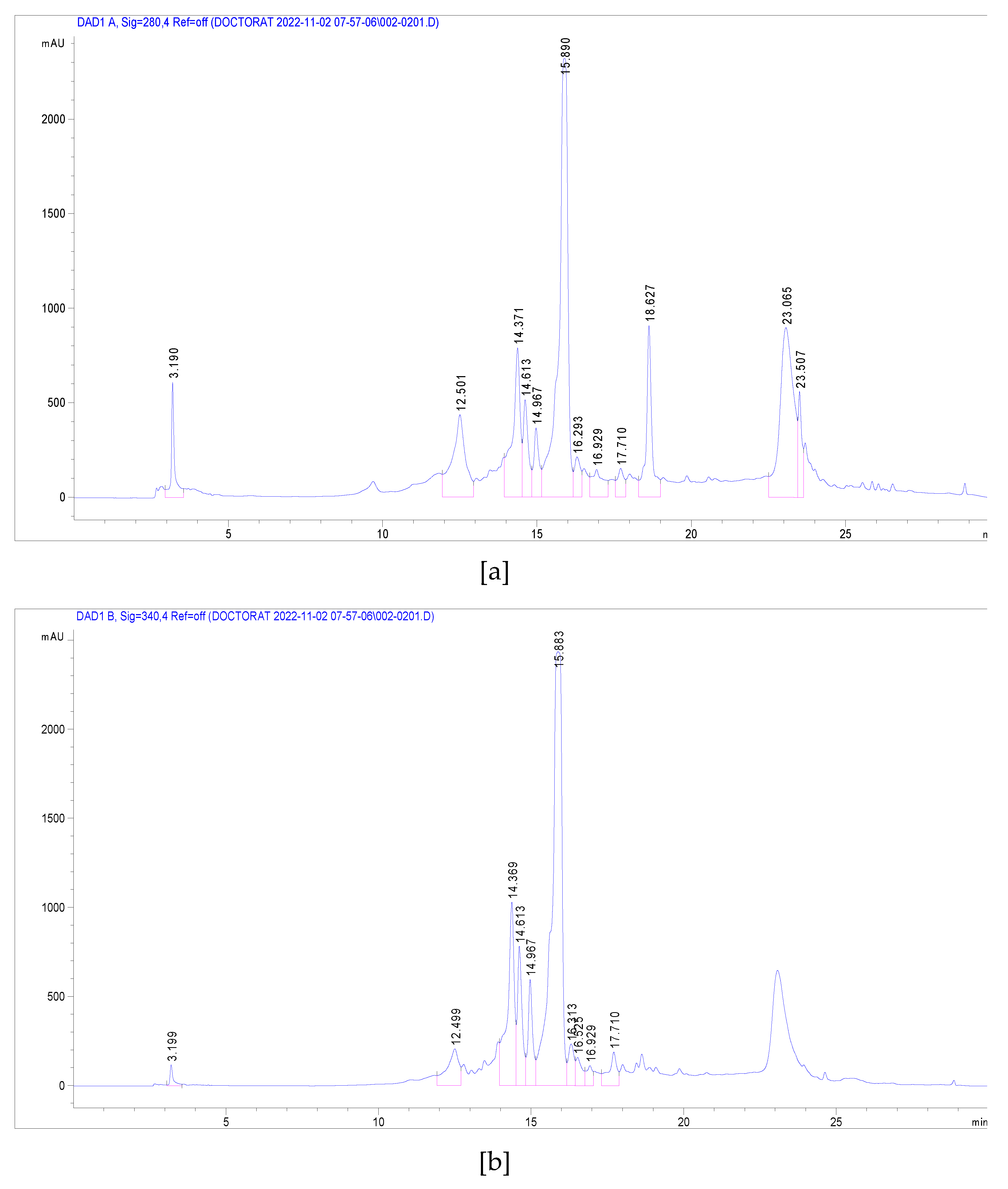
Mass spectrometry methodology was used to identify polyphenolic compounds present in methanol J. multifida leaves extract. These compounds were identified through the fragmentation of the molecules in the sample. Chromatograms were recorded at wavelength λ = 280, 350 nm (Figure 2). Therefore, the identification and quantification of the bioactive compounds from J. multifida leaves extract was shown in Table 3. Indeed, different phenolic and flavonoid compounds namely, 2-Hydroxybenzoic acid, o-Coumaroylquinic acid, Apigenin-apiosyl-glucoside, Luteolin-galactoside, Luteolin-glucoside, Luteolin-rhamnoside, Quercetin-glucoside, Quercetin-arabinoside, Dicaffeoyquinic acid, Kaempferol-rhamnoside were identify. Among these compounds, Luteolin-rhamnoside is the most abundant (19.73 mg/g) compound of J. multifida leaves extracts. This compound is followed by Apigenin-apiosyl-glucoside with a rate of 5.58 mg/g. Of all the compounds identified, three were identified at almost similar rates. There are: Luteolin-glucoside (2.17 mg.g), o-Coumaroylquinic acid (2.29 mg/g) and Luteolin-galactoside (2.89 mg/g). Dicaffeoyquinic acid, which belongs to the subclass of Hydroxycinnamic acid, is the compound identified with the lowest content (0.63 mg/g).

3.4. Antimicrobial Activity of J. multifida Leaves Extracts

3.4.1. Susceptibility of Reference Strains to J. multifida Leaves Extracts

The aqueous and ethanolic extracts were tested on the reference strains. The results are presented in Table 4. It is noted that these two extracts are active on all the reference strains at varying doses. For the aqueous extract, the MICs vary from 22.67 mg/mL (S. aureus) to 47.61 mg/ml (E. coli, S. enteridis, L. monocytogenes and C. albicans) while most of the MBCs are higher than 47.61 mg/ml except for S. aureus. Furthermore, for the ethanolic extract, the MICs vary from 5.14 mg/ml (S. aureus) to 34.54 mg/ml (E. coli) while the CMBs vary from 22.67 mg/ml (S. aureus, S. enteridis, L. monocytogenes and C. albicans) to 47.61 mg/ml (E. coli). The comparative action of the extracts shows that the aqueous extract has a bacteriostatic effect with reference strains while the ethanolic extract has a bactericidal effect on 4 reference strains except S. aureus.

3.4.2. Susceptibility of Maet Isolated Strains to J. multifida Leaves Extracts

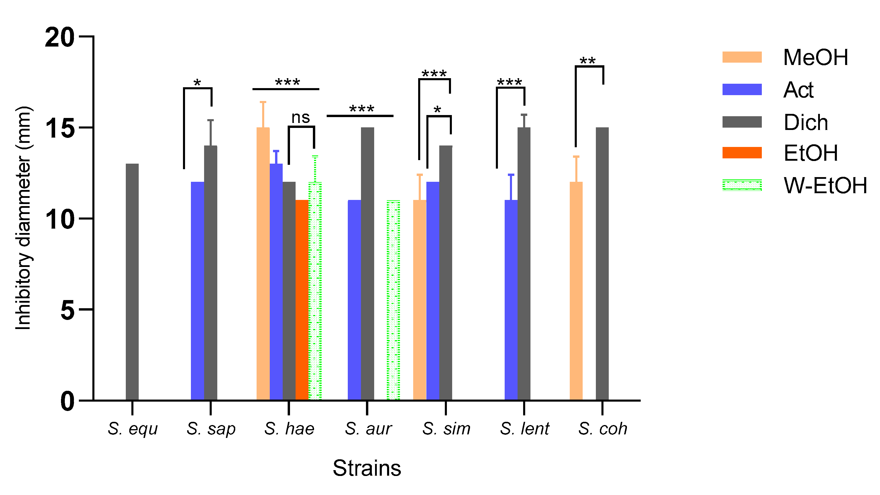
The diameters of the inhibition zones vary according to the extract type (p < 0.001) and the strain (p < 0.001). The aqueous extract has no action on the meat isolated strains. The dichloromethane extract has a broad spectrum action by inhibiting 100% of the strains, while the acetone extract inhibited 71.42%, followed by the methanol extract (42.85%), then the water-ethanolic extract extract (28.57%), and then ethanolic extract (14.28%). The greatest inhibition diameter (15.00±0.70 mm) was obtained with the dichloromethane extract on three strains (S. aureus, S. lentus and S. cohnii). Nevertheless, the methanolic extract also showed a diameter of 15.00±1.41 mm on S. haemoliticus strain. Apart from these observed performances, the dichloromethane extract inhibited S. saprophiticus strain with a diameter of 14.00±1.41 mm and also S. simulans strain with a diameter of 14.00±0.00 mm. Of all foodborne strains, S. haemoliticus is the most sensitive to all extracts while S. equorum is the most resistant in terms of extract action spectrum (Figure 3). For S. haemoliticus strain which is the most sensitive, the compared action of the extracts shows that the greatest variation in the inhibition diameters was obtained between the methanolic extract and the ethanolic extract (p < 0.001) while the lower variation was obtained between the acetone and methanol extract (p = 0.01).
Like the inhibition diameters, the MICs of the active extracts also vary according to the strains and the extract types. With the methanolic extract, the lowest MIC (10mg/ml) was obtained with S. cohnii strain as well as the lowest MBC (20mg/ml). The ratio between these two parameters shows that the methanolic extract has a bactericidal effect on S. cohnii strain (Table 5). As for the acetone extract, although it presented a MIC of 10 mg/ml, it has a CMB>20 mg/ml on all sensitive strains. For the dichloromethane extract, the MICs vary from 2.5 mg/ml to 5 mg/ml while the CMBs vary from 5 mg/ml to 20 mg/ml. Dichloromethane extract is the most active extract on foodborne strains. Indeed, the dichloromethane extract has a bactericidal effect with S. equorum, S. saprophiticus, S. haemoliticus and S. lentus strains. Although this extract exhibited a bacteriostatic effect with S. simulans strain, it recorded a CMB of 10mg/ml. The ethanolic extract meanwhile showed a bactericidal effect on S. haemoliticus strain while the water-ethanolic extract has no bactericidal effect on the strains which are sensitive to it.

3.5. Antioxydant Activity of J. multifida Leaves Extracts

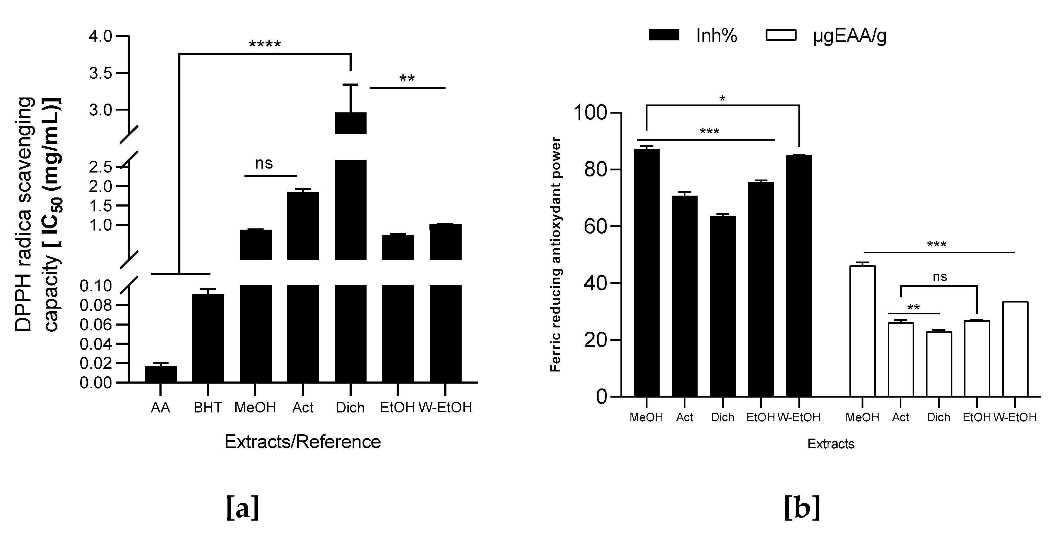
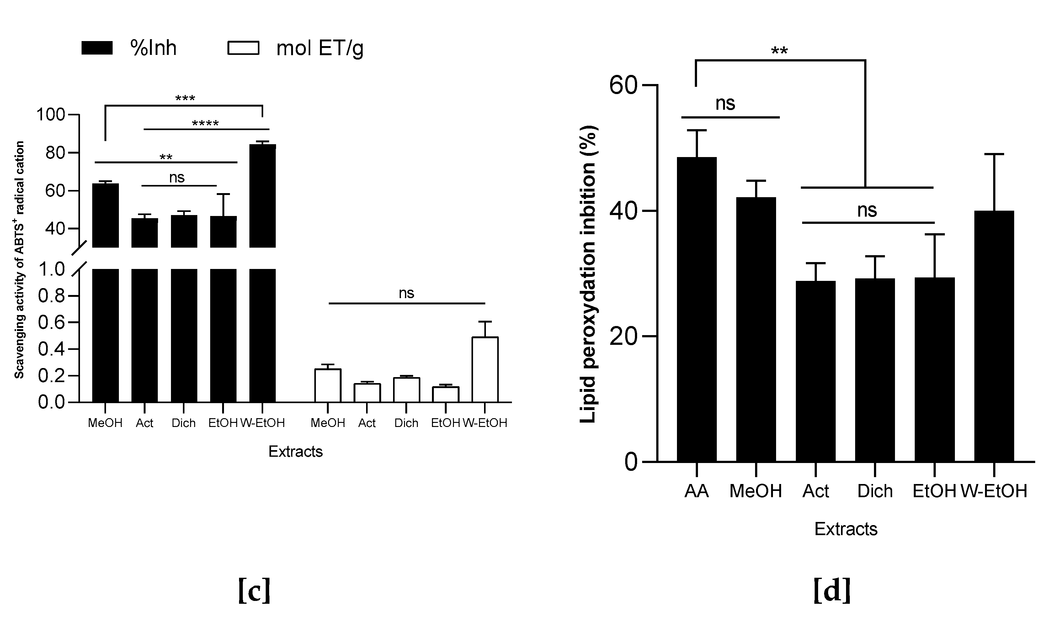
The antioxidant power of the extracts was evaluated by the reduction of the DPPH radical, the ABTS radical cation, the feric ion (FRAP) and the lipid peroxidation (LPO). The results are shown in Figure 4. The highest antioxidant power of J. multifida extracts using DPPH radical scavenging activity was observed with ethanolic extract because it has the lowest IC50 value (0.72±0.03 mg/ml), followed by methanolic extract who recorded an IC50 of 0.87±0.01 mg/ml. Besides, dichloromethane extract showed the lowest antioxydant activity with DPPH method because it has a high IC50 value (2.96±0.37 mg/ml). Depending on the IC50 obtained, we notice intermediate antioxidant powers of the water-ethanolic extracts (IC50 = 1.01±0.01 mg/ml) and acetone extract (IC50 = 1.85±0.08 mg/ml). Compared to our extracts, the two reference molecules showed strong DPPH radical reduction power with IC50 values ​​of 0.02±0.00 mg/ml (ascorbic acid) and 0.09±0.01 mg/ml for BHT (Figure 4a). Furthermore, the methanolic extract showed the highest percentage of ferric ion inhibition (87.28±1.01%) with a concentration of 46.23±1.10 µgEAA/g (Figure 4b). It is followed by the hydroethanolic extract (85.05±0.04%) with a concentration of 33.55±0.03 µgEAA/g of extract. On the other hand, the dichloromethane extract showed the lowest reduction capacity of the ferric ion (63.79 ± 0.54%) as observed with DPPH reduction. Considering the ABTS radical cation inhibition capacity (Figure 4c), the extracts activity (inhibition percentage) decreases as follows: acetone (45.49±2.10%) < ethanol (46.58±11.59%) < dichloromethane (47.15±2.02%) < methanol (63.78±1.16%) < water-ethanol (84.26±1.68%). Contrary to the FRAP method, the reducing power with ABTS method of water-ethanolic extract was greater (0.49±0.11 mol ET/g) than the one of methanolic extract (0.25 ± 0.03 mol ET/g). For lipid peroxidation essai, methanolic extract show the highest inhibition rate (42.19±2.60%) followed by water-ethanolic extract (40.00±9.07%). No difference (p > 0.05) was observed between these two extracts and the potency of ascorbic acid used as reference (48.59±4.21%). Acetone extract recorded the lowest inhibition rate (28.80±2.89%). Ascorbic acid show the hight lipid peroxidation inhibition compared to acetone extract (p = 0.0019), dichloromethane (p = 0.0024) and ethanol extract (p = 0.0026).

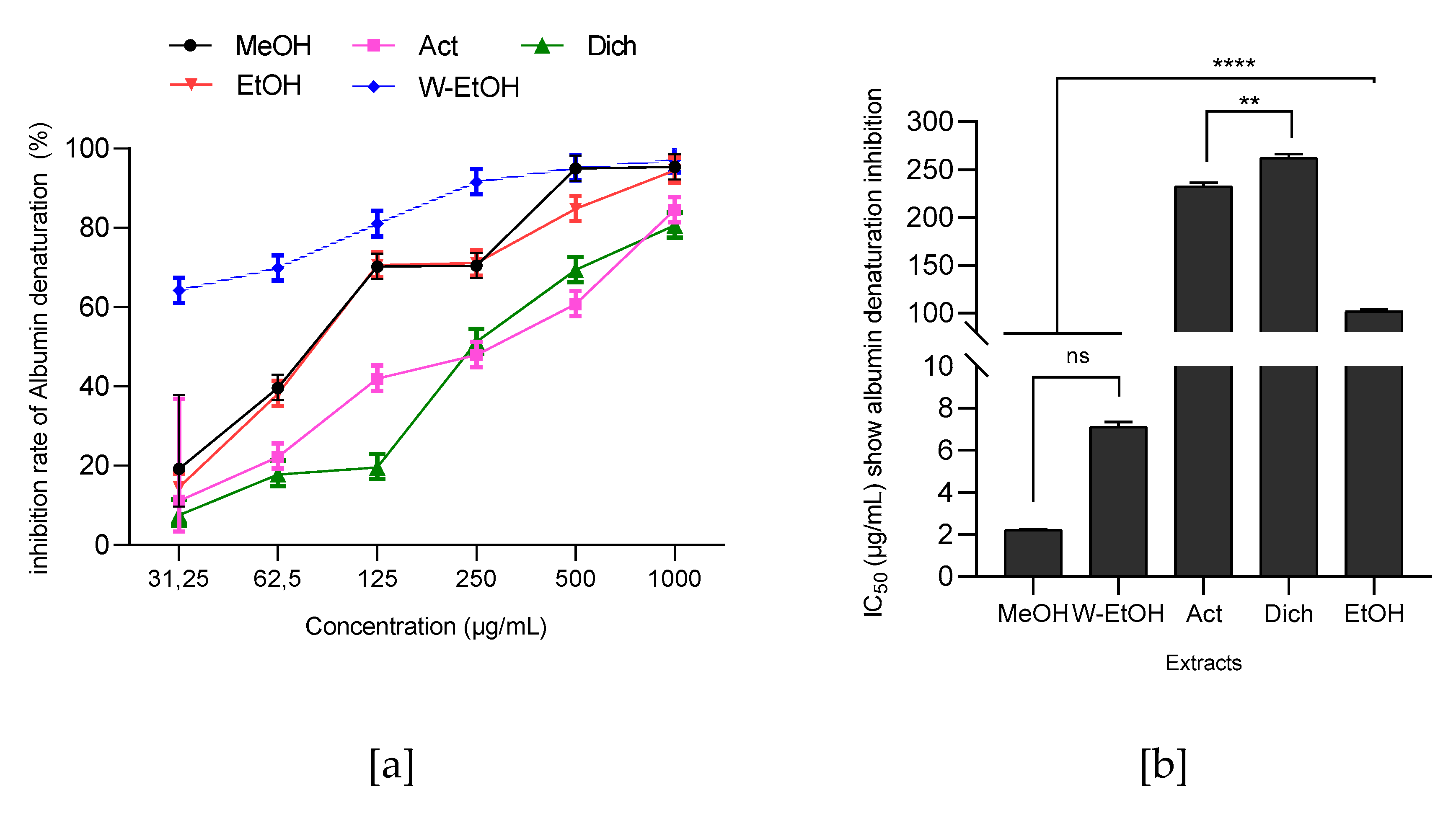
3.6. Antiinflammatory Activity of J. multifida Leaves Extracts

The capacity of J. multifida leaves extracts to prevent the thermal denaturation of protein (albumin) at 660nm was evaluated. A dose-dependent activity of J. multifida tested extracts is observed (Figure 5). The highest albumin denaturation inhibition percentage was observed with the water-ethanolic extract (97.31±0.35%) at 1000µg/ml. This extract is followed by the methanolic extract at 1000µg/ml which manages to inhibit the albumin denaturation at a rate of 95.35±1.05%. Moreover, the dichloromethane extract showed the lowest denaturation inhibition percentage (80.64 ± 1.32%). Although the methanolic extract presented the lowest IC50 (2.24±0.03 µg/ml), the same trend was observed for the extracts power action by considering the IC50 because the analysis of variance ANOVA shows that there is no difference (p > 0.05) between the methanolic and water-ethanolic extracts (7.12±0.24 μg/ml). The dichloromethane and acetone extracts showed the highest IC50 values respective ​​of 262.17±4.07 µg/ml and 232.51±3.96 µg/ml.

4. Discussion

Phytochemical screening showed that J. multifida leaves powder are source of secondary metabolite. The presence of these secondary metabolites in the powder suggest the probable biological activities of the tested extracts. The preliminary screening reveals, in nitrogen compunds group, the presence of alkaloïds. The work realized by Rampadarath et al. [23] and Hanafi et al. [24] led to the same result with J. multifida leaves collected respectively from different localities of Mauritius and Indonesia. It’s know that alkaloids have wide biological activities such as antiviral, antibacterial, anti-inflammatory, antioxydant and anticancer properties [25]. In this study we noted the presence of tanins, flavonoids and saponins. The presence of flavonoids and saponins have earlier been reported in Nigeria by Aiyelaagbe [26], in Niger by Nwokocha et al. [27] and in Thailand by Chokchaisiri et al. [28] during their work in the same plants. Tannins and flavonoids are reported to have antibacterial, antivirals, antifungals, and antioxidant potential. These compounds are also know to promote tissue regeneration in case of superficial burn injury [29]. In addition, terpenics compounds are found in J. multifida leaves, several study are also reported the presence of monoterpene, diterpenes and triterpenes [30,31]. Terpenes compound like others natural compounds have been reported by serveral authors to exert antimicrobial activities against antibiotic-resistant bacteria. Terpens compounds have the capacity to promote cell rupture and inhibit protein and DNA synthesis [32,33]. Zhao et al. [34] reported that terpene compounds play an important role in maintaining human health. They (terpene compounds) are known for their ability to treat several human diseases and are thus known as antimicrobial, anticancer, antiinflammatory, antioxidants, neuroprotective and many others agents. In the J. multifida leaves powder, the absence of cyanogenic derivatives, is very important because these are the causes of toxicity due to the production of cyanide ions and manifested by the consecutive massive poisoning to ingestion of derivatives cyanogenic to acceleration and amplification of respiratory rate, respiratory depression, dizziness, headache, disturbance of consciousness, coma etc [29]. The quantification of total phenolics and flavonoid, condensed and hydrolyzable tanins confirm the results obtained with qualitative tests. Mass spectrometry methodology with electrospray in positive ionization mode was used to identify polyphenolic compounds present in methanol J. multifida leaves extract. Different phenolic and flavonoid compounds namely, 2-Hydroxybenzoic acid, o-Coumaroylquinic acid, Apigenin-apiosyl-glucoside, Luteolin-galactoside, Luteolin-glucoside, Luteolin-rhamnoside, Quercetin-glucoside, Quercetin-arabinoside, Dicaffeoyquinic acid, Kaempferol-rhamnoside were identify. This study is one of the few works that have used this technic (LC-MS-ESI) for the analysis of J. multifida compounds unlike the species J. curcas and J. gossipifolia. However, recent works [24] have used the GC-MS technic to analysis volatile compounds of J. multifida and thus demonstrate the diversity of secondary metabolites sources of biological activities of J. multifida extracts. Indeed, the tested extracts shows antimicrobial activity with bactericidal effect through ethanolic extract on the used reference strains (E. coli, S. enteridis, L. monocytogenes and C. albicans). In addition, the dichloromethane extract has a bactericidal effect with on the meat isolated strains (S. equorum, S. saprophiticus, S. haemoliticus and S. lentus). Others study in Indonesia and Togo [23,35] are shows the antimicrobial activity of J. multifida extracts. This antimicrobial activity can be attribuated to the tanins who exhibit antibacterial effects on Gram+ and Gram- bacteria. It has been reported in litterature [36] that tannins exert their antibacterial activity by several mechanisms including the complexation with enzymes or bacterial substrates but also by permeability of the cell membrane. One the other hand, antimicrobial activity of these extracts can be attribuated to the alkaloids and Dicaffeoyquinic acid found in the J. multifida leaves. The membrane of bacterial cells being made up of lipid bilayer and glycoproteins, it has been reported that one of the mechanisms action of alkaloids is part of them insertion into the lipid bilayer and their interaction with the part of sugars present followed by a chemical rearrangement resulting in cell lysis [37]. Dicaffeoylquinic acids derived from Youngia japonica, a biannual medicinal herb and the others plants, exhibited antibacterial activities [38,39,40].
Outside of antimicrobial activity, J. multifida leaves extracts shown antioxydant and antinflammatory activity. All of the tested extracts display a good antioxydant activity by the reduction of the DPPH radical, the ABTS radical cation, the feric ion (FRAP) and the lipid peroxidation (LPO). This activity can be link to the identified compounds in this study by LC-MS-ESI technic. Indeed, Luteolin-rhamnoside is the most abundant (19.73 mg/g) compound of J. multifida leaves extracts. Also the compound such as Luteolin-glucoside, Quercetin-glucoside, Quercetin-arabinoside, and Kaempferol-rhamnoside are identified in the tested extrcat. The group of flavones includes luteolin, Luteolin-glucoside and those of flavonols such as quercetin and kaempferol have know for their various benefical activities due to their ability to modify their structure through chemical reactions such as hydroxylation, O-/C-glycosylation, O-methylation, and acylation [41]. There are also know as anti-inflammatory and antioxidant agents [42,43]. Considering soluble messengers that ensure communications between immune system cells, compounds like luteolin and luteolin-glucoside found in J. multifida extracts have been reported by several authors [43,44] to have the ability to regulate the production of proinflammatory cytokines like TNF-α, IL-1β, IL-6. This regulation therefore acts directly on the inflammatory pathways [43,44], thereby conferring anti-inflammatory activity of these molecules (luteolin and luteolin-glucoside). Although this mechanism was not evaluated in our study, extracts of J. multifida showed good anti-inflammatory activities through the inhibition of protein denaturation (albumin) with IC50 of 2.21±0.03 µg/mL. Other studies on experimental animal models and HEKn cells repported by Palombo et al. [45] and Caporali et al. [46] have also revealed the anti-inflammatory power of this molecule (luteolin-glucoside) found in J. multifida leaves extracts. All these observations confirm the anti-inflammatory activity of J. multifida extracts
Through four methods (DPPH, FRAP, ABTS and LPO), J. multifida leaves extracts showed good antioxidant activity. This antioxidant potential would be linked to its composition in compounds identified in the present study like Kaempferol, luteolin, Qercetin which are known for their remarkable antioxidant activity. Among these compounds found in J. multifida leaves extracts, the antioxidant properties of luteolin have been tested in vitro and in vivo models by other authors [47] using NRK-52E rat kidney cells incubated with ochratoxin A (OTA). These authors came to the conclusion that luteolin treatment restor the antioxidant capacity of kidney cells through the activation of Nrf2 [47]. Moreover, Alekhya et al. [48] showed the dual action of this compound on the reduction of lipid peroxidation linked to the production of pro-inflammatory lipids, as well as DNA damage by upregulating HO-1 (heme oxygenase) alongside Nrf2, both necessary against oxidative stress. Luteolin-glucoside, Kaempferol derivated and Quercetin and its glucosides has also been shown their ability to inhibit the harmful action of free radicals inducing oxidative stress, a precursor to several diseases in different cellular systems [49,50,51].

5. Conclusions

Qualitative and quantitative phytochemical analysis of secondary metabolites showed the presence of various secondary metabolites such as polyphenols and flavonoids. Ten compounds (2-Hydroxybenzoic acid, o-Coumaroylquinic acid, Apigenin-apiosyl-glucoside, Luteolin-galactoside, Luteolin-glucoside, Luteolin-rhamnoside, Quercetin-glucoside, Quercetin-arabinoside, Dicaffeoyquinic acid, Kaempferol-rhamnoside) were identified by the LC-MS-ESI technic. These different compounds identified from J. multifida leaf extracts have shown good biological activities, in particular the antimicrobial, antioxidant and anti-inflammatory activity.

Author Contributions

“Conceptualization, D.N.D; and M.C.; methodology, D.N.D; M.C.; M.N.; J.B.H.; A.E.A. software, D.N.D.; M.C. and S.H validation, B.M.L, S.R.; B.M.F.; A.C. J.F.M. and A.F.; formal analysis, D.N.D; and M.C.; R.V.; S.C.H.; investigation, M.N.; J.B.H. resources, D.N.D.; M.C.; B.M.L.; S.R. data curation, B.M.L, S.R.; B.M.F.; S.C.H.; R.V.; S.H.; A.C., J.F.M. and A.F.; writing—original draft preparation, D.N.D; M.C..; A.E.A.; J.B.H.; M.N.; writing—review and editing, D.N.D.; M.C.; S.H.; visualization, D.N.D.; M.C.; S.H.; R.V.; S.C.H.; supervision, B.M.L, S.R.; B.M.F.; A.C., J.F.M. and A.F.; project administration, B.M.L, S.R.; B.M.F.; A.C. and A.F.; funding acquisition, D.N.D; M.C.; S.R.; and B.M.L. All authors have read and agreed to the published version of the manuscript.”

Funding

This research was funded by Francophony Universitary Agency (AUF) and Romania Government through Eugen Ionescu mobility programme.

Institutional Review Board Statement

“Not applicable”

Informed Consent Statement

“Not applicable.”

Acknowledgments

Authors are indebted to the University of University of Abomey-Calavi, Benin; University of Agricultural Sciences and Veterinary Medicine Cluj-Napoca, Romania.

Conflicts of Interest

The authors declare no conflict of interest

References

  1. Caniça, M.; Manageiro, V.; Abriouel, H.; Moran-Gilad, J.; Franz, C.M.A.P. Antibiotic resistance in foodborne bacteria. Trends Food Sci Technol, 2019, 84, 41–44. [Google Scholar] [CrossRef]
  2. O’Neill, J. Antimicrobial Resistance: Tackling a Crisis for the Health and Wealth of Nations. 2014. Available online: https://www.who.int/news/item/29-04-2019-new-report-calls-for-urgent-action-to-avert-antimicrobial-resistance-crisis.
  3. Zuo, L.; Prather, E.R.; Stetskiv, M.; Garrison, D.E.; Meade, J.R.; Peace, T.I.; Zhou, T. Inflammaging and Oxidative Stress in Human Diseases: From Molecular Mechanisms to Novel Treatments. Int. J. Mol. Sci. 2019, 20, 4472. [Google Scholar] [CrossRef]
  4. Wong, R.S.Y. Role of Nonsteroidal Anti-Inflammatory Drugs (NSAIDs) in Cancer Prevention and Cancer Promotion. Adv. Pharmacol. Pharmaceut. Sci. 2019, 2019, 1–10. [Google Scholar] [CrossRef]
  5. Hartmann, T. From waste products to ecochemicals: Fifty years research of plant secondary metabolism. Phytochem. 2007, 68, 2831–2846. [Google Scholar] [CrossRef]
  6. Pang, Z.; Chen, J.; Wang, T.; Gao, C.; Li, Z.; Guo, L.; Xu, J.; Cheng, Y. Linking Plant Secondary Metabolites and Plant Microbiomes: A Review. Front. Plant Sci. 2021, 12, 621–276. [Google Scholar] [CrossRef]
  7. Akoègninou, A.; van der Burg, W.J.; van der Maesen, L.J.G. Flore analytique du Bénin; Backhuys Publishers: Leiden, Netherlands, 2006; 1034 p. [Google Scholar]
  8. Shoji, M.; Woo, S.Y.; Masuda, A.; Win, N.N.; Ngwe, H.; Takahashi, E.; Kido, H.; Morita, H.; Ito, T.; Kuzuhara, T. Anti-influenza virus activity of extracts from the stems of Jatropha multifida Linn. collected in Myanmar. BMC Complement Altern Med. 2017, 17, 1–7. [Google Scholar] [CrossRef]
  9. Houghton, P.J.; Raman, A. Laboratory Hand Book for the Fractionation of Natural Extracts; Chapman and Hall: London, UK, 1998. [Google Scholar]
  10. Dicko, M.H.; Hilhorst, R.; Gruppen, H.; Traore, A.S.; Laane, C.; van Berkel, W.J.H.; Voragen, A.G.J. Comparison of Content in Phenolic Compounds, Polyphenol Oxidase, and Peroxidase in Grains of Fifty Sorghum Varieties from Burkina Faso. J. Agric. Food Chem. 2002, 50, 3780–3788. [Google Scholar] [CrossRef]
  11. Cudalbeanu, M.; Ghinea, I.O.; Furdui, B.; Dah-Nouvlessounon, D.; Raclea, R.; Costache, T.; Cucolea, I.E.; Urlan, F.; Dinica, R.M. Exploring New Antioxidant and Mineral Compounds from Nymphaea alba Wild-Grown in Danube Delta Biosphere. Molecules. 2018, 23, 1–16. [Google Scholar] [CrossRef]
  12. Belem-Kabré, W.L.M.E.; Kaboré, B.; Compaoré-Coulibaly, A.; Traoré, T.K.; Thiombiano, E.A.M.; Nebié-Traoré, M.; Compaoré, M.; Kini, F.B.; Ouédraogo, S.; Kiendrebeogo, M.; Ouédraogo, N. Phytochemical and biological investigations of extracts from the roots of Cocos nucifera L. (Arecaceae) and Carica papaya L. (Caricaceae), two plants used in traditional medicine. Afr. J. Biochem. Res. 2021, 15, 28–35. [Google Scholar]
  13. Attien, P.; Sina, H.; Moussaoui, W.; Dadié, T.; Chabi, K.; Djéni, T.; Bankole, H.S.; Kotchoni, S.O.; Edoh, V.; Prévost, G.; Djè, M.; Baba-Moussa, L. Prevalence and antibiotic resistance of Staphylococcus strains isolated from meat products sold in Abidjan streets (Ivory Coast),” Afr. J. Microbiol. Res. 2011, 7, 3285–3293. [Google Scholar]
  14. Semeniuc, C.A.; Pop, C.R.; Rotar, A.M. Antibacterial activity and interactions of plant essential oil combinations against Gram-positive and Gram-negative bacteria. J. Food. Drug. Analys. 2017, 25, 403–408. [Google Scholar] [CrossRef] [PubMed]
  15. Bauer, A.W.; Kirby, W.M.M.; Sherris, T.C.; Truck, M. Antibiotic susceptibility testing by a standardized single disc method,” Amer. J. Clin. Pathol. 1966, 45, 493–496. [Google Scholar] [CrossRef]
  16. Parvekar, P.; Palaskar, J.; Metgud, S.; Maria, R.; Dutta, S. The minimum inhibitory concentration (MIC) and minimum bactericidal concentration (MBC) of silver nanoparticles against Staphylococcus aureus. Biomater Investig Dent. 2020, 23, 105–109. [Google Scholar] [CrossRef]
  17. Chokki, M.; Cudalbeanu, M.; Zongo, C.; Dah-Nouvlessounon, D.; Otilia Ghinea, I.; Furdui, B.; Raclea, R.; Savadogo, A.; Baba-Moussa, L.; Avamescu, S.M.; Dinica, R.M.; Baba-Moussa, F. Exploring Antioxidant and Enzymes (A-Amylase and B-Glucosidase) Inhibitory Activity of Morinda lucida and Momordica charantia leaves from Benin. Foods 2020, 9, 434. [Google Scholar] [CrossRef]
  18. Schmeda-Hirschmann, G.; Rodriguez, J.A.; Theoduloz, C.; Astudillo, S.L.; Feresin, G.E.; Tapia, A. Free-radical scavengers and antioxidants from Peumus boldus Mol. (‘Boldo’). Free. Rad. Res. 2003, 37, 447–452. [Google Scholar] [CrossRef] [PubMed]
  19. Scherer, R.; Godoy, H.T. Antioxidant activity index (AAI) by the 2,2-diphenyl-1-picrylhydrazyl method. Food. Chem. 2009, 112, 654–658. [Google Scholar] [CrossRef]
  20. Cudalbeanu, M.; Furdui, B.; Cârâc, G.; Barbu, V.; Iancu, A.V.; Marques, F.; Leitão, J.H.; Sousa, S.A.; Dinica, R.M. Antifungal, Antitumoral and Antioxidant Potential of the Danube Delta Nymphaea alba Extracts. Antibiotics (Basel). 2019, 9(1), 7. [Google Scholar] [CrossRef]
  21. Jones, A.; Pravadali-Cekic, S.; Dennis, G.R.; Bashir, R.; Mahon, P.J. Shalliker R.A. Ferric reducing antioxidant potential (FRAP) of antioxidants using reaction flow chromatography. Anal. Chim. Acta. 2017, 967, 93–101. [Google Scholar] [CrossRef]
  22. Kabré, W.J.D.; Dah-Nouvlessounon, D.; Hama, F.; Kohonou, N.A.; Sina, H.; Senou, M.; Baba-Moussa, L. Savadogo A. Anti-Inflammatory and Anti-Colon Cancer Activities of Mung Bean Grown in Burkina Faso. Evid Based Complement Alternat Med 2022, 9, 7873572. [Google Scholar] [CrossRef]
  23. Rampadarath, S.; Puchooa, D.; Ranghoo-Sanmukhiya, V.M. Antimicrobial, phytochemical and larvicidal properties of Jatropha multifida Linn. Asian Pac J Trop Med 2014, 7, S380–S383. [Google Scholar] [CrossRef]
  24. Hanafi, H.R.; Septilina, M.S.; Lintannisa, R.; Sri, R.S. Gas Chromatography-Mass Spectrometry (GC-MS) Analysis of Volatile components from Coral “BETADINE” (Jatropha multifida Linn) Leaves. Res. J. Pharm. Technol. 2022, 15, 4633–4636. [Google Scholar] [CrossRef]
  25. Adamski, Z.; Blythe, L.L.; Milella, L.; Bufo, S.A. Biological Activities of Alkaloids: From Toxicology to Pharmacology. Toxins 2020, 12, 210. [Google Scholar] [CrossRef] [PubMed]
  26. Aiyelaagbe, O. Antibacterial activity of Jatropha multifida roots. Fitoterapia, 2001, 72, 544–546. [Google Scholar] [CrossRef]
  27. Nwokocha Blessing, A.; Agbagwa, I.O.; Okoli, B.E. Comparative phytochemical screening of Jatropha L. species in the Niger Delta. Res J Phytochem 2011, 5, 107–114. [Google Scholar]
  28. Chokchaisiri, R.; Srijun, J.; Chaichompoo, W.; Cheenpracha, S.; Ganranoo, L.; Suksamrarn, A. Anti-herpes simplex type-1 (HSV-1) activity from the roots of Jatropha multifida L. Med. Chem. Res. 2019, 29, 328–333. [Google Scholar] [CrossRef]
  29. Bruneton, J. Phytochimie, plantes médicinales Les tanins. 3th ed. Editions TEC. Et DOC., Editions médicales internationales, Paris 1999.
  30. Cavalcante, N.B.; Diego da Conceição Santos, A.; Guedes da Silva Almeida, J.R. The genus Jatropha (Euphorbiaceae): A review on secondary chemical metabolites and biological aspects. Chem-Biologic. Int. 2020, 108976. [Google Scholar] [CrossRef] [PubMed]
  31. Zhang, J.-S.; Zhang, Y.; Li, S.; Ahmed, A.; Tang, G-H.; Yin, S. Cytotoxic macrocyclic diterpenoids from Jatropha multifida. Bioorganic Chem. 2018, 80, 511–518. [Google Scholar] [CrossRef]
  32. Masyita, A.; Sari, R.M.; Astuti, A.D.; Yasir, B.; Rumata, N.R.; Emran, T.B.; Nainu, F.; Simal-Gandara, J. Terpenes and terpenoids as main bioactive compounds of essential oils, their roles in human health and potential application as natural food preservatives. Food. Chem. X. 2022, 13, 100217. [Google Scholar] [CrossRef]
  33. Alvarez-Martínez, F.J.; Barrajon-Catalan, E.; Herranz-Lopez, M.; Micol, V. Antibacterial plant compounds, extracts and essential oils: An updated review on their effects and putative mechanisms of action. Phytomedicine, 2021, 90, 153626. [Google Scholar] [CrossRef]
  34. Zhao, D.-D.; Jiang, L.L.; Li, H.Y.; Yan, P.F.; Zhang, Y.L. Chemical components and pharmacological activities of terpene natural products from the genus paeonia. Molecules 2016, 21, 1362. [Google Scholar] [CrossRef]
  35. Anani, K.; Adjrah, Y.; Ameyapoh, Y.; Karou, S.D.; Agbonon, A.; de Souza, C.; et al. Antimicrobial, Anti-inflammatory and antioxidant activities of Jatropha multifida L. (Euphorbiaceae). Phcog Res 2016, 8, 142–146. [Google Scholar] [CrossRef] [PubMed]
  36. Banso, A.; Adeyemo, S.O. Evaluation of antibacterial properties of tannins isolated from Dichrostachys cinerea. Afr J Biotechnol 2007, 6, 1785–1787. [Google Scholar] [CrossRef]
  37. Carson, C.F.; Hammer, K.A.; Riley, T.V. Melaleuca alternifolia (Tea Tree) oil: A review of antimicrobial and other medicinal properties. Clin. Microbiol. Rev. 2006, 19, 50–62. [Google Scholar] [CrossRef] [PubMed]
  38. Ooi, L.S.M.; Wang, H.; He, Z.; Ooi, V.E.C. Antiviral activities of purified compounds from Youngia japonica (L.) DC (Asteraceae, Compositae). J. Ethnopharmacol. 2006, 106, 187–191. [Google Scholar] [CrossRef]
  39. Mijangos-Ramos, I.F.; Zapata-Estrella, H.E.; Ruiz-Vargas, J.A.; Escalante-Erosa, F.; Gómez-Ojeda, N.; García-Sosa, K.; Peña-Rodríguez, L.M. Bioactive dicaffeoylquinic acid derivatives from the root extract of Calea urticifolia. Rev. Bra. Farmaco. 2018, 28, 339–343. [Google Scholar] [CrossRef]
  40. Kim, S.; Lee, E.-Y.; Hillman, P.F.; Ko, J.; Yang, I.; Nam, S.-J. Chemical Structure and Biological Activities of Secondary Metabolites from Salicornia europaea L. Molecules 2021, 26, 2252. [Google Scholar] [CrossRef]
  41. Andersen, O.; Markham, R. Flavonoids, Chemistry, Biochemistry and Applications; CRC Press: Boca Raton, FL, USA, 2005; Volume 1, p. 1256. [Google Scholar]
  42. Kang, K.A.; Piao, M.J.; Ryu, Y.S.; Hyun, Y.J.; Park, J.E.; Shilnikova, K.; Zhen, A.X.; Kang, H.K.; Koh, Y.S.; Jeong, Y.J.; et al. Luteolin induces apoptotic cell death via antioxidant activity in human colon cancer cells. Int. J. Oncol. 2017, 51, 1169–1178. [Google Scholar] [CrossRef]
  43. De Stefano, A.; Caporali, S.; Di Daniele, N.; Rovella, V.; Cardillo, C.; Schinzari, F.; Minieri, M.; Pieri, M.; Candi, E.; Bernardini, S.; et al. Anti-Inflammatory and Proliferative Properties of Luteolin-7-O-Glucoside. Int. J. Mol. Sci 2021, 22, 1321. [Google Scholar] [CrossRef]
  44. Huang, W.C.; Liou, C.J.; Shen, S.C.; Hu, S.; Hsiao, C.Y.; Wu, S.J. Luteolin Attenuates IL-1beta-Induced THP-1 Adhesion to ARPE-19 Cells via Suppression of NF-kappaB and MAPK Pathways. Mediat. Inflamm 2020, 2020, 9421340. [Google Scholar] [CrossRef]
  45. Palombo, R.; Savini, I.; Avigliano, L.; Madonna, S.; Cavani, A.; Albanesi, C.; Mauriello, A.; Melino, G.; Terrinoni, A. Luteolin-7-glucoside inhibits IL-22/STAT3 pathway, reducing proliferation, acanthosis, and inflammation in keratinocytes and in mouse psoriatic model. Cell Death Dis. 2016, 7, e2344. [Google Scholar] [CrossRef]
  46. Caporali, S.; De Stefano, A.; Calabrese, C.; Giovannelli, A.; Pieri, M.; Savini, I.; Tesauro, M.; Bernardini, S.; Minieri, M.; Terrinoni, A. Anti-Inflammatory and Active Biological Properties of the Plant-Derived Bioactive Compounds Luteolin and Luteolin 7-Glucoside. Nutrients 2022, 14, 1155. [Google Scholar] [CrossRef] [PubMed]
  47. Liu, M.; Cheng, C.; Li, X.; Zhou, S.; Hua, J.; Huang, J.; Li, Y.; Yang, K.; Zhang, P.; Zhang, Y.; Tian, J. Luteolin alleviates ochratoxin A induced oxidative stress by regulating Nrf2 and HIF-1alpha pathways in NRK-52E rat kidney cells. Food Chem. Toxicol. 2020, 141, 111436. [Google Scholar] [CrossRef] [PubMed]
  48. Alekhya Sita, G.J.; Gowthami, M.; Srikanth, G.; Krishna, M.M.; Rama Sireesha, K.; Sajjarao, M.; Nagarjuna, K.; Nagarjuna, M.; Chinnaboina, G.K.; Mishra, A.; SreeHarsha, N. Protective role of luteolin against bisphenol A-induced renal toxicity through suppressing oxidative stress, inflammation, and upregulating Nrf2/ARE/ HO-1 pathway. IUBMB Life. 2019, 71, 1041–1047. [Google Scholar] [CrossRef]
  49. Palombo, R.; Caporali, S.; Falconi, M.; Iacovelli, F.; Morozzo Della Rocca, B.; Lo Surdo, A.; Campione, E.; Candi, E.; Melino, G.; Bernardini, S.; Terrinoni, A. Luteolin-7-O-beta-d-Glucoside Inhibits Cellular Energy Production Interacting with HEK2 in Keratinocytes. Int. J. Mol. Sci. 2019, 20, 2689. [Google Scholar] [CrossRef]
  50. Kluska, M.; Juszczak, M.; Żuchowski, J.; Stochmal, A.; Woźniak, K. Effect of Kaempferol and Its Glycoside Derivatives on Antioxidant Status of HL-60 Cells Treated with Etoposide. Molecules 2022, 27, 333. [Google Scholar] [CrossRef] [PubMed]
  51. Tatsimo, S.J.N.; Tamokou, J.D.; Havyarimana, L.; Csupor, D.; Forgo, P.; Hohmann, J.; Kuiate, J.R.; Tane, P. Antimicrobial and antioxidant activity of kaempferol rhamnoside derivatives from Bryophyllum pinnatum. BMC Res Notes 2012, 5, 158. [Google Scholar] [CrossRef]
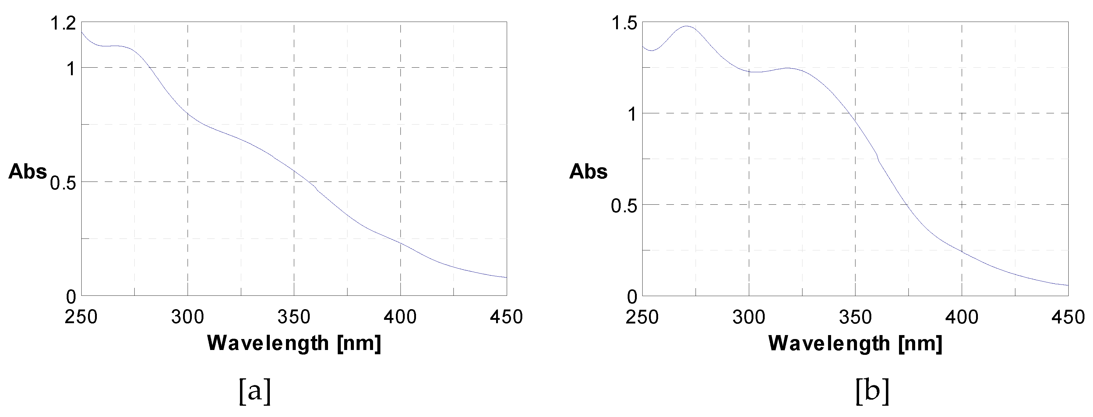
Figure 1. UV-vis spectrum showing the presence of polyphenols at 280 nm [a] and flavonoids at 340 nm [b].
Preprints 80592 g001
Figure 2. Chromatographic profile (LC-ESI-MS) at 280 nm [a] and 340 nm [b] of J. multifida methanolic extract in electrospray positive ionization mode.
Preprints 80592 g002
Figure 3. Medium inhibitory diameter zone of J. multifida leaves extracts on meat isolated Staphylococcus strains. MeOH : Methanol, Act : Acetone ; Dich : Dichlorometane ; EtOH : Ethanol ; W-EtOH : Water-Ethanol ; S. equ : Staphylococcus equorum ; S. sap : Staphylococcus saprophiticus ; S. hae : Staphylococcus haemoliticus ; S. aur : Staphylococcus aureus ; S. sim : Staphylococcus simulans ; S. len : Staphylococcus lentus ; S. coh : Staphylococcus cohnii. * : p < 0.05 ; ** : p < 0.01 ; *** : p < 0.001; ns: p > 0.05.
Preprints 80592 g003
Figure 4. Antioxydant activity of J. multifida extract using DPPH [a], FRAP [b], ABTS [c] and LPO d] methods. EAA: equivalent ascorbic acid, ET: equivalent trolox, MeOH : Methanol, Act : Acetone ; Dich : Dichlorometane ; EtOH : Ethanol ; W-EtOH : Water-Ethanol ; * : p < 0.05 ; ** : p < 0.01 ; *** : p < 0.001; *** : p = 0.0001; ns: p > 0.05.
Preprints 80592 g004aPreprints 80592 g004b
Figure 5. J. multifida extracts inhibition rate of albumin denaturation [a] and their IC50 [b]. MeOH : Methanol, Act : Acetone ; Dich : Dichlorometane ; EtOH : Ethanol ; W-EtOH : Water-Ethanol ; ** : p < 0.01 ; *** : p = 0.0001; ns: p > 0.05.
Preprints 80592 g005
Table 1. Phytochemical constituents of J. multifida leaves powdered samples.
Group of compounds Class J. multifida
Nitrogenous compound Alkaloids +
Poly-phenolics compound Tanin catéchique -
Tanin gallique +
Flavonoids +
Anthocyans +
Leuco-anthocyane +
Coumarin -
Quinonics derivate +
Terpenic compound Triterpenoids +
Steroids -
Cardenolids -
Heterosides Saponosids (IM) + (112)
Cyanogenics derivate -
Reducing compounds -
Free anthracénics -
Mucilags +
(+): Presence of secondary metabolite. (-): Absence of secondary metabolite. (IM): Index mouss.
Table 2. Phenolic, flavonoid, condensed and hydrolyzable tanins content of J. multifida leaves extracts.
Solvents Polyphenols (mgEqGA/g) Flavonoids (mgEqQ/g) Condensed Tanins (mgEqC/g) Hydrolysable Tanins (mgEqGA/g)
Water 11.25±1.37 6.58±1.36 2.05±0.28 2.18±0.15
Ethanol 23.03±6.9 7.18±2.85 5.49±0.35 3.03±0.38
Water-Ethanol 21.57±2.20 7.43±0.12 4.78±0.42 2.11±0.12
Methanol 45.01±11.87 3.65±0.09 6.49±0.46 3.31±0.52
Acetone 26.83±0.27 0.90±0.03 8.58±0.45 3.19±0.11
Dichloromethane 12.86±0.44 0.32±0.01 6.79±0.34 4.12±0.29
Table 3. Phenolic compounds from J. multifida leaves extracts identified by LC-DAD-MS-ESI+ analysis.
PeakNo. Rt (min) UVλmax (nm) [M+H]+(m/z) Phenolic Compound Subclass Content(mg/g)
1 3.19 270 139 2-Hydroxybenzoic acid Hydroxybenzoic acid 1.250
2 12.50 333 339 o-Coumaroylquinic acid Hydroxycinnamic acid 2.291
3 14.37 330, 280 565, 271 Apigenin-apiosyl-glucoside Flavone 5.587
4 14.61 350, 260 449, 287 Luteolin-galactoside Flavone 2.890
5 14.96 350, 260 449, 287 Luteolin-glucoside Flavone 2.173
6 15.88 350, 260 433, 287 Luteolin-rhamnoside Flavone 19.732
7 16.31 360, 260 465, 303 Quercetin-glucoside Flavonol 1.092
8 16.52 360, 260 434, 303 Quercetin-arabinoside Flavonol 0.779
9 16.93 332 517 Dicaffeoyquinic acid Hydroxycinnamic acid 0.636
10 17.71 350, 250 433, 287 Kaempferol-rhamnoside Flavonol 1.223
Total phenolics 37.652
Table 4. Minimum Inhibitory (MIC) and Bactericidal/Fungicidal Concentration (MBC/MFC) of J. multifida extracts.
Extracts Parameters (mg/mL) Staphylococcus aureus ATCC 6538P Escherichia coli ATCC 25922 Salmonella enteritidis ATCC 13076 Listeria monocytogenes ATCC 19114. Candida albicans
ATCC 10231
Water MIC 22.67 47.61 47.61 47.61 47.61
MBC 47.61 >47.61 >47.61 >47.61 >47.61
MBC/MIC 2.10 nd nd nd nd
Ethanol MIC 5.14 34.54 22.67 22.67 22.67
MBC 22.67 47.61 22.67 22.67 22.67
MBC/MIC 4.41 1.37* 1* 1* 1*
The ratio MBC/MIC value with *= Bactericidal effects and without * = Bacteriostatical effects.
Table 5. Minimum Inhibitory Concentrations (MIC) and Bactericidal (MBC) of J. multifida extracts on meat isolated Staphylococcus.
Extracts Parameters (mg/mL) S. equ S. sap S. hae S. aur S. sim S. len S. coh
MeOH MIC - - 20 - 20 - 10
MBC - - >20 - >20 - 20
MBC/MIC - - - - - - 2*
Act MIC - 10 20 20 20 20 -
MBC - >20 >20 >20 >20 >20 -
MBC/MIC - - - - - - -
Dich MIC 2.5 2.5 5 2.5 2.5 2.5 5
MBC 5 5 5 20 10 5 20
MBC/MIC 2* 2* 1* 8 4 2* 4
EtOH MIC - - 10 - - - -
MBC - - 20 - - - -
MBC/MIC - - 2* - - - -
W-EtOH MIC - - 20 20 - - -
MBC - - >20 >20 - - -
MBC/MIC - - - - - - -
MeOH : Methanol, Act : Acetone ; Dich : Dichlorometane ; EtOH : Ethanol ; W-EtOH : Water-Ethanol ; S. equ : Staphylococcus equorum ; S. sap : Staphylococcus saprophiticus ; S. hae : Staphylococcus haemoliticus ; S. aur : Staphylococcus aureus ; S. sim : Staphylococcus simulans ; S. len : Staphylococcus lentus ; S. coh : Staphylococcus cohnii. The ratio MBC/MIC value with *= Bactericidal effects and without * = Bacteriostatical effects.
Disclaimer/Publisher’s Note: The statements, opinions and data contained in all publications are solely those of the individual author(s) and contributor(s) and not of MDPI and/or the editor(s). MDPI and/or the editor(s) disclaim responsibility for any injury to people or property resulting from any ideas, methods, instructions or products referred to in the content.
Copyright: This open access article is published under a Creative Commons CC BY 4.0 license, which permit the free download, distribution, and reuse, provided that the author and preprint are cited in any reuse.
Prerpints.org logo

Preprints.org is a free preprint server supported by MDPI in Basel, Switzerland.

Subscribe

© 2024 MDPI (Basel, Switzerland) unless otherwise stated