Preprint
Article

Windows into Canine Leishmaniasis: In Vivo Diagnosis and In Vitro Promises of Extracellular Vesicles

Submitted:

08 August 2023

Posted:

09 August 2023

You are already at the latest version

Abstract
Leishmaniases are zoonotic vector-borne diseases caused by a wide variety of Leishmania species with complex transmission cycles involving different reservoirs, potential new hosts and vectors. Similarly, to other eukaryotes, Leishmania use released extracellular vesicles (LEVs) to play important initial interactions that are crucial to modulate the subsequent systemic immune response on the establishment of infection in humans and other important hosts like dogs. Recent studies in endemic areas of Brazil concluded that canine infections were predominantly due to L. amazonensis and not restricted to L. infantum (syn. Leishmania chagasi). Under these premises, the diagnosis of canine leishmaniasis needs to be improved, including the identification of current etiological agent, the clinical differential diagnosis and the histopathologic features. In this way, the dual aim of that study is to register collected observations for the diagnosis of natural canine infections and to insert in vitro results in the field of LEVs that still research gaps to be filled to understand the mechanisms and biological aspects involving the parasite-host interactions. Therefore, the future studies of Parasitology research for both of these fields are very important for the interventions for the prevention, control, elimination and eradication worldwide.
Keywords: 
Subject: 
Biology and Life Sciences  -   Parasitology

1. Introduction

Leishmania is the genus of a very successful group of parasitic protozoans of medical and veterinary importance divided into Old and New World species adapted to enormous diversity of hosts such as dogs (the main reservoirs to these parasites), rodents, and other vertebrates, being transiently infectious in humans (Figure 1) [1]. These hemoflagellates parasites have a high level of genetic variability in vivo and a propensity for rapid evolution in vitro, establishing infection by heterogeneous extracellular vesicles released containing a large number of molecules to modulate the host immune responses [1]. According WHO, their high ability to infect multiple hosts has facilitated the spread of Leishmania in 4 eco-epidemiological regions of the world: the Americas, East Africa, North Africa and West and South-east Asia [1,2]. Despite the development of some veterinary vaccines, the lack of effective vaccines and drug treatments for humans has allowed the increase of this major health problem worldwide [1,2,3]. However, there is increasing interest in the isolation and biological and functional characterization of the lipoproteic vesicles released by Leishmania (LEVs), given their apparent potential for the development of effective diagnostic and therapeutic approaches, including the prediction of the outcome of the interaction between cells [1,2,3,4]. Despite some advances, the mechanisms of the selective packaging of these extracellular vesicles are still poorly understood, and there is no consensus on the differential isolation and characterization of the extracellular vesicles or the ultrasensitive detection of specific extracellular vesicle subtypes, their specific biomarkers or their biogenesis [1,2,3,4]. Given these considerations, we present this work, with emphasis on the diagnosis of natural canine infections and to insert in vitro results of extracellular activity of Leishmania, complementing the set of studies we are developing during the last years and providing a reference base for future applied nanotechnological research towards in One Health.

2. Leishmaniotic dog: the infected animal, but healthy and the sick dog

Leishmania is an obligate intracellular parasite transmitted to dogs or humans via the bite of female sand flies to the genera Phlebotomus or Lutzomyia, that are most active in humid environment during the warmer months and at night from dusk to dawn [1,2,3,4,5,6]. The transmission may occur from domestic or wild animals to sand flies to human, or by non-sand fly transmission (infection through transfused blood products, from blood donors which are carriers of infection, transplacental transmission and venereal transmission) [3,4,5,6,7,8]. As well as also humans and mice can transmit the parasite between each other thought blood transfusion, vertical and sexual transmission of Leishmania [8,9,10]. However, there are hardly any case studies about direct dog-to-dog Leishmania transmission by wounds or dog bites [11,12]. After transmission or risk of transmission, is essential the veterinarian will can be able to identify the clinical signs and pathological abnormalities that can appear several months following Leishmania infection (it seems shorter for cutaneous forms), despite the incubation period is difficult to establish [3,4,5,6,7,8,9,10,11,12,13]. The clinical signs of leishmaniasis are directly related to the immune response of the infected dog, however it is estimated that more than half of infected dogs do not manifest clinical signs of the disease [14]. In susceptible animals, the organisms can spread from the skin to the local lymph node, spleen, and bone marrow within a few hours [15]. In resistant dogs, the parasite remains restricted to the skin and draining lymph node - either the animal remains healthy or develops a mild, self-limiting disease. In contrast, susceptible dogs mount a Th2 response characterized by high antibody levels but poor cell-mediated immunity [15]. These differences were attributed to the activities of IL-10-producing Treg lymphocytes. Furthermore, the parasite can actively suppress transcription of the IL-12 gene, ensuring that the Th2 response predominates [15]. Of course, all of these immune challenges affect the balance between progression to clinical disease and maintaining sub-clinical disease. Like this during a chronic infection, a progressive disease develops in susceptible dogs [15]. Vaccines and immunotherapies targeted at recovering or maintaining T and B cell function can be important factors in mending the immune balance required to survive canine leishmaniosis [16]. In the veterinary practice, animals with clinical leishmaniosis can present suggestive signs, but dogs with subclinical infection or infected but clinically healthy present neither clinical signs, nor clinicopathological abnormalities, however have a confirmed Leishmania infection [3]. In other side, is important to consider that the use of anti-Leishmania therapeutic protocols is known to reduce the parasite load and hence infectiousness by treated animals, however presenting only temporary efficacy [5].

3. Inserts of Leishmania infection during the activity of canine immune system (from newborns to seniors)

Dogs are an extraordinary heterogeneity in phenotype through the establishment of pure breeds; a change which has largely occurred over the past 200 years. With such selective inbreeding comes recognition that there is likely to be great diversity in the functioning of the immune system between breeds [17]. This has been clear for many years, based on the unique susceptibility of particular dog breeds to immune-mediated, infectious disease [16]. The immune response is crucial in the unfolding of the infectious process and in the establishment of the disease front the mechanisms of adaptive and innate immunity of dogs [18,19]. Understanding the mechanisms of the immune system of the hosts is an important factor to comparatively elucidate what happens to the individual’s organism during the progression of the disease [20]. Following the knowledge of Immunology Veterinary, the Pattern Recognition Receptors (PRRs) are a class of receptors that can directly recognize the specific molecular structures on the surface of pathogens [15]. The most important of the soluble C-type lectins is mannose-binding lectin (MBL) present at high levels in serum and which has multiple carbohydrate-binding sites that bind to oligosaccharides such as N-acetylglucosamine, mannose, glucose, galactose and N-acetylgalactosamine [15]. Although the binding is relatively weak, the multiple binding sites confer high functional activity [15]. Thus, MBL binds very strongly to different pathogens including parasites such as Leishmania, playing an important role in the activation of the complement system [15]. The surface of phagocytic cells is also covered by many PRRs that can interact with their ligands on the surface of infectious agents [15]. Another important mechanism that promotes contact between pathogens and neutrophils suspended in plasma is the capture [15]. If, however, the pathogen is trapped by a neutrophil and another immune cell, and so not can get away, it can be quickly ingested by phagocytosis [15]. Thus, neutrophils can undergo a form of cell death called NETose as an alternative to apoptosis or necrosis [15]. After activation by CXCL8 or lipopolysaccharides, neutrophils can release the contents of their nuclei, with extrusion of large strands of decondensed nuclear ADN and associated proteins in the extracellular fluid [15]. This forms networks of extracellular fibers called networks extracellular neutrophils (NETs) [15]. The NETs They are abundant at sites of acute inflammation. These networks trap and kill several pathogens such as L. amazonensis) [15]. NETs can be very important in containing microbial invaders by acting as physical barriers, capturing large numbers of parasites and thus prevent its spread) [15]. When promastigote forms of this parasite are injected by sandflies in the skin of a dog, they are quickly phagocytosed by the neutrophils [15]. When neutrophils go into apoptosis, parasites are released and then engulfed by macrophages and dendritic cells, in which organisms become differentiate in amastigotes. Leishmania amastigotes are intracellular parasites obligators that divide in macrophages until the cells rupture, and when released into the body, they are phagocytosed by adjacent cells [15]. Depending on the degree of host immunity, parasites can be restricted to the skin (skin disease); alternatively, dendritic cells may migrate to the lymph nodes or enter the circulation and lodge in the internal organs, leading to visceral spread of the disease. Although the disease is widely spread in endemic areas, most dogs is resistant to Leishmania, and only 10% to 15% develop the visceral form of the disease [15]. Macrophages are the main host cell for Leishmania and effector cells for the death of the parasite. Parasites divide into infected macrophage phagolysosomes [15]. Its resistance to intracellular destruction is the result of multiple mechanisms, including genetic factors - comparative studies of 245 macrophage genes demonstrated that 37% were suppressed by Leishmania infection [15]. Leishmania lipophosphoglicans delay the maturation of the phagosome, preventing the production of NO and inhibiting the response of macrophages to cytokines [15]. These parasites also reduce the presenting of macrophage antigen by suppressing the expression of the class II major histocompatibility (MHC), when the parasites stimulate chronic inflammation. Thus, initially will be characterized by granulocytic invasion, this is followed by macrophages, lymphocytes and NK cells that collectively form granulomas [15]. Additionally, one important factor that determine the success or failure of an infection is the availability of iron [15]. Innate resistance to many intracellular organisms such as Leishmania is controlled, in part, by a gene called Slc11a1 (short for solute carrier family 11), member 1a; formerly called Nramp1) [15]. By definition, parasites are able to evade the host’s immune response by long enough for at least parasitic reproduction to occur. In general, antibody-mediated immune responses protect against extracellular protozoa, while cell-mediated one’s control intracellular protozoa [15]. Parasitic protozoa employ some fairly sophisticated techniques to ensure its survival in the face of an animal’s immune response [15]. The Th1-mediated responses that result in macrophage activation are important in many diseases caused by protozoa, in which organisms are resistant to intracellular destruction [15]. One of the most significant routes of destruction in the M1 cells is the production of nitric oxide (NO) [15]. The nitrogen radicals formed by interaction of NO with oxidants are lethal to many intracellular protozoa [15]. However, protozoa are also experts at surviving inside macrophages; for example, Leishmania and Trypanosoma cruzi can migrate to safe intracellular vacuoles by blocking the maturation of phagosome. Leishmania and T. cruzi can suppress the production of oxidants or cytokines [15].
The ontogeny of the canine immune organs was reviewed, it is known hematopoietic and immune cells arise from a common bone marrow stem cell. Thereafter, B cells undergo maturation in the fetal liver and bone marrow, which represent successive primary lymphoid organs. B cells maturation involves the acquisition of BCR and selection to ensure that only B cells that express functional BCR (positive selection) and do not ligate self-antigens (negative selection) survive. On the other hand, immature T cells are exported to the thymus for final maturation. Although the puppy was considered immunocompetent between 6–12 weeks of age, it is not possible to accurately predict the onset of immunocompetence, since it depends on the presence of MDA [21]. Increased life span allowed the recognition of age-related higher susceptibility to infectious, inflammatory, autoimmune, and neoplastic diseases. Age-related changes include impairment of the cell-mediated immune response, as demonstrated by the reduction of proliferative response of blood lymphocytes to mitogens and the reduction of cutaneous delayed type hypersensitivity [21]. Moreover, there is a decline in the humoral immune response probably related to the decreased functionality of Th cells. The ability to mount humoral immune responses seems to prevail, as demonstrated by the persistence of protective vaccine antibody titers, and respond to booster vaccination with elevation in titer [21]. Although the currently adopted triennial re-vaccination program, instead of the prior annual re-vaccination, offers adequate protection to young and adult dogs, this vaccination scheme may not confer protection to geriatric dogs [21]. Older dogs commonly present an impairment of immune responses to novel antigenic challenges, such as infections and vaccines, which probably is related to the reduction of the peripheral pool of naïve T cells and low diversity of the repertoire of T cell receptors [21]. The key genetic elements of immune responsiveness lie within the genes of the major histocompatibility complex (MHC); present as the dog leukocyte antigen (DLA) and feline leukocyte antigen (FLA) systems in the species under discussion. This would suggest that specific dog breeds have genetically determined immune function, and recent studies concern breed-specific serological response patterns to vaccination Such genetic background is also likely to impinge on maturation of the immune system in these species [17]. In dogs, C-reactive protein (CRP) is the main acute phase protein and its levels increase about a hundred times in infectious diseases such as babesiosis, leishmaniasis, parvovirus and colibacillosis. Acute phase protein levels increase moderately in canine inflammatory bowel disease. Concentrations of CRP, haptoglobin, and SAA are significantly elevated in the cerebrospinal fluid and serum of dogs with corticosteroid-responsive arthritis and meningitis. In pregnant dogs, the levels of haptoglobin, ceruloplasmin and fibrinogen are moderately higher [15]. Some studies showed, dogs in the asymptomatic and symptomatic groups with an outcome of heterogeneity in Cu, Zn, and Fe concentrations compared with the control group, emphasize the important roles of trace elements (TEs) in leishmaniasis [21]. Suggesting, TEs could be assessed as a prognosis factor in leishmaniasis, and/or an adjuvant for the treatment of leishmaniasis [22]. Is clear that, susceptible dogs mount a Th2 response characterized by high levels of antibodies, but showing a poor cell-mediated immunity [15]. These differences were attributed to the activities of IL-10-producing Treg lymphocytes [15]. In addition, also is consensus that the parasite can actively suppress IL-12 gene transcription, ensuring that the response Th2 predominates [15]. In that connection, a chronic and progressive disease develops in susceptible dogs as well as the macrophages loaded with parasites accumulate, with continuous multiplication in the organism, spreading throughout the body, and resulting in disseminated infection [15].

4. Leishmania diagnosis: some findings of natural canine infections

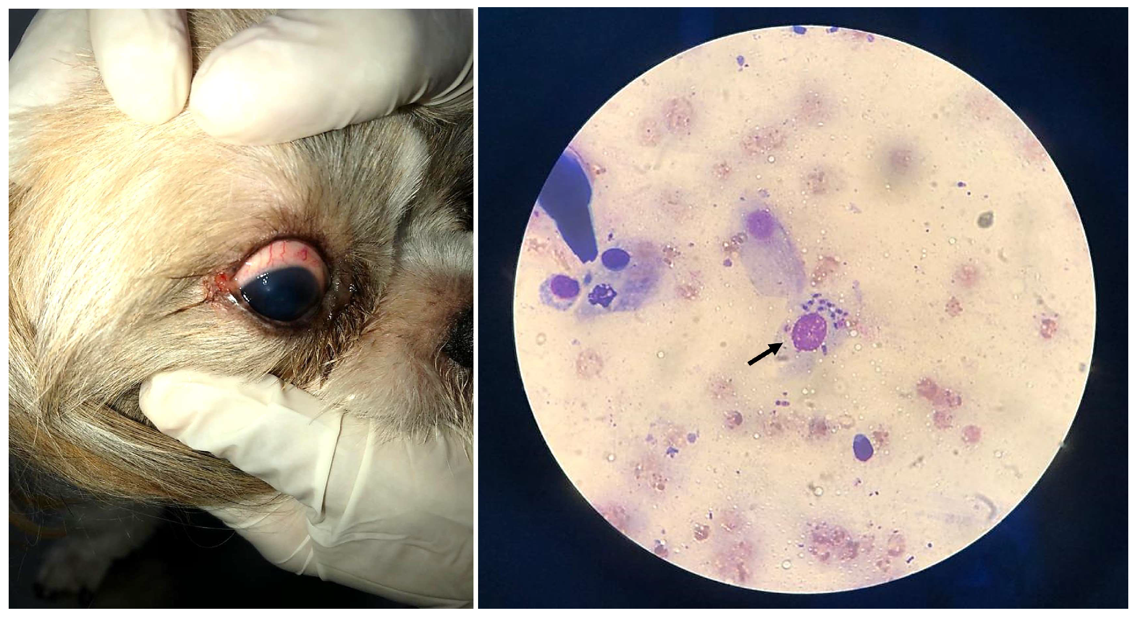
We present in that section the information’s case report of “Jimmy”, a healthy dog (Shih Tzu), male, 1 year old that had presented eye infection (Figure 2) during clinical examination.

3.1. Molecular results of clinical samples by Polymerase Chain Reaction PCR

PCR is based on the amplification of the number of copies of fragment(s) of Deoxyribonucleic Acid - DNA and/or Ribonucleic Acid of the researched microorganism (Leishmania and others). The POSITIVE result indicates the presence of the microorganism in the biological material analyzed, while the NEGATIVE result indicates its absence. - This test may present, although rarely, false-negative and false-positive results, which is a characteristic of the method. Animals with low parasitemia are more subject to false-negative results, especially when the test is performed from a peripheral blood sample. PCR test results that are at odds with the clinical status of the animal should be repeated with a new biological sample. Indirect immunofluorescence assay (IFAT) and quantitative real-time PCR (qPCR) are alternative methods that aid diagnosis.

3.1.1. Results of PCR for the biological samples collected from “Jimmy”

The biological samples were collected and analyzed according established protocols used in Molecular Biology of the Biomolecular Technology Laboratory of Institute of Biological Sciences of Federal University of Pará UFPA Brazil. The followed results were obtained:
  • Auditive duct swabs: (PCR) Leishmania sp. NEGATIVE
  • Mucous membrane of the anus swabs: (PCR) Leishmania sp. POSITIVE
  • Oral Mucosa swabs: (PCR) Leishmania sp. POSITIVE
  • Whole Blood*: (PCR) Anaplasma platys NEGATIVE
  • Whole Blood*:(PCR) Babesia vogeli NEGATIVE
  • Whole Blood*: (PCR) Ehrlichia canis NEGATIVE
  • Whole Blood*: (PCR) Leishmania sp. POSITIVE
  • Whole Blood*: (PCR) Mycoplasma sp. NEGATIVE
  • Whole Blood*: (PCR) Rangelia vitalli NEGATIVE
  • Preputial secretion swabs: (PCR) Leishmania sp. POSITIVE
  • Ocular Swab: (PCR) Leishmania sp. POSITIVE
* The whole blood sample was collected into Ethylenediaminetetraacetic acid (EDTA) tubes.

3.2. Hemogram and ELISA analyses

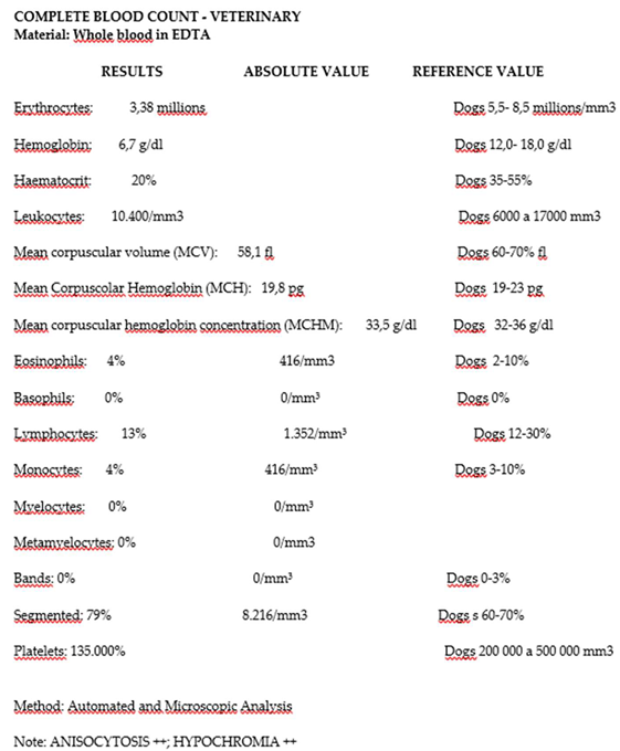
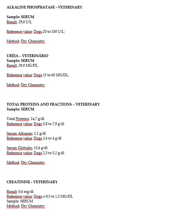
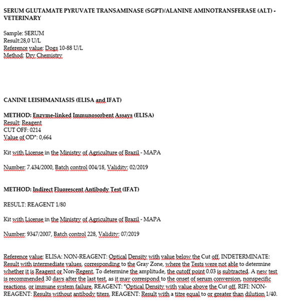
The results obtained from the samples of infected animal confirmed the Leishmania infection (Table 1).

5. An emerging focus on lipids in Leishmania extracellular vesicles

Extracellular vesicles contain a lipid bilayer membrane that protects the encapsulated material, such as proteins, nucleic acids, lipids and metabolites, from the extracellular environment. These vesicles are released from cells via different mechanisms. During recent years extracellular vesicles have been studied as possible biomarkers for different diseases, as biological nanoparticles for drug delivery, and in basic studies as a tool to understand the structure of biological membranes and the mechanisms involved in vesicular trafficking. Lipids are essential molecular components of extracellular vesicles, but at the moment our knowledge about the lipid composition and the function of lipids in these vesicles is limited. However, the interest of the research community in these molecules is increasing as their role in extracellular vesicles

3.1. Quantification of lipid bodies CLs in promastigote of Leishmania (Leishmania) amazonensis and endocytosis and exocytosis process of Leishmania

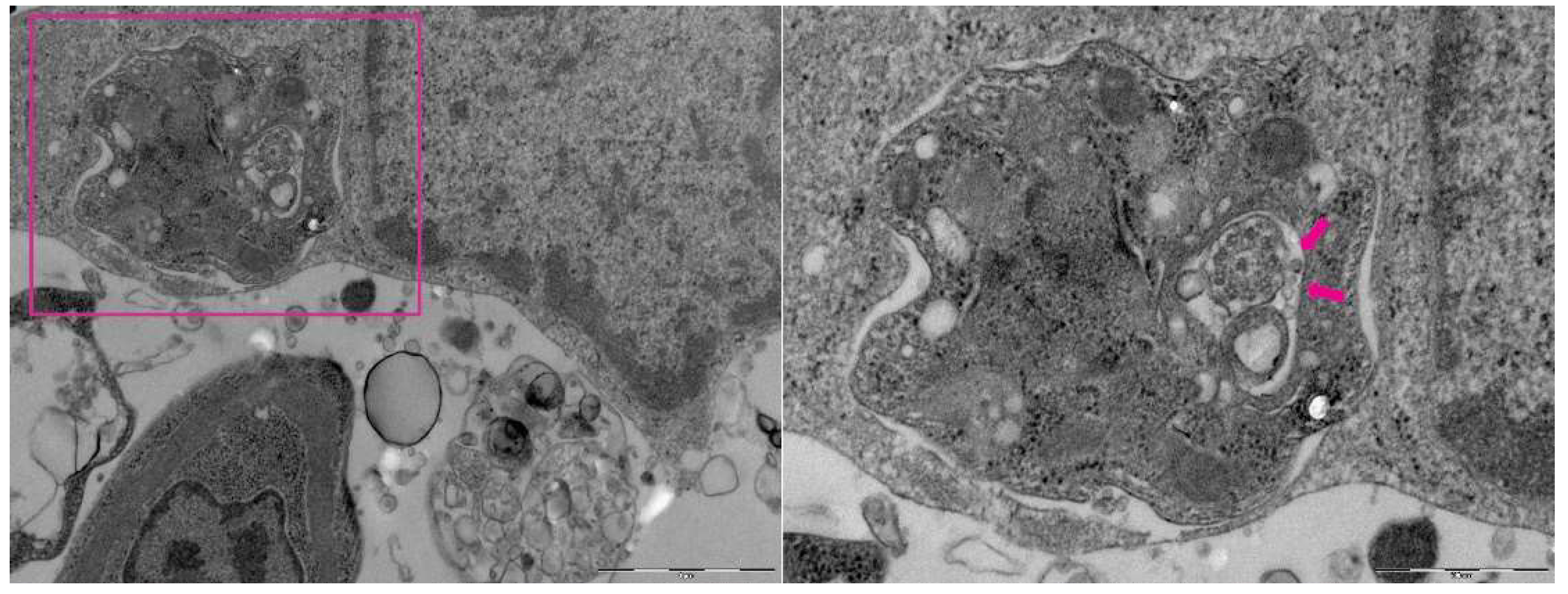
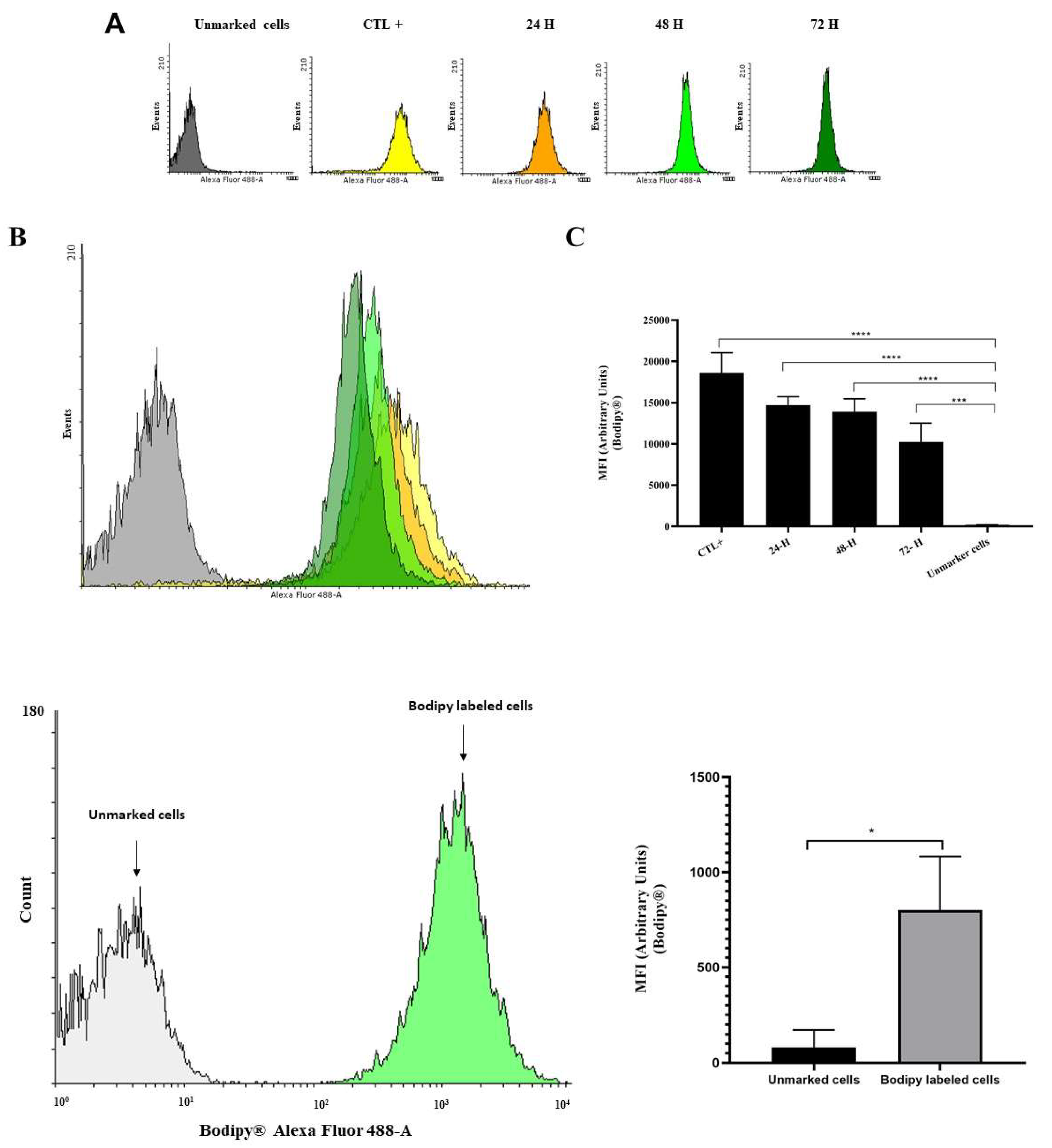
The knowledge of specific cargo molecules identified within LEVs suggest their function as adjuvant-like in the immune responses, with a possible key advantage for the infection establishment and disease progression, inducing at the same time quantitative and qualitative changes in the protein content of infected host cells extracellular vesicles (Figure 3 and Figure 4).

3.1.1. Methodology

Supplementary contents 1 and 2

3.2. Macrophages infected with Leishmania (Leishmania) amazonensis examined by Transmission Electronic Microscopy TEM

6. The immune expected effects of approved vaccines for canine leishmaniosis and some steps for the next-generation therapies following LEVs research advances

Effective vaccines are available against leishmaniasis. The most effective use purified fractions of Leishmania, including enriched fraction of glycoproteins, also called fucose-mannose ligands [15]. This vaccine not only prevents the development of the disease, but also serves as an immunotherapeutic agent, producing clinical improvement in dogs with disseminated disease [15]. An alternative vaccine containing excretory and secretory products of L. infantum promastigotes with an adjuvant Muramyl dipeptide also seems to work well. Experimental vaccines, including attenuated and ADN have shown promising results for Veterinary Medicine [15]. For additional Leishmania control mechanisms and tools are needed, including new drugs, vaccines, diagnostics, and vector control agents and strategies [23]. Considering these important points, for the future of prevention and treatment of leishmaniases we can include all advances resulting from the LEVs research in the world that represent variety of advantages over live biotherapeutics (next-generation therapies) [23]. Over the past years, isolation and analysis of extracellular vesicles from Leishmania is challenging. The protocols are not standardized yet, like we refer in our Guidelines for exosomal research published in 2021 [23], as many variants with potential effect on the outcome are used and published. Is important to refer that several protocols are labour intensive, requiring costly equipment, increase risks for loss of heterogeneous extracellular vesicles and do not discriminate well between exosomes and contaminating structures such as lager vesicles and protein/lipid aggregates [24]. We referred also that direct isolation with magnetic beads would can be used for LEVs research, requiring minimal hands-on provides highly pure exosomes with minimal loss and enables future automation opportunities in an immunoassay format and others lab experiments improved in the last years in the Ketil Winther Pedersen’s Lab. To this end, multidisciplinary expertise cooperation is very important to further our understanding of the Leishmania-host-cell interactions, a broad-scale analysis of the cargo of their production of extracellular molecules and create very promising perspectives for the development of innovative applications following the Leishmania virulence factors that include lipophosphoglycan (LPG), surface acid proteinase (GP63), glycoinositolphospholipids (GIPLs), proteophosphoglycan (PPG), A2 protein, the kinetoplastid membrane protein (KMP-11), nucleotidases, heat-shock proteins (HSPs), and transmembrane transporters, which support the survival and propagation of the parasite in the host cell [1].

7. Discussion

The clinical signs of leishmaniasis are directly related to the immune response of the infected dog and we can account the disease into four stages based on serological status, clinical signs, laboratory findings and type of therapy and prognosis for each stage [3,4,5,6,7,8,9,10,11,12,13,14,15]. In susceptible animals, organisms can spread from skin to the local lymph node, spleen and bone marrow in a few hours [15]. In resistant dogs, the parasite remains restricted to the skin and draining lymph node [15]. According these considerations the first-choice samples should be used for PCR are: bone marrow, lymph node, spleen, skin and conjunctival swabs and others, however samples of blood, buffy coat and urine are considered less sensitive [3]. In parallel, the study of LEVs of prokaryotes and eukaryotes has aroused considerable interest in the scientific community, due to the possible potential for the development of diagnostic and therapeutic methodologies [1]. Despite advances in Leishmania studies, the selective mechanisms of LEVs are still poorly understood, there is no consensus on the differential characterization or ultrasensitive detection of their specific subtypes, biomarkers or their biogenesis and how this knowledge can be effective for faster diagnosis and prevention [1]. Actually, the diagnostic methods for canine leishmaniasis include: parasitological (cytology/histology; immunohistochemistry and culture); molecular (conventional, nested and real-time PCR, considered the most sensitive technique) and serological quantitative (IFAT and ELISA) and quantitative (rapid tests) [3]. Increased transmission of the parasite and higher incidence of disease, as well as the emergence and re-emergence of this disease recorded in recent years may be related to many factors, among which we highlight the socio-economic conditions, climate and environmental change, closer contact between pets and wild ecosystems and the parasite resistance to drugs and insecticides vectors in use [1]. Concurrently with the spread of leishmaniasis recently studies reveal that dogs are at risk of acquiring coinfection with emerging zoonotic parasites [1]. In that sense, the most popular pet animals worldwide, such as dogs and cats can become infected by parasites [1]. Companion animals are important agents of different parasitic species, these infections can cause many complications with risk of high morbidity, including weight loss, anemia, and low immune resistance [25,26,27]. In turn, these conditions can lead to secondary infections and even death of these animals [27]. Many parasite species potentially threaten canine and feline health, while some dog and cat parasites are strictly associated to these animals, some of them may also infect humans causing zoonoses [27,28]. Protozoa and helminths are two major groups of organisms acting as etiologic agents of animal and human diseases with variable severity, especially for immunocompromised hosts resistance [28,29,30,31]. Parasitic infections caused by protozoa and helminths infect predominantly puppies, kittens, geriatric, chronically sick or immune-compromised animals and perhaps pregnant animals [32]. Older dogs and cats are mainly immune after previous infections and seldom show symptoms, however, may still be a source of transmission of infection [25,26,27,28,29,30,31,32,33]. Companion animals living in crowded conditions and poor sanitation or with access to the outdoors, may have a high risk of direct transmission of protozoan infections, for example the following: Giardia sp., Trichomonas sp., Cryptosporidium sp. and Cystoisospora sp. [25,26,27,28,29,30,31,32,33,34,35]. Dogs and cats may contact rodents or ingest raw meat are in risk to acquire infections caused by cyst-forming coccidia, i.e., Neospora sp., Hammondia sp., Toxoplasma sp. and Sarcocystis sp. [25,26,27,28,29,30,31,32]. Dogs and cats can also be infected with others gender and parasites species, like Ancylostoma sp. Angiostrongylus sp. [36,37,38]. Emerging infectious diseases may increase frequently in some regions, either due to increased importation of infected vertebrate hosts or by the establishment of pathogens and their vectors in previously non-endemic areas [39]. Cases of zoonotic infections such as leishmaniosis, babesiosis and dirofilariasis was detected in some non-endemic regions, mainly due to the expansion of the parasitic transmission area and their increasing occurrence in wildlife, which act as reservoir or hosts of their complex cycles [38,39,40,41,42]. Leishmaniasis affects humans and domestic companion animals and wild animals worldwide, involving reservoir and hosts such as rodents, marsupials, edentates, monkeys and wild canids [40,41,42,43]. More recent surveys reported from several countries in the world indicate that zoonotic parasites (Leishmania sp., Babesia sp., Toxoplasma sp., Neospora sp. and Dirofilaria sp.) are associated with infections and coinfections in companion animals [25,26,27,28,29,30,31,32,33,34,35,36,37,38]. Moreover, parasitic infections represent a serious public health threat, particularly in developing countries like Brazil where many unwanted animals are simply abandoned without clinical diagnosis to suffer and die on the streets of rural areas, but also in suburbs and large urban centers suffer deficient sanitation services [27,28,29,30,31,32,33,34,35,36,37,38,39,40,41,42,43,44,45] (Tables S1 and S2—Supplementary Contents 2) [1]. However individual case reports can add new parameters for the accuracy of diagnosis, to confirm the coinfection and the range of differential diagnoses - or if there the animal remains healthy or develops a mild, self-limiting illness [15]. Considering the canine leishmaniasis, these hardy dogs mount a weak antibody response, but a strong and effective Th1 response may have low antibody titers but produce IFN-γ in response to antigens parasitic, generate type I granuloma, mount a strong response of hypersensitivity of the late type, and eventually destroy the parasites [15]. The resistance to Leishmania has a strong genetic component; for example, dogs of the Podengo breed Ibicenco (Ibizan Hounds - antique hunter of rabbits) appear to be resistant to this parasite. There is also an association between resistance and certain MHC class II haplotypes, as well as certain Slc11a1 (Nramp) alleles in dogs [46]. Thus, the Ibizan Hounds may be an interesting canine model for the investigation of protective anti-Leishmania immune response [46]. Results of recent research show relevant differences between the cytokine serum profile and the data published for other canine breeds, and several genetic fixed variants in genes related to immune response, regulation of immune system, and genes encode cytokines and its receptors [47]. The most relevant genes that present such fixed polymorphisms were IFNG and IL6R [47]. Other variants with frequencies equal or above 0.7 were found in the genes ARHGAP18, DAPK1, GNAI2, MITF, IL12RB1, LTBP1, SCL28A3, SCL35D2, PTPN22, CIITA, THEMIS, CD180 [47]. Epigenetic regulatory genes as HEY2, L3MBTL3 show also intronic polymorphisms [47]. Future studies will can reinforce why the regulation of immune response is different in the Ibizan hound dogs compared to other breeds [47]. By other side, some dogs develop severe and generalized nodular dermatitis, lymphadenitis granulomatous, splenomegaly and hepatomegaly, exhibiting activation of polyclonal (occasionally monoclonal) B lymphocytes involving all four classes of IgG, as well as hypergammaglobulinemia, and develop lesions associated with hypersensitivity types II and III [15]. Additionally, excessive production of immunoglobulin can lead to the development of an immune-mediated hemolytic anemia, thrombocytopenia and the production of antinuclear antibodies [15]. The chronic deposition of Immune complexes can result in glomerulonephritis, uveitis, and synovitis, leading to failure renal and death [15]. The significant elevation of ant histone antibodies is a feature of some dogs with glomerulonephritis associated with leishmaniasis. There is a positive correlation between the levels of these ant histone autoantibodies and the protein/creatinine ratio once that antibodies increase the likelihood of the development of glomerulonephritis [15]. But, despite their antigenicity, parasitic protozoa manage to survive in their host using multiple evasion mechanisms acquired over many millions of years of co-evolution [1,2,3,4,5,6,7,8,9,10,11,12,13,14,15]. Following the reproducible results for lipid bodies of Leishmania labeled with BODIPY™ 493/503 we believe that the crucial 72 hours period has showed clear decrease of lipids released. This regression is in accord with Zhang (2021) in the way that amastigotes acquire most of their lipids from the host although they retain some capacity for de novo synthesis, differently of promastigotes that rely on de novo synthesis to produce the majority of their lipids including glycerophospholipids, sterols and sphingolipids [48].

8. Conclusions

Leishmania-infected dogs continue to be parasite reservoirs for sandfly vectors [1]. Therefore, new protocols are needed to achieve a better efficacy in the prevention and clinical treatment [1]. Different types of laboratory tests are available to diagnose parasitic infections, including conventional methods considered as gold standards and serological [43,44,45,46,47,48,49,50]. Several molecular diagnostic tools to detect parasites and new strains have been developed in the last decades [43,44,45,46,47,48,49,50]. Accurate diagnosis of zoonotic infections collaborates with the work of medical scientists, policy makers and public health officials planning to prevent the dissemination of these diseases, and establishing a world-wide network of surveillance for the coinfection of parasitic infections [49,50]. Actually, advanced techniques for the study and diagnosis of simultaneous infections are indicating that multi-parasitism is more common than single infections [51,52]. Evolutionarily, multi-parasite systems are ecologically dynamic, they involve key host species within multi-host parasite systems and their contribution to transmission [52]. The understanding of this complex relations is one of the highest priorities for biomedical sciences for the 21st century [53]. Clinically, coinfection of zoonotic parasites in companion animals may appear in its classical presentation, with acute aggressive evaluation or coming at long-standing infection, asymptomatic or sometimes non-specific, difficulting the clinical diagnosis [33,34,35,36,37,38,39,40,41,42,43,44,45,46,47,48,49,50,51,52,53]. Veterinarians and clinical researchers should consider the health status and background of the patients (animals or humans) to apply a better parasite management program [53]. Considering certain factors like resource-mediated processes most often influencing how, where and which co-infecting parasites interact, and may dictate more intensive monitoring for effective treatment, while others may suggest a less aggressive approach [32,33,34,35,36,37,38,39,40,41,42,43,44,45,46,47,48,49,50,51,52,53]. Furthermore, innovative strategies applying the knowledge about extracellular vesicles in their specific profiles, including proteic and lipidic data basis, on the studies of host-parasites mechanisms can be incorporated into immunotherapy to interfere with the dynamics of disease transmission and progression and the development of effective, safe and available vaccines against leishmaniasis in helping to protect puppies and dogs of different ages. Actually 4 vaccines against canine leishmaniasis are available on the market, Leishmine® and Leish-Tec® in Brazil, CaniLeish® and LetiFend® in Europe (the first vaccine based on purified excreted//secreted antigens of Leishmania has been licensed in Europe since 2011) [1,2,3,4,5,6,7,8,9,10,11,12,13,14,15,16,17,18,19,20,21,22,23,24,25,26,27,28,29,30,31,32,33,34,35,36,37,38,39,40,41,42,43,44,45,46,47,48,49,50,51,52,53,54]. Challenges with these vaccines include current manufacturer recommendations which require the vaccination of seronegative dogs. In countries where disease is endemic in both dogs and people, identification of healthy uninfected animals is less than 100% accurate due to challenges with current diagnostics. Adverse events were mild and site specific, so use of vaccines in healthy sub-clinical dogs may warrant a change in current recommendations regarding vaccination/immunotherapy in infected healthy animals [16]. Despite the available studies on licensed vaccines for canine leishmaniasis, they are still considered insufficient, given the lack of standardization of the study design, methodological deficiencies and substantial differences in the characteristics of the study populations are some of the issues that impede comparative analysis between the available vaccines. In addition, research is needed on other aspects of vaccination: xenodiagnostic studies to assess the infectivity of vaccinated and infected dogs and an adequate assessment of the potential interference of vaccination in the diagnosis of Leishmania infection are some examples. In addition, long-term pharmacological surveillance should be maintained after licensing any vaccine to provide reliable information to relevant organizations and the general public [54]. In this way are expectable, like we had indicated in previous publication of Guidelines for Exosomal Research [1], that more advances in the techniques and protocols for accuracy of isolation and characterization of LEVs and their activity on the host immune responses, including their lipid bilayer membrane that protects the encapsulated material, such lipids (essential molecular components of extracellular vesicles), from the extracellular environment. Actually, the knowledge about the lipid composition and the function of lipids in LEVs is very limited. Changes in the lipid profile and metabolism in both parasite and host during development of the disease depend on the presence of lipid bodies. Further research is required to fully understand the relationship between the interactions between lipid metabolism of host and parasite, immune response, and the prognosis of the disease [55]. However, we propose with our findings to enlarge the interest of the research community in these molecules release by Leishmania as their role in host-parasites extracellular interactions.

Supplementary Materials

The following supporting information can be downloaded at the website of this paper posted on Preprints.org, Methodology 1 Methodology 2, Tables S1 and S2: (open field for future research).

Author Contributions

The authors contributed equally to the work. Roles: resources, conceptualization, investigation, formal analysis, writing, review and editing, Á.M.G.; investigation, writing, review and editing, A.G.-P.; resources, investigation, formal analysis, writing, review and editing, W.L.A.P.; investigation, formal analysis, writing, review and editing, L.M.N.C. and K.W.P.; resources, formal analysis, supervision, writing, review and editing E.O.d.S. All authors have read and agreed to the published version of the manuscript.

Funding

This research was supported by the Coordination for the Improvement of Higher Education Personnel (CAPES) and by the Portuguese Foundation for Science and Technology (FCT), through the research project GHTM–UID/Multi/04413/2013 and Portugal-Brazil research project PTDC/SAU-PAR/28459/2017 EXOTRYPANO IHMT-NOVA/FMV-ULisboa/UFRN.

Institutional Review Board Statement

Ethics Committee on Animal Use of the Federal Rural University of Amazônia (CEUA-UFRA, authorization No. 034/2014).

Informed Consent Statement

Not applicable.

Data Availability Statement

The data presented in this study are available on request from the corresponding author (Á.M.G.). The data are not publicly available due to confidentiality.

Acknowledgments

All the authors of the manuscript acknowledge their respective laboratories/institutes/universities and would like to thank the anonymous reviewers for their thoughtful comments and efforts towards improving the manuscript. Á.M.G. gratefully acknowledge the Laboratory of Gabriela Santos-Gomes IHMT-UNL-Portugal for her permanent support, scientific guidance and thesis supervision. Also Á.M.G. gratefully acknowledge the Graduate Program in Biology of Infectious and Parasitic Agents (PPG BAIP) of Institute of Biological Sciences of Federal University of Pará Brazil and for Dr. Gilvandro Rodrigues Galvão of Federal Rural University of Amazon Brazil for contributes.

Conflicts of Interest

The authors declare no conflict of interest.

References

  1. Gabriel, M.; Galué-Parra, A.; Pereira, W.L.A.; Pedersen, K.W.; da Silva, E.O. Leishmania 360°: Guidelines for Exosomal Research. Microorganisms 2021, 9, 2081. [Google Scholar] [CrossRef] [PubMed]
  2. WHO. Control of Neglected Tropical Diseases. Available online: https://www.who.int/teams/control-of-neglected-tropical-diseases/interventions/strategies (accessed on 10 June 2023).
  3. Solano-Gallego, L.; Miró, G.; Koutinas, A.; Cardoso, L.; Pennisi, M.G.; Ferrer, L.; Bourdeau, P.; Oliva, G.; Baneth, G.; The LeishVet Group. LeishVet guidelines for the practical management of canine leishmaniosis. Parasit Vectors 2011, 4, 86–86. [Google Scholar] [CrossRef] [PubMed]
  4. Yakubovich, E.I.; Polischouk, A.G.; Evtushenko, V.I. Principles and Problems of Exosome Isolation from Biological Fluids. Biochem (Mosc) Suppl Ser A Membr Cell Biol. 2022, 16, 115–126. [Google Scholar] [CrossRef] [PubMed]
  5. Gizzarelli, M.; Bosco, A.; Foglia Manzillo, V.; Bongiorno, G.; Bianchi, R.; Giaquinto, D.; Ben Fayala, N.E.H.; Varloud, M.; Crippa, A.; Gradoni, L.; Cringoli, G.; Maurelli, M.P.; Rinaldi, L.; Oliva, G. Examining the Relationship of Clinical and Laboratory Parameters with Infectiousness to Phlebotomus perniciosus and Its Potential Infectivity in Dogs with Overt Clinical Leishmaniasis. Front. Vet. Sci. 2021, 8, 667290. [Google Scholar] [CrossRef] [PubMed]
  6. Wijerathna, T.; Gunathilaka, N. Diurnal adult resting sites and breeding habitats of phlebotomine sand flies in cutaneous leishmaniasis endemic areas of Kurunegala District, Sri Lanka. Parasites Vectors 2020, 13, 284. [Google Scholar] [CrossRef]
  7. Cecílio, P.; Cordeiro-Da-Silva, A.; Oliveira, F. Sand flies: Basic information on the vectors of leishmaniasis and their interactions with Leishmania parasites. Commun. Biol. 2022, 5, 305. [Google Scholar] [CrossRef]
  8. Dodd, R. Transmission of Parasites by Blood Transfusion. Vox Sang. 1998, 74 (Suppl. 2), 161–163. [Google Scholar] [CrossRef] [PubMed]
  9. Pangrazio, K.K.; Costa, E.A.; Amarilla, S.P.; Cino, A.G.; Silva, T.M.; Paixão, T.A.; Costa, L.F.; Dengues, E.G.; Diaz, A.A.; Santos, R.L. Tissue distribution of Leishmania chagasi and lesions in transplacentally infected fetuses from symptomatic and asymptomatic naturally infected bitches. Veterinary Parasitology 2009, 165, 327–331. [Google Scholar] [CrossRef]
  10. Avila-García, M.; Mancilla-Ramírez, J.; Segura-Cervantes, E.; Farfan-Labonne, B.; Ramírez-Ramírez, A.; Galindo-Sevilla, N. Transplacental transmission of cutaneous Leishmania mexicana strain in BALB/c mice. Am J Trop Med Hyg. 2013, 89, 354–358. [Google Scholar] [CrossRef] [PubMed]
  11. Karkamo, V.; Kaistinen, A.; Näreaho, A.; et al. The first report of autochthonous non-vector-borne transmission of canine leishmaniosis in the Nordic countries. Acta Veterinaria Scandinavica. 2014, 56, 84. [Google Scholar] [CrossRef]
  12. Naucke, T.J.; Amelung, S.; Lorentz, S. First report of transmission of canine leishmaniosis through bite wounds from a naturally infected dog in Germany. Parasites & Vectors 2016, 9, 256. [Google Scholar] [CrossRef]
  13. Miró, G.; López-Vélez, R. Clinical management of canine leishmaniosis versus human leishmaniasis due to Leishmania infantum: Putting “One Health” principles into practice. Veterinary Parasitology 2018, 254, 151–159. [Google Scholar] [CrossRef] [PubMed]
  14. do Rosário, C.J.; Dominici, M.F.; Braga, M.D.; Lima, C.A.; Pereira, J.G.; Melo, F.A. Quantification of IL-10 and IFN-γ in dogs with or without clinical signs of Leishmania (Leishmania) chagasi infection symptoms. Pesq. Vet. Bras. 2018, 38, 129–132. [Google Scholar] [CrossRef]
  15. Tizard, Ian R. Imunologia veterinária/Ian R. Tizard; By Luciana Medina, Mateus D. Luchese. - 9. ed. – Rio de Janeiro: Elsevier, 2014.
  16. Toepp, A.J.; Petersen, C.A. The balancing act: Immunology of leishmaniosis. Res Vet Sci. 2020, 130, 19–25. [Google Scholar] [CrossRef] [PubMed]
  17. Day, M.J. Immune system development in the dog and cat. J Comp Pathol. 2007, 137 (Suppl. 1), S10–S15. [Google Scholar] [CrossRef] [PubMed]
  18. Chaplin, D.D. Overview of the immune response. J Allergy Clin Immunol. 2010, 125 (Suppl. 2), S3–S23. [Google Scholar] [CrossRef] [PubMed]
  19. Hosein, S.; Blake, D.; Solano-Gallego, L. Insights on adaptive and innate immunity in canine leishmaniosis. Parasitology 2017, 144, 95–115. [Google Scholar] [CrossRef]
  20. Varadé, J.; Magadán, S.; González-Fernández, Á. Human immunology and immunotherapy: Main achievements and challenges. Cell Mol Immunol 2021, 18, 805–828. [Google Scholar] [CrossRef]
  21. Pereira, M.; Valério-Bolas, A.; Saraiva-Marques, C.; Alexandre-Pires, G.; Pereira da Fonseca, I.; Santos-Gomes, G. Desenvolvimento do Sistema Imunológico Canino: Do Útero ao Idoso. Vet Sci. 2019, 6, 83. [Google Scholar] [CrossRef]
  22. Taghipour, A.; Abdoli, A.; Ramezani, A.; et al. Leishmaniasis and Trace Element Alterations: A Systematic Review. Biol Trace Elem Res 2021, 199, 3918–3938. [Google Scholar] [CrossRef]
  23. Hotez, P.J.; Pecoul, B.; Rijal, S.; Boehme, C.; Aksoy, S.; Malecela, M.; et al. Eliminating the Neglected Tropical Diseases: Translational Science and New Technologies. PLoS Negl Trop Dis 2016, 10, e0003895. [Google Scholar] [CrossRef] [PubMed]
  24. Neurauter, A., Kierulf, B., Kullmann, Li, A., M., Manger, I. and Pedersen, K.W. From basic research to potential clinical applications: Adapting methods for EV enrichment and analysis. Available online: (PDF) From basic research to potential clinical applications: Adapting methods for EV enrichment and analysis (researchgate.net) (accessed on 10 June 2023).
  25. Baneth, G.; Thamsborg, S.M.; Otranto, D.; Guillot, J.; Blaga, R.; Deplazes, P.; Solano-Gallego, L. Major Parasitic Zoonoses Associated with Dogs and Cats in Europe. J. Comp. Pathol. 2016, 8, 1–21. [Google Scholar] [CrossRef]
  26. Vilhena, H.; Martinez-Díaz, V.L.; Cardoso, L.; Vieira, L.; Altet, L.; Francino, O.; Pastor, J.; Silvestre-Ferreira, A.C. Feline vector-borne pathogens in the north and centre of Portugal. Parasites & Vectors 2013, 6, 99–104. [Google Scholar] [CrossRef]
  27. Dantas-Torres, F.; Otranto, D. Dogs, cats, parasites, and humans in Brazil: Opening the black box. Parasites & Vectors 2014, 7, 22–47. [Google Scholar] [CrossRef]
  28. Woodhall, D.M.; Eberhard, M.L.; Parise, M.E. Neglected Parasitic Infections in the United States: Toxocariasis. Am J Trop Med Hyg. 2014, 90, 810–813. [Google Scholar] [CrossRef]
  29. World Health Organization. Accelerating work to overcome the global impact of neglected tropical diseases: A roadmap for implementation. WHO: Geneva, 2012. [Google Scholar]
  30. Akritidis, N. Parasitic, fungal and prion zoonoses: An expanding universe of candidates for human disease. Clinical Microbiology and Infection 2011, 17, 331–335. [Google Scholar] [CrossRef]
  31. Robertson, L.J.; van der Giessen, J.W.B.; Batz, M.B.; Kojima, M.; Cahill, S. Have foodborne parasites finally become a global concern? Trends in Parasitology 2013, 29, 101–103. [Google Scholar] [CrossRef]
  32. European Scientific Counsel Companion Animal Parasites. Control of Intestinal Protozoa in dogs and cats - Guideline 6 1rd Edition. Malvern: ESCCAP. 2011. Available online: 3sbvfy71_ESCCAP_Guide_6_spanish_version_def.pdf. (accessed on 10 June 2023).
  33. Laurenti, M.D.; Rossi, C.N.; da Matta, V.L.; Tomokane, T.Y.; Corbett, C.E.; Secundino, N.F.; Pimenta, P.F.; Marcondes, M. Asymptomatic dogs are highly competent to transmit Leishmania (Leishmania) infantum chagasi to the natural vector. Vet Parasitol. 2013, 196, 296–300. [Google Scholar] [CrossRef]
  34. Tangtrongsup, S.; Scorza, V. Update on the Diagnosis and Management of Giardia spp. Infections in Dogs and Cats Topics in Companion. Animal Medicine 2010, 25, 155–162. [Google Scholar] [CrossRef]
  35. Bouzid, M.; Halai, K.; Jeffreys, D.; Hunter, P.R. The prevalence of Giardia infection in dogs and cats, a systematic review and meta-analysis of prevalence studies from stool samples. Veterinary Parasitology 2015, 207, 181–202. [Google Scholar] [CrossRef]
  36. Braga, Í.A.; dos Santos, L.G.F.; Ramos, D.G.S.; Melo, A.L.T.; Mestre, G.L.C.; de Aguiar, D.M. Occurrence of Ancylostoma in dogs, cats and public places from Andradina city, São Paulo state, Brazil. Rev. Inst. Med. Trop. 2011, 53, 181–184. [Google Scholar] [CrossRef]
  37. Spratt, D.M. Species of Angiostrongylus (Nematoda: Metastrongyloidea) in wildlife: A review. International Journal for Parasitology: Parasites and Wildlife 2015, 4, 178–189. [Google Scholar] [CrossRef] [PubMed]
  38. Farrar, J.; White, N.J.; Hotez, P.J.; Junghanss, T.; Lalloo, D.; Kang, G. Manson’s Tropical Diseases. A Manual of the Diseases of Warm Climates, 23rd ed.; Elsevier Saunders: China, 2014. [Google Scholar]
  39. European Scientific Counsel Companion Animal Parasites. Control of Vector Borne Diseases in dogs and cats - Guideline 52rd Edition. ESCCAP: Malvern, 2012; Available online: https://www.esccap.org/guidelines/ (accessed on 10 June 2023).
  40. Mencke, N. The importance of canine leishmaniosis in non-endemic areas, with special emphasis on the situation in Germany. Berl Munch Tierarztl Wochenschr. 2011, 124, 434–442. [Google Scholar] [CrossRef] [PubMed]
  41. Palatnik-de-Sousa, C.B.; Day, M.J. One Health: The global challenge of epidemic and endemic leishmaniasis. Parasites & Vectors 2011, 4, 197–207. [Google Scholar] [CrossRef]
  42. Chargui, N.; Haouas, N.; Slama, D.; Gorcii, M.; Jaouadi, K.; Essabbah-Aguir, N.; Mezhoud, H.; Babba, H. Transmission of visceral leishmaniasis in a previously non-endemic region of Tunisia: Detection of Leishmania DNA in Phlebotomus perniciosus. Journal of Vector Ecology 2013, 38, 1–5. [Google Scholar] [CrossRef]
  43. Cargnelutti, D.E.; Borremans, C.G.; Tonelli, R.L.; Carrizo, L.C.; Salomon, M.C. Diagnosis of Leishmania infection in a nonendemic area of South America. Journal of Microbiology, Immunology and Infection 2014, 20, 1–4. [Google Scholar] [CrossRef]
  44. Maia, C.; Campino, L. Epidemiologia das leishmanioses em Portugal. Acta Med. Port. 2010, 23, 859–864. [Google Scholar] [CrossRef]
  45. Petersen, C.A. Leishmaniasis, An Emerging Disease Found in Companion Animals in the United States. Top Companion Anim Med. 2009, 24, 182–188. [Google Scholar] [CrossRef]
  46. Solano-Gallego, L.; Llull, J.; Ramos, G.; Riera, C.; Arboix, M.; Alberola, J.; Ferrer, L. The Ibizian hound presents a predominantly cellular immune response against natural Leishmania infection. Vet Parasitol. 2000, 90, 37–45. [Google Scholar] [CrossRef] [PubMed]
  47. Álvarez, L.; Marín-García, P.-J.; Llobat, L.; et al. Immune and genomic characterization of Ibizan hound and its relationship with Leishmania infantum infection, 02 September 2022, PREPRINT (Version 1) available at Research Square.
  48. Zhang, K. Balancing de novo synthesis and salvage of lipids by Leishmania amastigotes. Current Opinion in Microbiology 2021, 63, 98–103. [Google Scholar] [CrossRef]
  49. Johansen, M.V.; Lier, T.; Sithithaworn, P. Progress in research and control of helminth infections in Asia Towards improve diagnosis of neglected zoonotic trematodes using a One Health approach. Acta Tropica 2015, 141, 161–169. [Google Scholar] [CrossRef] [PubMed]
  50. Al Dahouk, S.; Sprague, L.D.; Neubauer, H. New developments in the diagnostic procedures for zoonotic brucellosis in humans. Rev. Sci. Tech. Off. Int. Epiz. 2013, 32, 177–188. [Google Scholar] [CrossRef] [PubMed]
  51. Tavares, R.G.; Staggemeier, R.; Borges, A.L.P.; Rodrigues, M.T.; Castelan, L.A.; Vasconcelos, J.; Anschau, M.E.; Spalding, S.M. Molecular techniques for the study and diagnosis of parasite infection. J. Venom. Anim. Toxins Incl. Trop. Dis 2011, 17, 239–248. [Google Scholar] [CrossRef]
  52. Steinmann, P.; Utzinger, J.; Du, Z.W.; Zhou, X.N. Multiparasitism: A neglected reality on global, regional and local scale. Advances in Parasitology 2010, 73, 21–50. [Google Scholar] [CrossRef]
  53. Griffiths, E.C.; Pedersen, A.B.; Fenton, A.; Petchey, O.L. Analysis of a summary network of coinfection in humans reveals that parasites interact most via shared resources. Proc. R. Soc. B. 2014, 281, 1–9. [Google Scholar] [CrossRef]
  54. Velez, R.; Gállego, M. Commercially approved vaccines for canine leishmaniosis: A review of available data on their safety and efficacy. Trop Med Int Health. 2020, 25, 540–557. [Google Scholar] [CrossRef] [PubMed]
  55. Martínez, C.R.; Ruiz, C.J. Alterations in Host Lipid Metabolism Produced During Visceral Leishmaniasis Infections. Curr Trop Med Rep 2019, 6, 250–255. [Google Scholar] [CrossRef]
  56. Gocze, P.M.; Freeman, D.A. Factors underlying the variability of lipid droplet fluorescence in MA-10 Leydig tumor cells. Cytometry: The Journal of the International Society for Analytical Cytology 1994, 17, 151–158. [Google Scholar] [CrossRef] [PubMed]
  57. Plotkowski, M.C.; Brandão, B.A.; de Assis, M.C.; Feliciano, L.F.P.; Raymond, B.; Freitas, C.; Saliba, A.M.; Zahm, J.M.; Touqui, L.; Bozza, P.T. Lipid body mobilization in the ExoU-induced release of inflammatory mediators by airway epithelial cells. Microbial pathogenesis 2008, 45, 30–37. [Google Scholar] [CrossRef]
  58. Da Silva, B.J.M.; Souza-Monteiro, J.R.; Rogez, H.; Crespo-López, M.E.; do Nascimento, J.L.M.; Silva, E.O. Selective effects of Euterpe oleracea (açai) on Leishmania (Leishmania) amazonensis and Leishmania infantum. Biomedicine & Pharmacotherapy 2018, 97, 1613–1621. [Google Scholar] [CrossRef]
  59. Lopes, M.G.; Mendonça, I.L.; Fortes, K.P.; Amaku, M.; Pena, H.d.F.; Gennari, S.M. Presence of antibodies against Toxoplasma gondii, Neospora caninum and Leishmania infantum in dogs from Piauí. Rev Bras Parasitol Vet. 2011, 20, 111–114. [Google Scholar] [CrossRef] [PubMed]
  60. Sharifdini, M.; Mohebali, M.; Keshavarz, H.; Hosseininejad, M.; Hajjaran, H.; Akhoundi, B.; Foroushani, A.R.; Zarei, Z.; Charehdar, S. Iran Neospora caninum and Leishmania infantum Co-Infection in Domestic Dogs (Canis familiaris) in Meshkin-Shahr District, Northwestern Iran. J Arthropod Borne Dis. 2011, 5, 60–68. [Google Scholar]
  61. Coelho, W.M.; do Amarante, A.F.; Apolinário, J.d.C.; Coelho, N.M.; de Lima, V.M.; Perri, S.H.; Bresciani, K.D. Seroepidemiology of Toxoplasma gondii, Neospora caninum, and Leishmania spp. infections and risk factors for cats from Brazil. Parasitol Res. 2011, 109, 1009–1013. [Google Scholar] [CrossRef]
  62. Sobrinho, L.S.V.; Rossi, C.N.; Vides, J.P.; Braga, E.T.; Gomes, A.A.D.; de Lima, V.M.F.; Biondo, A.W. Coinfection of Leishmania chagasi with Toxoplasma gondii, Feline Immunodeficiency Virus (FIV) and Feline Leukemia Virus (FeLV) in cats from an endemic area of zoonotic visceral leishmaniasis. Veterinary Parasitology 2012, 187, 302–306. [Google Scholar] [CrossRef]
  63. Ayllón, T.; Diniz, P.P.V.; Breitschwerdt, E.B.; Villaescusa, A.; Rodríguez-Franco, F.; Sainz, A. Vector-borne diseases in client-owned and stray cats from Madrid, Spain. Vector-Borne and Zoonotic Diseases 2012, 12, 143–150. [Google Scholar] [CrossRef] [PubMed]
  64. Cardia, D.F.F.; Camossi, L.G.; da Silveira Neto, L.; Langoni, H.; Bresciani, K.D.S. Prevalence of Toxoplasma gondii and Leishmania spp. infection in cats from Brazil. Veterinary Parasitology 2013, 197, 634–637. [Google Scholar] [CrossRef]
  65. Sakamoto, K.P.; de Melo, G.D.; Machado, G.F. T and B lymphocytes in the brains of dogs with concomitant seropositivity to three pathogenic protozoans: Leishmania chagasi, Toxoplasma gondii and Neospora caninum. BMC Res Notes 2013, 8, 226–233. [Google Scholar] [CrossRef]
  66. Paulan, S.d.C.; Lins, A.G.; Tenório, M.d.S.; Silva, D.T.; Pena, H.F.; Machado, R.Z.; Gennari, S.M.; Buzetti, W.A. Seroprevalence rates of antibodies against Leishmania infantum and other protozoan and rickettsial parasites in dogs. Rev Bras Parasitol Vet. 2013, 22, 162–166. [Google Scholar] [CrossRef]
  67. Zanette, M.F.; Lima, V.M.; Laurenti, M.D.; Rossi, C.N.; Vides, J.P.; Vieira, R.F.; Biondo, A.W.; Marcondes, M. Serological cross-reactivity of Trypanosoma cruzi, Ehrlichia canis, Toxoplasma gondii, Neospora caninum and Babesia canis to Leishmania infantum chagasi tests in dogs. Rev Soc Bras Med Trop. 2014, 47, 105–107. [Google Scholar] [CrossRef]
  68. Sousa, K.C.M.D.; Herrera, H.M.; Domingos, I.H.; Campos, J.B.V.; Santos, I.M.C.D.; Neves, H.H.; André, M.R. Serological detection of Toxoplasma gondii, Leishmania infantum and Neospora caninum in cats from an area endemic for leishmaniasis in Brazil. Revista Brasileira de Parasitologia Veterinária 2014, 23, 449–455. [Google Scholar] [CrossRef]
  69. Braga, A.R.C.; Corrêa, A.P.F.L.; Camossi, L.G.; Silva, R.C.D.; Langoni, H.; Lucheis, S.B. Coinfection by Toxoplasma gondii and Leishmania spp. in domestic cats (Felis catus) in State of Mato Grosso do Sul. Revista da Sociedade Brasileira de Medicina Tropical 2014, 47, 796–797. [Google Scholar] [CrossRef] [PubMed]
  70. Seabra, N.M.D.; Pereira, V.F.; Kuwassaki, M.V.; Benassi, J.C.; Oliveira, T.M.F.D.S. Toxoplasma gondii, Neospora caninum and Leishmania spp. serology and Leishmania spp. PCR in dogs from Pirassununga, sp. Revista Brasileira de Parasitologia Veterinária 2015, 24, 454–458. [Google Scholar] [CrossRef] [PubMed]
  71. Willi, B.; Spiri, A.M.; Meli, M.L.; Grimm, F.; Beatrice, L.; Riond, B.; Bley, T.; Jordi, R.; Dennler, M.; Hofmann-Lehmann, R. Clinical and molecular investigation of a canine distemper outbreak and vector-borne infections in a group of rescue dogs imported from Hungary to Switzerland. BMC Vet Res. 2015, 16, 154–169. [Google Scholar] [CrossRef] [PubMed]
  72. Maia, C.; Ramos, C.; Coimbra, M.; Cardoso, L.; Campino, L. Prevalence of Dirofilaria immitis antigen and antibodies to Leishmania infantum in cats from southern Portugal. Parasitology international 2015, 64, 154–156. [Google Scholar] [CrossRef] [PubMed]
  73. Maia, C.; Coimbra, M.; Ramos, C.; Cristóvão, J.M.; Cardoso, L.; Campino, L. Serological investigation of Leishmania infantum, Dirofilaria immitis and Angiostrongylus vasorum in dogs from southern Portugal. Parasit Vectors 2015, 8, 152–155. [Google Scholar] [CrossRef]
  74. René-Martellet, M.; Lebert, I.; Chêne, J.; Massot, R.; Leon, M.; Leal, A.; Badavelli, S.; Chalvet-Monfray, K.; Ducrot, C.; Abrial, D.; Chabanne, L.; Halos, L. Diagnosis and incidence risk of clinical canine monocytic ehrlichiosis under field conditions in Southern Europe. Parasit Vectors 2015, 6, 3–13. [Google Scholar] [CrossRef]
  75. Paşa, S.; Vardarlı, A.T.; Erol, N.; Karakuş, M.; Töz, S.; Atasoy, A.; Özbel, Y. Detection of Leishmania major and Leishmania tropica in domestic cats in the Ege Region of Turkey. Veterinary parasitology 2015, 212, 389–392. [Google Scholar] [CrossRef]
  76. Pantchev, N.; Schnyder, M.; Vrhovec, M.G.; Schaper, R.; Tsachev, I. Current Surveys of the Seroprevalence of Borrelia burgdorferi, Ehrlichia canis, Anaplasma phagocytophilum, Leishmania infantum, Babesia canis, Angiostrongylus vasorum and Dirofilaria immitis in Dogs in Bulgaria. Parasitology research 2015, 114, 117–130. [Google Scholar] [CrossRef]
  77. Krawczak, F.D.S.; Reis, I.A.; Silveira, J.A.D.; Avelar, D.M.; Marcelino, A.P.; Werneck, G.L.; Paz, G.F. ; Leishmania, Babesia and Ehrlichia in urban pet dogs: Co-infection or cross-reaction in serological methods? Revista da Sociedade Brasileira de Medicina Tropical 2015, 48, 64–68. [Google Scholar] [CrossRef]
  78. Cardinot, C.B.; Silva, J.E.; Yamatogi, R.S.; Nunes, C.R.M.; Biondo, A.W.; Vieira, R.F.; Marcondes, M. Detection of Ehrlichia canis, Babesia vogeli and Toxoplasma gondii and dna in the brain of dogs naturally infected with Leishmania infantum. Journal of Parasitology 2016, 102, 275–279. [Google Scholar] [CrossRef]
  79. Hamel, D.; Shukullari, E.; Rapti, D.; Silaghi, C.; Pfister, K.; Rehbein, S. Parasites and vector-borne pathogens in client-owned dogs in Albania. Blood pathogens and seroprevalences of parasitic and other infectious agents. Parasitol Res. 2016, 115, 489–499. [Google Scholar] [CrossRef] [PubMed]
Figure 1. Tracking the global distribution of endemic areas in the tropics, subtropics, and southern Europe: human visceral leishmaniasis (green areas) and cutaneous leishmaniasis (purple dots) Both zoonotic forms present expanding geographic ranges and rapid adaptation influenced by risk factors, with new epidemiological scenarios emerging in previously disease-free areas [1].
Preprints 81961 g001
Figure 2. Ocular infection in canine leishmaniasis and light microscopy examination of a conjunctival cytological sample showing a macrophage containing multiple Leishmania amastigotes (arrow) (Authored by Dr. Gilvandro Rodrigues Galvão - UFRA).
Preprints 81961 g002
Figure 3. Tubular structures and vesicles (arrows) participating in the endocytosis and exocytosis process can be seen in the flagellar pocket (FP) of L. (L.) amazonensis promastigotes (MHOM/BR/2009/M26361 strain). F = flagellum; K = kinetoplast; M = mithocondria; N = nucleus. Scale-bar 1 µ and 500 nm.
Preprints 81961 g003
Figure 4. Quantification of lipid bodies in Leishmania (L.) amazonensis at different culture times; (A) Histogram of each experimental group; (B) Overlay histogram demonstrating increased fluorescence intensity of BODIPY™ 493/503-labeled cells at 24, 48 and 72 hours of culture and (C) Graphic representation of BODIPY™ 493/503-labeling in Leishmania promastigotes *** p < 0.001 (ANOVA and Tukey post-hoc test).
Preprints 81961 g004
Table 1. Hemogram blood and Serological results obtained from the samples of the dog “Jimmy” (Archive of LABPAT - UFRA).
Preprints 81961 i001
Preprints 81961 i002
Preprints 81961 i003
Disclaimer/Publisher’s Note: The statements, opinions and data contained in all publications are solely those of the individual author(s) and contributor(s) and not of MDPI and/or the editor(s). MDPI and/or the editor(s) disclaim responsibility for any injury to people or property resulting from any ideas, methods, instructions or products referred to in the content.
Copyright: This open access article is published under a Creative Commons CC BY 4.0 license, which permit the free download, distribution, and reuse, provided that the author and preprint are cited in any reuse.
Alerts
Prerpints.org logo

Preprints.org is a free preprint server supported by MDPI in Basel, Switzerland.

Subscribe

© 2025 MDPI (Basel, Switzerland) unless otherwise stated