Preprint
Case Report

Motor Recovery after Surgical Neurolysis in Brachial Plexus Neuropathy: A Case series and Systematic Review

Altmetrics

Downloads

182

Views

155

Comments

0

Submitted:

12 August 2023

Posted:

15 August 2023

You are already at the latest version

Alerts
Abstract
Surgical neurolysis is a procedure designed to liberate an injured nerve from scar tissue or adjacent structure, thereby facilitating nerve regeneration in cases of brachial plexus neuropathy (BPN). This study presents a case series of patients diagnosed with BPN who underwent surgical neurolysis. The primary focus was on the clinical assessment of recovery using the British Medical Research Council motor grading scale (BMRC). Additionally, a comprehensive literature review was conducted to analyze motor recovery outcomes related to surgical neurolysis for BPN. 18 patients with BPN who underwent surgical neurolysis where included. The results experienced a notable increase of 58% in muscle strength as assessed by the BMRC. The average preoperative state of 2.17±1.15 improved significantly to a postoperative condition of 3.44±1.34 (p = 0.003, d= 0.913) The systematic review identified 2298 articles which were selected for qualitative analysis, demonstrated that surgical neurolysis was associated with favorable motor recovery outcomes in 75.82% of the patients. Both the case series and the literature review reveal significant motor recovery following surgical neurolysis. It is crucial to conduct well-designed, adequately powered, randomized, and blinded clinical trials. Such studies will provide robust evidence to support or refute the utility of this approach in motor recovery.
Keywords: 
Subject: Medicine and Pharmacology  -   Neuroscience and Neurology

1. Introduction

Brachial plexus injuries pose significant disability, and their surgical management is intricate due to diverse treatment options and injury patterns [1]. Surgical neurolysis is a procedure aimed at liberating an injured nerve from scar tissue or neighboring structures, facilitating regeneration [2,3]. This technique allows the nerve to adapt to mechanical stress by gliding against surrounding tissue. Nonetheless, it has been displaced by alternative interventions like nerve grafts, nerve transfers, and muscle/tendon transfers, partly due to negative results reported by some authors [4]. Additionally, it may not be effective in cases of nerve avulsion or transection. However, a recent systematic review comparing motor outcomes of various surgical techniques for brachial plexus injuries demonstrated that neurolysis presented the highest proportion of motor recovery (85%) compared to other techniques, which showed recovery rates below 73% [5]. Conversely, a study by Morgan R. et al. (2020) evaluating 21 patients with distal brachial plexus injury who underwent surgical neurolysis and open fasciotomy observed an improvement in the motor component, and other studies have shown effectiveness in pain relief and sensory recovery [6,7]. Although the study by Morgan R. et al. did not evaluate surgical neurolysis as an isolated technique, it provides valuable insights into its potential usefulness. The current perception of surgical neurolysis among peripheral nerve surgeons often regards it as a preparatory technique preceding other interventions [8]. Nevertheless, Guang-Yao Li et al. (2019) reported on one of the largest series related to the surgical management of brachial plexus injuries, suggesting that surgical neurolysis might be suitable for patients with preserved nerve continuity and conduction presenting compressive neuropathy [9]. Nonetheless, the true usefulness of this technique remains uncertain, underscoring the importance of assessing its impact on motor recovery in patients with brachial plexus neuropathy (BPN). Therefore, the objective of this study is to evaluate the motor outcomes of patients managed with surgical neurolysis at the neurosurgery department of a tertiary referral hospital. Furthermore, a comprehensive literature review will be conducted to define the overall usefulness of this technique in managing brachial plexus injuries.

2. Materials and Methods

Case series
A total of 18 patients received treatment for BPN at the Neurosurgery Service of the General Hospital of Mexico in Mexico City over the last 15 years (2007-2022). The study included adult patients of both genders, aged 18 to 65, with BPN injury diagnosed through preoperative electromyography showing a neurogenic pattern with positive fibrillations, polyphasic units, and an increased firing rate [10]. The inclusion criteria comprised patients with a high level of compromise (proximal third of the upper extremity) and motor impairment (British Medical Research Council motor grading scale BMRC score less than 5).
Patients with avulsion, preganglionic injury, pre-cervical lesion, and nerve transection were excluded based on magnetic resonance imaging, electrodiagnosis studies, and intraoperative findings for all patients. Data extraction focused on demographic information (age, gender), etiology, anatomical location of the injury, affected side, interval injury-surgery, and average follow-up period (last moment of clinical motor status evaluation).
Clinical evaluation involved collecting pre- and postoperative data on the motor component using BMRC [11]. Statistical analysis included calculating significant differences between pre-operative and post-operative BMRC scores using the Wilcoxon signed-rank test, and effect size was assessed using Cohen’s d, with adjustments for small sample sizes. Subgroup analysis was conducted based on etiology. Data analysis was performed using SPSS 25.0 for Windows software (SPSS, Inc., Chicago, IL), considering a p-value < 0.05 as statistically significant.
The study series consisted of 18 patients. Among them, males predominated, accounting for 61% of the total. The average age at the time of injury was 34.06 ± 13.01 years. The primary etiology of brachial plexus neuropathy (BPN) was post-traumatic in 11 cases (61%), followed by outlet thoracic syndrome (OTS) (22%), tumor (11%), and radiotherapy (6%). The most commonly affected region was the upper trunk (33%), followed by complete root lesions (C5-T1) at 27.8%. The mean interval between injury and surgical intervention was 10 ± 4.89 months, and the average follow-up period was 41.94 ± 39.84 months. For a summary of the clinical characteristics of the included patients, refer to Table 1.
Surgical technique
The surgical approach involved a “V-shaped” incision in the supraclavicular fossa, tracing along the posterior border of the sternocleidomastoid and the inferior border of the clavicle (Figure 1 A-B). Care was taken to lift the platysma while preserving the external jugular vein. The guiding point of the approach, the omohyoid muscle, was observed and gently displaced using a surgical rubber band without being sectioned. The transverse cervical artery was identified and ligated. Next, a dissection of the anterior interscalene triangle aponeurosis was performed, taking care to protect the phrenic nerve. This allowed exposure of the upper (C5-C6), middle (C7), and lower trunks (C8-T1) of the brachial plexus (Figure 1 C). During the surgical procedure, all trunks of the brachial plexus were thoroughly explored, and no muscles were sectioned. All available surgical corridors were utilized to perform the procedure. The main focus of the surgery was external neurolysis and decompression. This involved releasing the fascia, muscle, tendon, and vascular structures that were compressing the nerve. Scar tissue around the nervous structures was carefully removed (Figure 1 D). Additionally, external neurolysis was performed, which consisted of creating longitudinal cuts along the epineural area of the nerves. The extent of neurolysis was determined based on the observed compression sites during the surgery. It is worth noting that no magnifying loupes or surgical microscope were utilized during the procedure, as they were considered unnecessary for nerve decompression. The entire surgical process was performed by the corresponding author.
Systematic Review
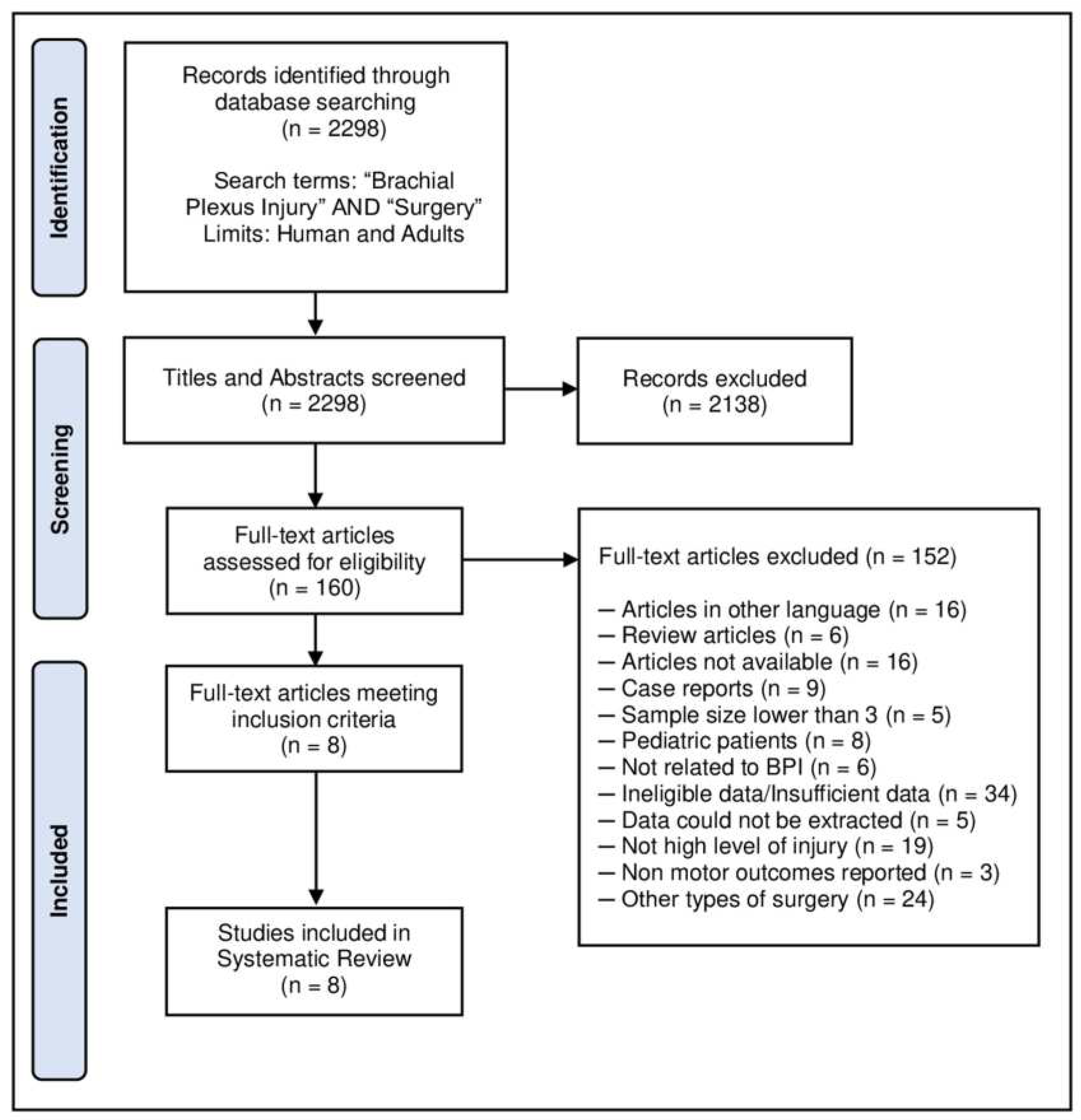
This systematic review was conducted following the Preferred Reporting Items for Systematic Reviews and Meta-Analyses (PRISMA) guidelines [12]. The primary objective was to determine the extent of motor status changes, as measured by the BMRC scale, after surgical neurolysis in adult patients with BPN. We included studies reporting pre- and post-operative motor clinical assessments of adult patients diagnosed with BPN who underwent primary surgical neurolysis. The review excluded pediatric populations with obstetric brachial plexus palsy, injuries located distally (arm, elbow, forearm, wrist, hand), case reports, basic science research, review articles, and publications in languages other than English. PubMed’s advanced search was used with Mesh terms “Brachial Plexus Injury” as the main topic and “Surgery” as a subtopic, along with additional filters for “Humans” and “Adults” to avoid animal models and pediatric populations. This search yielded a total of 2298 articles published between 1968 and 2022, with no restrictions on study design, year of publication, or publication status. Figure 2 provides a summary of the selection criteria, search process, and data extraction. Motor outcomes were collected by obtaining preoperative and postoperative motor status data according to the BMRC scale from each study. The systematic review results were analyzed to determine the proportion of motor recovery reported in each study.
Through a comprehensive bibliographic review, a total of 2298 articles published between 1968 and 2022 were initially identified. These articles underwent a screening process based on the review of titles and abstracts. Subsequently, 160 articles were selected in full-text format due to their relevance to brachial plexus surgery. Two reviewers (AAS and JLNO) conducted a detailed evaluation of these articles. In cases of disagreement, a third reviewer (JDCR) was involved to make decisions regarding the inclusion process.
In both the case series and the systematic review, the muscle selected for analysis was chosen based on its representation of the injury pattern. Specifically, the most representative muscle was identified for each cervical root as follows: Deltoid (86%) for C5, Biceps (71%) for C6, Triceps (100%) for C7, and either first dorsal interosseous (100%) or Extensor indicis proprius (100%) for C8-T1. The percentage in parentheses indicates the extent of innervation received by the respective cervical root, and this information was derived from Tsao B.’s study in 2007. The muscle with the highest percentage of innervation (i.e., the most representative according to the lesion pattern) was chosen for the analysis [13].

3. Results

Case series
The outcomes of the surgical intervention in the motor component are summarized in Table 2. In the long-term follow-up (mean 41.94±39.84 months) after the surgical intervention, a significant increase of 58.52% in motor recovery, as assessed by BMRC, was observed compared to the preoperative status. The average preoperative BMRC score was 2.17 ± 1.15, which improved to 3.44 ± 1.34 postoperatively. These changes were found to be statistically significant (p = 0.003), indicating a relevant motor recovery after the surgical intervention. The effect measure (d = 0.913) showed a large effect size. Notably, there were no observed postoperative complications, and the surgery did not affect sensory function. Furthermore, a subgroup analysis based on the etiology of BPN (traumatic and miscellaneous- OTS, tumor, radiotherapy) was conducted. Regardless of the etiology, patients demonstrated significant motor improvement after the surgical intervention (p < 0.05) (as shown in Table 2). Regarding the location of the lesions, motor improvement was observed across all lesion patterns. The most substantial recovery was observed across all lesion patterns. The most substantial recovery was observed in C5-T1 injuries, with an improvement of 64.2%, followed by C7-C8-T1 (41.1%, C5-C6-C7 (39.9%), and C5-C6 (14.28%). Importantly, none of the patients experienced an isolated motor deficit or sensory impairment after surgery.
All patients in the series opted for surgery using neurolysis alone, as none of them exhibited signs of nerve transection during the intraoperative period (Figure 1C). Moreover, anatomical structures compressing the brachial plexus were identified in all cases (Figure 1D). Out of the 18 patients who underwent surgical management, 11 (61%) experienced improvement, 5 (28%) showed no change, and 2 (11%) exhibited a slight worsening after the intervention.
Systematic Review
A total of eight studies published between 1995 and 2020 were included in this analysis [5,8,14,15,16,17,18,19], which are summarized in Table 3. These studies involved the evaluation of 240 patients regarding motor recovery outcomes. The most frequent study types were retrospective chart reviews and case series. However, no complete clinical trials (controlled, randomized, and blinded) were found in the included studies. Regarding the location of the lesions, they were found to be heterogeneous. Similarly, the delay in surgical management varied across studies, ranging from relatively early management between two weeks and four months [14,16] to others that extended up to 10-11 months [6]. One significant limitation observed was the lack of clear reporting on the preoperative motor status in all studies published before 2019. However, postoperative motor status was reported in most studies, showing a proportion of motor recovery ≥ M3 in 75% of cases.

4. Discussion

Regarding motor outcomes in the case series, two patients (patient number 12 and 18) experienced worsening after the intervention. These patients shared similar clinical-demographic characteristics, being male patients in their third decade of life with right neuropathy due to a post-traumatic injury. Both underwent surgery 8 months after the injury, and their lesions were located in the upper trunks (C5-C6). However, upon analysis, none of these shared characteristics seem to correlate with the clinical worsening, as the group that demonstrated motor recovery also includes patients with similar clinical-demographic characteristics. Therefore, we hypothesize that the clinical worsening may be attributed to other factors, such as the impact of the nerve injury on muscle innervation (trophism) or deterioration resulting from neural/vascular insult during neurolysis [20,21]. Additionally, a possible considerable decrease in nerve conduction, indicated by a proximal compound motor action potential (cMAP) of more than 50% of the distal cMAP amplitude (<50% conduction) [2], might have contributed to the outcome. Unfortunately, we were unable to conduct an electrophysiological study during surgery due to lack of available equipment for intraoperative analysis. Conducting such studies in the future could be relevant to determine an appropriate nerve conduction cut-off value in adults, enabling a better assessment of when it would be more suitable to perform a nerve graft instead of surgical neurolysis. Patients with similar characteristics could potentially benefit from nerve graft or nerve transfer procedures.
Furthermore, there exists a logical connection between surgical intervention using neurolysis and clinical recovery, as the patients who underwent surgical management presented with compressive neuropathy. This compressive neuropathy was observed in various etiologies, such as outlet thoracic syndrome (OTS), tumor presence, or the development of fibrosis secondary to trauma or radiotherapy. Regardless of the etiology, these conditions share a common pathophysiological mechanism [15,22,23] that involves nerve strangulation, leading to reduced blood flow and consequent motor impairment [24]. Surgical neurolysis effectively addresses this pathophysiological mechanism by releasing the compressed nervous structures through separation from the surrounding tissues. This phenomenon explains the observed improvement in patients, irrespective of the specific etiology, as all these conditions share the underlying pathophysiology associated with compressive neuropathy (as summarized in Table 3).
Limitations
Several limitations must be considered when interpreting the findings from the case series and the systematic review. Firstly, the heterogeneity of the follow-up periods in the included patients, ranging from two to three months up to 156 months, could lead to potential under or overestimation of motor recovery. Secondly, due to some limitations, intraoperative electrophysiology could not be performed in all cases, introducing biases related to the assessment of nerve integrity. Regarding the systematic review, a notable limitation is the relatively scarce and highly heterogeneous with a low level of evidence. Additionally, some studies with higher levels of evidence discussing motor outcomes in surgical neurolysis were conducted in pediatric populations, limiting their applicability to adult patients [2,4]. Furthermore, in the qualitative synthesis of the reviewed articles, only one study evaluated the efficacy of the technique as the main objective [6], while the remaining studies considered patients subjected to various techniques.
Upon analyzing the articles included in Table 3, it is interesting to note that Gousheh J. et al. (1995) and Altaf F. et al. (2012) reported motor recovery percentages greater than 95% with a BMRC >3. Notably, their studies had a shorter lesion-surgery interval compared to the other studies. Regarding the injury-surgery interval, Giuffre, J. et al. (2010) suggested that the optimal time window for surgical intervention, regardless of the surgical technique, should not exceed six months.Neurolysis is commonly performed in conjunction with other techniques such as nerve transfer or nerve graft. For such combined surgeries, the literature suggests that the surgical interval can be reduced, starting between eight weeks and up to a maximum of six months. This is because between two and eight weeks, scar tissue undergoes an inflammatory process that can hinder the surgery [25,26,27].
Further considerations
Current trends in the surgical management of peripheral nerve injuries of the upper extremity primarily focus on evaluating relatively more complex reconstructive techniques, such as muscle/tendon transfers and nerve transfers [22]. One of the main objectives of this study was to highlight the scarcity of information regarding a technique that has been largely replaced by these new alternatives. This reality makes it challenging to draw robust conclusions about the usefulness and indications for performing surgical neurolysis as an isolated technique based on the existing literature. Most of the studies included in this analysis lack a sufficient level of scientific evidence and necessary methodological rigor and standardization to make a compelling statement about the ideal candidates for surgical neurolysis. As a result, reaching firm conclusions about the specific group of patients who could benefit most from this technique remains difficult.
To address the current research gap, we propose a study design with the following characteristics: (1) Based on the observed effect size in the case series (d = 0.913), we calculated the required sample size using G * Power 3.1.9.7 for Windows XP, considering α = 0.05 and β = 0.01 for a T-test for dependent groups. The analysis indicates that a sample size of 21 patients per group is needed to achieve a statistical power of 99%. (2) The study will be designed as a randomized trial, where participants will be randomly assigned to two treatment groups: surgical neurolysis and other surgical alternatives (e.g., nerve graft). (3) Motor clinical outcomes will be defined by assessing strength using the BMRC scale [15], active range of motion [28,29], functional impact with the Disabilities of the Arm, Shoulder, and Hand (DASH) questionnaire [30], and objective strength assessment using the push and pull-dynamometer [31]. (4) The study will include a self-controlled design, evaluating both the healthy and compromised sides of the patients. (5) Six measurements will be conducted: one preoperative assessment and subsequent evaluations at one month, three months, six months, 12 months, and 24 months [32]. Additionally, quality of life will be assessed using the SF-36 questionnaire [33]. (6) If feasible, blinding will be implemented for patients, treating physicians, and the individuals performing statistical analyses. (7) Potential sources of bias, such as demographic characteristics, mechanism of injury (etiology), severity, and extension of the injury, interval between injury and surgery [34], location of the injury, preservation of muscle trophism, and integrity of muscle/nervous structures defined through electromyographic and nerve conduction analysis [2,35], will be carefully considered and addressed in the study design.
Clinical considerations
The main objective of this study is not to establish surgical neurolysis as the superior technique or advocate its exclusive use over other surgical alternatives. We recognize that many surgeons view it as a preparatory procedure preceding other interventions such as nerve grafts or transfers, and it is not typically considered in isolation. Nevertheless, our analysis of the case series, combined with the existing literature, suggests that surgical neurolysis may offer valuable benefits for motor restoration in specific patient scenarios.
In light of these findings, we believe that surgical neurolysis should be regarded as an integral part of the surgical armamentarium, even as a standalone technique. While we acknowledge the need for more comprehensive evidence, the current results indicate its effectiveness in terms of motor recovery for patients with preserved nerve continuity and conduction, as well as those with compressive neuropathy.

5. Conclusions

Contrary to conventional assumptions, the findings of this case series and systematic review prompt us to reconsider the potential of surgical neurolysis for motor recovery in adult patients with brachial plexus neuropathy. Nevertheless, given the absence of complete clinical trials, these results underscore the necessity to re-evaluate the utility of this technique through well-designed, adequately powered, randomized, and blinded clinical trials. Such trials are essential to substantiate the apparent effectiveness of surgical neurolysis in achieving motor restoration.

Author Contributions

Armas-Salazar, Carrillo-Ruiz, Cid-Rodríguez and González-Morales wrote the manuscript with support from García-Jerónimo and Navarro-Olvera. Carrillo-Ruiz and Armas-Salazar conceived of the presented idea. Armas-Salazar, Navarro-Olvera, García-Jerónimo and Abarca-Rojano performed the analysis and measurements. Carrillo-Ruiz verified the analytical methods and contributed to the interpretation of the results. Rodríguez reviewed the hole manuscript and made the corrections as and English native speaker. Carrillo-Ruiz supervised the project and carried out all the surgical interventions. All authors discussed the results and contributed to the final manuscript.

Funding

This research received no specific grant from any funding agency in the public, commercial, or not-for-profit sectors

Institutional Review Board Statement

Prior to participation in this study, written informed consent was obtained from each patient. The study protocol and research procedures were reviewed and approved by the hospital’s Research and Bioethics Committee under protocol number DI/16/403/03/152. All aspects of the study were conducted in compliance with the principles outlined in the Declaration of Helsinki and other relevant ethical guidelines to safeguard the rights, confidentiality, and well-being of the participants.

Data Availability Statement

The datasets generated during the current study are available from the corresponding author on reasonable request.

Conflicts of Interest

The authors declare that the research was conducted in the absence of any commercial or financial relationships that could be construed as a potential conflict of interest.

References

  1. Limthongthang R, Bachoura A, Songcharoen P, Osterman AL. Adult brachial plexus injury: evaluation and management. Orthop Clin North Am 2013;44:591-603. [CrossRef]
  2. Andrisevic E, Taniguchi M, Partington MD, Agel J, Van Heest AE. Neurolysis alone as the treatment for neuroma-in-continuity with more than 50% conduction in infants with upper trunk brachial plexus birth palsy. J Neurosurg Pediatr 2014;132:229-237. [CrossRef]
  3. Millesi H, Rath T, Reihsner R, Zoch G. Microsurgical neurolysis: its anatomical and physiological basis and its classification. Microsurgery. 1993;14:430-439. [CrossRef]
  4. Clarke HM, Al-Qattan MM, Curtis CG, Zuker RM. Obstetrical brachial plexus palsy: results following neurolysis of conducting neuromas-in-continuity. Plast Reconstr Surg 1997;97:974-984. [CrossRef]
  5. Armas-Salazar A, García-Jerónimo AI, Villegas-López FA, Navarro-Olvera JL, Carrillo-Ruiz JD. Clinical outcomes report in different brachial plexus injury surgeries: a systematic review. Neurosurg Rev 2022;45:411-419. [CrossRef]
  6. Morgan R, Elliot I, Banala V, Dy C, Harris B, Ouellette EA. Pain Relief after Surgical Decompression of the Distal Brachial Plexus. J Brachial Plex Peripher Nerve Inj 2020;15:e22-e32. [CrossRef]
  7. Armas-Salazar A. Téllez-León N. García-Jerónimo AI. Villegas-López FA. Navarro-Olvera JL. Carrillo-Ruiz JD (2022) Neuropathic pain relief after surgical neurolysis in patients with traumatic brachial plexus injuries: A preliminary report. Pain Res Manag (Accepted).
  8. Midha R, Grochmal J. Surgery for nerve injury: current and future perspectives. J Neurosurg 2019;130:675-685. [CrossRef]
  9. Li GY, Xue MQ, Wang JW, Zeng XY, Qin J, Sha K. Traumatic brachial plexus injury: a study of 510 surgical cases from multicenter services in Guangxi, China. Acta Neurochir (Wien) 2019;161:899-906. [CrossRef]
  10. Rempel DM, Diao E. Entrapment neuropathies: pathophysiology and pathogenesis. J Electromyogr Kinesiol 2004;14:71-75. [CrossRef]
  11. James MA. Use of the Medical Research Council muscle strength grading system in the upper extremity. J Hand Surg Am 2007;32:154-156. https://doi.org/10.1016/j.jhsa.2006.11.008. [CrossRef]
  12. McInnes MDF, Moher D, Thombs BD, et al. Preferred Reporting Items for a Systematic Review and Meta-analysis of Diagnostic Test Accuracy Studies: The PRISMA-DTA Statement [published correction appears in JAMA. 2019 Nov 26;322(20):2026]. JAMA 2018;319:388-396. [CrossRef]
  13. Tsao B. The electrodiagnosis of cervical and lumbosacral radiculopathy. Neurol Clin 2007;25:473-494. [CrossRef]
  14. Altaf F, Mannan K, Bharania P, Sewell MD, Di Mascio L, Sinisi M. Severe brachial plexus injuries in rugby. Injury 2012;43:272-273. [CrossRef]
  15. Dubuisson AS, Kline DG. Brachial plexus injury: a survey of 100 consecutive cases from a single service. Neurosurgery 2002;51:673-683.
  16. Gousheh J. The treatment of war injuries of the brachial plexus. J Hand Surg Am 1995;20:S68-S76. https://doi.org/10.1016/s0363-5023(95)80173-1. [CrossRef]
  17. Gutkowska O, Martynkiewicz J, Mizia S, Bąk M, Gosk J. Results of Operative Treatment of Brachial Plexus Injury Resulting from Shoulder Dislocation: A Study with A Long-Term Follow-Up. World Neurosurg 2017;105:623-631. [CrossRef]
  18. Kim DH, Han K, Tiel RL, Murovic JA, Kline DG. Surgical outcomes of 654 ulnar nerve lesions. J Neurosurg 2003;98:993-1004. [CrossRef]
  19. Stewart MP, Birch R. Penetrating missile injuries of the brachial plexus. J Bone Joint Surg Br 2001;83:517-524. [CrossRef]
  20. Gordon T, Tyreman N, Raji MA. The basis for diminished functional recovery after delayed peripheral nerve repair. J Neurosci 2011;31:5325-5334. [CrossRef]
  21. Martin E, Senders JT, DiRisio AC, Smith TR, Broekman MLD. Timing of surgery in traumatic brachial plexus injury: a systematic review [published online ahead of print, 2018 ]. J Neurosurg 2018;1-13. [CrossRef]
  22. Čebron U, Mayer JA, Lu C, Daigeler A, Prahm C, Kolbenschlag J. Treatment Trends of Adult Brachial Plexus Injury: A Bibliometric Analysis. Plast Reconstr Surg Glob Open 2021;9:e3803. [CrossRef]
  23. Warade AC, Jha AK, Pattankar S, Desai K. Radiation-induced brachial plexus neuropathy: A review. Neurol India 2019;67(Supplement):S47-S52. [CrossRef]
  24. Baxter B, Gillingwater TH, Parson SH. Rapid loss of motor nerve terminals following hypoxia-reperfusion injury occurs via mechanisms distinct from classic Wallerian degeneration. J Anat 2008;212:827-835. [CrossRef]
  25. Giuffre JL, Kakar S Bishop AT, Spinner RJ, Shin AY. Current Concepts of the Treatment of Adult Brachial Plexus Injuries. American Society for Surgery of the Hand, 2010;35A:678–688. [CrossRef]
  26. Hems TEJ. Timing of surgical reconstruction for closed traumatic injury to the supraclavicular brachial plexus. Abstract. The Journal of Hand Surgery (European Volume), 2015;40E:568-572. [CrossRef]
  27. Noland SS, Bishop AT, Spinner, RJ, Shin AY. Adult Traumatic Brachial Plexus Injuries. Journal of the American Academy of Orthopaedic Surgeons, 2019;27:705-716. [CrossRef]
  28. Doğan M, Koçak M, Onursal Kılınç Ö, Ayvat F, Sütçü G, Ayvat E. et al. Functional range of motion in the upper extremity and trunk joints: Nine functional everyday tasks with inertial sensors. Gait Posture 2019;70:141-147. [CrossRef]
  29. Lea RD, Gerhardt JJ. Range-of-motion measurements. J Bone Joint Surg Am 1995;77:784-798. [CrossRef]
  30. Roy JS, MacDermid JC, Woodhouse LJ. Measuring shoulder function: a systematic review of four questionnaires. Arthritis Rheum 2009;61:623-632. [CrossRef]
  31. Tsai YJ, Su FC, Hsiao CK, Tu YK. Comparison of objective muscle strength in C5-C6 and C5-C7 brachial plexus injury patients after double nerve transfer. Microsurgery 2015;35:107-114. [CrossRef]
  32. Martins RS, Siqueira MG, Heise CO, Foroni L, Teixeira MJ. A prospective study comparing single and double fascicular transfer to restore elbow flexion after brachial plexus injury. Neurosurgery 2013;72:709-715. [CrossRef]
  33. Kitajima I, Doi K, Hattori Y, Takka S, Estrella E. Evaluation of quality of life brachial plexus injury patients after reconstructive surgery. Hand Surgery, 2006;11:103-107. [CrossRef]
  34. West DC, Shaw DM, Lorenz P, Adzick NS, Longaker MT. Fibrotic healing of adult and late gestation fetal wounds correlates with increased hyaluronidase activity and removal of hyaluronan. Int J Biochem Cell Biol 1997;29:201-210. [CrossRef]
  35. Brophy RH, Wolfe SW. Planning brachial plexus surgery: treatment options and priorities. Hand Clin 2005;21:47-54. [CrossRef]
Figure 1. Surgical technique. A. Surgical approach performed in the supraclavicular fossa B. “V-shaped” incision following the posterior border of the sternocleidomastoid and the inferior border of the clavicle. C. Exposure of the neuroma-in-continuity located in the upper trunk (1) and its branches; supraclavicular nerve (1.c), lateral cord (1.b), and the upper trunk division to the posterior cord (1.a). D. Number 1 represents a preserved nerve structure, contrasting the structures highlighted with numbers 3 and 2, which indicate the contact zone (arrow) that was released after surgical neurolysis.
Preprints 82280 g001
Figure 2. Flow Diagram preferred reporting items for systematic review and meta-analysis (PRISMA) search strategy. BPN: Brachial Plexus Neuropathy.
Preprints 82280 g002
Table 1. Clinical and demographic characteristics of the patients included.
BMRC
motor grading scale
No. of Patient Gender Age at time of surgery Etiology
(mechanism)
Location of injury Side affected Interval
Injury-Surgery (mos)
Follow-up
(mos)
Pre-op Post-op
1 Female 29 Post-Traumatic (VT) C5-C6 Left 12 24 2 4
2 Female 62 Radiotherapy C5-T1 Right 5 48 2 2
3 Male 43 Post-Traumatic (VT) C5-C6-C7 Left 19 108 2 4
4 Female 21 Outlet thoracic Sx. C7-C8-T1 Left 14 12 1 4
5 Male 20 Post-Traumatic (VT) C5-T1 Right 6 60 0 4
6 Male 41 Tumor (E) C5-T1 Left 4 60 1 4
7 Male 29 Outlet thoracic Sx. C7-C8-T1 Right 10 12 3 5
8 Male 46 Outlet thoracic Sx. C7-C8-T1 Right 16 12 3 3
9 Female 28 Outlet thoracic Sx. C7-C8-T1 Right 11 36 3 5
10 Male 35 Post-Traumatic (VT) C5-T1 Right 7 156 0 0
11 Female 22 Post-Traumatic (VT) C5-T1 Right 9 18 2 4
12 Male 22 Post-Traumatic (IT) C5-C6 Right 8 3 3 2
13 Male 21 Post-Traumatic (VT) C5-C6-C7 Right 20 12 2 2
14 Female 56 Tumor (A) C5-C6 Left 5 48 4 5
15 Female 52 Post-Traumatic (VT) C5-C6 Left 6 48 4 4
16 Male 32 Post-Traumatic (VT) C5-C6 Left 6 72 2 4
17 Male 26 Post-Traumatic (VT) C5-C6-C7 Right 14 24 2 4
18 Male 28 Post-Traumatic (SI) C5-C6 Right 8 2 3 2
*Mean ± SD 34.06 ± 13.01 10 ± 4.89 41.94 ± 39.84 2.17 ± 1.15 3.44 ± 1.34
Median (IQR) 29 (21.75) 8.5 (8) 30 (48) 2 (1.25) 4 (2)
BMRC: British Medical Research Council. VT: Vehicular trauma. IT: Industrial trauma. SI: Stab injury. E: Ependymoma. A: Astrocytoma. SD: Standard deviation. IQR: Interquartile range. *The data were represented as mean and standard deviation despite being a non-parametric sample because in the literature they are usually represented in this manner.
Table 2. Clinical outcomes after surgical neurolysis and demographic characteristics according to etiology.
Etiology (group) Global Traumatic Miscellaneous*
Sample size (n) 18 11 7
Demographics
Age (yrs) 34.06 ± 13.01 30 ± 10.05 40.42 ± 15.28
Gender () 61.11% 72.7% 42.85%
Location
C5-C6 6 (33.3%) 5 (45.46%) 1 (14.28%)
C5-C6-C7 3 (16.7%) 3 (27.27%) 0
C8-T1 4 (22.2%) 0 4 (57.15%)
C5-T1 (complete) 5 (27.8% 3 (27.27%) 2 (28.57%)
Side affected
Left 7 (38.9%) 4 (36.36%) 3 (42.85%)
Right 11 (61.1%) 7 (63.64%) 4 (57.15%)
Interval Injury-Surgery (mos) 10 4.89 10.45 ± 5.14 9.28 ± 4.75
Follow-up (mos) 41.94 39.84 47.90 ± 48.44 32.57 ± 20.45
Clinical outcomes
BMRC
Pre-operative status 2.17 ± 1.15 2 ± 1.18 2.42 ± 1.13
Post-operative status 3.44 ± 1.34 3.09 ± 1.37 4 ± 1.15
Statistical analysis
p-valuea 0.003 0.045 0.017
Effect size (d)b 0.913 0.703 0.977
Delta (∆)c ↑ 58.52% ↑ 35.27% ↑ 39.5%
Data are shown as mean ± standard deviation or percentage. a Wilcoxon rank-sum test to compare the changes after the intervention. b Effect size calculated through Cohen’s D with a correction factor for small sample sizes. c Increase (↑) or decrease (↓) relative to preoperative status. BMRC: British medical research council motor grading scale. *The group called miscellaneous considers etiologies such as thoracic outlet syndrome, tumors, or post-radiotherapy lesions.
Table 3. Summary of articles included in the systematic review of motor outcomes in surgical neurolysis for BPI.
Author’s & year
(group/subgroups)
No. of subjects Study design Location of Compromise/Injury Interval
injury-surgery
BMRC motor grading scale Proportion of motor recovery
(≥M3)
Follow-up
Preoperative Post-operative
<M3 ≥M3 <M3 ≥M3
Current study 18 CS C5-C6: 33.3%, C5-C7: 16.6%,
C7-T1: 22.3%, C5-T1: 27.8%
10 (4.89) mos b 11 7 5 13 72.2% 41.9 (39.8) b
Morgan R.
et al. (2020) [23]
21 RCR Distal Brachial Plexus (cord branches at the level of bicipital groove). 11 mos c 3 18 1 20 95.2% 10 (6-85) mos c
Guang-Yao Li.
et al. (2019) [17]
73 a CR C5: 19.18%, C5-C7: 17.81%, C5-T1: 35.62%, Cord branches: 19.18%, Supra & infraclavicular: 8.22% NM 73 NM 15 58 79.4% 47.95 (25–68) mos c
Gutkowska O.
et al. (2017) [13]
33 a RCS Cord branches at high level
(proximal third of upper extremity)
9.03 mos b 33 NM 21 11 33.3% 5.1 yrs b
Altaf F. (2012)
et al. [1]
6 a STI C5-T1: 38.46%, C5-C6: 23.07%, C5-C7: 38.46% 12 days to 4.5 mos NM NM NM 6 100% between 4 and 6 mos
Kim D.
et al. (2003) [15]
20 a RCR High (proximal third of upper extremity) ulnar nerve injuries NS 20 NM 4 16 80% NM
Dubuisson A.
et al. (2002) [10]
11 a RCR C5-C6: 36.36%, C5-T1: 18.18%, Cord branches: 45.45% 7 mos c 11 NM 2 9 81.8% 3 yrs of more
Stewart M.
et al. (2000) [26]
14 a CS C5-C6: 14.28%, C7: 7.14%, C5-T1: 21.42%, Cord branch: 57.14% 7.35 mos b NM NM 8 6 42.8% at least two yrs
Gousheh J.
et al. (1995) [12]
44 a U C5: 27.27%, C6: 34.09%, C7: 18.18%, C8: 11.36%, C5-T1: 9.09%. 3 wks to 4 mos 44 NM 1 43 97.7% 5 yrs or more
Total/Mean 240 75.82%
Studies in which multiple techniques were performed, among them the motor outcomes of surgical neurolysis were evaluated. b Mean (Standard deviation). c Median (Ranges). BMRC: British Medical Research Council. RCR: Retrospective chart review. CR: Consecutive recruitment. RCS: Retrospective case series. STI: Structured telephone interview. U: Unclear. NM: Not mentioned.
Disclaimer/Publisher’s Note: The statements, opinions and data contained in all publications are solely those of the individual author(s) and contributor(s) and not of MDPI and/or the editor(s). MDPI and/or the editor(s) disclaim responsibility for any injury to people or property resulting from any ideas, methods, instructions or products referred to in the content.
Copyright: This open access article is published under a Creative Commons CC BY 4.0 license, which permit the free download, distribution, and reuse, provided that the author and preprint are cited in any reuse.
Prerpints.org logo

Preprints.org is a free preprint server supported by MDPI in Basel, Switzerland.

Subscribe

© 2024 MDPI (Basel, Switzerland) unless otherwise stated