Preprint
Article

Synergistic Antimicrobial and Antibiofilm Activity of Panax Ginseng, Symphytum Officinale, and Metronidazole against P. gingivalis

Altmetrics

Downloads

227

Views

93

Comments

0

Submitted:

18 August 2023

Posted:

21 August 2023

You are already at the latest version

Alerts
Abstract
Background: The biofilm-forming bacteria Porphyromonas gingivalis (P. gingivalis) is primarily responsible for periodontal disorders, which continue to be a global health issue. Despite the widespread use of antibiotics like metronidazole, other potent therapeutic a crucial quorum sensing molecule implicated in the formation of P. gingivalis biofilm. Approaches are now being explored due to the growing challenge of antibiotic resistance. Objective: In particular, chemicals from Symphytum Officinale (S) and Panax Ginseng (G) plants will be examined in this study to assess if they have the ability to inhibit biofilm and to prevent the development of acylated homoserine lactones (AHLs), a quorum sensing molecule. Materials and Methods: With Metronidazole serving as a control, the antibacterial effects of these substances (Symphytum Officinale and Panax Ginseng) were examined against a standard strain and a clinical isolate of P. gingivalis. Dilutions of these plant extracts were used either alone (G+S) or in conjunction with metronidazole (G+S+F), to assess their antibacterial activity using antibacterial susceptibility test (inhibition zone), biofilm inhibition and disruption assay (optical density) and quorum sensing inhibition assay (AHL). Results: The combinations of Symphytum officinale, Panax Ginseng and metronidazole (S+G+F) showed the maximum effectiveness with highest zone of inhibition (25.000±0.001 mm) and biofilm inhibition (98.46%), and were comparable to G+S (inhibition zone of 24.341±0.593 mm and biofilm inhibition of 97.76%), and significantly different in the degree of biofilm inhibition with different treatment scenarios. Additionally, the plant's extracts and combinations are specific concentrations had a significant effect on the suppression of the generation of AHL (p < 0.05). Conclusion: According to preliminary research, these plant-derived substances, especially when paired with metronidazole, significantly inhibited the growth of P. gingivalis biofilm providing valuable information for the development of innovative therapeutic approaches for periodontitis and other biofilm-associated illnesses.
Keywords: 
Subject: Medicine and Pharmacology  -   Dentistry and Oral Surgery

1. Introduction

Periodontal conditions impact a sizeable section of the world's population and, if ignored, can lead to tooth loss [1]. The gram-negative, anaerobic bacterium P. gingivalis is usually considered as one of the pathogens connected to the periodontal problems because of its ability to form biofilms on oral tissues [2]. An organized bacterial colony that is protected by a self-produced polymeric matrix and a biofilm has an advantage over the host's defenses and conventional antibiotic therapies [3].
The bacterial cell to cell communication mechanism known as quorum sensing (QS) is essential for the development of biofilms and other virulence traits [4]. In Gram-negative bacteria like P. gingivalis, acylated homoserine lactones (AHLs) play a crucial signaling function in the quorum sensing process [5]. In order to treat chronic disorders like periodontitis, approaches to stop AHL production may lower the activity of bacteria and prevent the formation of biofilms. [6].
Metronidazole, a nitroimidazole antibiotic, is an excellent treatment option for bacterial infections caused by P. gingivalis, are also considered .It functions by damaging bacterial DNA and obstructing protein synthesis [7]. However, the rise in antibiotic resistance threatens the antibiotic's long-term usefulness, necessitating the search for alternative therapies [8].
In this respect, compounds made from plants have attracted a lot of interest because of their shown efficacy against several bacterial infections. Comfrey (Symphytum Officinale) and Panax Ginseng have shown significant antibacterial properties in earlier studies [9]. Comfrey has a number of bioactive compounds with anti-inflammatory, antibacterial, and wound-healing activities, including allantoin, rosmarinic acid, and mucilage [10]. On the other hand, Panax Ginseng containing ginsenosides and saponins, a herb used in East Asian traditional medicine, has been found to have strong antibacterial and anti-inflammatory properties [11]. Ginsenosides are known for having potent antibacterial and anti-inflammatory properties [13]. The examination of these organic compounds for their capacity to inhibit quorum sensing and biofilm growth may pave the way for the development of innovative approaches to treating periodontal disease [12,14].
Given the growing interest in phytochemicals as potential therapeutic agents, the findings of this study may help in the development of more effective and long-lasting techniques for treating periodontal problems [18]. Given the threat that antibiotic resistance poses to the global health, it is more crucial than ever to find novel plant-based antibacterial medicines [19]. Studying how these two herbs interact with P. gingivalis may thus indicate the potential therapeutic value of Symphytum Officinale and Panax Ginseng in the management and treatment of periodontal disease [20].
However, little is known about the specific ways that Panax ginseng and Symphytum Officinale interact with P. gingivalis, particularly in relation to the inhibition of biofilm development and the production of AHLs [14]. This is despite the fact that both Panax ginseng and Symphytum Officinale have well-established antibacterial abilities [15]. Furthermore, the potential synergistic effects of mixing these plant extracts with typical antibiotics like metronidazole have also just been briefly studied in only a few studies [16]. Because the bulk of research so far have focused on the effects of individual plant extracts, there is a big gap in our understanding of the potential benefits of a combined therapeutic strategy [17].
The goal of the current study was to determine if these extracts might effectively treat P. gingivalis whether used alone or in combination with Metronidazole. Additionally, the impact of these extracts on AHL synthesis and biofilm formation was explored. This approach has the potential to significantly contribute to the creation of more effective choices for treating periodontal disease, and this research may be the first step towards that direction.

2. Materials and Methods

2.1. Isolation and Culturing of Bacteria

The primary bacterium for this study, P. gingivalis (standard strain 33277), was procured from the American Type Culture Collection (ATCC). Additionally, clinical isolates of P. gingivalis were obtained from diagnosed patients at local dental clinics in collaboration with local health authorities. Further details of the clinical isolate collection procedure can be found in [21]. The bacteria were grown anaerobically at 37°C in in a brain-heart infusion agar medium (BHI-A) supplemented with hemin and vitamin k [22] as shown in Figure 1. The process involved in obtaining clinical isolate was conducted following the approval of the Local Ethical Committee (Ethical approval Reference number 382). All participants provided informed consent, and the study was performed in accordance with the Declaration of Helsinki.
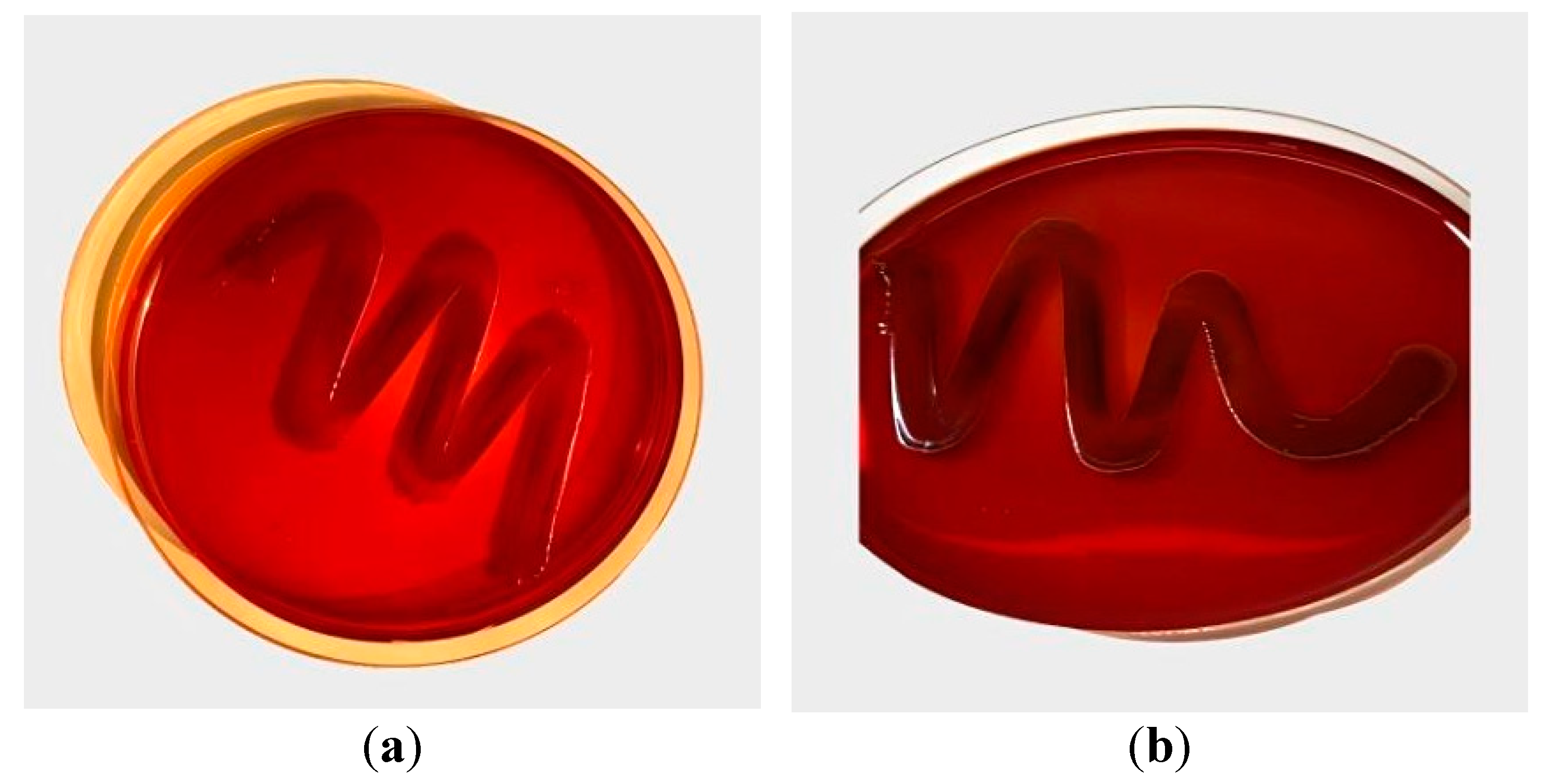
Detection of biofilm production by both the strains of P. gingivalis was assessed qualitatively by Congo Red Agar (CRA) media which composes of Brain heart infusion broth (37 gms/L), sucrose (50 gms/L), agar no.1 (10 gms/L) and congo red stain (0.8 gms/L, Himedia) [23]. The appearance of black stain indicated strong ability of quality biofilm formation (Figure 2). The clinical isolates were largely similar to those of the standard strain. Furthermore, 3 out 3 standard strains formed biofilm while 10 out of 11 clinical isolates form biofilm. This indicates that both the strains have good quality to form biofilm.

2.2. Supplement of Plant Extracts

Plant materials from Symphytum officinale (Comfrey) and Panax ginseng were sourced from certified vendors (Rejuvica Health, Gilbert, AZ, USA). The base concentrations for Comfrey, ginseng and Metronidazole were 330 mg/ml, 1000mg/ml and 500 mg/mL for without any alcohol or additive materials. Workable dilutions are prepared from the base concentrations by dissolving in Dimethyl sulfoxide (DMSO) and using systematic two-fold serial dilution technique. Eight dilutions were prepared for a G+S and G+S+F according to Table 1.
Total combination concentration of G+S (c3) was determined by using Equation 1.
v1 × c1 + v2 × c2 = v3 × c3
where v1/v2 is 1.0 mL as the volume of a single element (G or S), c1/c2 is the base concentration of a single element, and v3 is the combined volume of 5 mL. Volume of G or S is given as v1 = v2 = 1 mL, the baseline concentration of G, c1 = 1000 mg/mL, the baseline concentration of S, c2 = 330 mg/mL and the combined volume v3 = 5 ml, and the combined concentration of G+ S is calculated to be 266 mg/ml according to the above equation. Based on a two-fold serial dilution, half of this concentration (D1: first dilution for G+S) is 133 mg/mL.
Total combination concentration G+S+F (c3) was determined by using the same equation. Here v1/v2 is 1.0 mL as volume of a single element (G + S or F), c1/c2 is base concentration of a single element, and v3 is combination volume of 5 mL. Volume of G + S or F, v1 = v2 = 1 mL, base concentration of G+S, c1 = 266 mg/mL, base concentration of F, c2 = 500 mg/mL, and combination volume v3 = 5 ml were considered, and the combination concentration is calculated as 153.2 mg/mL. According to the two-fold serial dilution, half of this concentration (D1: first dilution for G+S+F) is 76.6 mg/mL.

2.3. Antibacterial Susceptibility Test

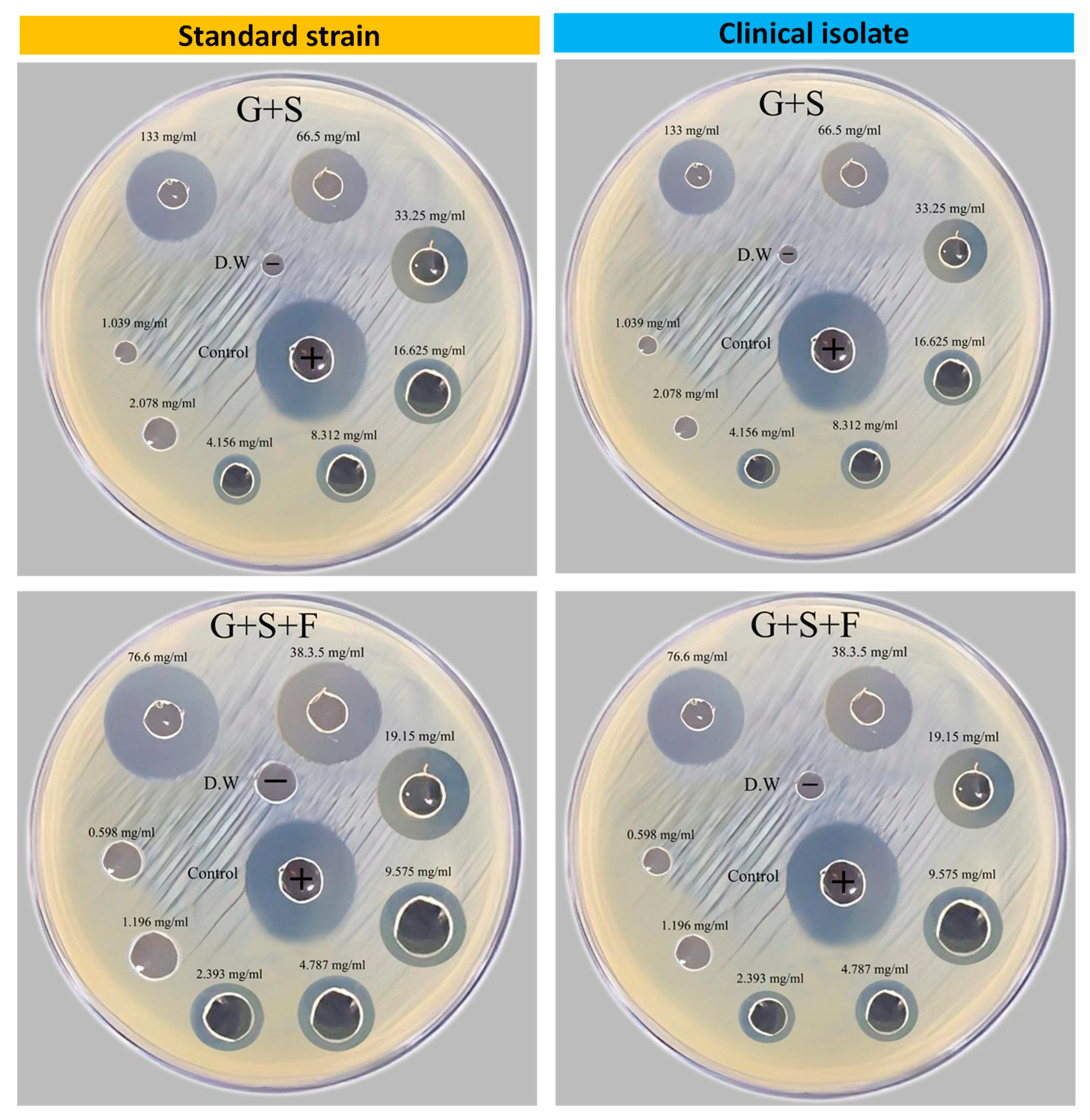
The antibacterial properties of the extracts from the plants and metronidazole, a common antibiotic used for treating infections by P. gingivalis, was assessed using the agar disk diffusion method. The test was performed by inoculating the bacteria on Brain Heart Infusion (BHI)-A plates supplemented with hemin and vitamin k to grow the bacteria. Subsequently, Mueller Hinton Agar (MHA) impregnated with different concentrations of plant extracts, metronidazole, and a combination of both extracts and metronidazole were placed onto the inoculated plates, which were then incubated anaerobically at 37°C for 48 hours. Zones of inhibition were measured to determine antibacterial activity (Figure 3).

2.4. Minimum Inhibitory Concentration (MIC) determination protocol

By making successive dilutions of the plant extracts and metronidazole, the MIC was determined. Individual wells or tubes containing the P. gingivalis inoculum received various dosages of the test chemicals. The samples were then placed in an incubator with the right conditions (5% H2, 10% CO2, and 85% N2 at 37 °C) to promote bacterial growth. The lowest dose of each test chemical that prevented discernible bacterial growth was determined by visually inspecting the plates or tubes after incubation. The MIC value was regarded as being at this concentration.

2.5. Biofilm Inhibition and Disruption Assay

Using a microtiter plate biofilm test, the capacity for biofilm formation by P. gingivalis was evaluated in the presence and absence of plant extracts and metronidazole. Both the strains of P. gingivalis were suspended in BHI containing 1% glucose and adjusted to 0.5 McFarland (1.5×10⁸ CFU/mL). Inoculate 20 μL of the bacterial suspension and 180 μL of BHI supplemented with 1% glucose into sterile flat-bottomed polystyrene microtiter plates [24]. The microplate was then incubated anaerobically at 37°C for 24 hours, and the degree to which the biofilm was inhibited was assessed using the crystal violet staining [25]. Then, the microplate was spectrophotometrically (UV-V, Germany) analyzed with a microplate reader to determine the optical density (OD). If the OD was greater than 0.240, the strain would be considered strong, moderate if the OD was between 0.120 and 0.240, and weak if the OD was less than 0.120 [26].

2.6. Quorum Sensing Inhibition Assay

The potential of the plant extracts to block the activity of quorum sensing was evaluated using a colorimetric method that produces orange coloration when combined with AHL, a molecule that is used as a quorum sensing tool. The reporter strain was cultivated alongside P. gingivalis in the presence and absence of the plant extracts. The degree of orange coloration was used as a means of measuring the activity of AHL, which was accomplished spectrophotometrically. The isolated organisms were considered to have a weak or negative association with AHLs if the OD was less than 0.98, and a strong or positive association with AHLs if the OD was greater than 0.98. [27].

2.7. Statistical Analysis

All experiments were performed in triplicate, and the data were expressed as mean ± standard deviation. Statistical analyses were performed using one-way ANOVA, followed by Tukey's multiple comparison tests. P values < 0.05 were considered statistically significant.

3. Results

3.1. Antibacterial Activity of Plant Extracts and Metronidazole

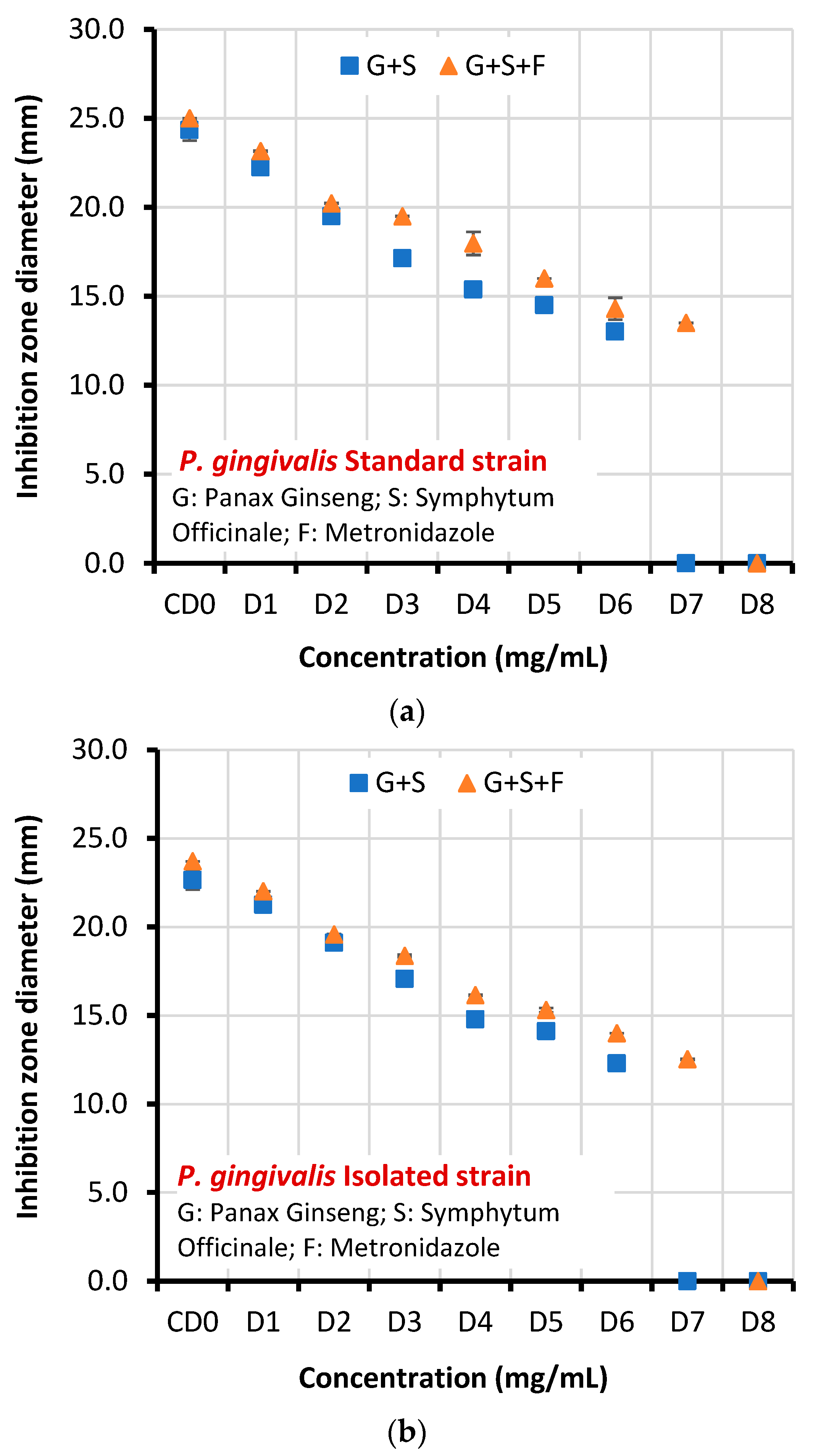
The antibacterial activities of both G+S and G+S+F against P. gingivalis in terms of inhibition zone diameters are presented in Table 2 and Figure 4. The mean inhibition-zone diameter increased with increasing concentrations of plant extracts. For (G+S), the zones of inhibition against strain ATCC (33277) ranged from 24.341±0.593 mm at 266 mg/mL to 13.022±0.013 at 4.156 mg/mL and against the clinical isolate, they ranged from 22.654±0.534mm at 266 mg/mL to 12.301±0.060 at 4.156 mg/mL. For (G+S+F), the zones of inhibition against the standard strain ranged from 25.000±0.001 mm at 153.2 mg/mL to 13.502±0.011 mm at 1.196 mg/mL and against the clinical isolate they ranged from 23.712±0.004 mm at 153.2 mg/mL to 12.533±0.033 mm at 1.196 mg/mL.
When the plant extracts were used in combination with Metronidazole, a noticeable enhancement of antibacterial activity was observed. The zones of inhibition increased significantly, suggesting a synergistic effect. For example, when Panax ginseng (1000 mg/ml) Symphytum officinale (330 mg/ml) and Metronidazole (500 mg/ml) were combined (G+S+F), the zones of inhibition were 25.000±0.001 mm and 23.712±0.004 mm against the standard strain and clinical isolate, which were significantly greater (P<0.05) than the zones produced by extracts alone (G+S).

3.2. MIC determination

The mean MICs of both G+S and G+S+F against P. gingivalis of the standard strain and the clinical isolate are listed in Table 3.

3.3. Biofilm disruption of P. gingivalis

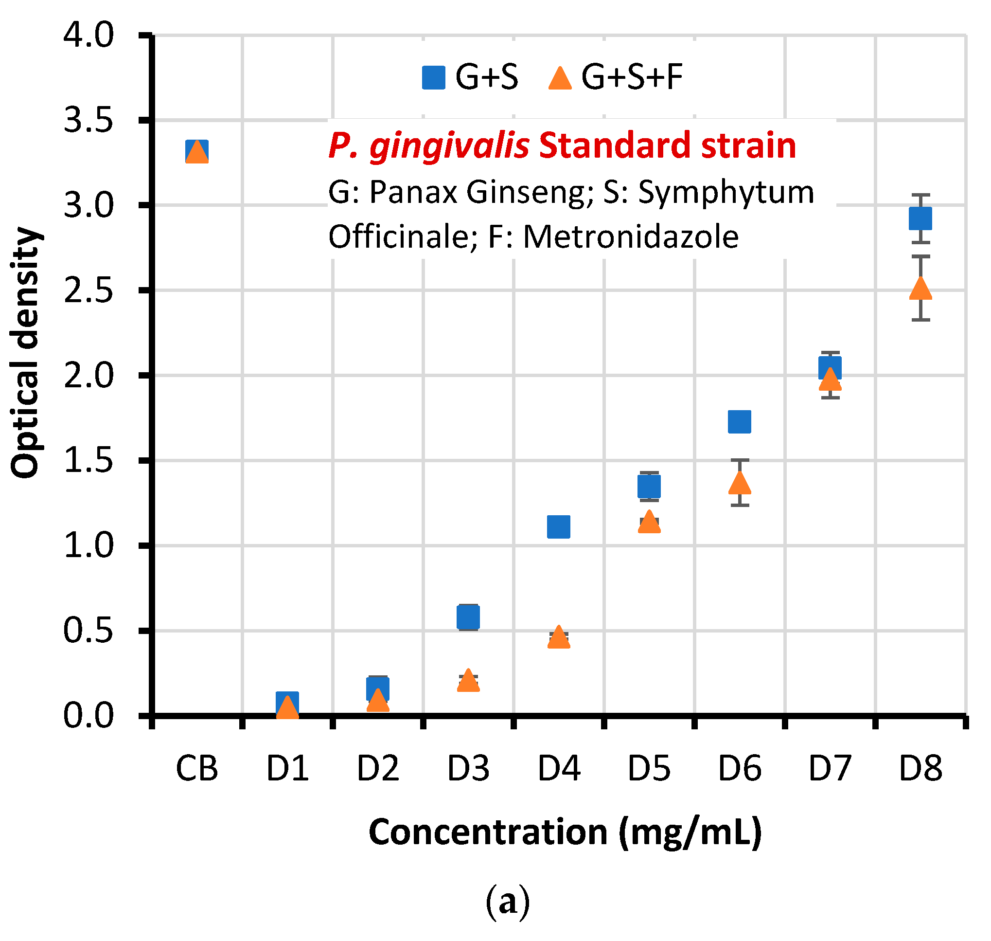
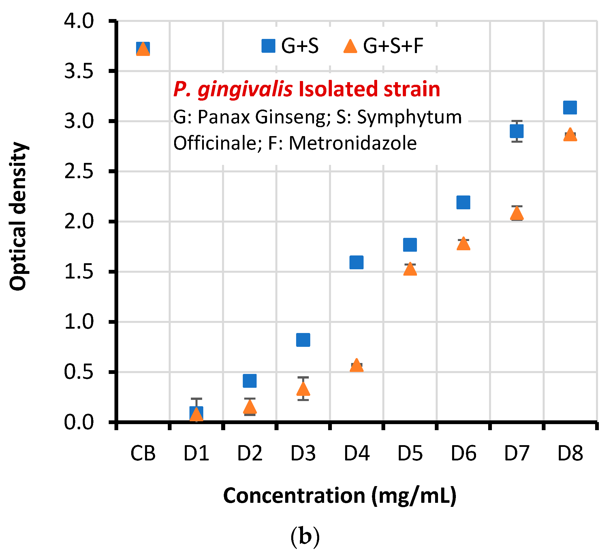
After being impacted by several experimental extracts, the effectiveness of these extracts in inhibiting biofilms formed by P. gingivalis was assessed. The antibacterial activity of the extracts tested was demonstrated by a significant difference in the optical density of the biofilms in the various treatments (p < 0.05) (Table 4 and Figure 5). It is important to recognize that even the D8 (lowest concentration) had some antibacterial properties. For both the strains, the OD values without any extracts and Metronidazole were higher than 0.240 (CB: 3.315 and 3.722), hence the strains are considered strong.
Biofilm disruption results showed that established biofilms were also sensitive to the extracts. G+S at a concentration of D5: 8.312 mg/mL disrupted about 59.36% and 52.55% of the established biofilm for the standard strain and clinical isolate respectively (Table 5). On the other hand, G+S+F at a concentration of D6: 2.393 mg/mL disrupted about 58.64% and 52.12% of the established biofilm for the standard strain and clinical isolate respectively. These concentrations were considered as the minimum dose that inhibits the bacteria.

3.4. Quorum Sensing Inhibition

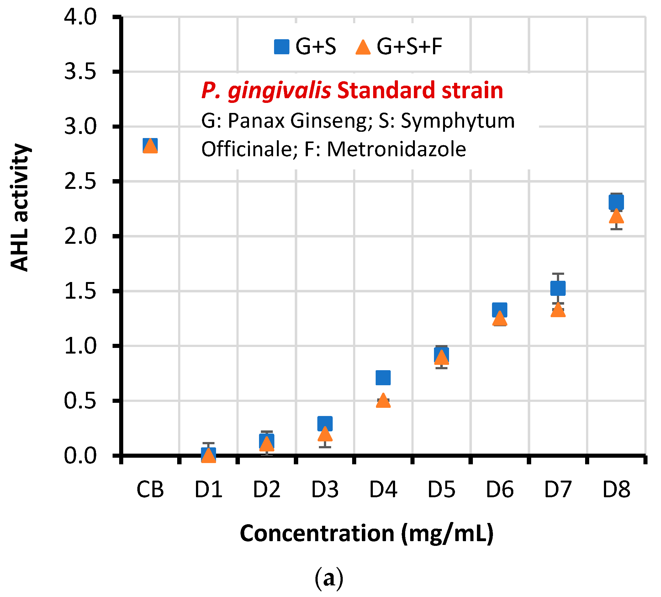
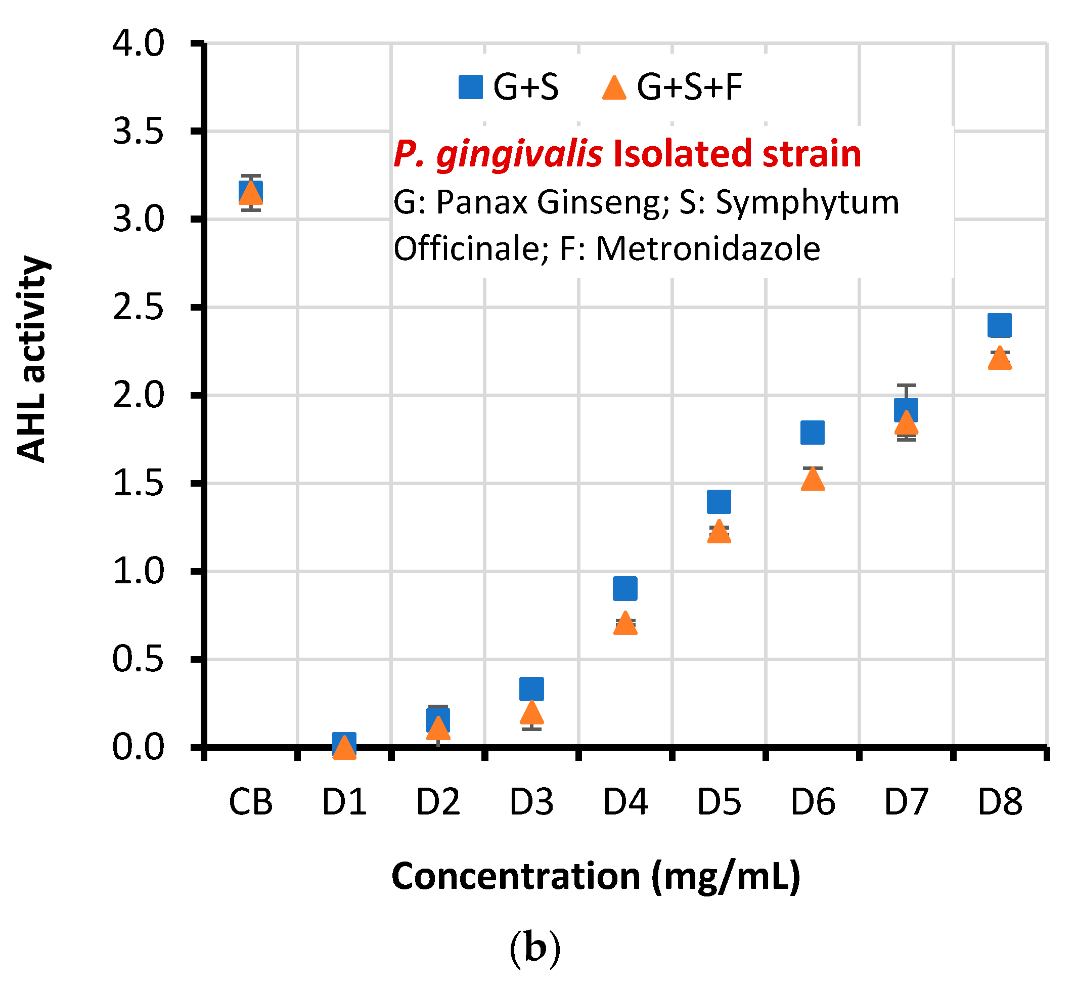
Combination of Symphytum officinale and Panax ginseng (G+S) significantly reduced the production of AHLs, as indicated by a decrease in orange pigmentation in the reporter strain (Table 6 and Figure 6). A dose-dependent decrease in AHL production was observed with increasing concentrations of both extracts (Table 7). With S+G at the concentration D5: 8.312 mg/mL reduced AHL production by approximately 67.55% and 55.76 mg/mL for the standard strain and clinical isolate respectively. While with G+S+F at a concentration of D6: 2.393 mg/ml disrupted about 55.62% and 51.53% of the established biofilm for the standard strain and clinical isolate respectively. These concentrations were considered as the minimum amount of dose that inhibit the bacteria.
The data strongly suggest that both plant extracts, especially when used in combination with Metronidazole, not only show antibacterial properties but also inhibit biofilm formation and quorum sensing against P. gingivalis. The ability of these extracts to disrupt established biofilms further augments their potential as promising therapeutic agents for the control of periodontal infections caused by P. gingivalis.

4. Discussion

The results of the current investigation showed that the extracts of Panax ginseng and Symphytum officinale had significant antibacterial actions against P. gingivalis, limiting the generation of quorum-sensing molecules and preventing the creation of new biofilms [1]. They demonstrated a synergistic interaction with the antibiotic Metronidazole, greatly boosting its antibacterial effectiveness [2]. This information may be significant because using plant extracts [28] in conjunction with antibiotics may help to combat the rising issue of antibiotic resistance [3].
The effectiveness of the plant extracts against both the standard and clinical isolates of P. gingivalis points to the possibility of using them as therapeutic agents to treat periodontal disorders [29,30]. It is crucial to keep in mind that these results are preliminary and more research is required to completely comprehend the mechanisms of action of these plant extracts and to confirm their efficiency in vivo [4].
In particular, those connected to biofilm formation and quorum sensing, the study offers encouraging insights into the potential of plant-derived chemicals in creating more efficient methods for managing P. gingivalis infections. Future research should [31,32], however, focus on identifying the precise bioactive substances that are in charge of these antibacterial activities as well as assessing the effectiveness and safety of these extracts in human clinical trials [5]. Additionally, research into additional plant-derived substances with comparable qualities may result in the identification of brand-new antibacterial substances that might significantly improve the capacity to manage periodontal disorders [6].
According to the current study's findings, Panax ginseng and Symphytum officinale both have strong antibacterial activity against P. gingivalis, both in terms of preventing growth and dissolving existing biofilms [7]. The bacteria that were isolated from patients were more active and energetic than the conventional strain, as evidenced by the smaller size of the inhibition zone (Table 2), increased optical density (Table 4), and increased activity of AHL (Table 6).This also corroborates with other studies that claim clinical isolates may display unique characteristics [11,12,13]. Additionally, there was increasing in inhibition zone diameter by increasing extracts concentrations and synergy when Metronidazole was combined with plant extracts (Table 2), suggesting a possible method for boosting the effectiveness of already prescribed antibiotics [8].
This work provides new insights into a growing body of research supporting the use of natural extracts for enhanced antimicrobial effectiveness when compared to our previous study (21). Where inhibition zone diameter of G+S (24.341±0.593) was greater than G, S, or F alone (G= 20.5120 ± 0.014, S=18.6111 ± 0.147, F= 20.4119 ± 0.114) and more synergistic effects were observed when combining the extracts with metronidazole, the inhibition zone of G+S+F (25.000±0.001) was greater than that of G+F (G+F=24.0125 ± 0.321) or S+F (22.0367 ± 0.014).
Additionally, both plant extracts shown a notable capacity to suppress the synthesis of AHLs, a crucial quorum sensing molecule involved in P. gingivalis biofilm development [9]. This is particularly intriguing since it shows that these plant extracts may not only directly kill bacteria but also interfere with the intricate networks of communication that P. gingivalis use to coordinate the creation of biofilms and other virulence traits [10].
The bioactive substances responsible for the reported antibacterial activity have not yet been identified, despite these encouraging results. Because Symphytum officinale and Panax ginseng are both known to contain a range of bioactive substances [14,15], the antibacterial effects that have been reported may be the result of one or more of these substances working alone or in combination [11].
The safety and effectiveness of these plant extracts must also be examined in clinical studies [33,34] before they can be taken into consideration for therapeutic application, despite the fact that the in vitro results are encouraging [13]. Additionally, since the current study only looked at two plant species, investigating additional plant-derived substances with comparable qualities may result in the identification of brand-new antibacterial chemicals, considerably boosting the ability to treat periodontal diseases [14,19,21,35].
In particular, those connected to biofilm formation and quorum sensing, the study offers encouraging insights into the potential of plant-derived chemicals [16,17,18] in creating more efficient methods for managing P. gingivalis infections. Future research should, however, focus on identifying the precise bioactive substances that are in charge of these antibacterial activities as well as assessing the effectiveness and safety of these extracts in human clinical trials [14].
Future study should concentrate on in vivo confirmation of the efficacy of Panax ginseng and Symphytum officinale in relation to P. gingivalis, building on the current findings. It would also be helpful to investigate any potential negative effects or toxicities connected with these extracts, particularly when used over an extended period of time. In order to create new, more potent medications to treat P. gingivalis, it is crucial to pinpoint the precise chemical components in these extracts that are responsible for the antibacterial action.
Understanding the methods by which these plant extracts prevent P. gingivalis from forming biofilms and using quorum sensing might also provide light on the pathogenicity of this bacterium and the larger significance of these processes in periodontal disease. It could also result in the creation of more specialised and successful treatment approaches.

5. Conclusions

This study assessed the effectiveness of Panax ginseng and Symphytum officinale, two plant-derived substances, in reducing periodontal infections caused on by P. gingivalis bacteria. The production of AHLs, a key quorum sensing molecule in P. gingivalis, decreased in the presence of the extracts and they showed inhibitory effects on the development of biofilms as well as destruction of already-formed biofilms. Curiously, a synergistic effect was seen when Metronidazole was coupled with these plant extracts, further increasing their antibacterial activity against P. gingivalis. Plant Extracts and Metronidazole showed a higher zone of inhibition, lower optical density and lower AHL production compared to the plant extracts alone at all tested concentrations affirming their synergistic effect. Given the problems with antibiotic resistance, plant extracts or their combination with Metronidazole will open a new avenue for treating periodontal disease and improve the quality of life.

Funding

This research was supported by the University of Iraqi's Ministry of Higher Education and Scientific Research. This grant was numbered as [ Protocol-3 ].

Acknowledgments

The Department of Periodontics personnel and technicians at the College of Dentistry, University of Baghdad are acknowledged by the authors for their exceptional support and cooperation throughout the study. The local dental clinics are thanked by the authors for extracting the P. gingivalis clinical isolate. Finally, the Ministry of Higher Education and Scientific Research, Republic of Iraq, is acknowledged for provided funding for this investigation.

References

  1. Adler et al. Helicobacter pylori and oral pathology: relationship with the gastric infection. World J. Gastroenterol. WJG 2014, 20, 9922. [CrossRef] [PubMed]
  2. Bazaz, M.R.; Rahman, Z.; Qadir, I.; Pasam, T.; Dandekar, M.P. Importance of Gut Microbiome-Based Therapeutics in Cancer Treatment. In Targeted Cancer Therapy in Biomedical Engineering; Springer: Berlin/Heidelberg, Germany, 2023; pp. 831–885. [Google Scholar]
  3. Cha, J.-D.; Jeong, M.-R.; Choi, K.-M.; Park, J.-H.; Cha, S.-M.; Lee, K.-Y. Synergistic effect between cryptotanshinone and antibiotics in oral pathogenic bacteria. Adv. Biosci. Biotechnol. 2013, 4, 283–294. [Google Scholar] [CrossRef]
  4. Cha, S.M.; Han, S.B.; Lee, Y.S.; Cha, J.D. Synergistic Effect of the Ethanol Extract of Alismatis rhizoma against Oral Pathogens. J. Oral. Bio 2015, 2, 7. [Google Scholar]
  5. Duan, L.; Dou, L.-L.; Guo, L.; Li, P.; Liu, E.-H. Comprehensive Evaluation of Deep Eutectic Solvents in Extraction of Bioactive Natural Products. ACS Sustain. Chem. Eng. 2016, 4, 2405–2411. [Google Scholar] [CrossRef]
  6. Eng-Chong, T.; Yean-Kee, L.; Chin-Fei, C.; Choon-Han, H.; Sher-Ming, W.; Li-Ping, C.T.; Gen-Teck, F.; Khalid, N.; Rahman, N.A.; Karsani, S.A.; et al. Boesenbergia rotunda: From Ethnomedicine to Drug Discovery. Evidence-Based Complement. Altern. Med. 2012, 2012, 1–25. [Google Scholar] [CrossRef] [PubMed]
  7. Ermolenko, E.; Koroleva, I.; Suvorov, A. Microbial Therapy with Indigenous Bacteria: From Idea to Clinical Evidence. In Microbiome in 3P Medicine Strategies: The First Exploitation Guide; Springer: Berlin/Heidelberg, Germany, 2023; pp. 251–274. [Google Scholar]
  8. García-Montoya, I.A.; Cendón, T.S.; Arévalo-Gallegos, S.; Rascón-Cruz, Q. Lactoferrin a multiple bioactive protein: An overview. Biochim. Biophys. Acta Gen. Subj. 2012, 1820, 226–236. [Google Scholar] [CrossRef] [PubMed]
  9. Huang, C.C.; Lai, T.; Huang, R.Y.; Su, K.W.; Lai, S.R.; Lan, A. Effect of an herbal preparation fermented by Lactobacillus reuteri LR107 in preventing periodontal inflammation in an experimental gingivitis model. Asian J Complement Altern. Med. 2014, 2, 12–18. [Google Scholar]
  10. Kim, T.-H.; Kim, S.-C.; Jung, W.-K. Therapeutic effect of marine bioactive substances against periodontitis based on in vitro, in vivo, and clinical studies. Fish. Aquat. Sci. 2023, 26, 1–23. [Google Scholar] [CrossRef]
  11. Juiz, P.J.L.; Lucchese, A.M.; Gambari, R.; Piva, R.; Penolazzi, L.; DI Ciano, M.; Uetanabaro, A.P.T.; Silva, F.; Avila-Campos, M.J. Essential oils and isolated compounds from Lippia alba leaves and flowers: Antimicrobial activity and osteoclast apoptosis. Int. J. Mol. Med. 2014, 35, 211–217. [Google Scholar] [CrossRef]
  12. Menchicchi, B.; Hensel, A.; Goycoolea, F. Polysaccharides as Bacterial Antiadhesive Agents and “Smart” Constituents for Improved Drug Delivery Systems Against Helicobacter pylori Infection. Curr. Pharm. Des. 2015, 21, 4888–4906. [Google Scholar] [CrossRef]
  13. Meresta, A.; Folkert, J.; Gaber, T.; Miksch, K.; Buttgereit, F.; Detert, J.; Pischon, N.; Gurzawska, K. Plant-derived pectin nanocoatings to prevent inflammatory cellular response of osteoblasts following Porphyromonas gingivalis infection. Int. J. Nanomed. 2017, ume 12, 433–445. [Google Scholar] [CrossRef]
  14. Mihm, C. Beeinflussung der Adhärenz und er Internalisierung von Porphyromonas gingivalis durch. Proteinaseinhibitoren. Dissertation, Friedrich-Schiller-Universität Jena, Jena, 2011. [Google Scholar]
  15. Fauzi, F.M.; Mischon, A.; Zain, N.M.; Baharuddin, I. The Therapeutic Potential of Plant Extraction in Oral Health - A Systematic Review. Compend. Oral Sci. 2022, 9, 88–104. [Google Scholar] [CrossRef]
  16. Nchiozem-Ngnitedem, V.-A.; Mukavi, J.; Omosa, L.K.; Kuete, V. Phytochemistry and antibacterial potential of the genus Garcinia. In Advances in Botanical Research; Elsevier: Amsterdam, The Netherlands, 2023; pp. 105–175. [Google Scholar] [CrossRef]
  17. E. O. F. H. O. N. PERIODONTITIS–A and S. GUM. International Journal of Pharmacology Research.
  18. Salas-Jara, M.J.; Sanhueza, E.A.; Retamal-Díaz, A.; González, C.; Urrutia, H.; García, A. Probiotic Lactobacillus fermentum UCO-979C biofilm formation on AGS and Caco-2 cells and Helicobacter pylori inhibition. Biofouling 2016, 32, 1245–1257. [Google Scholar] [CrossRef] [PubMed]
  19. Subbiah, U.; Elango, S.; Jayesh, R. Herbals and green synthesized nanoparticles in dentistry. In Nanobiomaterials in Clinical Dentistry; Elsevier: Amsterdam, The Netherlands, 2019; pp. 617–646. [Google Scholar] [CrossRef]
  20. Yin, Y.; Yang, S.; Ai, D.; Qin, H.; Sun, Y.; Xia, X.; Xu, X.; Ji, W.; Song, J. Rational Design of Bioactive Hydrogels toward Periodontal Delivery: From Pathophysiology to Therapeutic Applications. Adv. Funct. Mater. 2023, 33. [Google Scholar] [CrossRef]
  21. Mahdi Ibrahim, S.; sabri Al-mizragchi, A.; Haider, J. Metronidazole Potentiation by Panax Ginseng and Symphidium Officinale: A New Strategy for P. gingivalis Infection Control. 2023. [Google Scholar] [CrossRef]
  22. Ibraheem, D.R.; Hussein, N.N.; Sulaiman, G.M. Antibacterial Activity of Silver nanoparticles Against Pathogenic Bacterial Isolates from Diabetic Foot Patients. Iraqi Journal of Science 2023, 64, 2223–2239. [Google Scholar] [CrossRef]
  23. Hassan, A.; Usman, J.; Kaleem, F.; Omair, M.; Khalid, A.; Iqbal, M. Evaluation of different detection methods of biofilm formation in the clinical isolates. Brazilian journal of infectious diseases 2011, 15, 305–311. [Google Scholar] [CrossRef]
  24. Christensen, G.D.; A Simpson, W.; Younger, J.J.; Baddour, L.M.; Barrett, F.F.; Melton, D.M.; Beachey, E.H. Adherence of coagulase-negative staphylococci to plastic tissue culture plates: a quantitative model for the adherence of staphylococci to medical devices. J. Clin. Microbiol. 1985, 22, 996–1006. [Google Scholar] [CrossRef] [PubMed]
  25. Al-Hayanni, H.S. The Antibacterial and anti-biofilm effects of Sumac (Rhus coriaria L) fruits extracts against some multidrug-resistant pathogenic bacteria. Journal of the Faculty of Medicine Baghdad 2022, 64, 183–188. [Google Scholar]
  26. Bakir, S.H.; Fattma, A.A. Comparison of different methods for detection of biofilm production in multi-drug resistance bacteria causing pharyngotonsillitis. International journal of research in pharmacy and biosciences 2016, 3, 13–22. [Google Scholar]
  27. Baldiris, R.; TeherÃ, V; Vivas-Reyes, R.; Montes, A.; Arzuza, O. Anti-biofilm activity of ibuprofen and diclofenac against some biofilm producing Escherichia coli and Klebsiella pneumoniae uropathogens. African Journal of Microbiology Research 2016, 10, 1675–1684. [Google Scholar]
  28. Shallal, L.F.; A Ahmed, M. Experimental In vitro Study to Assess the Antibacte-rial Activity of Thymus vulgaris Oil on Streptococ-cus Sanguinis. J. Baghdad Coll. Dent. 2022, 34, 17–27. [Google Scholar] [CrossRef]
  29. M’ebid, A.K. Application of Combined Chlorhexidine and Hydrogen Peroxide In Periodontal Pockets. Bacteriological and Clinical Outcomes.
  30. Salah, R.; Abdulbaqi, H.R.; Mohammed, A.; Abdulkareem, A.A. Four-day randomized controlled crossover trial evaluating the antiplaque effect of a combination of green tea and Salvadora persica L. mouthwash. J. Herb. Med. 2020, 23, 100357. [Google Scholar] [CrossRef]
  31. Enad, H.H.; Nahidh, M. Salivary Cortisol as a Stress Biomarker and Total Viable Count of Salivary Bacterial Microbiome among COVID-19 Patients. J. Baghdad Coll. Dent. 2021, 33, 6–10. [Google Scholar] [CrossRef]
  32. Kadhum, W.N.; Al-Ogaidi, I.A.Z. Evaluation of Chitosan-Alginate Nanoparticle as A Stable Antibacterial Formula in Biological Fluids. Iraqi J. Sci. 2022, 2398–2418. [Google Scholar] [CrossRef]
  33. A Abdulkareem, A.; Abdulbaqi, H.R.; Milward, M.R. In Vitro Homeostasis of Rat Oral Epithelial Cell Cultures Following Withdrawal of Periodontal Pathogens. Braz. Dent. J. 2020, 31, 135–142. [Google Scholar] [CrossRef]
  34. Alaamery, S.K.; Al-Hayanni, H.S.A. Antibacterial and antibiofilm effect of sumac (Rhus coriaria L) fruits extracts against some multidrug_resistant pathogenic bacteria. J. Fac. Med. Baghdad 2022, 64, 183–188. [Google Scholar] [CrossRef]
  35. Zhang, Z.; Yu, Y.; Zhu, G.; Zeng, L.; Xu, S.; Cheng, H.; Ouyang, Z.; Chen, J.; Pathak, J.L.; Wu, L.; et al. The Emerging Role of Plant-Derived Exosomes-Like Nanoparticles in Immune Regulation and Periodontitis Treatment. Front. Immunol. 2022, 13, 896745. [Google Scholar] [CrossRef]
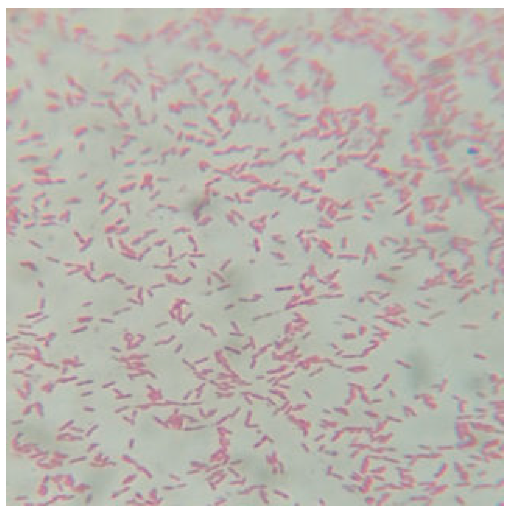
Figure 1. P. gingivalis appeared as Gram-negative coccobacilli under ×100 light microscope.
Preprints 82736 g001
Figure 2. Biofilm formation of P. gingivalis by Congo red agar (Qualitative method) (a) standard strain and (b) clinical isolate.
Preprints 82736 g002
Figure 3. Inhibition zones created by (G+S) and (G+S+F) in competition with the standard strain and clinical isolate. of P. gingivalis.
Preprints 82736 g003
Figure 4. The inhibitory zones for bacteria, which are created by plant extracts and metronidazole at different concentrations against the two types of P. gingivalis (a) the standard strain and (b) the clinical isolate.
Preprints 82736 g004
Figure 5. Biofilm inhibition activity (optical density) at different does against P. gingivalis (a) standard strain and (b) clinical isolate.
Preprints 82736 g005aPreprints 82736 g005b
Figure 6. Acylated Homoserine Lacticones (AHLs) activity to inhibit the growth of P. gingivalis in a dose-dependent manner (a) standard strain and (b) clinical isolate.
Preprints 82736 g006aPreprints 82736 g006b
Table 1. Total concentrations of plant extracts and Metronidazole.
Dilution Total concentrations (mg/mL)
G+S G+S+F
CD0: Control 266 153.2
D1: first dilution 133 76.6
D2: second dilution 66.5 38.3
D3: third dilution 33.25 19.15
D4: fourth dilution 16.625 9.575
D5: fifth dilution 8.312 4.787
D6: sixth dilution 4.156 2.393
D7: seventh dilution 2.078 1.196
D8: eighth dilution 1.039 0.598
Table 2. Antibacterial activities of plant extracts and Metronidazole against P. gingivalis.
Microorganisms Concentration Mean inhibition zone diameter ± SD (mm) T-test P-value
G+S G+S+F
Standard strain CD0 24.341±0.593 25.000±0.001 13.618 0.000
D1 22.247±0.382 23.154±0.030
D2 19.503±0.205 20.221±0.012
D3 17.136±0.361 19.503±0.020
D4 15.381±0.146 17.970±0.645
D5 14.500±0.004 16.005±0.001
D6 13.022±0.013 14.302±0.620
D7 0.0 13.502±0.011
D8 0.0 0.0
Clinical isolate CD0 22.654±0.534 23.712±0.004 12.796 0.000
D1 21.254±0.125 22.0189±0.013
D2 19.105±0.010 19.5890±0.013
D3 17.071±0.004 18.3801±0.074
D4 14..791±0.015 16.155±0.033
D5 14.123±0.105 15.311±0.123
D6 12.301±0.060 14.002±0.001
D7 0.0 12.533±0.033
D8 0.0 0.0
Note: CD0 is control group concentration; D1 to D8 is 2-fold serial dilution (Table 1); 0.0 means no inhibition zone; Panax Ginseng and Symphytum Officinale (G+S), Panax Ginseng. Symphytum Officinale and metronidazole (G+S+F).
Table 3. MIC against P. gingivalis (standard strain and clinical isolate).
Microorganisms Minimum Inhibitory Concentration (MIC) mg/mL (Doses)
G+S G+S+F
Standard strain 8.312 (D5) 2.393 (D6)
Clinical Isolate 8.312 (D5) 2.393 (D6)
Table 4. Mean optical density (OD) of the biofilm formed by P. gingivalis.
Microorganism Concentration Mean optical density (OD) of biofilm ±SD T-test P-value
G+S G+S+F
Standard strain CB 3.315±0.098 3.315±0.098 3.859 0.005
D1 0.074±0.004 0.051±0.022
D2 0.157±0.070 0.094±0.002
D3 0.579±0.069 0.211±0.021
D4 1.109±0.037 0.466±0.015
D5 1.347±0.081 1.144±0.010
D6 1.727±0.047 1.371±0.133
D7 2.043±0.092 1.980±0.112
D8 2.921±0.14 2.513±0.187
Clinical isolate CB 3.722±0.069 3.722±0.069 4.402 0.002
D1 0.090±0.145 0.080±0.001
D2 0.411±0.001 0.155±0.081
D3 0.820±0.040 0.334±0.113
D4 1.591±0.005 0.571±0.008
D5 1.766±0.208 1.530±0.041
D6 2.189±0.050 1.782±0.034
D7 2.900±0.103 2.085±0.068
D8 3.135±0.022 2.870±0.011
Note: CB is control group with biofilm without any extract; D1 to D8 is 2-fold serial dilution (Table 1); Panax Ginseng and Symphytum Officinale (G+S), Panax Ginseng + Symphytum Officinale+ Metronidazole (G+S+F).
Table 5. Percentage inhibition of biofilm formation by P. gingivalis (strain ATCC 33277) and isolate.
Microorganism Concentration Percentage inhibition of biofilm formation
[(1-(OD with dilution/OD with control))×100]
G+S G+S+F
Standard strain D1 97.76 98.46
D2 95.26 97.16
D3 82.53 93.63
D4 66.54 85.94
D5 59.36 65.49
D6 47.90 58.64
D7 38.37 40.27
D8 11.88 24.19
Clinical isolate D1 97.58 97.85
D2 88.95 95.83
D3 77.96 91.02
D4 57.25 84.65
D5 52.55 58.89
D6 41.18 52.12
D7 22.08 43.98
D8 15.77 22.89
X2 3.814
p value 0.00021
Table 6. Influence of plant extracts and Metronidazole on AHLs production.
Microorganism Concentration AHL activity ± SD T-test P-value
G+S G+S+F
Standard strain CB 2.826±0.037 3.264 0.011
D1 0.006±0.110 0.003±0.001
D2 0.130±0.033 0.109±0.110
D3 0.290±0.031 0.201±0.122
D4 0.709±0.001 0.506±0.004
D5 0.917±0.025 0.898±0.100
D6 1.327±0.039 1.254±0.062
D7 1.524±0.135 1.332±0.004
D8 2.310±0.078 2.187±0.122
Clinical isolate CB 3.151±0.098
D1 0.017±0.002 0.000 3.204 0.013
D2 0.152±0.011 0.111±0.120
D3 0.331±0.003 0.201±0.098
D4 0.901±0.014 0.709±0.013
D5 1.394±0.010 1.229±0.020
D6 1.787±0.009 1.527±0.058
D7 1.915±0.143 1.847±0.101
D8 2.397±0.060 2.215±0.029
Note: CB is control group with biofilm without any extract; D1 to D8 is 2-fold serial dilution (Table 1); Panax Ginseng and Symphytum Officinale (G+S), Panax Ginseng + Symphytum Officinale+ Metronidazole (G+S+F).
Table 7. Percentage inhibition of Acylated Homoserine Lactones (AHLS) activity by P. gingivalis (strain ATCC 33277) and isolate.
Microorganism Concentration Percentage inhibition of AHL activity
[(1-(AHL with dilution/AHL with control))×100]
G+S G+S+F
Standard strain (ATCC 33277) D1 99.78 99.89
D2 95.39 96.14
D3 89.73 92.88
D4 74.91 82.09
D5 67.55 68.22
D6 53.04 55.62
D7 46.07 52.86
D8 18.25 22.61
Clinical isolate D1 99.46 100
D2 95.17 96.47
D3 89.49 93.62
D4 71.40 77.49
D5 55.76 60.99
D6 43.28 51.53
D7 39.22 41.38
D8 23.92 29.70
X2 3.291
p value 0.00013
Disclaimer/Publisher’s Note: The statements, opinions and data contained in all publications are solely those of the individual author(s) and contributor(s) and not of MDPI and/or the editor(s). MDPI and/or the editor(s) disclaim responsibility for any injury to people or property resulting from any ideas, methods, instructions or products referred to in the content.
Copyright: This open access article is published under a Creative Commons CC BY 4.0 license, which permit the free download, distribution, and reuse, provided that the author and preprint are cited in any reuse.
Prerpints.org logo

Preprints.org is a free preprint server supported by MDPI in Basel, Switzerland.

Subscribe

© 2024 MDPI (Basel, Switzerland) unless otherwise stated