Preprint
Article

Preparation, Characterization of Magnetic Chitosan nanoparticles Coated with Fe3O4 Prepared by Ex-situ Co-precipitation Method

Altmetrics

Downloads

246

Views

88

Comments

0

This version is not peer-reviewed

Submitted:

30 August 2023

Posted:

31 August 2023

You are already at the latest version

Alerts
Abstract
The suspension cross-linking technique has been utilized to create the magnetic chitosan nanoparticles, which were then applied to the magnetic carrier method at 80°C. First the FeCl2 and FeCl3 solution co-precipitated and synthesized Fe3O4 for utilization in the formation of magnetic chitosan nanoparticles, it has been characterized by utilizing DLS (Dynamic light scattering), XRD (X-ray diffraction spectrometer), FTIR (Fourier transform infrared spectroscopy), which identified structure, size, FE-SEM (Field Emission Scanning Electron Microscope) and HR-TEM (High-resolution transmission electron microscopy) which identified structure and particle size whereas magnetic behavior of chitosan nanoparticles was determined by VSM (Vibrating sample magnetometer). According to the results collected, the magnetic chitosan nanoparticles have been spherical in form and size ranging from around 250 to 400 nm.
Keywords: 
Subject: Chemistry and Materials Science  -   Nanotechnology

1. Introduction

A cationic polysaccharide is chitosan, it is a macromolecule formed by the repetition of D-glucosamine which was easily obtained by partial deacetylation of chitin, chitin is a widely occurring bio-polymer in nature after cellulose [1] a variety of eukaryotic organisms, including crustacea, fungi, and insects, it is a polymer of N-acetyl-D-glucosamine.[2] Given that heavy metal ions are poisonous and non-biodegradable, which represent a severe danger to ecological systems and public health[3], the adsorption technique has been utilized to remove ions of the heavy metal by the water solutions. Therefore, it is essential to eliminate such harmful metal ions in the wastewater.[4] In this investigation, Fe3O4 magnetic chitosan nanoparticles have been created, assessed, and used to remove metals from a water solution.[5] We have created a chitosan magnetic nano-adsorbent employing chitosan as ionic exchange groups and Fe3O4 nanoparticles as the cores.[6] Most chitosan-based adsorbents are sub-micron to micron-sized, and to offer a sufficient surface area for adsorption, they need large internal porosities. The adsorption rate and the available capacity are reduced by the diffusion constraint inside the particles. This particular kind of magnetic adsorbent, which has favorable compatibility, a cheap price, quick and simple extraction or regeneration, and convenient operation, can be helpful for future study and actual metal removal applications.[7] Several benefits have led to the adsorption process being regarded as the most widely utilized approach (efficient, eco-friendly, economical, simple, and reversible).[8] Here, chitosan-coated with magnetic nanoparticles of Fe3O4 was effectively created as a new nano-adsorbent. Metals' adsorption kinetics and absorption capability were examined.[9] We systematically looked at how adsorbent dosage, pH, and contact duration affected the results. Additionally, the metals adsorbent's reusability and adsorption process were investigated. Additionally, the adsorbent's ability to remove metals from a water solution was assessed.[10]

2. Materials and methods

2.1. Materials

The chitosan form of a shrimp shells purchased from Sigma-Aldrich, which was about 75% (deacetylated) and ranged in molecular weight from 100 kDa to 1200 kDa, depending on the production method and product quality.[11] acetic acid, hydrochloric acid (ACS reagent, 37%), additionally, analytical grade reagents that are readily accessible on the market, such as FeCl2 and FeCl3, were utilized without additional purification, along with double-distilled water for preparing the solution.

2.2. Preparation of magnetic chitosan nanoparticles

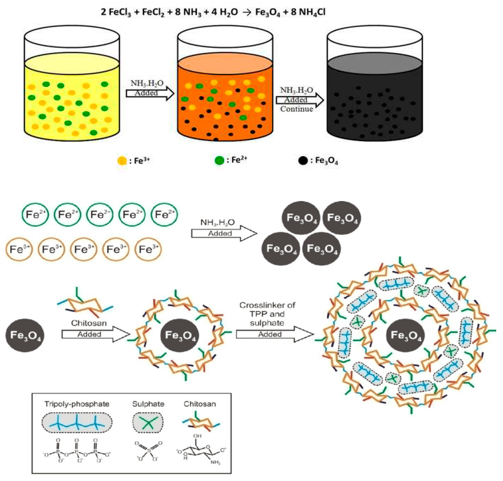
In order to obtained ferrogel solution for examined, 0.65 g of chitosan was first dissolved in 100mL of 1 % (v/v) acetic acid to prepared 0.6 % w/v chitosan solution.[12] This solution was then continuously stirred for an 1 hour at room temperature having the pH of 4 increased with 10 N solution of NaOH, and 10 mL of Fe3O4 in aqueous solution (by the “mixing of FeCl2.4H2O (1.90 g) and FeCl3.6H2O (4.50 g) salts in de-ionized water” of 50 mL) was then dropwise added into chitosan solution and then sonicated for half an hour.[13] After adding tripolyphosphate, the (Tripolyphosphate) TPP was combined for a further hour after being dissolved in water to a concentration of 1 percent under magnetic stirring at 2,000 rpm. (The weight-to-volume ratio of chitosan to TPP is 5:1 and 2:1, respectively.) [14,15] The particles have been incubated for 30 minutes at room temperature. All magnetic chitosan nanoparticles were resuspended in ultrapure water, the products have been rinsed with ethanol, and they have been drying in a vacuum oven at a temperature of 500°C for 24 H after being filtered by centrifugation at 9,000 g for 50 min at 5° [16]. [17] The electrostatic attraction among the positively charged chitosan ion and the negatively charged area around the surface of Fe3O4 led to the chitosan-coated Fe3O4. Then, tripolyphosphate and sulphate were used to create an ionic crosslinked chitosan complex, which delayed the formation of a Fe3O4 coating and improved the biodegradability & biocompatibility of the chitosan nanoparticles. The combination of these crosslinking created chitosan-Fe3O4 nanoparticles with a spherical morphology, and interactions between the two occurred through electrostatic forces.[18] After drying, the nanoparticles were characterized to look at their physical character and magnetic properties.

2.3. Characterizations of magnetic chitosan nanoparticles
TEM 200CX, Quanta 400 FESEM, and EDS have been utilized to study the shape and size of dried chitosan nanoparticles. [18] The electrons are launched in a strong electric field gradient after being liberated from a source of field emission. The socalled primary electrons are focused and refracted bent into a narrow scan beam within the high vacuum column, where it bombards the target. A detector then picks up the secondary electrons and produces an electronic signal, [19] and signal displays the FESEM images. Emitted electrons from an electron source at the top of the microscope's column fill a vacuum.[20].
Figure 1. The formation mechanism of chitosan-Fe3O4 nanoparticles by ex-situ method.
Preprints 83669 g001
The electrons are focused using electromagnetic lenses into a very narrow beam that is then directed into the specimen of interest. An energy-dispersive spectrometer (EDS) within the TEM may also detect X-ray emission as the findings of the main electron beam's interaction using the sample.[21] In DLS analysis, a sample suspension is irradiated through a beam of laser, which causes the laser light to scatter in all directions. A alternative name for DLS is photon correlation spectroscopy.[22] Over time, the light dispersion is seen at a certain angle. The randomized Brownian motion of the particles is what causes the signal fluctuation.[23] The Stokes-Einstein equation uses the angular intensity distribution to calculate the particle size. On the basis of Faraday's Law of Induction, the VSM is a scientific tool used to detect magnetic characteristics.[24] The VSM works on the premise that when the sample material is exposed to a homogeneous field of magnet, In the sample, a dipole moment is produced that is proportional to the sample susceptibility product as well as the applied field.[25]

3. Results and discussions

3.1. Nanoparticles with magnetic properties are described

3.1.1. Morphological characterization

With an almost limitless depth of focus, the FESEM gives information on topography and elements at magnifications of
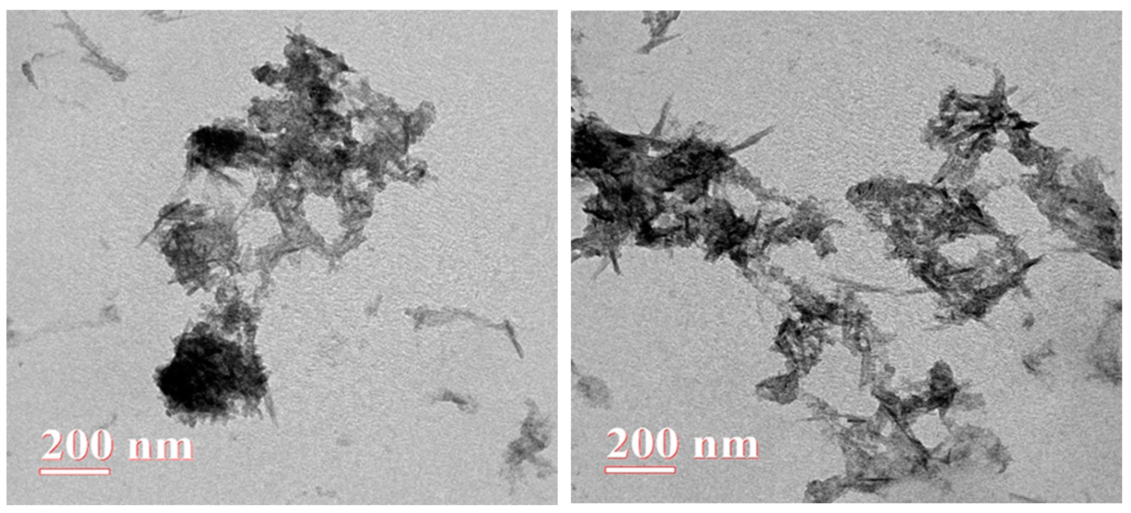
10x to 300,000x. [26] FESEM focuses on the capability to evaluate smaller-area contamination areas at electron accelerating voltages compatible with EDS and with mapping, as shown in Figure 2. [27] Because electrons have a shorter de Broglie wavelength than light, HRTEM can image at a much greater resolution than light microscopes. [28] The goal of TEM imaging of nanomaterials is to capture many calibrated transmission electron pictures that faithfully depict the nanomaterial on the HRTEM images in Figure 3.[29]

3.1.2. Magnetic properties

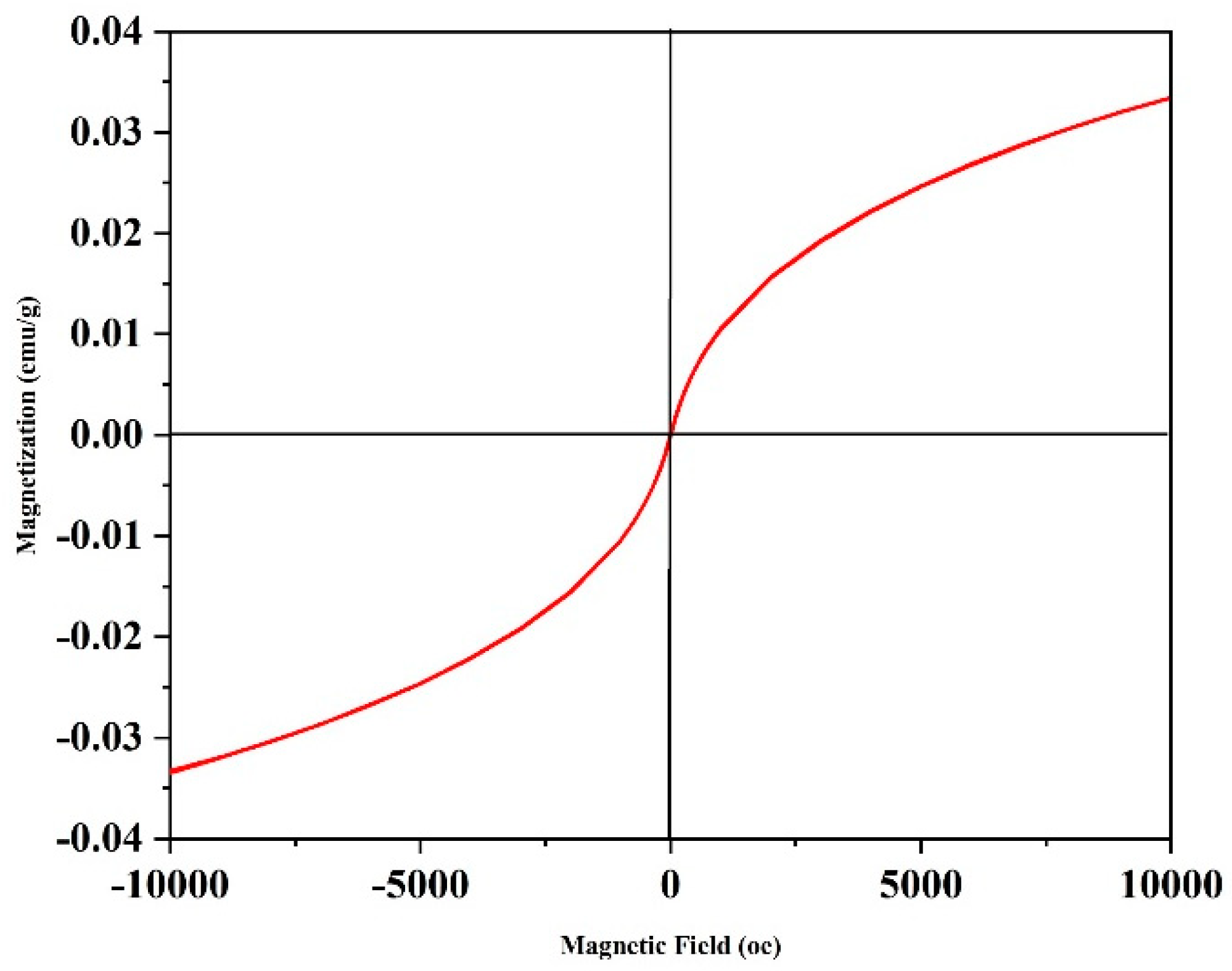
The “magnetic characteristics of materials like a function of temperature, time, and magnetic field are measured using VSM (vibrating sample magnetometer) instrument, as shown in Figure 4 [30] The magnetization of Fe3O4 chitosan nanoparticles was shown, and the VSM predicted the improved magnetic behaviour of nanoparticles. All samples were shown to possess superparamagnetic capabilities.[31] The graph displays the Fe3O4 chitosan nanoparticles' saturation magnetization values, all of which are sufficient for a practical magnetic separation. The VSM is the tool used to measure the magnetic moment, which is the most basic property of solid sample magnetism. The magnetization spiral for magnetite nanoparticles at 25°C temperature is shown in Figure 4. [32]

3.1.3. DLS analysis

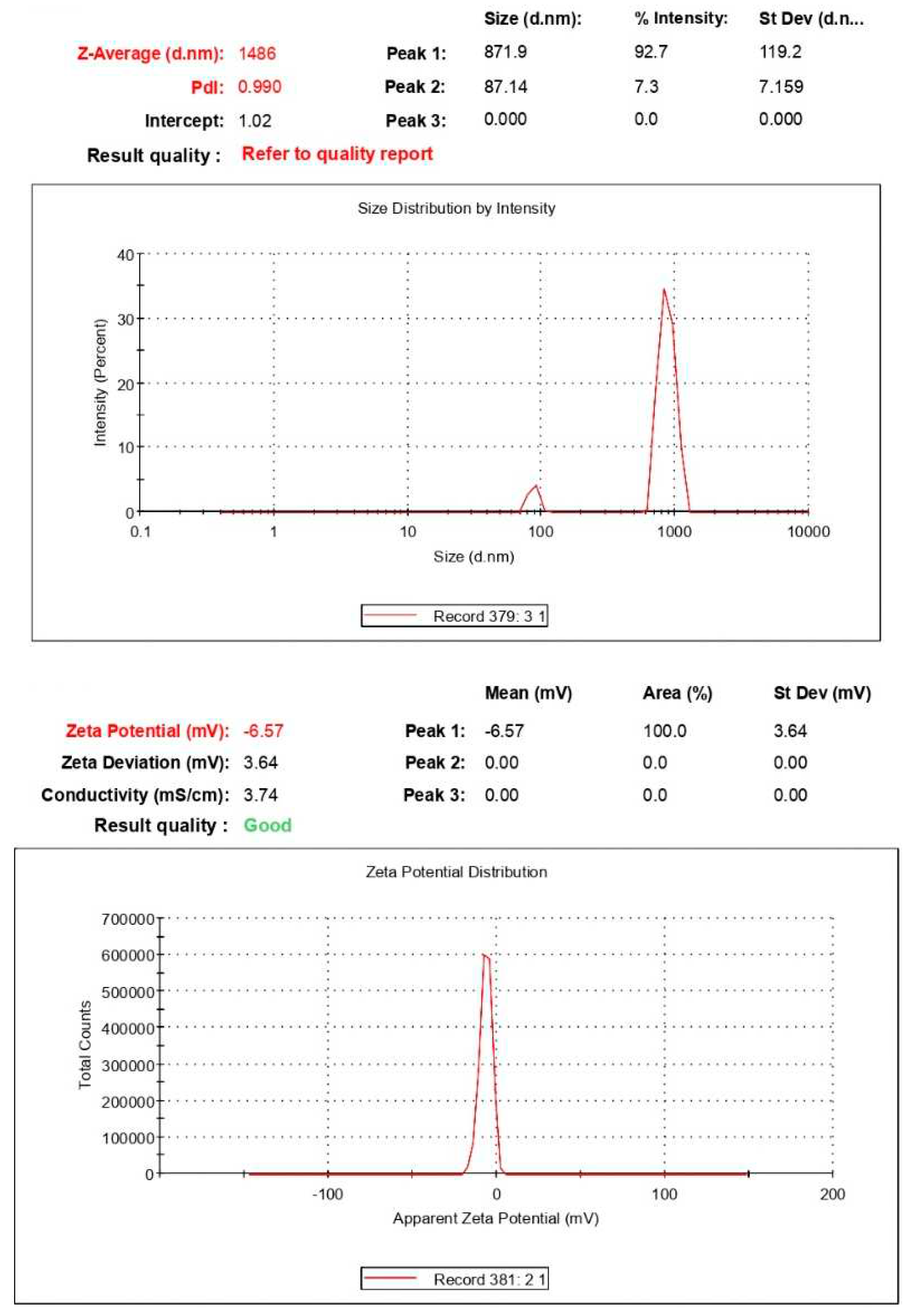
Particle size may be determined by observing the inconsistent changes in light intensity that are scattered from a suspension or solution.[33] As seen in Figure 5, this approach is often in relation as DLS. The most popular approach for analyzing nanoparticles and figuring out their size distribution is DLS.[34] DLS is a very efficient and quick method for determining the particles size distribution in the fields of physics, chemistry, and biochemistry, particularly for monomodally dispersed spherical particles. By combining DLS with methods like AFM and TEM, one can quickly gain a thorough understanding of the analyte's size distribution. [35]

3.1.4. XRD analysis

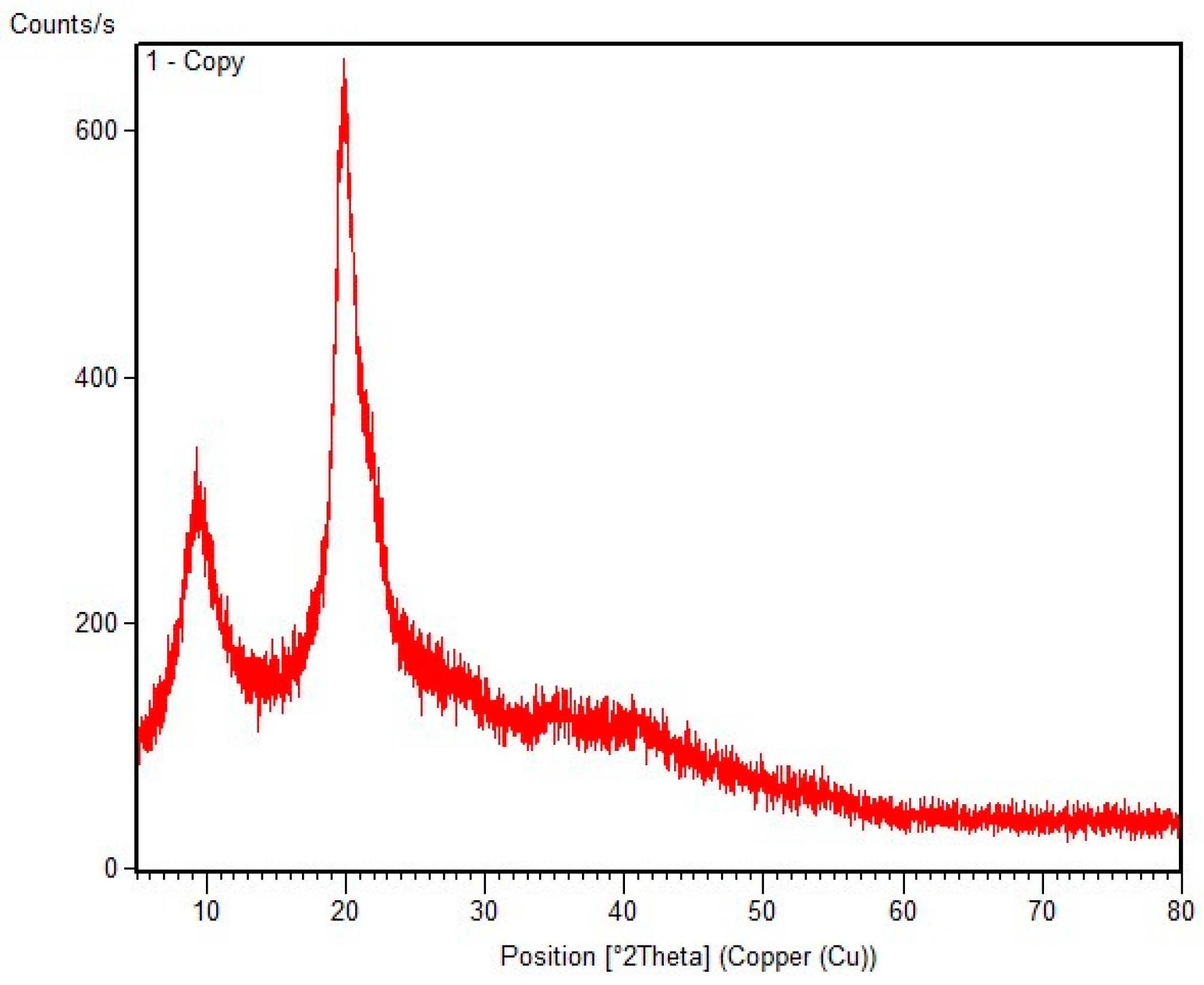
Particles of Fe3O4 with magnetic characteristics and the ability to be utilized for magnetic separation are shown by the XRD patterns of the pure magnetic chitosan nanoparticles displayed in Figure 6. Three prominent distinctive peaks for magnetic chitosan nanoparticles were identified by XRD at 2θ = 9.4°, 20.0°, and 35.4° in the graph. [36].

3.1.5. FTIR analysis

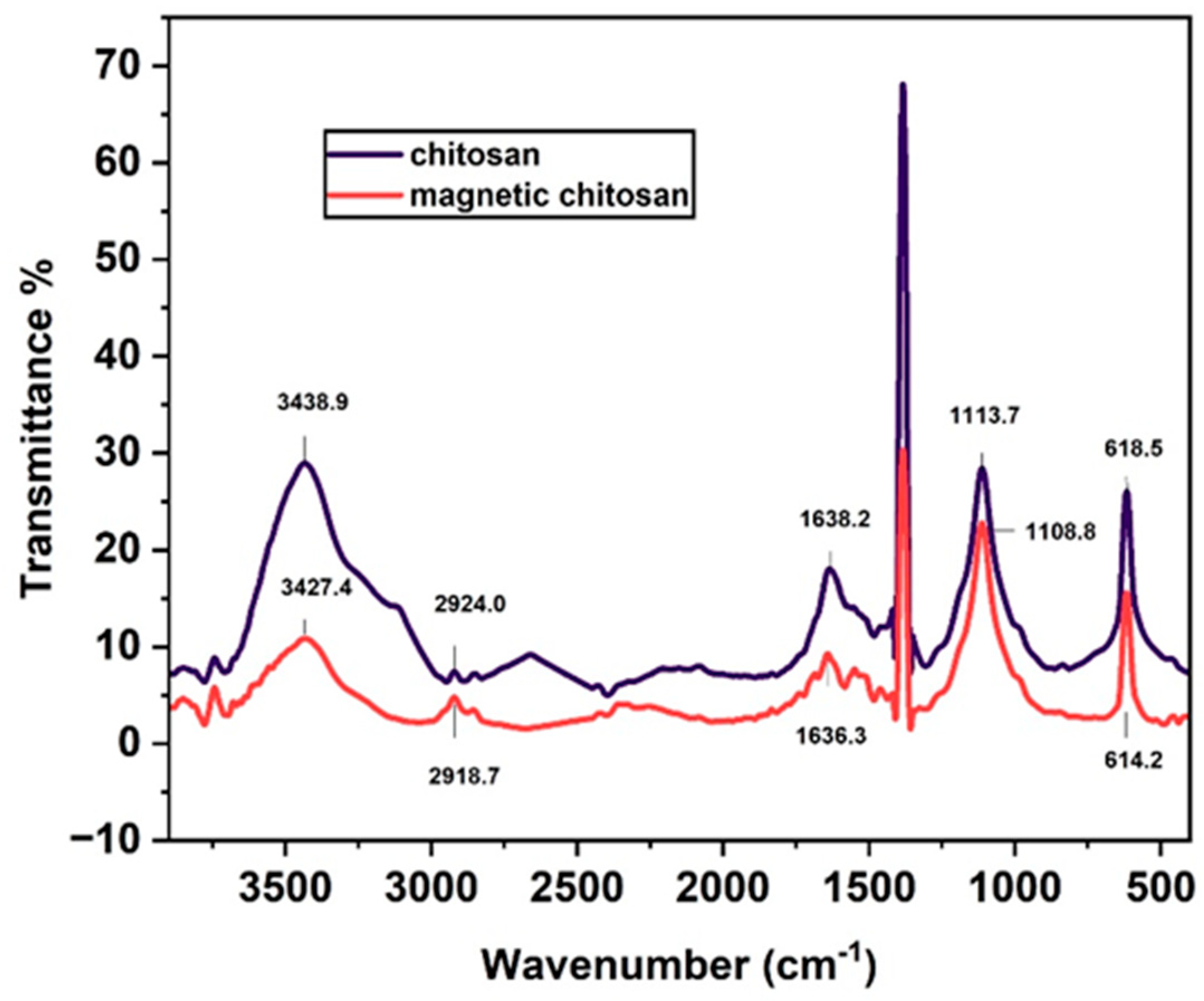
Spectra “of FTIR for pure chitosan and magnetic chitosan nanoparticles have been plotted to verify the presence of the surface Fe3O4 coating. [37] For chitosan, the peak at 3438.9 cm-1 is attributed to O-H stretching vibration, and the peak at 2924 cm-1 is expressed for C-H stretching vibration” [38]. In contrast, 1113.7 cm-1 was observed for primary alcoholic group C-O stretching, 1638.2 cm-1 was observed for -NH2 in amide group, and 3438.9 cm-1 was also seen for N-H bond in chitosan. [39] However, for magnetic chitosan nanoparticles, O-H “stretching vibration and C-H stretching vibration peak at 3427.4 cm-1 and 2918.7 cm-1, respectively [40]. For -NH2 in amide group, magnetic chitosan nanoparticles peak at 1636.3 cm-1.” [41],[15]
FTIR spectroscopy examined by nicolet iS50 FTIR tri-detector, which was gold flex spectrometer-gold optics with 0.09 cm-1 resolution and using the DLaTGS detector with KBR window and scanning range was 4000-400 cm-1. especially Far-IR is typically defined as radiation between range of 500 cm-1 and 20 cm-1, whereas mid-IR between 4000 cm-1 and 500 cm-1 and NIR as usually between 10,000 and 4,000 cm-1

3.1.6. Applications of magnetic chitosan nanoparticles

The magnetic chitosan nanoparticles are a unique material with optimal heavy metal adsorption behavior, [42] Because of their efficient elimination of heavy metals by magnetic fields and strong metal chelating capacity, magnetic chitosan nanoparticles are a significant class of heavy metal adsorbents. [43] Chitosan and Fe3O4 were combined in a sufficient ratio to create chitosan magnetite particles through the amine group via reverse-phase suspension cross-linking technique, which is relevant in a field’s varieties, like wastewater treatment. In comparison to other separation techniques, [44] advances in magnetic separation are efficient, quick, and economical. The magnetic chitosan's adsorption efficiency on the basis of the utilizing the chemical groups for the extracting the heavy metals like arsenic and mercury. [45] This method has made the separation process simpler since it eliminates the need for centrifugation or filtering, making it simple to separate magnetic adsorbents from samples of any size.[46] Capacity of adsorption, the active sites numbers, and the adsorbent surface area are used to measure the effectiveness of heavy metal removal. As adsorbent concentrations were raised, the adsorption capacity went up, then dropped.[47] The adsorbent surface area is increased and abundance of adsorption sites for heavy metals including arsenic, mercury, and lead are responsible for this. Additionally, strong magnetization and adsorption capability of magnetic chitosan nanoparticles towards heavy metals from wastewater are shown.[48]

4. Conclusions

Chitosan has been linked to the Fe3O4 nanoparticles surface to create the magnetic Fe3O4 chitosan nanoparticles. Chitosan is highly effective in nanoparticles production or magnetic nanoparticles formation. The magnetic chitosan nanoparticles exhibit very steady magnetization capabilities, as shown in Figure 3. In this research, ionic gelation was used to create magnetic chitosan nanoparticles. Size, magnetic properties and morphology of the magnetic chitosan nanoparticles have been assessing by utilizing the FESEM, TEM, DLS and VSM methods. In FESEM the energy dispersive spectroscopy (EDS) or elemental mapping method popularly utilized for qualitative evaluation of materials but it is able to give the semi-quantitative results as well. The HR-TEM investigations showed the presence of Fe3O4 nanoparticles with lateral sizes of about 10 to 200 nm. The magnetic chitosan nanoparticles, which ranging size from 200 to 400 nm, seemed to be separated. Additionaly, more chitosan and Fe3O4 increased the molecular weight of the magnetic nanoparticles, which in turn increased their size. DLS is often utilized to examine the Zeta potential as well as size of magnetic nanoparticles, which was 871.9 nm, despite the fact that the rise in Fe3O4 concentration likewise improved the “magnetic property of the magnetic chitosan nanoparticles. The FTIR spectrum of magnetic chitosan nanoparticles revealed that the peak of O-H stretching vibration was considerably broader than pure chitosan, and the XRD patterns of the magnetic chitosan nanoparticles” analysed two primary peaks.

References

  1. Peter, Sherin, Nathalie Lyczko, Deepu Gopakumar, Hanna J. Maria, Ange Nzihou, and Sabu Thomas. Chitin and chitosan based composites for energy and environmental applications: A review. Waste and Biomass Valorization 12, no. 9 (2021): 4777-4804. [CrossRef]
  2. Joseph, Sherin M., Srinivasan Krishnamoorthy, R. Paranthaman, J. A. Moses, and C. Anandharamakrishnan. A review on source-specific chemistry, functionality, and applications of chitin and chitosan. Carbohydrate Polymer Technologies and Applications 2 (2021): 100036. [CrossRef]
  3. Wang, Xiangtao, Yifei Guo, Li Yang, Meihua Han, Jing Zhao, and Xiaoliang Cheng. Nanomaterials as sorbents to remove heavy metal ions in wastewater treatment. J. Environ. Anal. Toxicol 2, no. 7 (2012): 154-158. [CrossRef]
  4. Qasem,Naef AA,Ramy H.Mohammed,and Dahiru U. Lawal. Removal of heavy metal ions from wastewater:A comprehensive and critical review. Npj Clean Water 4,no.1 (2021):1-15. [CrossRef]
  5. Qin, H., C. M. Wang, Q. Q. Dong, L. Zhang, X. Zhang, Z. Y. Ma, and Q. R. Han. Preparation and characterization of magnetic Fe3O4–chitosan nanoparticles loaded with isoniazid. Journal of Magnetism and Magnetic Materials 381(2015):120-126. [CrossRef]
  6. Chang, Yang-Chuang, and Dong-Hwang Chen. Adsorption kinetics and thermodynamics of acid dyes on a carboxymethylated chitosan-conjugated magnetic nano-adsorbent. Macromolecular bioscience 5, no. 3 (2005): 254-261. [CrossRef]
  7. Hosseini, Fatemeh, Somayeh Sadighian, Hassan Hosseini-Monfared, and Niyaz Mohammad Mahmoodi. Dye removal and kinetics of adsorption by magnetic chitosan nanoparticles. Desalination and Water Treatment 57, no. 51 (2016):24378-24386. [CrossRef]
  8. Carolin, C. Femina, P. Senthil Kumar, A. Saravanan, G. Janet Joshiba, and Mu Naushad. Efficient techniques for the removal of toxic heavy metals from aquatic environment: A review. Journal of environmental chemical engineering 5, no.3(2017):2782-2799. [CrossRef]
  9. Prabu, D., P. Senthil Kumar, B. Senthil Rathi, S. Sathish, K. Vijai Anand, J. Aravind Kumar, Osama B. Mohammed, and P. Silambarasan. Feasibility of magnetic nano adsorbent impregnated with activated carbon from animal bone waste: Application for the chromium (VI) removal. Environmental Research 203(2022):111813. [CrossRef]
  10. Zhang, Lei, Yuexian Zeng, and Zhengjun Cheng. Removal of heavy metal ions using chitosan and modified chitosan: A review. Journal of Molecular Liquids 214 (2016):175-191. [CrossRef]
  11. Trung, Trang Si, Nguyen Van Tan, Nguyen Van Hoa, Nguyen Cong Minh, Phan Thanh Loc, and Willem F. Stevens. Improved method for production of chitin and chitosan from shrimp shells. Carbohydrate research 489 (2020): 107913. [CrossRef]
  12. Mahdavinia, Gholam Reza, Moslem Soleymani, Mohammad Sabzi, Hamidreza Azimi, and Ziba Atlasi. Novel magnetic polyvinyl alcohol/laponite RD nanocomposite hydrogels for efficient removal of methylene blue. Journal of environmental chemical engineering 5, no. 3(2017):2617-2630. [CrossRef]
  13. Sánchez-Aguinagalde, O. , Ainhoa Lejardi, Emilio Meaurio, Rebeca Hernández, Carmen Mijangos, and Jose-Ramon Sarasua. Novel hydrogels of chitosan and poly (Vinyl alcohol) reinforced with inorganic particles of bioactive glass. Polymers 13, no. 5 (2021): 691. [CrossRef]
  14. Kaloti, Mandeep, and H. B. Bohidar. Kinetics of coacervation transition versus nanoparticle formation in chitosan–sodium tripolyphosphate solutions. Colloids and Surfaces B: Biointerfaces 81, no. 1 (2010): 165-173. [CrossRef]
  15. Janes, K. A. , and M. J. Alonso. Depolymerized chitosan nanoparticles for protein delivery: preparation and characterization. Journal of applied polymer science 88, no. 12 (2003): 2769-2776. [CrossRef]
  16. Janes, Kevin A., Marie P. Fresneau, Ana Marazuela, Angels Fabra, and Marı́a José Alonso. Chitosan nanoparticles as delivery systems for doxorubicin. Journal of controlled Release 73, no. 2-3 (2001): 255-267. [CrossRef]
  17. Yu, Liang, Jie Gong, Changfeng Zeng, and Lixiong Zhang. Synthesis of monodisperse zeolite A/chitosan hybrid microspheres and binderless zeolite A microspheres. Industrial & engineering chemistry research 51,no.5(2012):2299-2308. [CrossRef]
  18. McGinniss, V. D., F. A. Sliemers, D. K. Landstrom, and S. G. Talbert. Compendium Of information on identification and testing of materials for plastic solar thermal collectors No. DOE/CS/30171-1. Battelle Columbus Labs., OH (USA), 1980. [CrossRef]
  19. Zhou, Weilie, Robert Apkarian, Zhong Lin Wang, and David Joy. Fundamentals of scanning electron microscopy (SEM). In Scanning microscopy for nanotechnology, pp. 1-40.(2006)Springer,NewYork,NY. [CrossRef]
  20. Bertrand, Loïc, Marine Cotte, Marco Stampanoni, Mathieu Thoury, Federica Marone, and Sebastian Schöder. Development and trends in synchrotron studies of ancient and historical materials. Physics Reports 519, no. 2 (2012): 51-96. [CrossRef]
  21. Pine, D. J., D. A. Weitz, J. X. Zhu, and E. Herbolzheimer. Diffusing-wave spectroscopy: dynamic light scattering in the multiple scattering limit. Journal de Physique 51, no. 18 (1990):2101-2127. [CrossRef]
  22. Clark, Noel A., Joseph H. Lunacek, and George B. Benedek. A study of Brownian motion using light scattering. American Journal of Physics 38, no. 5 (1970): 575-585. [CrossRef]
  23. Vinod, Sithara, and John Philip. Experimental evidence for the significant role of initial cluster size and liquid confinement on thermo-physical properties of magnetic nanofluids under applied magnetic field. Journal of MolecularLiquids 257(2018):1-11. [CrossRef]
  24. Pathmamanoharan, Chellappah, and Albert P. Philipse. Preparation and properties of monodisperse magnetic cobalt colloids grafted with polyisobutene. Journal of colloid and interfacescience 205,no.2(1998):340-353. [CrossRef]
  25. Maji, Ambarish, and Gautam Choubey. Microstructure and mechanical properties of alumina toughened zirconia (ATZ). Materials Today: Proceedings 5, no. 2 (2018): 7457-7465. [CrossRef]
  26. Khaleda, MZ Farah, Vengadaesvaran Balakrishnan, A. Syafiq, Nasrudin Abd Rahim, and A. K. Pandey. SnO2 and TiO2 coatings toward decreasing the surface temperature of solarcells. (2018):50-5. [CrossRef]
  27. Kalita, op choudhary pc, pj doley, and a. kalita. 2. uses of transmission electron microscope in microscopy and its advantages and disadvantages by op choudhary, pc kalita, pj doley and a. kalita. Life Sciences Leaflets 85 (2017): 8-to.
  28. Bogner, Agnes, P-H. Jouneau, Gilbert Thollet, D. Basset, and Catherine Gauthier. A history of scanning electron microscopy developments: Towards wet-STEM imaging. Micron 38,no.4(2007):390-401. [CrossRef]
  29. Niazi, Asad, Pankaj Poddar, and A. K. Rastogi. A precision, low-cost vibrating sample magnetometer. CURRENT SCIENCE-BANGALORE- 79, no. 1 (2000): 99-109.
  30. Kirupakar, B. R., B. A. Vishwanath, and M. Padma Sree. Vibrating sample magnetometer and its application in characterisation of magnetic property of the anti cancer drug magnetic microspheres. International Journal of Pharmaceutics and Drug Analysis (2016): 227-233.
  31. Li, Gui-Yin, Yu-Ren Jiang, Ke-Long Huang, Ping Ding, and Li-Li Yao. Kinetics of adsorption of Saccharomyces cerevisiae mandelated dehydrogenase on magnetic Fe3O4–chitosan nanoparticles. Colloids and Surfaces A: Physicochemical and Engineering Aspects 320, no. 1-3 (2008): 11-18. [CrossRef]
  32. Brar, Satinder K., and M. Verma. Measurement of nanoparticles by light-scattering techniques. TrAC Trends in AnalyticalChemistry 30,no.1(2011):4-17. [CrossRef]
  33. Filipe, Vasco, Andrea Hawe, and Wim Jiskoot. Critical evaluation of Nanoparticle Tracking Analysis (NTA) by NanoSight for the measurement of nanoparticles and protein aggregates. Pharmaceutical research 27, no. 5 (2010): 796-810. [CrossRef]
  34. Caputo, F., J. Clogston, L. Calzolai, M. Rösslein, and A. Prina-Mello. Measuring particle size distribution of nanoparticle enabled medicinal products, the joint view of EUNCL and NCI-NCL. A step by step approach combining orthogonal measurements with increasing complexity. Journal of Controlled Release 299 (2019): 31-43. [CrossRef]
  35. Mahdavi, Mahnaz, Mansor Bin Ahmad, Md Jelas Haron, Farideh Namvar, Behzad Nadi, Mohamad Zaki Ab Rahman, and Jamileh Amin. Synthesis, surface modification and characterisation of biocompatible magnetic iron oxide nanoparticles for biomedical applications. Molecules 18, no. 7(2013):7533-7548. [CrossRef]
  36. Ding, Yongling, Shirley Z. Shen, Huadong Sun, Kangning Sun, Futian Liu, Yushi Qi, and Jun Yan. Design and construction of polymerized-chitosan coated Fe3O4 magnetic nanoparticles and its application for hydrophobic drug delivery. Materials Science and Engineering: C 48 (2015): 487-498. [CrossRef]
  37. Abdollahi, M., S. Zeinali, S. Nasirimoghaddam, and S. Sabbaghi. Effective removal of As (III) from drinking water samples by chitosan-coated magnetic nanoparticles. Desalination and Water Treatment 56, no. 8 (2015):2092-2104. [CrossRef]
  38. Raeiatbin, P. A. R. I. N. A. Z. , and Y. Sağ Açıkel. Removal of tetracycline by magnetic chitosan nanoparticles from medical wastewaters. Desalin Water Treat 73 (2017): 380-388. [CrossRef]
  39. Kang, Xin, Yuxiang Gan, Renpeng Chen, and Chao Zhang. Sustainable eco-friendly bricks from slate tailings through geopolymerization: synthesis and characterization analysis. Construction and Building Materials 278 (2021): 122337. [CrossRef]
  40. Samal, R. K., S. Acharya, M. Mohanty, and M. C. Ray. FTIR spectra and physico-chemical behavior of vinyl ester participated transesterification and curing of jute. Journal of applied polymer science 79, no. 4 (2001): 575-581. https://doi.org/10.1002/1097-4628(20010124)79:4%3C575::AID-APP10%3E3.0.CO;2-U. [CrossRef]
  41. Wulandari, I. O., D. J. D. H. Santjojo, R. A. Shobirin, and A. Sabarudin. Characteristics and magnetic properties of chitosan-coated Fe3O4 nanoparticles prepared by ex-situ co-precipitation method. Rasayan J. Chem 10 (2017): 1348-1358. [CrossRef]
  42. Zhang, Meng, Zhi Zhang, Yazhou Peng, Li Feng, Xuhao Li, Chuanliang Zhao, and Khan Sarfaraz. Novel cationic polymer modified magnetic chitosan beads for efficient adsorption of heavy metals and dyes over a wide pH range. International journal of biological macromolecules 156(2020):289-301. [CrossRef]
  43. Pu, Shengyan, Yaqi Hou, Chun Yan, Hui Ma, Hongyan Huang, Qingqing Shi, Sandip Mandal, Zenghui Diao, and Wei Chu. In situ coprecipitation formed highly water-dispersible magnetic chitosan nanopowder for removal of heavy metals and its adsorption mechanism. ACS Sustainable Chemistry & Engineering 6, no. 12 (2018): 16754-16765. [CrossRef]
  44. Hazra, S. , and N. N. Ghosh. Preparation of nanoferrites and their applications. Journal of nanoscience and nanotechnology 14, no. 2 (2014): 1983-2000. [CrossRef]
  45. Shaumbwa, Veino Risto, Dagang Liu, Bright Archer, Jinlei Li, and Fan Su. Preparation and application of magnetic chitosan in environmental remediation and other fields: A review. Journal of Applied Polymer Science 138, no. 42 (2021): 51241. [CrossRef]
  46. Liu, Changkun, and Renbi Bai. Recent advances in chitosan and its derivatives as adsorbents for removal of pollutants from water and wastewater. Current Opinion in Chemical Engineering 4 (2014): 62-70. [CrossRef]
  47. Ngah, WS Wan, and MAK Megat Hanafiah. Removal of heavy metal ions from wastewater by chemically modified plant wastes as adsorbents: a review. Bioresource technology 99,no.10(2008):3935-3948. [CrossRef]
  48. El-Dib, Fawzia I., Dalia E. Mohamed, Omnia AA El-Shamy, and Marwa R. Mishrif. Study the adsorption properties of magnetite nanoparticles in the presence of different synthesized surfactants for heavy metal ions removal. Egyptian Journal of Petroleum 29, no. 1 (2020): 1-7. [CrossRef]
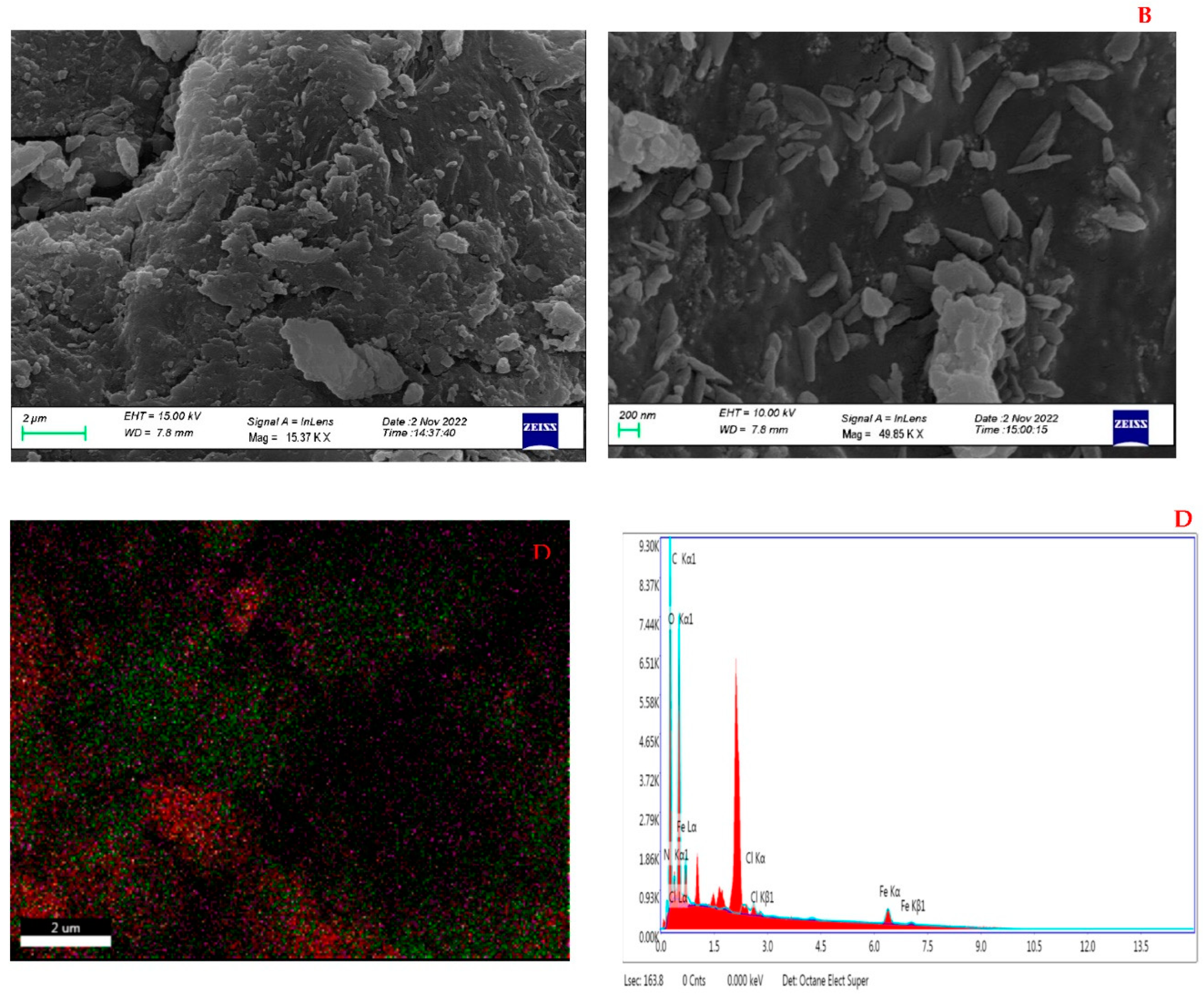
Figure 2. FESEM images (A and B) of magnetic chitosan nanoparticles and mapping (C) with EDS (D).
Preprints 83669 g002
Figure 3. HRTEM images of magnetic chitosan nanoparticles.
Preprints 83669 g003
Figure 4. Magnetic properties of magnetic chitosan nanoparticles in VSM curve.
Preprints 83669 g004
Figure 5. Size distribution and Zeta potential distribution of magnetic chitosan nanoparticles by DLS.
Preprints 83669 g005
Figure 6. Chitosan nanoparticles with a magnetic XRD pattern.
Preprints 83669 g006
Figure 7. FTIR spectra of chitosan and magnetic chitosan nanoparticles.
Preprints 83669 g007
Disclaimer/Publisher’s Note: The statements, opinions and data contained in all publications are solely those of the individual author(s) and contributor(s) and not of MDPI and/or the editor(s). MDPI and/or the editor(s) disclaim responsibility for any injury to people or property resulting from any ideas, methods, instructions or products referred to in the content.
Copyright: This open access article is published under a Creative Commons CC BY 4.0 license, which permit the free download, distribution, and reuse, provided that the author and preprint are cited in any reuse.
Prerpints.org logo

Preprints.org is a free preprint server supported by MDPI in Basel, Switzerland.

Subscribe

© 2024 MDPI (Basel, Switzerland) unless otherwise stated