Submitted:
30 August 2023
Posted:
01 September 2023
You are already at the latest version
Abstract
Keywords:
1. Introduction
2. Materials and Methods
2.1. Bacterial strains and growth conditions
2.2. DNA manipulations
2.3. Construction of the lipoprotein maturation isogenic mutants
2.4. Complementation of the mutants
2.5. Growth analysis
2.6. Bacterial surface hydrophobicity assay
2.7. Bacterial adhesion and invasion assays using porcine brain microvascular endothelial and tracheal epithelial cells
2.8. Biofilm assay
2.9. Generation of bone marrow-derived dendritic cells (bmDCs
2.10. Preparation of heat-killed S. suis
2.11. Preparation of bacterial supernatant
2.12. S. suis activation of bmDCs
2.13. S. suis virulence mouse model of systemic infection
2.13. Measurement of plasma (systemic) pro-inflammatory mediators
2.14. Statistical analyses
3. Results
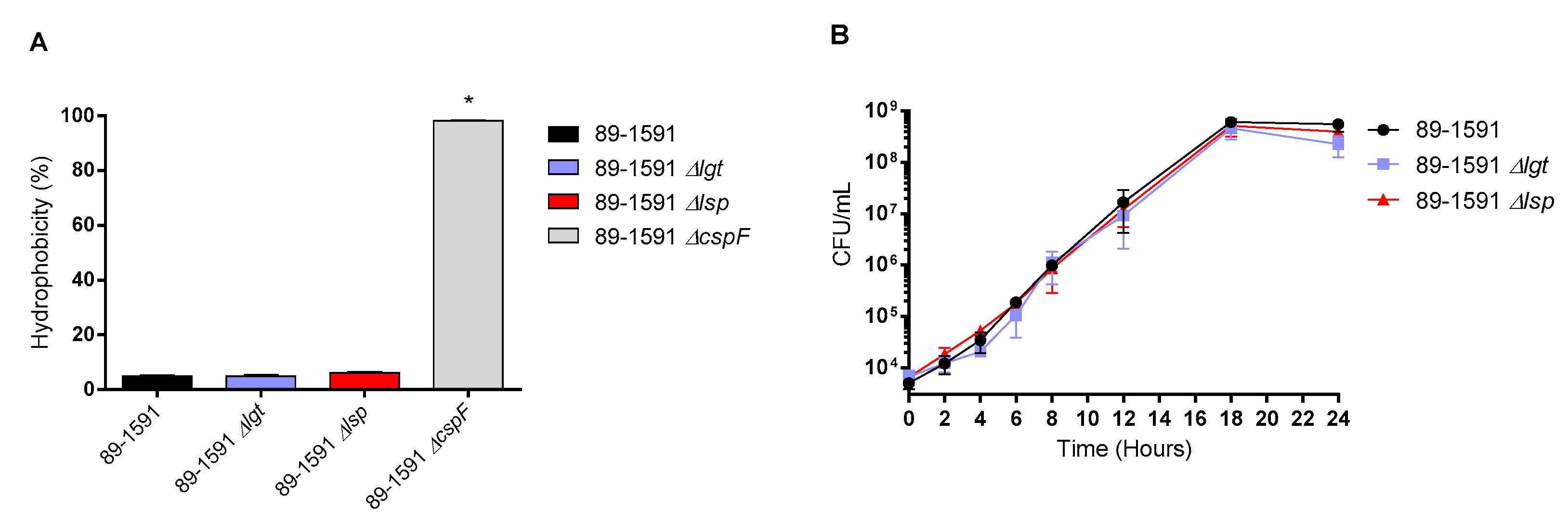
3.1. Characteristics of the Δlgt and Δlsp mutants derived from the ST25 89-1591 strain
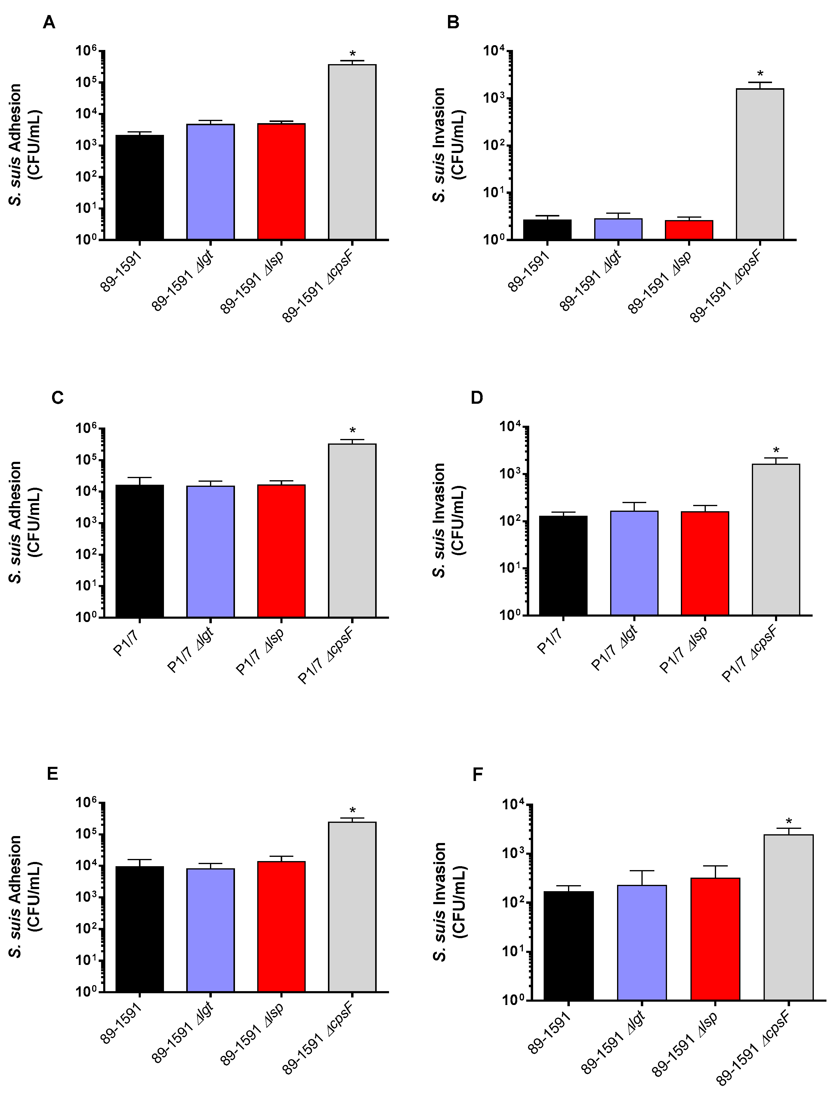
3.2. Lack of lipoprotein maturation enzymes does not impair adhesion to and invasion of respiratory epithelial and brain microvascular endothelial swine cells regardless of the sequence type of the strain
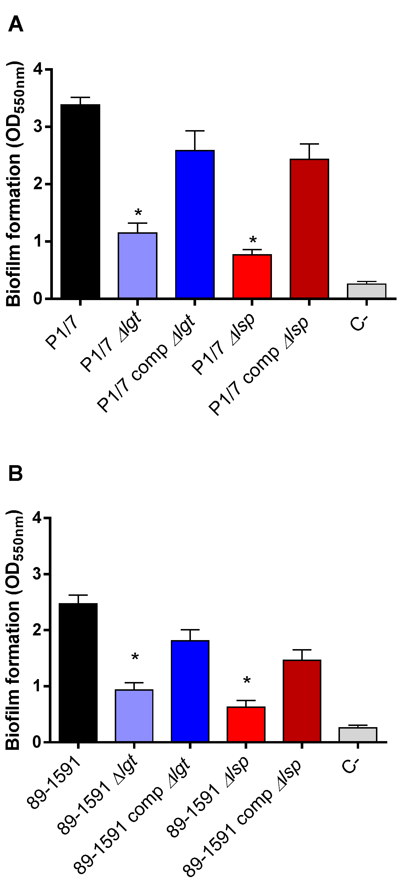
3.3. Lgt and Lsp enzymes are important for S. suis biofilm formation regardless of the sequence type of the strain
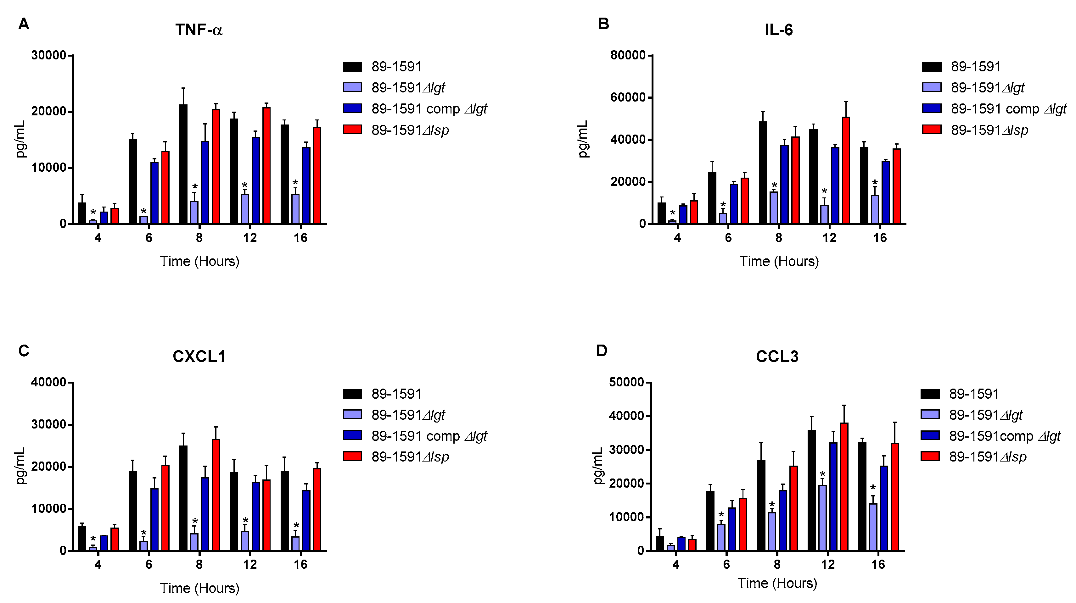
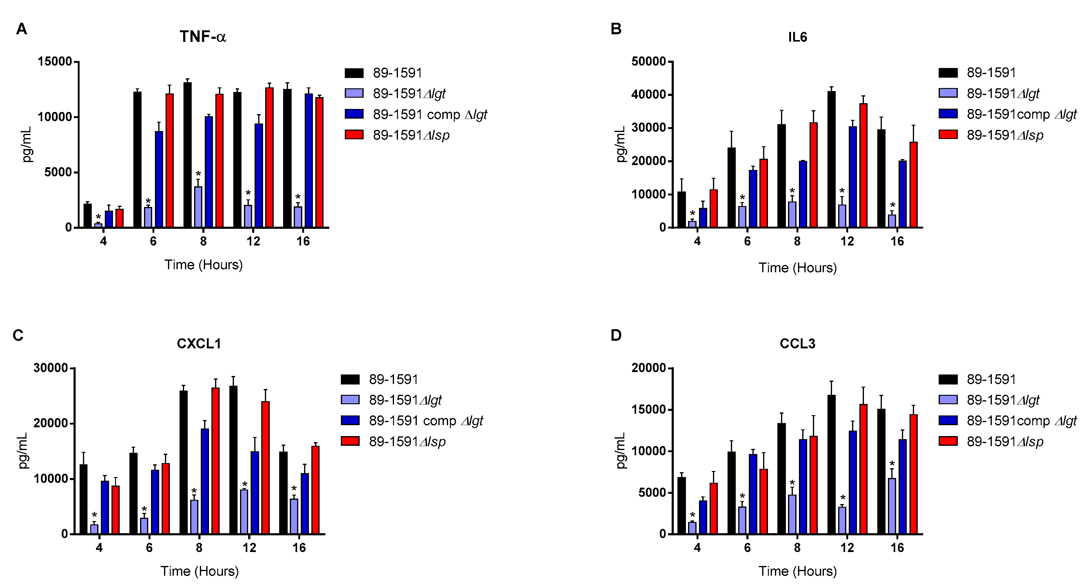
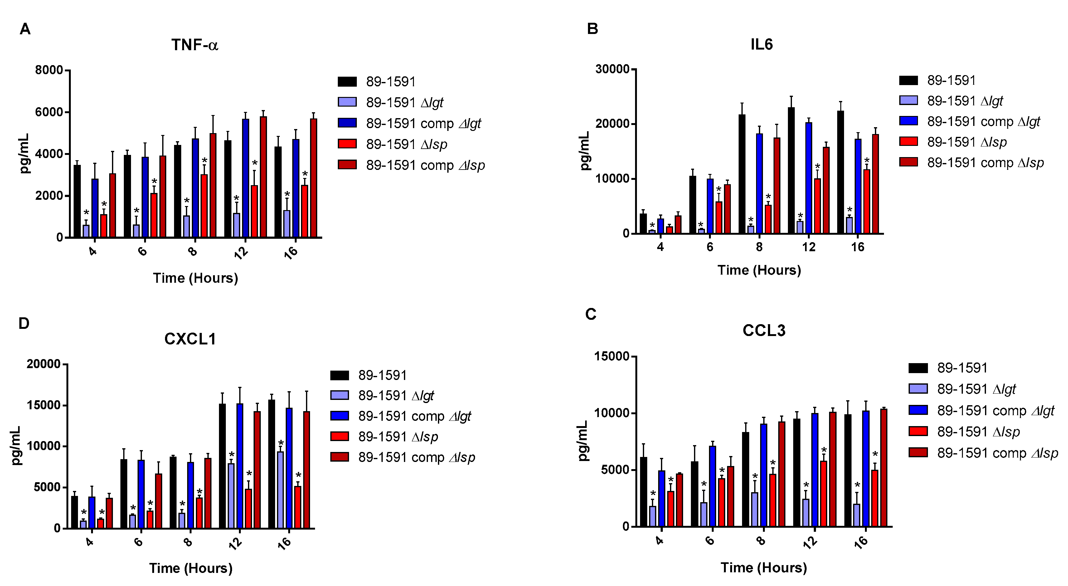
3.4. The diacyl motif and the peptide signal cleavage are important for the recognition by innate immune cells of periplasmic and/or secreted lipoproteins of S. suis serotype 2 ST25 strain
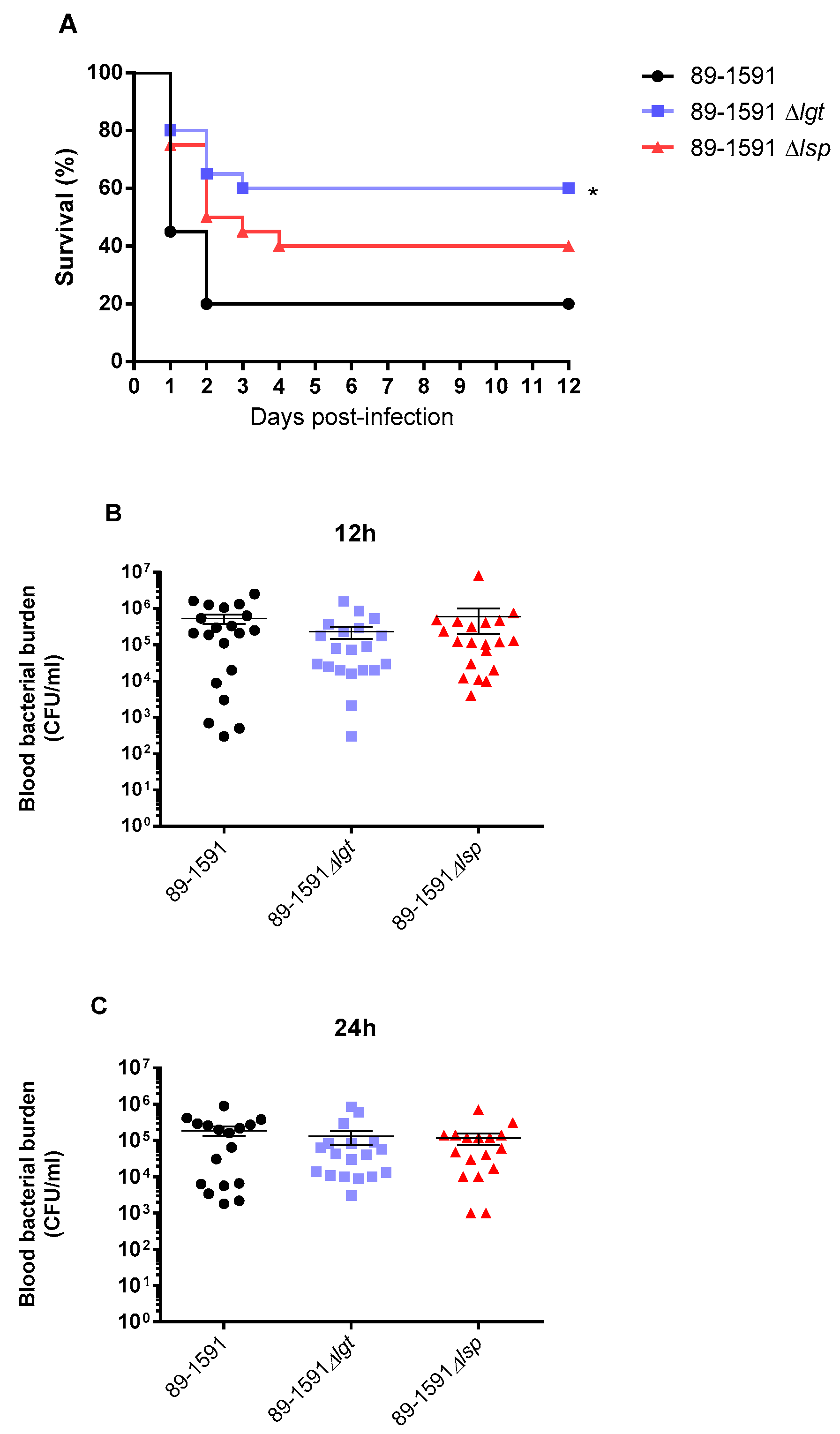
3.5. The absence of Lgt enzyme, but not Lsp, significantly affects the virulence of S. suis serotype 2 ST25
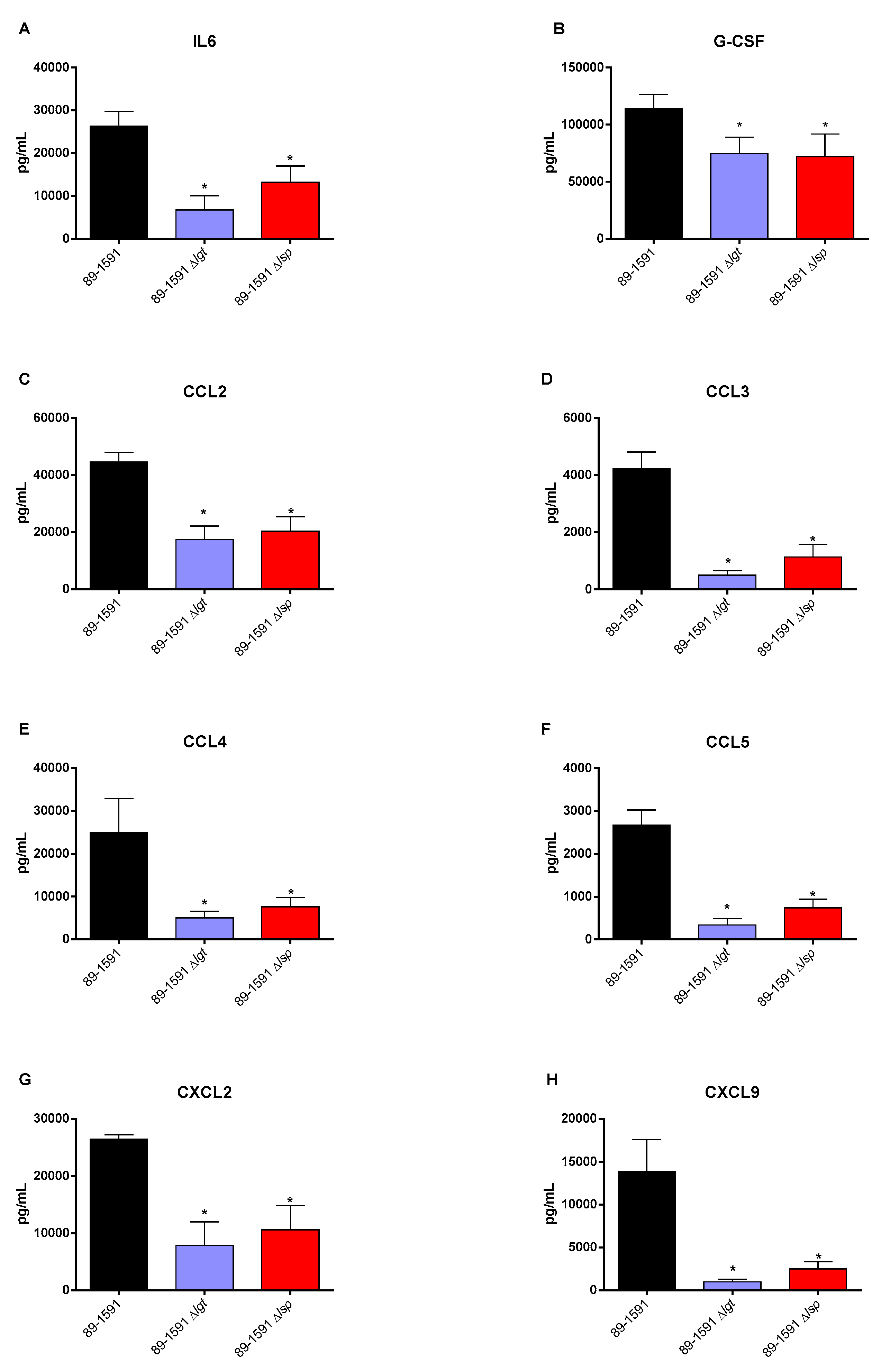
3.6. Absence of the Lgt or Lsp enzymes significantly reduces the in vivo inflammatory response of mice infected with either the wild type S. suis serotype 2 strain ST25 or its respective Δlgt or Δlsp mutant
4. Discussion
5. Conclusions
6. Patents
Author Contributions
Funding
Institutional Review Board Statement
Informed Consent Statement
Data Availability Statement
Acknowledgments
Conflicts of Interest
References
- Gottschalk, M.; Xu, J.; Calzas, C.; Segura, M. Streptococcus suis: a new emerging or an old neglected zoonotic pathogen? Future Microbiol 2010, 5, 371–391. [Google Scholar] [CrossRef] [PubMed]
- Goyette-Desjardins, G.; Auger, J.P.; Xu, J.; Segura, M.; Gottschalk, M. Streptococcus suis, an important pig pathogen and emerging zoonotic agent-an update on the worldwide distribution based on serotyping and sequence typing. Emerg Microbes Infect 2014, 3, e45. [Google Scholar] [CrossRef] [PubMed]
- Yu, H.; Jing, H.; Chen, Z.; Zheng, H.; Zhu, X.; Wang, H.; Wang, S.; Liu, L.; Zu, R.; Luo, L.; et al. Human Streptococcus suis outbreak, Sichuan, China. Emerg Infect Dis 2006, 12, 914–920. [Google Scholar] [CrossRef] [PubMed]
- Berthelot-Herault, F.; Gottschalk, M.; Morvan, H.; Kobisch, M. Dilemma of virulence of Streptococcus suis: Canadian isolate 89-1591 characterized as a virulent strain using a standardized experimental model in pigs. Can J Vet Res 2005, 69, 236–240. [Google Scholar]
- Lacouture, S.; Olivera, Y.R.; Mariela, S.; Gottschalk, M. Distribution and characterization of Streptococcus suis serotypes isolated from January 2015 to June 2020 from diseased pigs in Quebec, Canada. Can J Vet Res 2022, 86, 78–82. [Google Scholar] [PubMed]
- Fittipaldi, N.; Segura, M.; Grenier, D.; Gottschalk, M. Virulence factors involved in the pathogenesis of the infection caused by the swine pathogen and zoonotic agent Streptococcus suis. Future Microbiol 2012, 7, 259–279. [Google Scholar] [CrossRef]
- Payen, S.; Roy, D.; Boa, A.; Okura, M.; Auger, J.P.; Segura, M.; Gottschalk, M. Role of maturation of lipoproteins in the pathogenesis of the infection caused by Streptococcus suis serotype 2. Microorganisms 2021, 9, 2386. [Google Scholar] [CrossRef]
- Kovacs-Simon, A.; Titball, R.W.; Michell, S.L. Lipoproteins of bacterial pathogens. Infect Immun 2011, 79, 548–561. [Google Scholar] [CrossRef]
- Kohler, S.; Voss, F.; Gomez Mejia, A.; Brown, J.S.; Hammerschmidt, S. Pneumococcal lipoproteins involved in bacterial fitness, virulence, and immune evasion. FEBS Lett 2016, 590, 3820–3839. [Google Scholar] [CrossRef]
- Sander, P.; Rezwan, M.; Walker, B.; Rampini, S.K.; Kroppenstedt, R.M.; Ehlers, S.; Keller, C.; Keeble, J.R.; Hagemeier, M.; Colston, M.J.; et al. Lipoprotein processing is required for virulence of Mycobacterium tuberculosis. Mol Microbiol 2004, 52, 1543–1552. [Google Scholar] [CrossRef]
- Henneke, P.; Dramsi, S.; Mancuso, G.; Chraibi, K.; Pellegrini, E.; Theilacker, C.; Hubner, J.; Santos-Sierra, S.; Teti, G.; Golenbock, D.T.; et al. Lipoproteins are critical TLR2 activating toxins in group B streptococcal sepsis. J Immunol 2008, 180, 6149–6158. [Google Scholar] [CrossRef]
- Hashimoto, M.; Tawaratsumida, K.; Kariya, H.; Kiyohara, A.; Suda, Y.; Krikae, F.; Kirikae, T.; Gotz, F. Not lipoteichoic acid but lipoproteins appear to be the dominant immunobiologically active compounds in Staphylococcus aureus. J Immunol 2006, 177, 3162–3169. [Google Scholar] [CrossRef] [PubMed]
- Hutchings, M.I.; Palmer, T.; Harrington, D.J.; Sutcliffe, I.C. Lipoprotein biogenesis in Gram-positive bacteria: knowing when to hold 'em, knowing when to fold 'em. Trends Microbiol 2009, 17, 13–21. [Google Scholar] [CrossRef] [PubMed]
- Auger, J.P.; Dolbec, D.; Roy, D.; Segura, M.; Gottschalk, M. Role of the Streptococcus suis serotype 2 capsular polysaccharide in the interactions with dendritic cells is strain-dependent but remains critical for virulence. PLoS One 2018, 13, e0200453. [Google Scholar] [CrossRef] [PubMed]
- Lavagna, A.; Auger, J.P.; Girardin, S.E.; Gisch, N.; Segura, M.; Gottschalk, M. Recognition of lipoproteins by Toll-like receptor 2 and DNA by the AIM2 inflammasome is responsible for production of interleukin-1beta by virulent suilysin-negative Streptococcus suis serotype 2. Pathogens 2020, 9, 147. [Google Scholar] [CrossRef] [PubMed]
- Lecours, M.P.; Segura, M.; Fittipaldi, N.; Rivest, S.; Gottschalk, M. Immune receptors involved in Streptococcus suis recognition by dendritic cells. PLoS One 2012, 7, e44746. [Google Scholar] [CrossRef]
- Gottschalk, M.; Higgins, R.; Boudreau, M. Use of polyvalent coagglutination reagents for serotyping of Streptococcus suis. J Clin Microbiol 1993, 31, 2192–2194. [Google Scholar] [CrossRef]
- Slater, J.D.; Allen, A.G.; May, J.P.; Bolitho, S.; Lindsay, H.; Maskell, D.J. Mutagenesis of Streptococcus equi and Streptococcus suis by transposon Tn917. Vet Microbiol 2003, 93, 197–206. [Google Scholar] [CrossRef]
- Lecours, M.P.; Gottschalk, M.; Houde, M.; Lemire, P.; Fittipaldi, N.; Segura, M. Critical role for Streptococcus suis cell wall modifications and suilysin in resistance to complement-dependent killing by dendritic cells. J Infect Dis 2011, 204, 919–929. [Google Scholar] [CrossRef]
- Gottschalk, M.; Higgins, R.; Jacques, M.; Dubreuil, D. Production and characterization of two Streptococcus suis capsular type 2 mutants. Vet Microbiol 1992, 30, 59–71. [Google Scholar] [CrossRef]
- Casadaban, M.J.; Cohen, S.N. Analysis of gene control signals by DNA fusion and cloning in Escherichia coli. J Mol Biol 1980, 138, 179–207. [Google Scholar] [CrossRef] [PubMed]
- Takamatsu, D.; Osaki, M.; Sekizaki, T. Thermosensitive suicide vectors for gene replacement in Streptococcus suis. Plasmid 2001, 46, 140–148. [Google Scholar] [CrossRef] [PubMed]
- Okura, M.; Osaki, M.; Fittipaldi, N.; Gottschalk, M.; Sekizaki, T.; Takamatsu, D. The minor pilin subunit Sgp2 is necessary for assembly of the pilus encoded by the srtG cluster of Streptococcus suis. J Bacteriol 2011, 193, 822–831. [Google Scholar] [CrossRef]
- Warrens, A.N.; Jones, M.D.; Lechler, R.I. Splicing by overlap extension by PCR using asymmetric amplification: an improved technique for the generation of hybrid proteins of immunological interest. Gene 1997, 186, 29–35. [Google Scholar] [CrossRef]
- Takamatsu, D.; Osaki, M.; Sekizaki, T. Construction and characterization of Streptococcus suis-Escherichia coli shuttle cloning vectors. Plasmid 2001, 45, 101–113. [Google Scholar] [CrossRef] [PubMed]
- Wang, Y.; Gagnon, C.A.; Savard, C.; Music, N.; Srednik, M.; Segura, M.; Lachance, C.; Bellehumeur, C.; Gottschalk, M. Capsular sialic acid of Streptococcus suis serotype 2 binds to swine influenza virus and enhances bacterial interactions with virus-infected tracheal epithelial cells. Infect Immun 2013, 81, 4498–4508. [Google Scholar] [CrossRef]
- Vanier, G.; Segura, M.; Friedl, P.; Lacouture, S.; Gottschalk, M. Invasion of porcine brain microvascular endothelial cells by Streptococcus suis serotype 2. Infect Immun 2004, 72, 1441–1449. [Google Scholar] [CrossRef]
- Bonifait, L.; Grignon, L.; Grenier, D. Fibrinogen induces biofilm formation by Streptococcus suis and enhances its antibiotic resistance. Appl Environ Microbiol 2008, 74, 4969–4972. [Google Scholar] [CrossRef]
- Segura, M.; Su, Z.; Piccirillo, C.; Stevenson, M.M. Impairment of dendritic cell function by excretory-secretory products: a potential mechanism for nematode-induced immunosuppression. Eur J Immunol 2007, 37, 1887–1904. [Google Scholar] [CrossRef]
- Segura, M.; Stankova, J.; Gottschalk, M. Heat-killed Streptococcus suis capsular type 2 strains stimulate tumor necrosis factor alpha and interleukin-6 production by murine macrophages. Infect Immun 1999, 67, 4646–4654. [Google Scholar] [CrossRef]
- Lachance, C.; Gottschalk, M.; Gerber, P.P.; Lemire, P.; Xu, J.; Segura, M. Exacerbated type II interferon response drives hypervirulence and toxic shock by an emergent epidemic strain of Streptococcus suis. Infect Immun 2013, 81, 1928–1939. [Google Scholar] [CrossRef] [PubMed]
- Auger, J.P.; Fittipaldi, N.; Benoit-Biancamano, M.O.; Segura, M.; Gottschalk, M. Virulence studies of different sequence types and geographical origins of Streptococcus suis serotype 2 in a mouse model of infection. Pathogens 2016, 5. [Google Scholar] [CrossRef] [PubMed]
- Donlan, R.M.; Costerton, J.W. Biofilms: survival mechanisms of clinically relevant microorganisms. Clin Microbiol Rev 2002, 15, 167–193. [Google Scholar] [CrossRef] [PubMed]
- Peng, M.; Xu, Y.; Dou, B.; Yang, F.; He, Q.; Liu, Z.; Gao, T.; Liu, W.; Yang, K.; Guo, R.; et al. The adcA and lmb genes play an important role in drug resistance and full virulence of Streptococcus suis. Microbiol Spectr 2023, 11, e0433722. [Google Scholar] [CrossRef]
- Auger, J.P.; Chuzeville, S.; Roy, D.; Mathieu-Denoncourt, A.; Xu, J.; Grenier, D.; Gottschalk, M. The bias of experimental design, including strain background, in the determination of critical Streptococcus suis serotype 2 virulence factors. PLoS One 2017, 12, e0181920. [Google Scholar] [CrossRef]
- Tenenbaum, T.; Asmat, T.M.; Seitz, M.; Schroten, H.; Schwerk, C. Biological activities of suilysin: role in Streptococcus suis pathogenesis. Future Microbiol 2016, 11, 941–954. [Google Scholar] [CrossRef]
- Fittipaldi, N.; Fuller, T.E.; Teel, J.F.; Wilson, T.L.; Wolfram, T.J.; Lowery, D.E.; Gottschalk, M. Serotype distribution and production of muramidase-released protein, extracellular factor and suilysin by field strains of Streptococcus suis isolated in the United States. Vet Microbiol 2009, 139, 310–317. [Google Scholar] [CrossRef]
- Stoll, H.; Dengjel, J.; Nerz, C.; Gotz, F. Staphylococcus aureus deficient in lipidation of prelipoproteins is attenuated in growth and immune activation. Infect Immun 2005, 73, 2411–2423. [Google Scholar] [CrossRef]
- Chimalapati, S.; Cohen, J.M.; Camberlein, E.; MacDonald, N.; Durmort, C.; Vernet, T.; Hermans, P.W.; Mitchell, T.; Brown, J.S. Effects of deletion of the Streptococcus pneumoniae lipoprotein diacylglyceryl transferase gene lgt on ABC transporter function and on growth in vivo. PLoS One 2012, 7, e41393. [Google Scholar] [CrossRef]
- Pribyl, T.; Moche, M.; Dreisbach, A.; Bijlsma, J.J.; Saleh, M.; Abdullah, M.R.; Hecker, M.; van Dijl, J.M.; Becher, D.; Hammerschmidt, S. Influence of impaired lipoprotein biogenesis on surface and exoproteome of Streptococcus pneumoniae. J Proteome Res 2014, 13, 650–667. [Google Scholar] [CrossRef]
- Reglier-Poupet, H.; Frehel, C.; Dubail, I.; Beretti, J.L.; Berche, P.; Charbit, A.; Raynaud, C. Maturation of lipoproteins by type II signal peptidase is required for phagosomal escape of Listeria monocytogenes. J Biol Chem 2003, 278, 49469–49477. [Google Scholar] [CrossRef] [PubMed]
- Hamilton, A.; Robinson, C.; Sutcliffe, I.C.; Slater, J.; Maskell, D.J.; Davis-Poynter, N.; Smith, K.; Waller, A.; Harrington, D.J. Mutation of the maturase lipoprotein attenuates the virulence of Streptococcus equi to a greater extent than does loss of general lipoprotein lipidation. Infect Immun 2006, 74, 6907–6919. [Google Scholar] [CrossRef] [PubMed]
- Chuzeville, S.; Auger, J.P.; Dumesnil, A.; Roy, D.; Lacouture, S.; Fittipaldi, N.; Grenier, D.; Gottschalk, M. Serotype-specific role of antigen I/II in the initial steps of the pathogenesis of the infection caused by Streptococcus suis. Vet Res 2017, 48, 39. [Google Scholar] [CrossRef] [PubMed]
- Segura, M.; Fittipaldi, N.; Calzas, C.; Gottschalk, M. Critical Streptococcus suis virulence factors: are they all really critical? Trends Microbiol 2017, 25, 585–599. [Google Scholar] [CrossRef]
- Segura, M.; Calzas, C.; Grenier, D.; Gottschalk, M. Initial steps of the pathogenesis of the infection caused by Streptococcus suis: fighting against nonspecific defenses. FEBS Lett 2016, 590, 3772–3799. [Google Scholar] [CrossRef]
- Zhao, L.; Gao, X.; Liu, C.; Lv, X.; Jiang, N.; Zheng, S. Deletion of the vacJ gene affects the biology and virulence in Haemophilus parasuis serovar 5. Gene 2017, 603, 42–53. [Google Scholar] [CrossRef]
- Xie, F.; Li, G.; Zhang, W.; Zhang, Y.; Zhou, L.; Liu, S.; Liu, S.; Wang, C. Outer membrane lipoprotein VacJ is required for the membrane integrity, serum resistance and biofilm formation of Actinobacillus pleuropneumoniae. Vet Microbiol 2016, 183, 1–8. [Google Scholar] [CrossRef]
- Mitrakul, K.; Loo, C.Y.; Gyurko, C.; Hughes, C.V.; Ganeshkumar, N. Mutational analysis of the adcCBA genes in Streptococcus gordonii biofilm formation. Oral Microbiol Immunol 2005, 20, 122–127. [Google Scholar] [CrossRef]
- Park, O.J.; Jung, S.; Park, T.; Kim, A.R.; Lee, D.; Jung Ji, H.; Seong Seo, H.; Yun, C.H.; Hyun Han, S. Enhanced biofilm formation of Streptococcus gordonii with lipoprotein deficiency. Mol Oral Microbiol 2020, 35, 271–278. [Google Scholar] [CrossRef]
- Nepper, J.F.; Lin, Y.C.; Weibel, D.B. Rcs phosphorelay activation in cardiolipin-deficient Escherichia coli reduces biofilm formation. J Bacteriol 2019, 201, e00804–18. [Google Scholar] [CrossRef]
- Wichgers Schreur, P.J.; Rebel, J.M.; Smits, M.A.; van Putten, J.P.; Smith, H.E. Differential activation of the Toll-like receptor 2/6 complex by lipoproteins of Streptococcus suis serotypes 2 and 9. Vet Microbiol 2010, 143, 363–370. [Google Scholar] [CrossRef]
- Nguyen, M.T.; Gotz, F. Lipoproteins of Gram-positive bacteria: key players in the immune response and virulence. Microbiol Mol Biol Rev 2016, 80, 891–903. [Google Scholar] [CrossRef]
- Petit, C.M.; Brown, J.R.; Ingraham, K.; Bryant, A.P.; Holmes, D.J. Lipid modification of prelipoproteins is dispensable for growth in vitro but essential for virulence in Streptococcus pneumoniae. FEMS Microbiol Lett 2001, 200, 229–233. [Google Scholar] [CrossRef]
- Fittipaldi, N.; Xu, J.; Lacouture, S.; Tharavichitkul, P.; Osaki, M.; Sekizaki, T.; Takamatsu, D.; Gottschalk, M. Lineage and virulence of Streptococcus suis serotype 2 isolates from North America. Emerg Infect Dis 2011, 17, 2239–2244. [Google Scholar] [CrossRef] [PubMed]
- Baums, C.G.; Valentin-Weigand, P. Surface-associated and secreted factors of Streptococcus suis in epidemiology, pathogenesis and vaccine development. Anim Health Res Rev 2009, 10, 65–83. [Google Scholar] [CrossRef] [PubMed]
- Lavagna, A.; Auger, J.P.; Dumesnil, A.; Roy, D.; Girardin, S.E.; Gisch, N.; Segura, M.; Gottschalk, M. Interleukin-1 signaling induced by Streptococcus suis serotype 2 is strain-dependent and contributes to bacterial clearance and inflammation during systemic disease in a mouse model of infection. Vet Res 2019, 50, 52. [Google Scholar] [CrossRef] [PubMed]
| Strain or plasmid | Characteristics | Reference |
|---|---|---|
| Streptococcus suis | ||
| P1/7 | Virulent serotype 2 ST1 strain isolated from a case of pig meningitis in the United Kingdom | [18] |
| P1/7Δlgt | Isogenic mutant derived from P1/7; in frame deletion of lgt gene | [7] |
| P1/7 Δlsp | Isogenic mutant derived from P1/7; in frame deletion of lsp gene | [7] |
| P1/7 comp Δlgt | Mutant Δlgt complemented with pMX1-lgt complementation vector | [7] |
| P1/7 comp Δlsp | Mutant Δlsp complemented with pMX1-lsp complementation vector | [7] |
| P1/7 ΔcpsF | Isogenic mutant derived from P1/7; in frame deletion of cpsF | [19] |
| 89-1591 | Virulent North American ST25 strain isolated from a case of pig sepsis in Canada | [20] |
| 89-1591 Δlgt | Isogenic mutant derived from SC84; in frame deletion of lgt gene | This study |
| 89-1591 Δlsp | Isogenic mutant derived from SC84; in frame deletion of lsp gene | This study |
| 89-1591 comp Δlgt | Mutant Δlgt complemented with pMX1-lgt complementation vector | This study |
| 89-1591 comp Δlsp | Mutant Δlsp complemented with pMX1-lsp complementation vector | This study |
| 89-1591 ΔcpsF | Isogenic mutant derived from 89-1591; in frame deletion of cpsF | [14] |
| Escherichia coli | ||
| TOP10 | F- mrcA Δ(mrr-hsdRMS-mcrBC) φ80 lacZΔM15 ΔlacX74 recA1 araD139 Δ(araleu) 7697 galU galK rpsL (Strr) endA1 nupG | Invitrogen |
| MC1061 | F- Δ(ara-leu)7697 [araD139]B/r Δ(codB-lacI)3 galK16 galE15 λ- e14- mcrA0 relA1 rpsL150(StrR) spoT1 mcrB1 hsdR2(r-m+)Host for pMX1 derivatives | [21] |
| Plasmids | ||
| pCR2.1 | Apr, Kmr, pUC ori, lacZΔM15 | Invitrogen |
| pSET4s | Spcr, pUC ori, thermosensitive pG+host3 ori, lacZΔM15 | [22] |
| pMX1 | Replication functions of pSSU1, MCS pUC19 lacZ Spcr, malX promoter of S. suis, derivative of pSET2 | [22,23] |
| p4Δlgt | pSET-4s carrying the construct for lgt allelic replacement | This study |
| p4Δlsp | pSET-4s carrying the construct for lsp allelic replacement | This study |
| pMX1-lgt (P1/7) | pMX1 carrying intact lgt gene | [7] |
| pMX1-lsp (P1/7) | pMX1 carrying intact lsp gene | [7] |
| pMX1-lgt (89-1591) | pMX1 carrying intact lgt gene | This study |
| pMX1-lsp (89-1591) | pMX1 carrying intact lsp gene | This study |
| Name | Sequence (5’ – 3’) | Construct |
|---|---|---|
| lgt-ID1 | GGAACGCTATGGAACAGGTC | p4Δlgt |
| lgt-ID2 | CACTCCATGAAAAGGCGACG | p4Δlgt |
| lgt-ID3 | CGTAGACGGCCAAAATTCC | p4Δlgt |
| lgt-ID4 | CGCTTATCTGCTGGATTCTCC | p4Δlgt |
| lgt-ID5 | GCCAATCGTCTGCATCAAGG | p4Δlgt |
| lgt-ID6 | GGGTTGATAGAATGGGATTGCATACCAACG | p4Δlgt |
| lgt-ID7 | CGTTGGTATGCAATCCCATTCTATCAACCC | p4Δlgt |
| lgt-ID8 | GACCGACTTGCTGGTCAAAC | p4Δlgt |
| lsp-ID1 | TGAGAAAACTGTTGTGGGTA | p4Δlsp |
| lsp-ID2 | AGAGCACCAGCAATCATCAA | p4Δlsp |
| lsp-ID3 | TTGATGATTGCTGGTGCTCT | p4Δlsp |
| lsp-ID4 | TAGACAGCGAACAGAGATAC | p4Δlsp |
| lsp-ID5 | TACGCTACGTTGTAGCCATTGC | p4Δlsp |
| lsp-ID6 | ACCTACACCAACTGTTAATACTACCATCAA | p4Δlsp |
| lsp-ID7 | TTGATGGTAGTATTAACAGTTGGTGTAGGT | p4Δlsp |
| lsp-ID8 | CGCGCTGCAGCCAAAGTGTAGTCACCAAAA | p4Δlsp |
| pMX1-lgt-F | CCGCCATGGACAGATGGGGTTTGATGCAAC | pMX1-lgt |
| pMX1-lgt-R | CGCGAATTCGGACAAGGCAATAATCAAGAC | pMX1-lgt |
| pMX1-lsp-F | GTGCCATGGACTTTATTGAAACCATGCAGG | pMX1-lsp |
| pMX1-lsp-R | ATCGAATTCAATACCACCAACCTCAACTCT | pMX1-lsp |
Disclaimer/Publisher’s Note: The statements, opinions and data contained in all publications are solely those of the individual author(s) and contributor(s) and not of MDPI and/or the editor(s). MDPI and/or the editor(s) disclaim responsibility for any injury to people or property resulting from any ideas, methods, instructions or products referred to in the content. |
© 2023 by the authors. Licensee MDPI, Basel, Switzerland. This article is an open access article distributed under the terms and conditions of the Creative Commons Attribution (CC BY) license (http://creativecommons.org/licenses/by/4.0/).


