Submitted:
03 September 2023
Posted:
05 September 2023
You are already at the latest version
Abstract
Keywords:
1. Introduction
2. Results
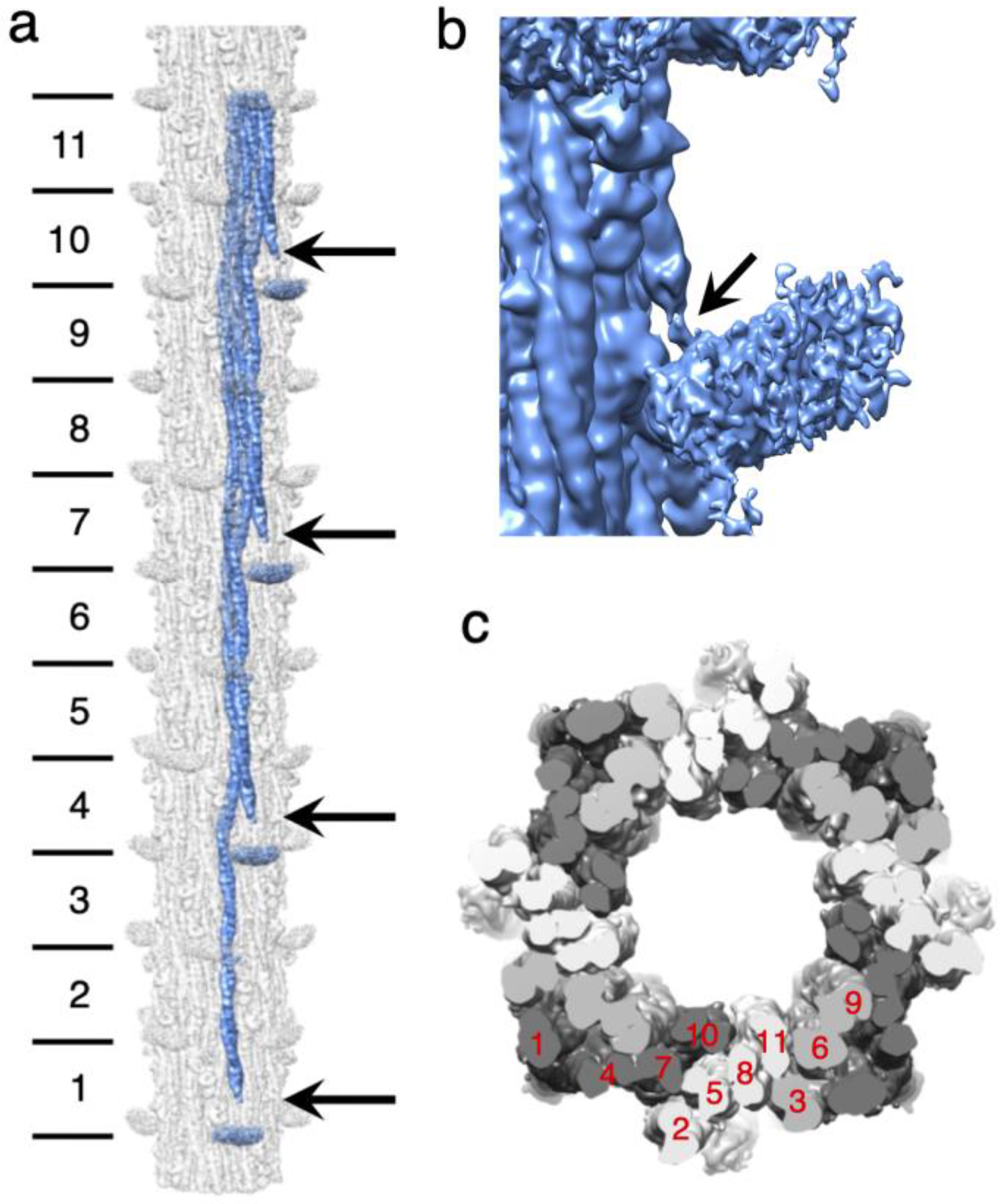
2.1. Myosin Tail Structure and Arrangement
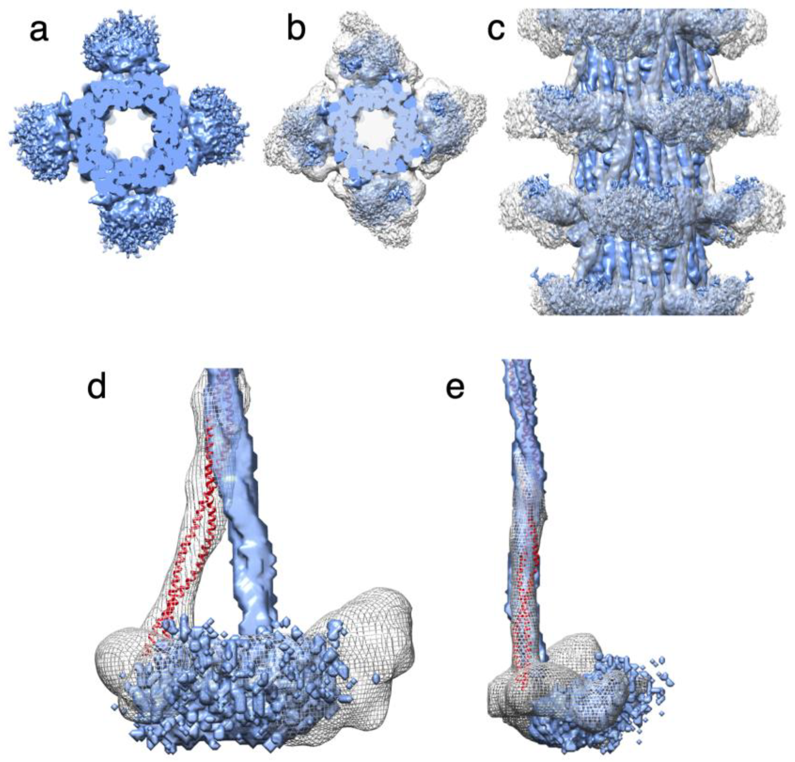
2.2. Myosin Heads and Proximal S2 Region
2.3. Homology Model of the Myosin Coiled-Coil
2.4. Non-myosin Proteins
2.4.1. Myofilin
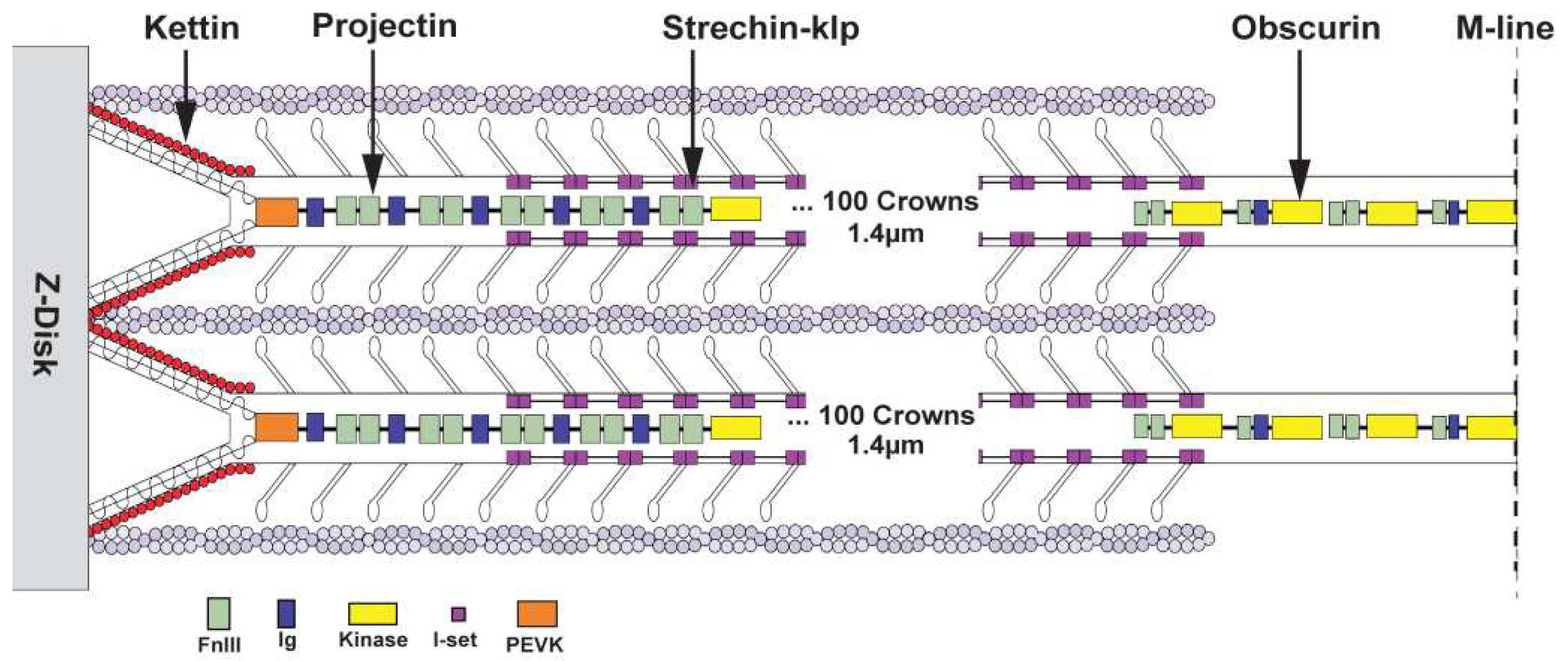
2.4.2. Stretchin-klp
2.4.3. Flightin
3. Discussion
3.1. Disordered Myosin Heads
3.2. Role of the Proximal S2 in Muscle Contraction
4. Conclusion
5. Materials and Methods
5.1. Thick filament preparation
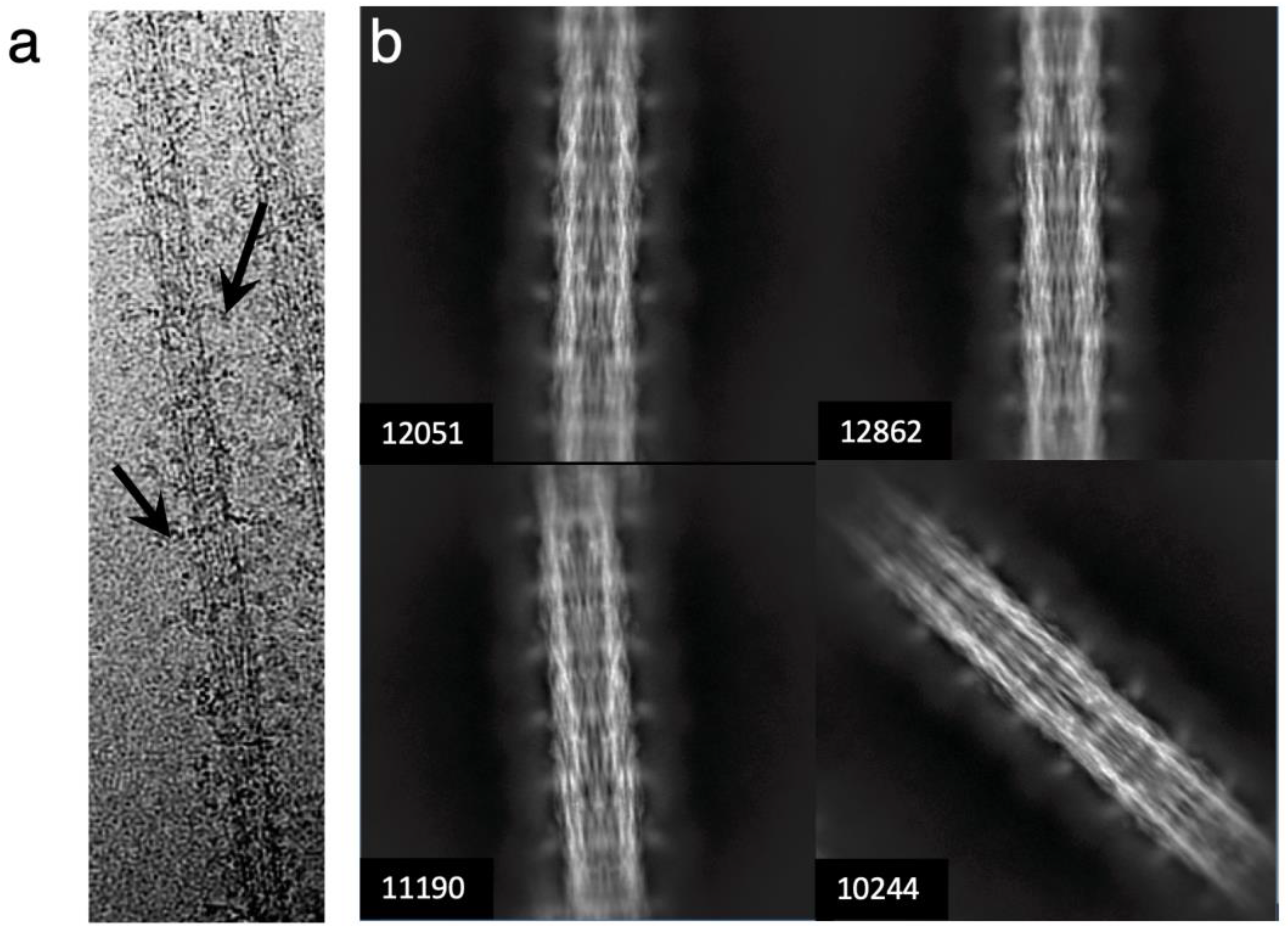
5.2. Electron microscopy
5.3. Data Analysis
Supplementary Materials
Author Contributions
Funding
Data Availability Statement
Acknowledgments
Conflicts of Interest
References
- Hooper, S. L.; Hobbs, K. H.; Thuma, J. B., Invertebrate muscles: thin and thick filament structure; molecular basis of contraction and its regulation, catch and asynchronous muscle. Prog Neurobiol 2008, 86, (2), 72-127. [CrossRef]
- Page, S. G.; Huxley, H. E., Filament Lengths in Striated Muscle. J. Cell Biol. 1963, 19, 369-90. [CrossRef]
- Al-Khayat, H. A.; Morris, E. P.; Kensler, R. W.; Squire, J. M., Myosin filament 3D structure in mammalian cardiac muscle. J Struct Biol 2008, 163, (2), 117-26. [CrossRef]
- Zoghbi, M. E.; Woodhead, J. L.; Moss, R. L.; Craig, R., Three-dimensional structure of vertebrate cardiac muscle myosin filaments. Proc Natl Acad Sci U S A 2008, 105, (7), 2386-90. [CrossRef]
- Dutta, D.; Nguyen, V.; Campbell, K.; Padron, R.; Craig, R., Cryo-EM structure of the human cardiac myosin filament. bioRxiv 2023, 2023.04.11.536274.
- Tamborrini, D.; Wang, Z.; Wagner, T.; Tacke, S.; Stabrin, M.; Grange, M.; Kho, A. L.; Rees, M.; Bennett, P.; Gautel, M.; Raunser, S., In situ structures from relaxed cardiac myofibrils reveal the organization of the muscle thick filament. bioRxiv 2023, 2023.04.11.536387.
- Squire, J. M., General model for the structure of all myosin-containing filaments. Nature 1971, 233, (5320), 457-62. [CrossRef]
- Sellers, J. R., Myosins. 2nd ed.; Oxford University Press: New York, NY, 1999; p 237.
- Harrington, W. F.; Rodgers, M. E., Myosin. Annu Rev Biochem 1984, 53, 35-73.
- Tregear, R. T.; Hoyland, J.; Sayers, A. J., The Repeat Distance of Myosin in the Thick Filaments of Various Muscles. J Mol Biol 1984, 176, 417-420. [CrossRef]
- Huxley, A. F., Muscular contraction. J Physiol 1974, 243, (1), 1-43.
- Reedy, M. K.; Holmes, K. C.; Tregear, R. T., Induced changes in orientation of the cross-bridges of glycerinated insect flight muscle. Nature 1965, 207, (5003), 1276-80. [CrossRef]
- Stewart, M.; Kensler, R. W.; Levine, R. J., Structure of Limulus telson muscle thick filaments. J Mol Biol 1981, 153, (3), 781-90.
- Crowther, R. A.; Padron, R.; Craig, R., Arrangement of the heads of myosin in relaxed thick filaments from tarantula muscle. J Mol Biol 1985, 184, (3), 429-39. [CrossRef]
- Huxley, H. E.; Brown, W., The low-angle x-ray diagram of vertebrate striated muscle and its behaviour during contraction and rigor. J Mol Biol 1967, 30, (2), 383-434. [CrossRef]
- Wendt, T.; Taylor, D.; Trybus, K. M.; Taylor, K., Three-dimensional image reconstruction of dephosphorylated smooth muscle heavy meromyosin reveals asymmetry in the interaction between myosin heads and placement of subfragment 2. Proc Natl Acad Sci U S A 2001, 98, (8), 4361-6. [CrossRef]
- Sellers, J. R., Regulation of cytoplasmic and smooth muscle myosin. Curr Opin Cell Biol 1991, 3, (1), 98-104. [CrossRef]
- Liu, J.; Wendt, T.; Taylor, D. W.; Taylor, K. A., Refined model of the 10S conformation of smooth muscle myosin by cryo-electron microscopy 3D image reconstruction. J Mol Biol 2003, 329, (5), 963-72. [CrossRef]
- Burgess, S. A.; Yu, S.; Walker, M. L.; Hawkins, R. J.; Chalovich, J. M.; Knight, P. J., Structures of smooth muscle myosin and heavy meromyosin in the folded, shutdown state. J Mol Biol 2007, 372, (5), 1165-78. [CrossRef]
- Scarff, C. A.; Carrington, G.; Casas-Mao, D.; Chalovich, J. M.; Knight, P. J.; Ranson, N. A.; Peckham, M., Structure of the shutdown state of myosin-2. Nature 2020, 588, (7838), 515-520.
- Yang, S.; Tiwari, P.; Lee, K. H.; Sato, O.; Ikebe, M.; Padron, R.; Craig, R., Cryo-EM structure of the inhibited (10S) form of myosin II. Nature 2020, 588, (7838), 521-525. [CrossRef]
- Heissler, S. M.; Arora, A. S.; Billington, N.; Sellers, J. R.; Chinthalapudi, K., Cryo-EM structure of the autoinhibited state of myosin-2. Sci Adv 2021, 7, (52), eabk3273. [CrossRef]
- Woodhead, J. L.; Zhao, F. Q.; Craig, R.; Egelman, E. H.; Alamo, L.; Padron, R., Atomic model of a myosin filament in the relaxed state. Nature 2005, 436, (7054), 1195-9.
- Jung, H. S.; Burgess, S. A.; Billington, N.; Colegrave, M.; Patel, H.; Chalovich, J. M.; Chantler, P. D.; Knight, P. J., Conservation of the regulated structure of folded myosin 2 in species separated by at least 600 million years of independent evolution. Proc Natl Acad Sci U S A 2008, 105, (16), 6022-6. [CrossRef]
- Zhao, F. Q.; Craig, R.; Woodhead, J. L., Head-head interaction characterizes the relaxed state of Limulus muscle myosin filaments. J Mol Biol 2009, 385, (2), 423-31. [CrossRef]
- Sulbaran, G.; Alamo, L.; Pinto, A.; Marquez, G.; Mendez, F.; Padron, R.; Craig, R., An invertebrate smooth muscle with striated muscle myosin filaments. Proc Natl Acad Sci U S A 2015, 112, (42), E5660-8. [CrossRef]
- Jung, H. S.; Komatsu, S.; Ikebe, M.; Craig, R., Head-head and head-tail interaction: a general mechanism for switching off myosin II activity in cells. Mol Biol Cell 2008, 19, (8), 3234-42. [CrossRef]
- Lee, K. H.; Sulbaran, G.; Yang, S.; Mun, J. Y.; Alamo, L.; Pinto, A.; Sato, O.; Ikebe, M.; Liu, X.; Korn, E. D.; Sarsoza, F.; Bernstein, S. I.; Padron, R.; Craig, R., Interacting-heads motif has been conserved as a mechanism of myosin II inhibition since before the origin of animals. Proc Natl Acad Sci U S A 2018, 115, (9), E1991-E2000. [CrossRef]
- Naber, N.; Cooke, R.; Pate, E., Slow myosin ATP turnover in the super-relaxed state in tarantula muscle. Journal of Molecular Biology 2011, 411, (5), 943-50.
- Trivedi, D. V.; Adhikari, A. S.; Sarkar, S. S.; Ruppel, K. M.; Spudich, J. A., Hypertrophic cardiomyopathy and the myosin mesa: viewing an old disease in a new light. Biophys Rev 2018, 10, (1), 27-48. [CrossRef]
- Hu, Z.; Taylor, D. W.; Reedy, M. K.; Edwards, R. J.; Taylor, K. A., Structure of myosin filaments from relaxed Lethocerus flight muscle by cryo-EM at 6 Å resolution. Sci Adv 2016, 2, (9), e1600058. [CrossRef]
- Bullard, B.; Burkart, C.; Labeit, S.; Leonard, K., The function of elastic proteins in the oscillatory contraction of insect flight muscle. J Muscle Res Cell Motil 2005, 26, (6-8), 479-85. [CrossRef]
- Daneshparvar, N.; Taylor, D. W.; O'Leary, T. S.; Rahmani, H.; Abbasiyeganeh, F.; Previs, M. J.; Taylor, K. A., CryoEM structure of Drosophila flight muscle thick filaments at 7 A resolution. Life Sci Alliance 2020, 3, (8), e202000823. [CrossRef]
- Menetret, J. F.; Schroder, R. R.; Hofmann, W., Cryo-electron microscopic studies of relaxed striated muscle thick filaments. J Muscle Res Cell Motil 1990, 11, (1), 1-11. [CrossRef]
- Farman, G. P.; Miller, M. S.; Reedy, M. C.; Soto-Adames, F. N.; Vigoreaux, J. O.; Maughan, D. W.; Irving, T. C., Phosphorylation and the N-terminal extension of the regulatory light chain help orient and align the myosin heads in Drosophila flight muscle. J Struct Biol 2009, 168, (2), 240-9. [CrossRef]
- Li, J.; Rahmani, H.; Abbasi Yeganeh, F.; Rastegarpouyani, H.; Taylor, D. W.; Wood, N. B.; Previs, M. J.; Iwamoto, H.; Taylor, K. A., Structure of the Flight Muscle Thick Filament from the Bumble Bee, Bombus ignitus, at 6 Å Resolution. Int J Mol Sci 2023, 24, (1), 377.
- Katzemich, A.; Kreiskother, N.; Alexandrovich, A.; Elliott, C.; Schock, F.; Leonard, K.; Sparrow, J.; Bullard, B., The function of the M-line protein obscurin in controlling the symmetry of the sarcomere in the flight muscle of Drosophila. J Cell Sci 2012, 125, (Pt 14), 3367-79. [CrossRef]
- Tskhovrebova, L.; Trinick, J., Roles of titin in the structure and elasticity of the sarcomere. J Biomed Biotechnol 2010, 2010, 612482. [CrossRef]
- Hu, D. H.; Matsuno, A.; Terakado, K.; Matsuura, T.; Kimura, S.; Maruyama, K., Projectin is an invertebrate connectin (titin): isolation from crayfish claw muscle and localization in crayfish claw muscle and insect flight muscle. J Muscle Res Cell Motil 1990, 11, (6), 497-511. [CrossRef]
- Lakey, A.; Ferguson, C.; Labeit, S.; Reedy, M.; Larkins, A.; Butcher, G.; Leonard, K.; Bullard, B., Identification and localization of high molecular weight proteins in insect flight and leg muscle. EMBO J 1990, 9, (11), 3459-67. [CrossRef]
- Lakey, A.; Labeit, S.; Gautel, M.; Ferguson, C.; Barlow, D. P.; Leonard, K.; Bullard, B., Kettin, a large modular protein in the Z-disc of insect muscles. EMBO J 1993, 12, (7), 2863-71. [CrossRef]
- Patel, S. R.; Saide, J. D., Stretchin-klp, a novel Drosophila indirect flight muscle protein, has both myosin dependent and independent isoforms. J Muscle Res Cell Motil 2005, 26, (4-5), 213-24. [CrossRef]
- Becker, K. D.; O'Donnell, P. T.; Heitz, J. M.; Vito, M.; Bernstein, S. I., Analysis of Drosophila paramyosin: identification of a novel isoform which is restricted to a subset of adult muscles. J. Cell Biol. 1992, 116, (3), 669-81. [CrossRef]
- Ayer, G.; Vigoreaux, J. O., Flightin is a myosin rod binding protein. Cell Biochem Biophys 2003, 38, (1), 41-54. [CrossRef]
- Qiu, F.; Brendel, S.; Cunha, P. M.; Astola, N.; Song, B.; Furlong, E. E.; Leonard, K. R.; Bullard, B., Myofilin, a protein in the thick filaments of insect muscle. J Cell Sci 2005, 118, (Pt 7), 1527-36. [CrossRef]
- Beinbrech, G.; Ashton, F. T.; Pepe, F. A., Invertebrate myosin filament: subfilament arrangement in the wall of tubular filaments of insect flight muscles. J Mol Biol 1988, 201, (3), 557-65. [CrossRef]
- He, S.; Scheres, S. H. W., Helical reconstruction in RELION. J Struct Biol 2017, 198, (3), 163-176. [CrossRef]
- Grant, T.; Rohou, A.; Grigorieff, N., cisTEM, user-friendly software for single-particle image processing. Elife 2018, 7.
- Squire, J. M., General model of myosin filament structure. 3. Molecular packing arrangements in myosin filaments. J Mol Biol 1973, 77, (2), 291-323. [CrossRef]
- Dickinson, M.; Farman, G.; Frye, M.; Bekyarova, T.; Gore, D.; Maughan, D.; Irving, T., Molecular dynamics of cyclically contracting insect flight muscle in vivo. Nature 2005, 433, (7023), 330-4.
- Rahmani, H.; Ma, W.; Hu, Z.; Daneshparvar, N.; Taylor, D. W.; McCammon, J. A.; Irving, T. C.; Edwards, R. J.; Taylor, K. A., The myosin II coiled-coil domain atomic structure in its native environment. Proc Natl Acad Sci U S A 2021, 118, (14), e202415111.
- Daneshparvar, N.; Rahmani, H.; Taylor, K., Homology model of Drosophila melanogaster myosin filaments. Microsc. Microanal. 2021, 27, 1704-1706. [CrossRef]
- Crick, F. H. C., The packing of α-helices: Simple coiled coils. Acta Crystallogr 1953, 6, 689–697.
- Vigoreaux, J. O.; Saide, J. D.; Valgeirsdottir, K.; Pardue, M. L., Flightin, a novel myofibrillar protein of Drosophila stretch-activated muscles. J. Cell Biol. 1993, 121, (3), 587-98. [CrossRef]
- Champagne, M. B.; Edwards, K. A.; Erickson, H. P.; Kiehart, D. P., Drosophila stretchin-MLCK is a novel member of the Titin/Myosin light chain kinase family. J Mol Biol 2000, 300, (4), 759-77. [CrossRef]
- Maroto, M.; Arredondo, J.; Goulding, D.; Marco, R.; Bullard, B.; Cervera, M., Drosophila paramyosin/miniparamyosin gene products show a large diversity in quantity, localization, and isoform pattern: a possible role in muscle maturation and function. J. Cell Biol. 1996, 134, (1), 81-92. [CrossRef]
- Soto-Adames, F. N.; Alvarez-Ortiz, P.; Vigoreaux, J. O., An evolutionary analysis of flightin reveals a conserved motif unique and widespread in Pancrustacea. J Mol Evol 2014, 78, (1), 24-37. [CrossRef]
- Jumper, J.; Evans, R.; Pritzel, A.; Green, T.; Figurnov, M.; Ronneberger, O.; Tunyasuvunakool, K.; Bates, R.; Zidek, A.; Potapenko, A.; Bridgland, A.; Meyer, C.; Kohl, S. A. A.; Ballard, A. J.; Cowie, A.; Romera-Paredes, B.; Nikolov, S.; Jain, R.; Adler, J.; Back, T.; Petersen, S.; Reiman, D.; Clancy, E.; Zielinski, M.; Steinegger, M.; Pacholska, M.; Berghammer, T.; Bodenstein, S.; Silver, D.; Vinyals, O.; Senior, A. W.; Kavukcuoglu, K.; Kohli, P.; Hassabis, D., Highly accurate protein structure prediction with AlphaFold. Nature 2021, 596, (7873), 583-589.
- Torices, R.; Munoz-Pajares, A. J., PHENIX: An R package to estimate a size-controlled phenotypic integration index. Appl Plant Sci 2015, 3, (5). [CrossRef]
- Wingfield, P. T., N-Terminal Methionine Processing. Curr Protoc Protein Sci 2017, 88, 6 14 1-6 14 3.
- Tanner, B. C.; Miller, M. S.; Miller, B. M.; Lekkas, P.; Irving, T. C.; Maughan, D. W.; Vigoreaux, J. O., COOH-terminal truncation of flightin decreases myofilament lattice organization, cross-bridge binding, and power output in Drosophila indirect flight muscle. Am J Physiol Cell Physiol 2011, 301, (2), C383-91. [CrossRef]
- Gasek, N. S.; Nyland, L. R.; Vigoreaux, J. O., The Contributions of the Amino and Carboxy Terminal Domains of Flightin to the Biomechanical Properties of Drosophila Flight Muscle Thick Filaments. Biology (Basel) 2016, 5, (2). [CrossRef]
- Kronert, W. A.; O'Donnell, P. T.; Fieck, A.; Lawn, A.; Vigoreaux, J. O.; Sparrow, J. C.; Bernstein, S. I., Defects in the Drosophila myosin rod permit sarcomere assembly but cause flight muscle degeneration. J Mol Biol 1995, 249, (1), 111-25. [CrossRef]
- Knupp, C.; Luther, P. K.; Squire, J. M., Titin organisation and the 3D architecture of the vertebrate-striated muscle I-band. J Mol Biol 2002, 322, (4), 731-9. [CrossRef]
- Kolmerer, B.; Clayton, J.; Benes, V.; Allen, T.; Ferguson, C.; Leonard, K.; Weber, U.; Knekt, M.; Ansorge, W.; Labeit, S.; Bullard, B., Sequence and expression of the kettin gene in Drosophila melanogaster and Caenorhabditis elegans. J Mol Biol 2000, 296, (2), 435-48. [CrossRef]
- Ayme-Southgate, A. J.; Southgate, R. J.; Philipp, R. A.; Sotka, E. E.; Kramp, C., The myofibrillar protein, projectin, is highly conserved across insect evolution except for its PEVK domain. J Mol Evol 2008, 67, (6), 653-69. [CrossRef]
- Mogami, K.; O'Donnell, P. T.; Bernstein, S. I.; Wright, T. R.; Emerson, C. P., Jr., Mutations of the Drosophila myosin heavy-chain gene: effects on transcription, myosin accumulation, and muscle function. Proceedings of the National Academy of Sciences of the United States of America 1986, 83, (5), 1393-7.
- Craig, R., Molecular structure of muscle filaments determined by electron microscopy. Appl Microsc 2017, 47, (4), 226-232. [CrossRef]
- Lowey, S.; Trybus, K. M., Common structural motifs for the regulation of divergent class II myosins. J Biol Chem 2010, 285, (22), 16403-7. [CrossRef]
- Liu, J.; Wu, S.; Reedy, M. C.; Winkler, H.; Lucaveche, C.; Cheng, Y.; Reedy, M. K.; Taylor, K. A., Electron tomography of swollen rigor fibers of insect flight muscle reveals a short and variably angled S2 domain. J Mol Biol 2006, 362, (4), 844-60. [CrossRef]
- Hvidt, S.; Nestler, F. H.; Greaser, M. L.; Ferry, J. D., Flexibility of myosin rod determined from dilute solution viscoelastic measurements. Biochemistry 1982, 21, (17), 4064-73. [CrossRef]
- Taylor, K. A.; Reedy, M. C.; Cordova, L.; Reedy, M. K., Three-dimensional reconstruction of rigor insect flight muscle from tilted thin sections. Nature 1984, 310, (5975), 285-91.
- White, D. C., Rigor contraction and the effect of various phosphate compounds on glycerinated insect flight and vertebrate muscle. J Physiol 1970, 208, (3), 583-605. [CrossRef]
- Reedy, M. K.; Reedy, M. C., Rigor crossbridge structure in tilted single filament layers and flared-X formations from insect flight muscle. J Mol Biol 1985, 185, (1), 145-76.
- Bullard, B.; Pastore, A., Through thick and thin: dual regulation of insect flight muscle and cardiac muscle compared. J Muscle Res Cell Motil 2019. [CrossRef]
- McLachlan, A. D.; Karn, J., Periodic charge distributions in the myosin rod amino acid sequence match cross-bridge spacings in muscle. Nature 1982, 299, (5880), 226-31. [CrossRef]
- Zheng, S. Q.; Palovcak, E.; Armache, J. P.; Verba, K. A.; Cheng, Y.; Agard, D. A., MotionCor2: anisotropic correction of beam-induced motion for improved cryo-electron microscopy. Nat Methods 2017, 14, (4), 331-332. [CrossRef]
- Zhang, K., Gctf: Real-time CTF determination and correction. J Struct Biol 2016, 193, (1), 1-12. [CrossRef]
- Scheres, S. H., Processing of Structurally Heterogeneous Cryo-EM Data in RELION. Methods Enzymol 2016, 579, 125-57. [CrossRef]
- Punjani, A.; Rubinstein, J. L.; Fleet, D. J.; Brubaker, M. A., cryoSPARC: algorithms for rapid unsupervised cryo-EM structure determination. Nat Methods 2017, 14, (3), 290-296.
- Vilas, J. L.; Gomez-Blanco, J.; Conesa, P.; Melero, R.; Miguel de la Rosa-Trevin, J.; Oton, J.; Cuenca, J.; Marabini, R.; Carazo, J. M.; Vargas, J.; Sorzano, C. O. S., MonoRes: Automatic and Accurate Estimation of Local Resolution for Electron Microscopy Maps. Structure 2018, 26, (2), 337-344 e4. [CrossRef]
- Ramirez-Aportela, E.; Vilas, J. L.; Glukhova, A.; Melero, R.; Conesa, P.; Martinez, M.; Maluenda, D.; Mota, J.; Jimenez, A.; Vargas, J.; Marabini, R.; Sexton, P. M.; Carazo, J. M.; Sorzano, C. O. S., Automatic local resolution-based sharpening of cryo-EM maps. Bioinformatics 2020, 36, (3), 765-772. [CrossRef]
- de la Rosa-Trevin, J. M.; Quintana, A.; Del Cano, L.; Zaldivar, A.; Foche, I.; Gutierrez, J.; Gomez-Blanco, J.; Burguet-Castell, J.; Cuenca-Alba, J.; Abrishami, V.; Vargas, J.; Oton, J.; Sharov, G.; Vilas, J. L.; Navas, J.; Conesa, P.; Kazemi, M.; Marabini, R.; Sorzano, C. O.; Carazo, J. M., Scipion: A software framework toward integration, reproducibility and validation in 3D electron microscopy. J Struct Biol 2016, 195, (1), 93-9. [CrossRef]
- Pettersen, E. F.; Goddard, T. D.; Huang, C. C.; Couch, G. S.; Greenblatt, D. M.; Meng, E. C.; Ferrin, T. E., UCSF Chimera--a visualization system for exploratory research and analysis. J Comput Chem 2004, 25, (13), 1605-12. [CrossRef]

| Domain | Residue Range | Domain Name | Domain Length | Linker length |
| 1 | 456 - 543 | Ig-like | 88 | 11 |
| 2 | 554 - 643 | Ig-like | 90 | 67 |
| 3 | 710 - 798 | Ig-like | 89 | 17 |
| 4 | 815 - 904 | Ig-like | 89 | 61 |
| 5 | 965 - 1056 | Ig-like | 92 | 12 |
| 6 | 1068 - 1153 | Ig-like | 86 | 93 |
| 7 | 1246 - 1333 | Ig-like | 88 | 12 |
| 8 | 1345 - 1434 | Ig-like | 90 | 174 |
| 9 | 1608 - 1693 | Ig-like | 86 | 27 |
| 10 | 1720 - 1808 | Ig-like | 89 | 110 |
Disclaimer/Publisher’s Note: The statements, opinions and data contained in all publications are solely those of the individual author(s) and contributor(s) and not of MDPI and/or the editor(s). MDPI and/or the editor(s) disclaim responsibility for any injury to people or property resulting from any ideas, methods, instructions or products referred to in the content. |
© 2023 by the authors. Licensee MDPI, Basel, Switzerland. This article is an open access article distributed under the terms and conditions of the Creative Commons Attribution (CC BY) license (http://creativecommons.org/licenses/by/4.0/).


