Preprint
Interesting Images

Genu Recurvatum and Rocker Bottom Foot Co-occurrence in a Unique Clinical Image of Edwards Syndrome.

Altmetrics

Downloads

93

Views

32

Comments

0

Submitted:

04 September 2023

Posted:

06 September 2023

You are already at the latest version

Alerts
Abstract
Edward syndrome, clinically known as trisomy 18 syndrome, is an autosomal chromosomal disorder characterized by an extra copy of chromosome 18. It belongs to the category of autosomal trisomy syndromes and is the second most common, with trisomy 21 being the only more frequent occurrence. In the context of this research, we present a case study focusing on a 3-month-old female infant admitted to the emergency department due to a complex array of symptoms. The primary clinical manifestations included a persistent cough, cold, and fever lasting for eight consecutive days. Comprehensive clinical evaluations, encompassing radiological and hematological investigations, confirmed the diagnosis of bronchiolitis along with viral bronchopneumonia. Chest X-rays revealed bilateral patchy opacities and diffuse haziness, primarily concentrated in the right hemithorax. Beyond respiratory issues, the infant displayed a range of distinctive physical attributes, including low-set ears, micrognathia, cranial anomalies, flattened nasal bridge, widely spaced nipples, overlapping fingers, abdominal distention, rocker-bottom feet, and recurring limb deformities known as recurvatum. Attempts to correct the limb deformity with a hip spica cast over an extended month-long period proved unsuccessful. Chronic villus sampling (CVS) ultimately confirmed the presence of Edward Syndrome (Trisomy 18), linked to the extra chromosome 18, leading to diverse physiological and developmental abnormalities. This case underscores the critical importance of timely and precise diagnosis, highlighting the challenges involved in addressing complex medical conditions marked by multifaceted clinical phenotypes.
Keywords: 
Subject: Public Health and Healthcare  -   Physical Therapy, Sports Therapy and Rehabilitation
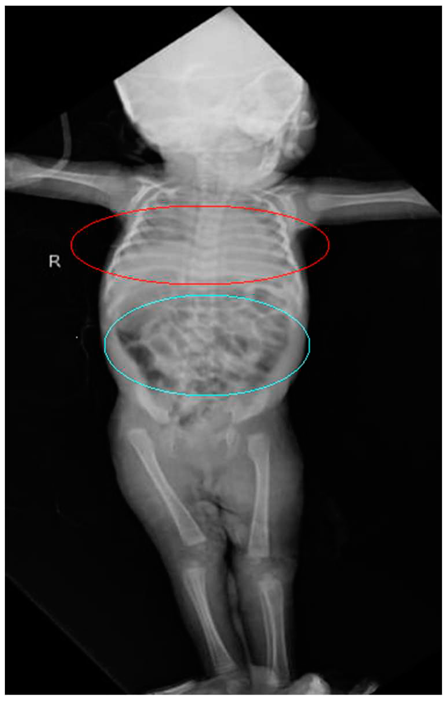
Figure 1. An X-ray examination of a 3-month-old female subject reveals several noteworthy observations.
Preprints 84136 g001
  • In the anterior-posterior view, there are discernible patchy opacities evident in both lungs, accompanied by a diffuse haziness within the right hemithorax (encircled in red).
  • The examination also discloses bowel dilation with the presence of gas. The distinctive bubbly gas pattern observed in the right lower quadrant signifies the presence of an intramural gas (encircled in blue) [1].
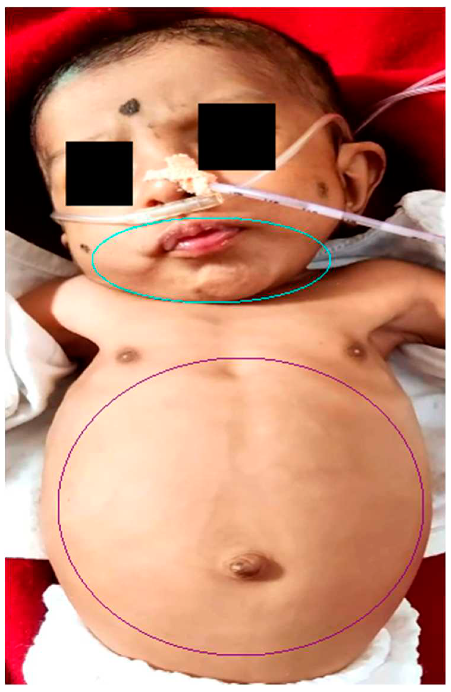
Figure 2. Shows micrognathia (encircled in blue) and a distended abdomen (encircled in purple) [2].
Preprints 84136 g002
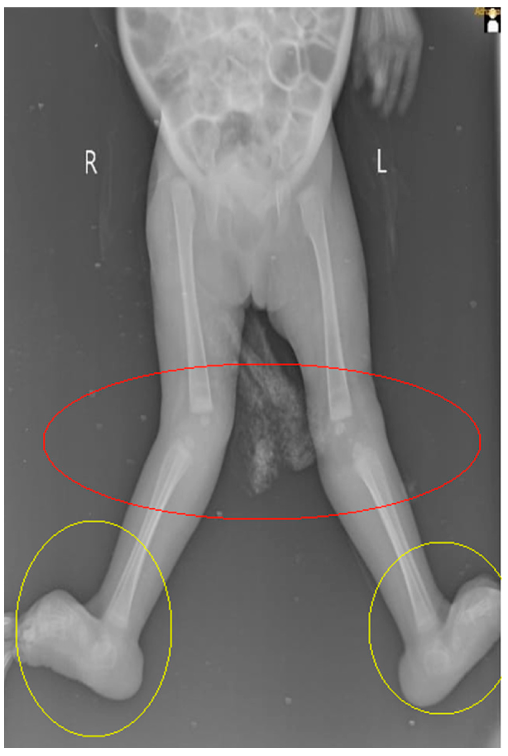
Figure 3. month-old female illustrates the presence of Genu recurvatum (encircled in red), in conjunction with bilateral rocker bottom foot deformity, (encircled in yellow).
Preprints 84136 g003
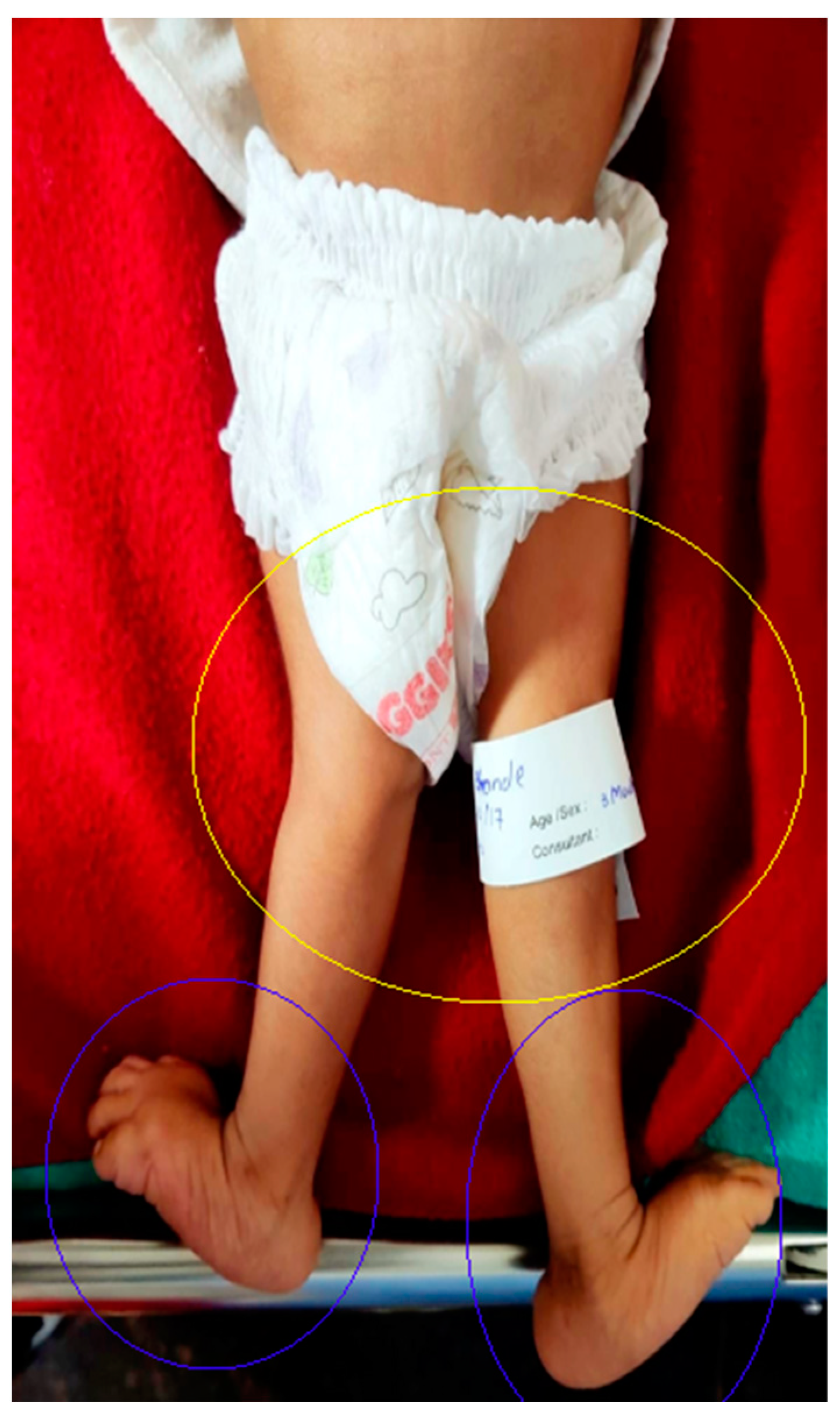
Figure 4. Depicts the occurrence of Genu recurvatum (encircled in red), concomitant with bilateral rocker bottom foot deformity with the prominent calcaneus, (encircled in blue) [3].
Preprints 84136 g004
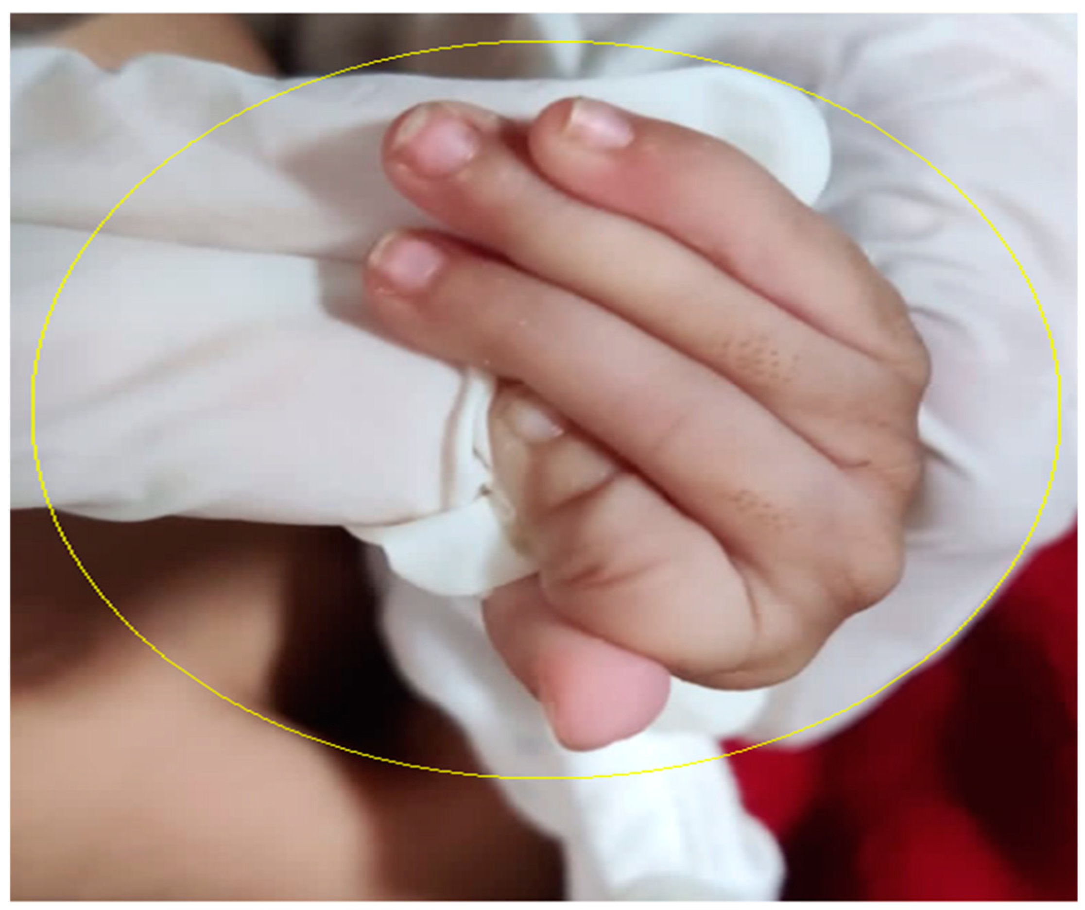
Figure 5. Demonstrates the distinctive clenched hand posture featuring camptodactyly (encircled in yellow) [2].
Preprints 84136 g005

Author Contributions

S.G. played a pivotal role in conceptualizing, composing, and initializing the primary draft of the manuscript. Subsequent to this, all authors collectively engaged in the comprehensive process of evaluating and refining the content. The ultimate rendition of the manuscript underwent thorough scrutiny and received the endorsement of all authors.

Funding

The current study got no external funding.

Institutional Review Board Statement

Not applicable.

Informed Consent Statement

Written consent was obtained from the patient’s caregivers.

Conflicts of Interest

The authors declare no conflict of interest.

References

  1. Epelman, M.; Daneman, A.; Navarro, O.M.; Morag, I.; Moore, A.M.; Kim, J.H.; Faingold, R.; Taylor, G.; Gerstle, J.T. Necrotizing Enterocolitis: Review of State-of-the-Art Imaging Findings with Pathologic Correlation. RadioGraphics 2007, 27, 285–305. [Google Scholar] [CrossRef] [PubMed]
  2. Adetunji, O.O.; Ferdinad, F.F.; Idowu, J.-M.V.; Ademola, O.G. Edwards Syndrome In A Neonate From A Developing Country; Reasons For Concern: A Case Report. The Internet Journal of Third World Medicine 2006, 4. [Google Scholar]
  3. Rathod, B.D.; Tamilarasan, P. Umbilical Cyst with Edward Syndrome.
Disclaimer/Publisher’s Note: The statements, opinions and data contained in all publications are solely those of the individual author(s) and contributor(s) and not of MDPI and/or the editor(s). MDPI and/or the editor(s) disclaim responsibility for any injury to people or property resulting from any ideas, methods, instructions or products referred to in the content.
Copyright: This open access article is published under a Creative Commons CC BY 4.0 license, which permit the free download, distribution, and reuse, provided that the author and preprint are cited in any reuse.
Prerpints.org logo

Preprints.org is a free preprint server supported by MDPI in Basel, Switzerland.

Subscribe

© 2024 MDPI (Basel, Switzerland) unless otherwise stated