Preprint
Article

Altered Expression of Vitamin D Metabolism Genes and Circulating MicroRNAs in PBMCs of Patients with Type 1 Diabetes: Their Association with Vitamin D Status and Ongoing Islet Autoimmunity

Altmetrics

Downloads

174

Views

87

Comments

0

A peer-reviewed article of this preprint also exists.

Submitted:

02 September 2023

Posted:

05 September 2023

Read the latest preprint version here

Alerts
Abstract
Background: Vitamin D (VD) acts as an immunomodulator through the vitamin D receptor (VDR) located on pancreatic and immune cells, and deficiency is related to the development of T1DM. There is no clear understanding of the molecular mechanism by which VD is down-regulated in type 1 diabetes. So, we investigated whether VD metabolism genes are expressed differently in T1DM patients and whether serum VD levels are correlated with those genes. Additionally, we sought to assess whether miRNAs affect the expression of VD metabolism genes in PBMCs from patients with T1DM. The study also examines whether altered VD metabolism genes and miRNAs impact autoimmunity. Methods: Real-time PCR was used to detect the expression profile of gene encoding 1-hydroxylases (CYP27B1), 24-hydroxylases (CYP24A1), as well as related miRNAs in PBMCs of 30 individuals with T1DM and 23 non-diabetic subjects. ELISA test was used to determine VD, GAD65, and IA-2 expression. Results: CYP27B1 mRNA levels were down-regulated, while CYP24A1 levels were not changed in PBMCs T1DM patients than in those of healthy controls (CYP27B1, p = 0.0005, CYP24A1, p=0.205, respectively). has-miR-216b-5p expression was significantly up-regulated in T1DM while has-miR-216b-5p down-regulated compared to the control. The expression of CYP27B1 in T1DM subjects was not correlated with levels of has-miR-216b-5p, has-miR-21-5p, and VD. Interestingly, CYP27B1 mRNA concentrations in PBMCs of T1DM subjects correlated negatively with IA2 but not GAD65. Conclusions: We observed down-regulation of CYP27B1 mRNA levels and up-regulated has-miR-216b-5p in PBMCs of T1DM. However, CYP27B1 expression was not correlated with the expression of has-miR-216-5p and VD status, suggesting other mechanisms may regulate CYP27B1 expression.
Keywords: 
Subject: Biology and Life Sciences  -   Biochemistry and Molecular Biology

1. Introduction

T1DM (T1DM) is a chronic autoimmune disease characterized by beta cell degeneration, leading to insulin deficiency [1]. T1DM incidence is strongly associated with vitamin D deficiency. In addition, vitamin D can prevent islet cell death and enhance insulin production. Several studies suggest low vitamin D levels may negatively affect beta cell regulation [2]. According to many epidemiological studies, vitamin D deficiency has been reported to be prevalent among Saudi children with T1DM, suggesting a strong correlation between them [3].
MicroRNAs (miRNAs) are small, non-coding RNA molecules that regulate gene expression post-transcriptionally, generally by preventing or triggering the degradation of mRNAs [4]. miRNAs that target genes involved in vitamin D metabolism may affect the level of circulating vitamin D in patients with type 1 diabetes. Various genes control the vitamin D metabolism pathway, including CYP2R1, CYP27B1 (activation), CYP24A1 (inactivation), VDR (action), and GC. 1,25(OH)2D concentrations are tightly regulated by both 1-hydroxylase and the catabolic enzyme 24-hydroxylase (CYP24A1 gene). The CYP24A1 enzyme catalyzes the hydroxylation reaction that results in the degradation of 1,25-dihydroxy vitamin D3, which is the physiologically active form of vitamin D. The hydroxylation of the side chain results in calcitroic acid and other metabolites being excreted in the bile. CYP24A1 is induced by both 1,25(OH)2D and 25(OH)D and is one of the most highly inducible genes in humans, capable of increasing its transcription by 20,000-fold [5]. The high inducibility of CYP24A1 is likely to be a critical factor in the large therapeutic window of vitamin D.
Several miRNAs have also been predicted to target CYP24A1, but only miR-125b-5p has been experimentally validated [6]. According to studies conducted on ovarian granulosa and breast cancer cells, over-expression or antisense knockdown of miR-125b-5p respectively suppresses and enhances the expression of CYP24A1 protein [6]. In addition to its effects on vitamin D catabolism, miR-125b-5p also targets the VDR. Reducing miR-125b-5p expression increases responses to 1,25(OH)2D in melanoma cell lines [7]. In contrast, an increase in miR-125b-5p suppresses endogenous levels of VDR protein in MCF-7 breast cancer cells, contributing to resistance to 1,25(OH)2D [8]. It has been suggested that other miRNAs may target VDR, but only one of them, miR-326, has been validated in the peripheral blood lymphocytes of people with T1DM [9]. Given the proposed role of vitamin D in protecting against autoimmunity [10], it is tempting to speculate that miR-326 may inhibit 1,25(OH)2D's immunomodulatory effects in preventing inflammatory and autoimmune disorders. Moreover, other studies have demonstrated that miR-125a-5p is differentially expressed in Treg cells isolated from the lymph nodes draining the pancreas of patients with T1DM, which might contribute to the reduced expression of the CCR2 gene [11].
The vitamin D-activation enzyme CYP27B1 is predicted to be targeted by multiple miRNAs, but only one of these has been validated experimentally. A number of studies have shown that miR-21 inhibits the expression of CYP27B1 in monocytes infected with Mycobacterium leprae (M. Leprae), and thereby inhibits downstream antibacterial responses induced by vitamin D intracrine signaling [12]. Other miRNAs predicate to target CYP27B1 is miR-216a-5p. T1D is associated with increased expression of miR-216a in pancreatic islets, which may be a compensatory mechanism [13]. miR-377 and miR-216a can be used as early biomarkers of nephropathy in children with type 1 diabetes. Their correlation with CIMT provides insight into the subclinical atherosclerotic process in diabetic nephropathy [14].
Accumulating evidence shows that miRNA is involved in T1DM pathogenesis through multiple mechanisms, including regulation of immune cell differentiation, development and activation, and disruption of immune system equilibrium. miR-34a overexpression in diabetic mice reduces the capacity for B lymphopoiesis and causes disturbance of pancreatic islet defense and sensitivity to damage [15]. Qualitative differential miRNA expression also affects the production of specific T lymphocytes. miR-26 and miR-101 have been linked with the differentiation of cells towards Th1 phenotype. In addition, miR-21, miR-93, miR-326, and miR-31 are believed to alter T cell functions and play a role in T1DM autoimmunity [16,17]. It has been found that miR-21 and miR-93 are essential for inflammatory and apoptosis signaling pathways and that their expression was downregulated in PBMCs of patients with T1DM [18]. Furthermore, miR-326 was overexpressed in PBMCs from T1DM patients [19,20], which indicates the role of miRNAs in T1DM autoimmunity, as it targets significant immune modulators—vitamin D receptor (VDR) and erythroblastosis virus E26 oncogenic homolog 1 (ETS-1).
Several miRNAs are associated with type 1 diabetes in peripheral blood mononuclear cells (PBMCs). There was significant downregulation of miR-21a and miR-93 in patients with recently diagnosed T1DM. These miRNAs target NF-KB signaling to regulate apoptosis and inflammation [18], while other miRNAs, such as miR-20a and miR-326, were up-regulated [20]. In addition, miRNA signatures in PBMCs were associated with autoantibodies in T1D patients, with increased levels of miR-326 correlated with antibodies against GAD and IA2 [20] and reduced levels of miR-146a correlated with antibodies against GAD [21]. The main advantage of detecting miRNA in peripheral blood mononuclear cells (PBMCs) is the ability to use them as biomarkers for monitoring the progression of the disease.
The cause of T1DM is not fully understood, but is believed to be the development of autoantibodies and autoreactive TH1 and CTL, which cause the immune system to destroy insulin-producing pancreatic cells [1,22]. Vitamin D deficiency appears to contribute to increased activation of B cells and autoantibody production, and long-term supplementation with vitamin D led to an increase in T-regulator cells in individuals with SLE [23,24].
Most human cells contain VDR, allowing vitamin D to exert its immunomodulatory effects via a genomic response and its ability to change gene transcription [21]. Many immune cells express vitamin D receptors (VDRs) and CYP27B1 enzymes; the latter enzyme's synthesis is controlled by several immune-specific signals [25]. Considering autoimmune diseases, this vitamin D metabolite plays a vital role in the downregulation of all mechanisms related to adaptive immunity and the induction of immunological tolerance and anti-inflammatory activity [26]. So, it is crucial to understand the molecular mechanism responsible for VD deficiency in T1DM patients.
The expressional activity of CYP27B1, CYP24A1, and VDR genes, all of which contain VDREs, can be modulated both by VD itself and other factors that play a significant role in epigenetic modifications, such as miRNA [15,16,17]. Our objective in this study was to determine whether VD metabolism genes are expressed differently in T1DM patients and whether serum VD levels are correlated with those genes. Furthermore, to assess whether miRNAs affect the expression of VD metabolism genes in PBMCs T1DM. The study also examines if altered VD metabolism genes and miRNAs may be related to autoimmunity.

2. Results

2.1. Comparison of the demographic characteristics of patients with type 1 diabetes and healthy controls and vitamin D levels

Table 1 shows patients' demographic characteristics and healthy controls' vitamin D levels. The mean age of T1DM patients was 10.71 ± 6.072 years, while the healthy controls were 9.296 ±3.006 years. The age, sex, and BMI of T1DM patients and healthy controls were not significantly different.
Vitamin D serum levels were significantly lower in T1DM patients than in healthy controls (24.37±17.14 vs. 43.18±50.89 ng/ml). 53.33% of patients with T1DM were classified as having insufficient vitamin D (<20 ng/ml), whereas only no deficient healthy controls was observed. There was a statistically significant difference in vitamin D deficiency between T1DM patients and healthy controls. In T1DM patients, there was a vitamin D deficiency of 3.33% compared with zero in healthy controls (p=0.0045) (Table 1).
Table 1. Demographic and clinical features of T1DM and healthy control groups.
Variable Control group n = 23 T1DM group n = 30 P-value
HbA1c (%) 5.126± 0.4158 8.957 ±1.353 <0.0001
IA2 (IU/mL) 0 140.4 (14.45–511.0) -
GAD65A (IU/mL) 2.62 (0.4–4.7) 79.55 (18.77–264.3) -
Age (years) 9.296 ±3.006 10.71 ± 6.072 0.3098
Sex: male/female 11/12 15/15 -
BMI 21.86 ±3.250 22.31±4.248 0.6775
Vitamin D 43.18±50.89 24.37±17.14 0.0045
Data are presented as medians and interquartile ranges or mean + standard deviation. Abbreviations: BMI, body mass index; T1DM, type 1 diabetes; HbA1c, glycated hemoglobin; GAD, glutamic acid decarboxylase; IA2, tyrosine phosphatase-like protein islet antigen-2. reference to the value of 5 IU/mL for GAD65, and 10 IU/mL for IA2; T1DM vs. Control p < 0.05.
Figure 1. The distribution of vitamin D status among study participants with Type 1 Diabetes and healthy controls.
Preprints 84074 g001

2.2. The expression of CYP27B1 mRNA but not CYP24A1 was downregulated in PBMCs from T1DM as compared with healthy controls
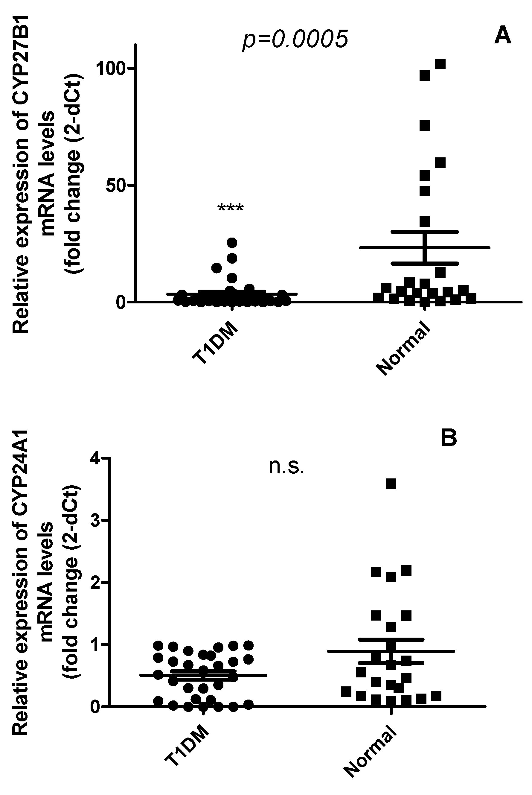
Since vitamin D levels are decreased in T1DM patients than healthy controls, we examined the expression profile of VD-metabolism genes in PBMCs of T1DM. CYP27B1 mRNA levels (Figure 2A) were significantly downregulated in T1DM patients, compared to non-diabetic subjects (p = 0.0005), while no changes were observed in CYP24A1 expression (Figure 2B) (p =0.205).
Figure 2. The expression of CYP27B1(a) is decreased in the PBMCs of patients with T1DM, while there is no significant change in the expression of CYP24A1(b) in n = 23 non-diabetic and n = 30 T1DM patients. Data are reported as mean ± SD of normalized to expression of acidic ribosomal phosphoprotein P0 (RPLP0) 2−ΔCT values. Statistical analysis using the Mann–Whitney U test, p < 0.05.
Preprints 84074 g002

2.3. Vitamin D does not correlate with the expression levels of CYP24A1 and CYP27B1 mRNA levels
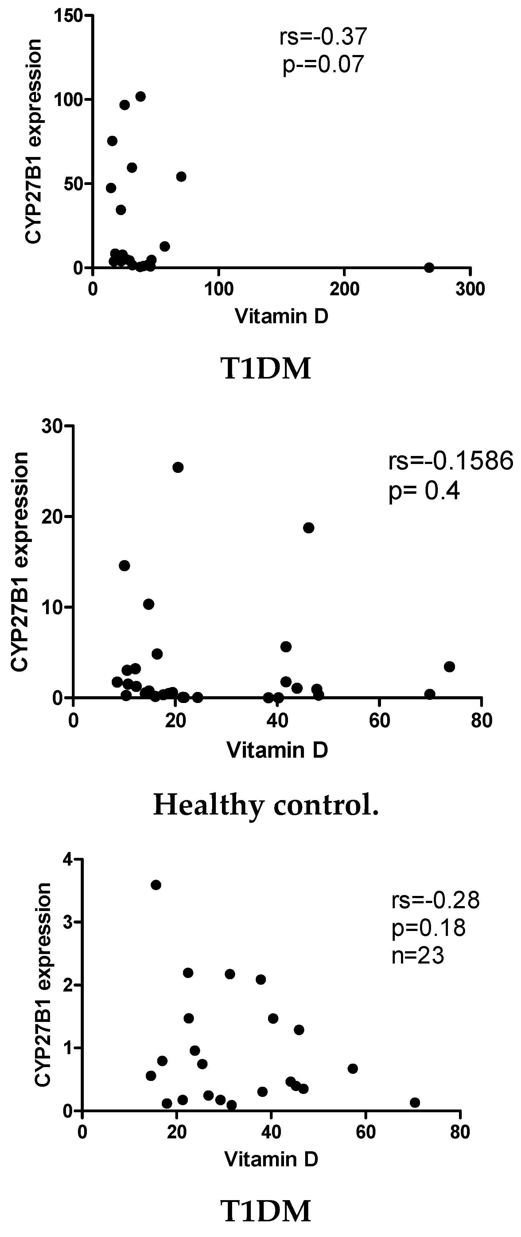
In order to determine whether VD status influences the expression of CYP24A1 and CYP27B1 in PBMCs of T1DM patients, we correlated serum 1,25(OH)2D3 levels with CYP24A1 and CYP27B1 levels. However, neither the T1DM group nor the control group demonstrated any significant correlations between VD and VD metabolism genes in both cases and controls.
Figure 3. The expression of CYP27B1 (a and b) and CYP24A1 (c and d) levels are not correlated with Serum 1,25(OH) D3 in T1DM patients and non-diabetic control subjects. Correlation analysis between CYP27B1 and CYP24A1 expression levels was reported as normalized 2−ΔCT values, and serum 1,25(OH) D3 was reported as pg/mL. Spearman R test was performed to evaluate r-values and p-values (p < 0.05).
Preprints 84074 g003aPreprints 84074 g003b

2.4. Circulating Levels of has-miR-21 and has-miR-216b-5p but not has-miR-125b are differentially expressed in PBMCs of T1D patients compared with healthy controls
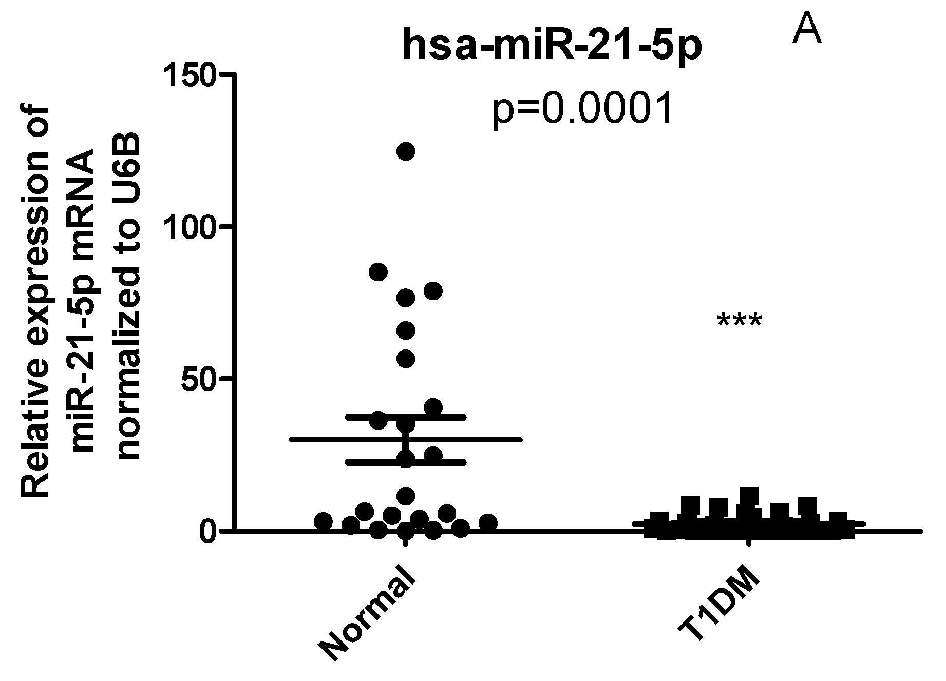
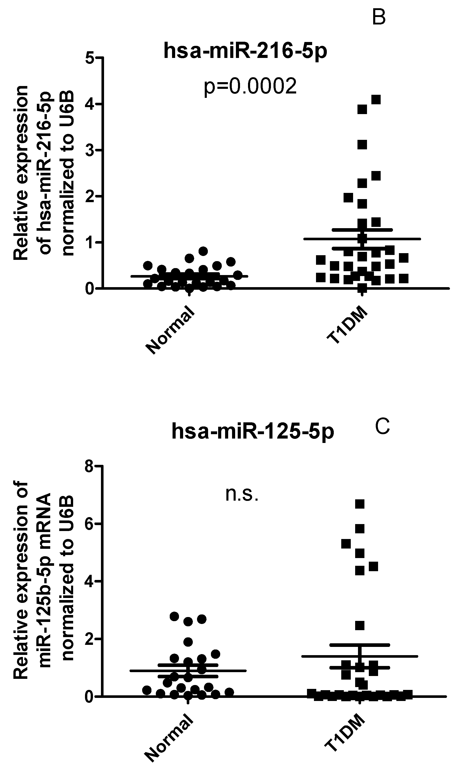
Based on our findings that serum 1,25(OH)2D3 concentrations were not correlated to CYP24A1 and CYP27B1 mRNA levels in T1DM PBMCs, we asked then whether epigenetic factors such as miRNAs might be involved in the regulation of CYP27B1 expression. The expression of hsa-miR-216b-5p was significantly up-regulated, while has-miR-21 down-regulated in PBMCs of T1DM compared to healthy controls. No significant differences between T1DM and controls was observed in the has-miR-125b expression.
Figure 4. Expression of miRNA in PBMCs from patients with T1DM and healthy controls. (A) The level of has-miR-216-5p (B) has-miRNA-21-5p, and (C) miRNA- has-miRNA-125-5p levels in PBMCs. The error bars represent the standard deviation (SD). * p<0.05 vs con.
Preprints 84074 g004aPreprints 84074 g004b

2.5. CYP27B1 not correlated with has-miR-216 and has-miR-21 in T1DM

Due to the differential expression of has-miR-216, has-miR-21, and CYP27B1 in T1DM, we performed a correlation analysis to determine whether miRNAs were correlated with CYP27B1 expression. In T1DM, we observed no significant correlations between CYP27B1 mRNA level and hsa-miR-216b-5p and has-miR-21 (P = 0. 6, rs = 0.09, Figure 5a and P = 0.2237, rs = -0.2289 respectively, Figure 5b).

2.6. The correlation between miRNAs, and CYP27B1 islet autoantibodies

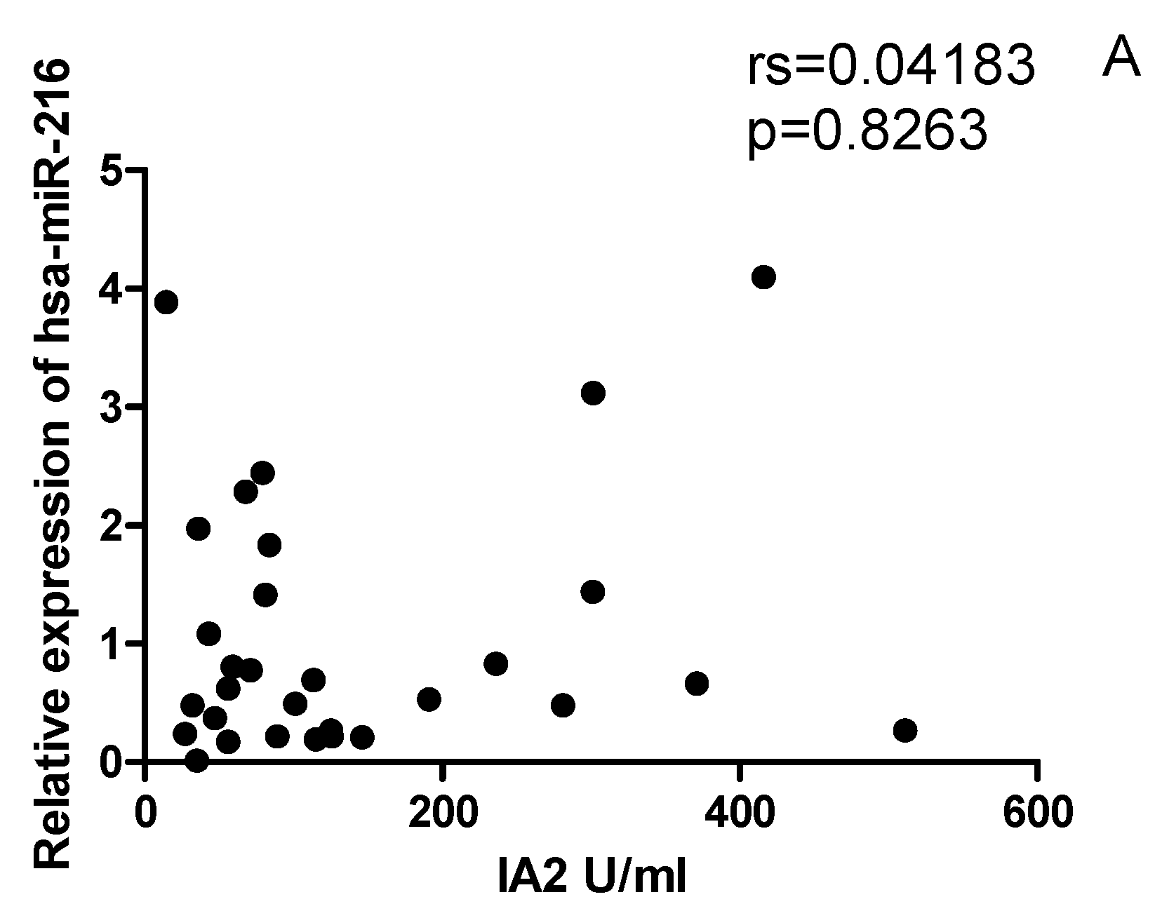
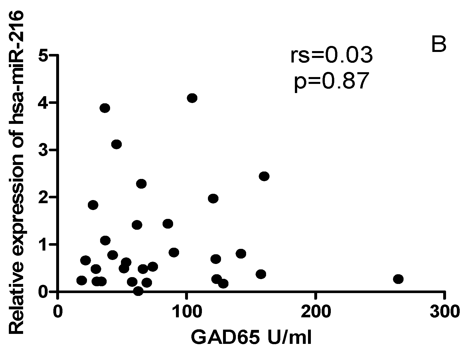
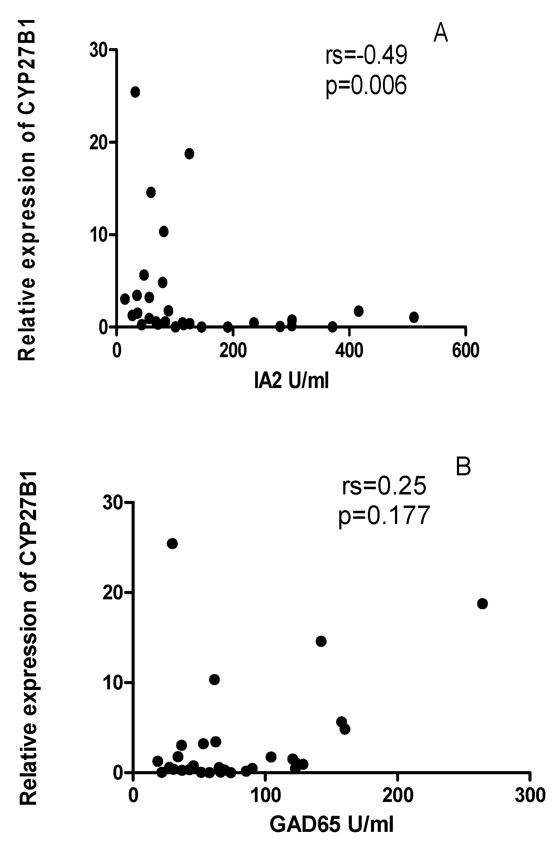
Due to the differential expression of miRNAs, and CYP27B1 in PBMCs of individuals with T1DM, we investigated which of these factors could correlate with islet autoantibodies such as GADA65 and IA2A. Analysis of associations between has-miR-216 expression and titers of islet autoantibodies (GADA, and IA2A) revealed no significant correlations (p<0.01, Figure 6A,B). IA2A titers correlated negatively with CYP27B1(Figure 7A), whereas GADA65 titers showed no correlations to CYP27B1 (Figure 7B).

3. Discussion

VD plays a critical role in immunomodulating autoimmune diseases, such as diabetes, and its deficiency contributes to T1DM pathogenesis. Therefore, we examined whether serum VD levels and VD metabolism genes are correlated in T1DM patients. Furthermore, to examine whether miRNAs influence the expression of VD metabolism genes in PBMCs T1DM. As miRNAs that target genes involved in vitamin D metabolism genes may affect the level of circulating vitamin D in T1DM patients. A number of key genes are involved in the metabolism of vitamin D, including CYP2R1, CYP27B1, CYP24A1, and VDR. To our knowledge, this is the first study that associates circulating miRNA expression to vitamin D metabolism gene alterations in PBMCs T1DM with VD status and ongoing islet autoimmunity.
T1DM incidence is strongly associated with vitamin D deficiency [32]. According to many epidemiological studies, vitamin D deficiency has been reported to be prevalent among Saudi children with T1DM, suggesting a strong correlation between them [3]. Based on the findings of this study, vitamin D deficiency was greater in patients with T1DM than in non-diabetic controls. Our results demonstrate that T1DM patients have significantly lower serum vitamin D levels than healthy controls. Our study revealed that 53.33% of T1DM patients had low levels of vitamin D. These results align with several reports indicating that vitamin D deficiency is a common occurrence in patients with type 1 diabetes [33]. It is unclear whether vitamin D deficiency is a consequence or a cause of type 1 diabetes.
To examine whether vitamin D deficiency in T1DM correlated with vitamin D metabolism genes, the expression of VD metabolism genes in PBMCs from patients with T1DM and normal subjects was compared. We found that CYP27B1 mRNA levels were significantly decreased in PBMCs of T1DM, while in the case of CYP24A1, no change was observed. Our results are coherent with previous studies' data that showed lower expression of CYP27B1 mRNA in T1DM compared with healthy controls [34]. This study showed that the CYP27B1 gene may influence T1DM pathology by modulating its mRNA expression and influencing 1,25(OH)2D3 serum levels via the −1260 C/A polymorphism [34]. We then asked whether CYP27B1 mRNA levels may correlate with serum 1,25(OH)2D3. Our findings show no significant correlation between them since our measurements were carried out at the mRNA level and might be correlated at protein levels.
We investigated other factors that might affect CYP7B1 expression since CYP7B1 was not correlated with VD status in T1DM. According to previous studies, epigenetic modifications, such as methylation or miRNAs of regulatory regions, may affect the expression of CYP7B1 genes. In light of the fact that miRNAs play an essential role in the pathogenesis in T1DM, as well as in the regulation of CYP27B1 expression in other tissues [35], We examined whether miRNA might play a role in altering the expression of CYP27B1.
According to bioinformatic analysis prediction, has-miRNA-216b-5p and has-miRNA-21 are predicted to target 3 UTR of the CYP27B1 while has-miRNA125b-5p predicated to target the 3 UTR of the CYP24A1. In this study, the variables known to influence miRNA expression, such as BMI and age, were similar between the two groups. Our results showed that has-miR-216b-5p was highly expressed while has-miR-21-5p was down-regulated, but not has-miR -125b-5p. The differential expression of miR-21-5p and has-miR-216-5p in T1DM has previously been reported in T1DM [13,18,27,29]. The present study confirms this differential expression and demonstrates no relationship between CYP27B1 and has-miRNA-216b-5p and has-miR-21-5p in PBMCs of T1DM. Since there is no correlation between CYP27B1 and miR-216b-5p and miR-21-5p, suggesting that there may be another mechanism responsible for the downregulation of CYP27B1 in T1DM, such as genetic polymorphism such as −1260 C/A polymorphism as reported in the previous study [34]. In addition, other mechanisms, including insulin resistance, must be responsible for the decreased CYP27B1 expression.
It is clear that miRNAs play a critical role in maintaining immune balance. However, the abnormal expression of specific miRNAs is thought to contribute to the development and progression of autoimmune diseases [36]. Evidence is mounting that miRNAs play a significant role in the pathogenesis of T1DM, including immune system functions and beta-cell metabolism [37]. Recent studies indicate that miRNAs play a crucial role in immune homeostasis, while aberrant expression of some miRNAs contributes to the initiation and progression of autoimmunity [36,37,38]. Several studies have reported altered miRNA expression in the pathology of T1DM [18,19,20]. The expression of miR-21 and miR-93 was downregulated in PBMCs of patients with T1DM and is essential for inflammatory and apoptosis signaling pathways [18]. This is in line with our findings that show the reduction of has-miR-21 in T1DM.
Several significant antibodies were found in T1DM patient's years before disease onset, including antibodies against IA-2, IA-2b, and glutamic acid decarboxylase (GAD). According to studies, miRNAs may contribute to the biosynthesis of set auto-antibodies since a cluster of 32 miRNAs affects the expression sequence of these T1DM auto-antibodies [39].
Finally, we investigated how decreases in CYP27B1 expression may influence autoantibodies in patients with T1DM. This was achieved by correlating CYP27B1 mRNA levels with autoantibodies, IA-2 and GAD65. A negative correlation was observed between the level of CYP27B1 mRNA and IA2 autoantibodies, indicating the role of CYP27B1 genes in the autoimmune process of T1DM. It has been showed that polymorphisms in codon 416 of the vitamin D-binding protein gene polymorphism are associated with T1DM autoimmune markers such as IA-2 antibodies [40]
Our study's main limitation is that gene expression measurements were performed at the mRNA level. In addition, the limited number of samples investigated may have also contributed to the results obtained. To clarify the role of VD and VD related genes in the development of T1DM, further studies on a greater number of individuals are required, followed by in vitro experiments.

4. Materials and Methods

4.1. Ethics and Consent

This research project was reviewed and approved by the Institutional Review Board, General Directorate of Health Affairs (GDHA) in Madinah (IRB Number: 276). This official government ethics committee issues ethical approvals on humans on behalf of the Ministry of Health (MOH). Ethical guidelines were followed in all human participant studies per the Declaration of Helsinki. Each participant provided written informed consent to participate in the study.

4.2. Study Design, T1DM patients' recruitment, inclusion and exclusion criteria

In this case-control study, patients with T1DM were evaluated for diabetes symptoms and a casual plasma glucose concentration of 7.0 mmol/L or a 2-hour post-load glucose concentration of 11.1 mmol/L during an oral glucose tolerance test. This was based on the American Diabetes Society criteria. The control age-matched children were selected from Maternity and Children's Hospital children. Inclusion criteria for cases: age group: 0 to 16 years, diagnosed cases of T1DM with classical symptoms (polyuria, polydipsia, and polyphagia) with a random plasma glucose of ≥ 200 mg/dl. Exclusion criteria for cases: patients with pancreatic disease, hepatic disease, renal disease, bone diseases, malignancy, and any history of drug use such as calcium and vitamin D.
Inclusion criteria for controls: Individuals with blood sugar within normal limits were included as controls. All controls must be in the normal range for growth and puberty and have no endocrine abnormalities or autoimmune conditions.
Exclusion criteria for controls: Subjects with pancreatic disease, hepatic disease, renal disease, bone disease, malignancy, and any history of use of drugs such as calcium and vitamin D were excluded from the controls.
Data was collected for each patient from the medical records in the Maternity and Children's Hospital. This includes demographic data, medical history, comorbidities, and medications.

4.3. Isolation of peripheral blood mononuclear cells (PBMCs)

5 ml of blood samples were collected from T1DM patients and healthy control in EDTA-containing tubes and diluted 1:1 in PBS. Then, the density gradient separation of diluted samples was performed using the Hypaque-Ficoll (Innotrain-Germany) medium. PBMC samples were isolated by centrifugation of the Hypaque-Ficoll gradient, and fresh PBMCs were immediately used for RNA extraction.

4.4. RNA and miRNA extraction from PBMCs

Total RNA was extracted from the purified PBMCs using the Qiagen miRNeasy Mini Kit (Qiagen, Valencia, CA, USA) according to the manufacturer's protocol. In order to determine the purity and concentration of RNA, OD260/280 readings were obtained using a dual-beam UV spectrophotometer (Eppendorf AG, Hamburg, Germany).

4.5. Reverse Transcription and Quantitative Real-Time PCR

Complementary DNA synthesis from miRNA samples was performed using the miScript II RT Kit and HiSpec Buffer (Qiagen) according to the manufacturer's protocol.
(RQ) RT-PCR was achieved using the miScript SYBR Green PCR Kit (Qiagen) in a 25 μl reaction containing 1 μl of cDNA, forward and reverse primer at the optimized concentration, and the volume was brought up to 25 μl with RNase-free water. The PCR was set up in a 96-well plate in triplicates in a UV-irradiated hood on an ABI 7700HT PCR machine (Applied Biosystems 7500 Fast Real-Time PCR System) (Applied Biosystems, USA). For each assay, minus RT and no-template controls were included. Cycling conditions were as follows: 50ºC for 2 min, 95ºC for 10 min followed by 40 cycles of 95ºC for 15 sec and 60ºC for 60 sec), and a melt curve stage (95 °C for 15 s, 60 °C for 1 min, 95 °C for 15 s). Table S1 shows all the primer sequences for the selected miRNAs and VD-related genes for real-time RT-PCR analyses.
Results from real-time PCR were calculated using an equation [2ˆ(-∆Ct)]. All CYP27B1 and CYP24A1 mRNA expression levels, were normalized to the housekeeping, an acidic ribosomal phosphoprotein P0 (RPLP0). The has- miR-U6B was used as a housekeeping gene for miRNAs to normalize across all Ct values.
All PCR reactions were run in triplicate. Ct values were calculated using the SDS software v.2.1 using manual baseline settings by fixing the same threshold for both target miRNAs and reference genes.

4.6. Serum sample collection and vitamin D level measurement

A blood sample of 3 ml was centrifuged (1500g for 10 minutes) to obtain serum samples from patients and controls. All serum samples were frozen at -80°C until required for analysis. Serum 1,25(OH) D3 levels were measured with a 1,25-hydroxy vitamin D Kit (IDS, UK) in the same laboratory. A concentration of 20 ng/ml or higher was considered to be a normal level of vitamin D. Levels between 10 and 20 ng/ml were classified as vitamin D insufficiency, whereas levels of less than 10 ng/ml were classified as vitamin D deficiency.

4.7. Serological analysis

Anti-GAD65 and anti-IA2 antibodies were determined through enzyme immunoassay (ELISA) using the Medizym commercial kits (Berlin, Germany). Antibody detection was carried out semi-quantitatively through reference to the value of 5 IU/mL for GAD65 and 10 IU/mL for IA2. HbA1c levels were measured using a commercially available automatic system (DCA 2000, Bayer Diagnostics, Tarrytown, NY, USA).

4.8. Target gene identification and bioinformatic analysis

An extensive literature search was conducted to identify a specific set of miRNAs whose expression has been previously associated with T1DM and those proposed to modulate the vitamin D signaling pathway.
Our study focused on three miRNAs that were found to be significantly dysregulated in serum, plasma, and PBMCs of patients with T1DM, namely has-miR-125b-5p[27,28,29], miR-216-5p [13,14], and miR-21-5p[30,31]—using bioinformatics tools (TargetScan Human available at http://www.targetscan.org). Based on the available literature covering in vitro studies, we selected miRNA potentially interfering with CYP24A1 and CYP27B1 3′UTR sequences (Supplementary Figure S1).
Among several possible microRNAs that potentially interact with the CYP24A1 3′UTR sequence, we selected hsa-miR-125b-5p. For CYP27B1 3′UTR, we selected has-miR-216b-5p and has-miR-21. In the case of all these miRNAs, we found significant differences in their expression between the T1DM cases and non-diabetic subjects for has-miR-216b-5p and has-miR-21 but not hsa-miR-125b-5p and their role in the regulation of CYP27B1 expression was confirmed by in vitro studies.

4.9. Statistical analysis

Statistical analysis was performed using GraphPad Prism 6.0 software (GraphPad Software, Inc. CA, USA). The clinical parameters were presented in mean ± standard deviation (SD). The unpaired t-test or Mann–Whitney U test was used to compare statistical significance between two groups (T1DM vs. healthy control). miRNA or VD-related gene expression was presented as mean ± standard error. Pearson or Spearman correlation testing was conducted to determine correlations between miRNA expression and mRNA of VD-related genes in all patients. A two-tailed p-value (p) < 0.05 was considered statistically significant for all statistical tests.

5. Conclusions

In summary, we observed down-regulation of CYP27B1 mRNA levels in PBMCs T1DM, which were not associated with the VD status of the investigated individuals. On the other hand, we observed a negative correlation between the mRNA levels of CYP27B1 and the IA-2 autoantibodies but none with miRNAs, indicating the role of the alteration of CYP27B1 expression in the autoimmune process of T1DM; however, further studies are essential to confirm this observation.

Supplementary Materials

The following supporting information can be downloaded at the website of this paper posted on Preprints.org.

Author Contributions

Conceptualization, H.A. and I.M.; methodology, H.A.; software, H.A; validation, H.A., I.M. and E.A.; formal analysis, H.A.; investigation.; resources, B.E.; data curation, A.A.; B.E and S.A. writing—original draft preparation, H.A.; writing—review and editing, H.A. I.M.; and J.F.visualization, H.A.; supervision,I.M.; project administration, H.A.; funding acquisition, S.A. All authors have read and agreed to the published version of the manuscript.

Funding

This research received no external funding.

Institutional Review Board Statement

This research project was reviewed and approved by the Institutional Review Board, General Directorate of Health Affairs (GDHA) in Madinah (IRB Number: 276). This official government ethics committee issues ethical approvals on humans on behalf of the Ministry of Health (MOH). Ethical guidelines were followed in all human participant studies per the Declaration of Helsinki. Each participant provided written informed consent to participate in the study.

Informed Consent Statement

Each participant provided written informed consent to participate in the study.

Conflicts of Interest

The authors declare no conflict of interest.

References

  1. J.-W. Yoon and H.-S. Jun, “Autoimmune destruction of pancreatic beta cells.,” American journal of therapeutics, vol. 12, no. 6, pp. 580–591, 2005. [CrossRef]
  2. P. Pozzilli et al., “Low levels of 25-hydroxyvitamin D3 and 1,25-dihydroxyvitamin D3 in patients with newly diagnosed type 1 diabetes.,” Hormone and metabolic research = Hormon- und Stoffwechselforschung = Hormones et metabolisme, vol. 37, no. 11, pp. 680–683, Nov. 2005. [CrossRef]
  3. Alghamdi, High Prevalence of Vitamin D Deficiency Among Saudi Children And Adolescents With Type 1 Diabetes In Albaha Region, Saudi Arabia, vol. 12. 2017. [CrossRef]
  4. J. Feng, W. Xing, and L. Xie, “Regulatory Roles of MicroRNAs in Diabetes,” International Journal of Molecular Sciences, vol. 17, no. 10, p. 1729, Oct. 2016. [CrossRef]
  5. K. Tashiro, T. Abe, N. Oue, W. Yasui, and M. Ryoji, “Characterization of vitamin D-mediated induction of the CYP 24 transcription.,” Molecular and cellular endocrinology, vol. 226, no. 1–2, pp. 27–32, Oct. 2004. [CrossRef]
  6. S. Komagata, M. Nakajima, S. Takagi, T. Mohri, T. Taniya, and T. Yokoi, “Human CYP24 Catalyzing the Inactivation of Calcitriol Is Post-Transcriptionally Regulated by miR-125b,” Molecular Pharmacology, 2009. [CrossRef]
  7. S. Essa et al., “VDR microRNA expression and epigenetic silencing of vitamin D signaling in melanoma cells.,” The Journal of steroid biochemistry and molecular biology, vol. 121, no. 1–2, pp. 110–113, Jul. 2010. [CrossRef]
  8. T. Mohri, M. Nakajima, S. Takagi, S. Komagata, and T. Yokoi, “MicroRNA regulates human vitamin D receptor.,” International journal of cancer, vol. 125, no. 6, pp. 1328–1333, Sep. 2009. [CrossRef]
  9. G. Sebastiani, F. A. Grieco, I. Spagnuolo, L. Galleri, D. Cataldo, and F. Dotta, “Increased expression of microRNA miR-326 in type 1 diabetic patients with ongoing islet autoimmunity,” Diabetes/Metabolism Research and Reviews, vol. 27, no. 8, pp. 862–866, 2011. [CrossRef]
  10. L. Adorini and G. Penna, “Control of autoimmune diseases by the vitamin D endocrine system.,” Nature clinical practice. Rheumatology, vol. 4, no. 8, pp. 404–412, Aug. 2008. [CrossRef]
  11. G. M. Gager et al., “Expression Patterns of MiR-125a and MiR-223 and Their Association with Diabetes Mellitus and Survival in Patients with Non-ST-Segment Elevation Acute Coronary Syndrome,” Biomedicines, vol. 11, no. 4, 2023. [CrossRef]
  12. P. T. Liu et al., “MicroRNA-21 targets the vitamin D-dependent antimicrobial pathway in leprosy.,” Nature medicine, vol. 18, no. 2, pp. 267–273, Jan. 2012. [CrossRef]
  13. P. Wang et al., “miR-216a-targeting theranostic nanoparticles promote proliferation of insulin-secreting cells in type 1 diabetes animal model.,” Scientific reports, vol. 10, no. 1, p. 5302, Mar. 2020. [CrossRef]
  14. M. H. El-Samahy et al., “Urinary miRNA-377 and miRNA-216a as biomarkers of nephropathy and subclinical atherosclerotic risk in pediatric patients with type 1 diabetes,” Journal of Diabetes and its Complications, vol. 32, no. 2, pp. 185–192, 2018. [CrossRef]
  15. K. Margaritis et al., “Micro-RNA Implications in Type-1 Diabetes Mellitus: A Review of Literature.,” International journal of molecular sciences, vol. 22, no. 22, Nov. 2021. [CrossRef]
  16. M. G. Scherm and C. Daniel, “miRNA-Mediated Immune Regulation in Islet Autoimmunity and Type 1 Diabetes.,” Frontiers in endocrinology, vol. 11, p. 606322, 2020. [CrossRef]
  17. Y. Zheng, Z. Wang, and Z. Zhou, “miRNAs: novel regulators of autoimmunity-mediated pancreatic β-cell destruction in type 1 diabetes,” Cellular &Amp; Molecular Immunology, vol. 14, p. 488, Mar. 2017, [Online]. [CrossRef]
  18. F. Salas-Pérez, E. Codner, E. Valencia, C. Pizarro, E. Carrasco, and F. Pérez-Bravo, “MicroRNAs miR-21a and miR-93 are down regulated in peripheral blood mononuclear cells (PBMCs) from patients with type 1 diabetes,” Immunobiology, vol. 218, no. 5, pp. 733–737, 2013. [CrossRef]
  19. G. Sebastiani, F. A. Grieco, I. Spagnuolo, L. Galleri, D. Cataldo, and F. Dotta, “Increased expression of microRNA miR-326 in type 1 diabetic patients with ongoing islet autoimmunity,” Diabetes/Metabolism Research and Reviews, vol. 27, no. 8, pp. 862–866, Nov. 2011. [CrossRef]
  20. Z. Azhir, F. Dehghanian, and Z. Hojati, “Increased expression of microRNAs, miR-20a and miR-326 in PBMCs of patients with type 1 diabetes.,” Molecular biology reports, vol. 45, no. 6, pp. 1973–1980, Dec. 2018. [CrossRef]
  21. K. Rak and M. Bronkowska, “Immunomodulatory Effect of Vitamin D and Its Potential Role in the Prevention and Treatment of Type 1 Diabetes Mellitus-A Narrative Review.,” Molecules (Basel, Switzerland), vol. 24, no. 1, Dec. 2018. [CrossRef]
  22. M. Li, L.-J. Song, and X.-Y. Qin, “Advances in the cellular immunological pathogenesis of type 1 diabetes.,” Journal of cellular and molecular medicine, vol. 18, no. 5, pp. 749–758, May 2014. [CrossRef]
  23. L. L. Ritterhouse et al., “B lymphocyte stimulator levels in systemic lupus erythematosus: higher circulating levels in African American patients and increased production after influenza vaccination in patients with low baseline levels.,” Arthritis and rheumatism, vol. 63, no. 12, pp. 3931–3941, Dec. 2011. [CrossRef]
  24. S. Piantoni et al., “Phenotype modifications of T-cells and their shift toward a Th2 response in patients with systemic lupus erythematosus supplemented with different monthly regimens of vitamin D.,” Lupus, vol. 24, no. 4–5, pp. 490–498, Apr. 2015. [CrossRef]
  25. R. Lin, “Crosstalk between Vitamin D Metabolism, VDR Signalling, and Innate Immunity.,” BioMed research international, vol. 2016, p. 1375858, 2016. [CrossRef]
  26. N. Charoenngam and M. F. Holick, “Immunologic Effects of Vitamin D on Human Health and Disease.,” Nutrients, vol. 12, no. 7, Jul. 2020. [CrossRef]
  27. R. Cheung et al., “Glucose-Dependent miR-125b Is a Negative Regulator of β-Cell Function.,” Diabetes, vol. 71, no. 7, pp. 1525–1545, Jul. 2022. [CrossRef]
  28. S. Januszewski et al., “Insulin micro-secretion in Type 1 diabetes and related microRNA profiles.,” Scientific reports, vol. 11, no. 1, p. 11727, Jun. 2021. [CrossRef]
  29. Y. Shen et al., “miR-34a and miR-125b are upregulated in peripheral blood mononuclear cells from patients with type 2 diabetes mellitus,” Exp Ther Med, vol. 14, no. 6, pp. 5589–5596, 2017. [CrossRef]
  30. G. E. Grieco et al., “Serum Levels of miR-148a and miR-21-5p Are Increased in Type 1 Diabetic Patients and Correlated with Markers of Bone Strength and Metabolism.,” Non-coding RNA, vol. 4, no. 4, Nov. 2018. [CrossRef]
  31. P. Morales-Sánchez et al., “Circulating miRNA expression in long-standing type 1 diabetes mellitus.,” Scientific reports, vol. 13, no. 1, p. 8611, May 2023. [CrossRef]
  32. M. Chakhtoura and S. T. Azar, “The role of vitamin d deficiency in the incidence, progression, and complications of type 1 diabetes mellitus.,” International journal of endocrinology, vol. 2013, p. 148673, 2013. [CrossRef]
  33. Bener et al., “High prevalence of vitamin D deficiency in type 1 diabetes mellitus and healthy children.,” Acta diabetologica, vol. 46, no. 3, pp. 183–189, Sep. 2009. [CrossRef]
  34. E. Ramos-Lopez, P. Brück, T. Jansen, J. M. Pfeilschifter, H. H. Radeke, and K. Badenhoop, “CYP2R1-, CYP27B1- and CYP24-mRNA expression in German type 1 diabetes patients,” The Journal of Steroid Biochemistry and Molecular Biology, vol. 103, no. 3, pp. 807–810, 2007. [CrossRef]
  35. R. Singh, V. Yadav, S. kumar, and N. Saini, “MicroRNA-195 inhibits proliferation, invasion and metastasis in breast cancer cells by targeting FASN, HMGCR, ACACA and CYP27B1,” Scientific Reports, vol. 5, no. 1, p. 17454, 2015. [CrossRef]
  36. L. P. Garo and G. Murugaiyan, “Contribution of MicroRNAs to autoimmune diseases.,” Cellular and molecular life sciences : CMLS, vol. 73, no. 10, pp. 2041–2051, May 2016. [CrossRef]
  37. T. S. Assmann, M. Recamonde-Mendoza, B. M. De Souza, and D. Crispim, “MicroRNA expression profiles and type 1 diabetes mellitus: systematic review and bioinformatic analysis.,” Endocrine connections, vol. 6, no. 8, pp. 773–790, Nov. 2017. [CrossRef]
  38. Gonzalez-Martin et al., “The microRNA miR-148a functions as a critical regulator of B cell tolerance and autoimmunity.,” Nature immunology, vol. 17, no. 4, pp. 433–440, Apr. 2016. [CrossRef]
  39. L. Abuhatzira, H. Xu, G. Tahhan, A. Boulougoura, A. A. Schäffer, and A. L. Notkins, “Multiple microRNAs within the 14q32 cluster target the mRNAs of major type 1 diabetes autoantigens IA-2, IA-2β, and GAD65.,” FASEB journal : official publication of the Federation of American Societies for Experimental Biology, vol. 29, no. 10, pp. 4374–4383, Oct. 2015. [CrossRef]
  40. J. C. Ongagna, M. Pinget, and A. Belcourt, “Vitamin D-binding protein gene polymorphism association with IA-2 autoantibodies in type 1 diabetes,” Clinical Biochemistry, vol. 38, no. 5, pp. 415–419, 2005. [CrossRef]
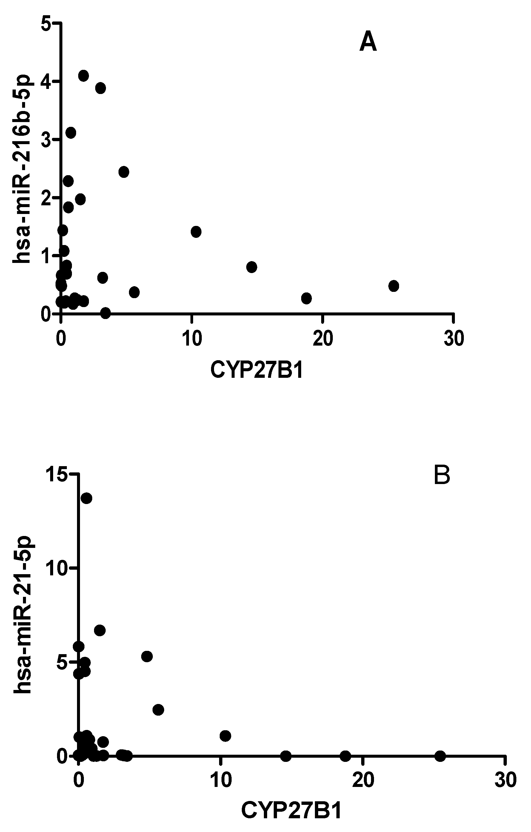
Figure 5. The expression of hsa-miR-216-5p (A) and miR-21-5p (B) levels are not correlated with CYP27B1 expression in T1DM patients. Correlation analysis, reported as normalized 2−ΔCT values. Spearman R test was performed to evaluate r-values and p-values (p < 0.05).
Preprints 84074 g005
Figure 6. Correlation of miRNA expression to islet autoantibodies within the T1DM group. A) IA2A, B) GADA65.
Preprints 84074 g006aPreprints 84074 g006b
Figure 7. Correlation of CYP27B1 expression to islet autoantibodies within the T1DM group. A) IA2, B) GADA65.
Preprints 84074 g007
Disclaimer/Publisher’s Note: The statements, opinions and data contained in all publications are solely those of the individual author(s) and contributor(s) and not of MDPI and/or the editor(s). MDPI and/or the editor(s) disclaim responsibility for any injury to people or property resulting from any ideas, methods, instructions or products referred to in the content.
Copyright: This open access article is published under a Creative Commons CC BY 4.0 license, which permit the free download, distribution, and reuse, provided that the author and preprint are cited in any reuse.
Prerpints.org logo

Preprints.org is a free preprint server supported by MDPI in Basel, Switzerland.

Subscribe

© 2024 MDPI (Basel, Switzerland) unless otherwise stated