Preprint
Article

Valorization of Prickly Pear Peel Residues (Opuntia ficus-indica) Using of Solid-State Fermentation to Accumulation of Tannins

Altmetrics

Downloads

140

Views

90

Comments

0

A peer-reviewed article of this preprint also exists.

Submitted:

13 September 2023

Posted:

14 September 2023

You are already at the latest version

Alerts
Abstract
The Opuntia ficus-indica (OFI) prickly pear peel, is a residue product and was used as a substrate in solid-state fermentation (SSF) to obtain bioactive compounds of interest due to its antimicrobial and antioxidant activity. A Box Hunter & Hunter design to evaluate the independent factors was used. These factors were temperature (°C), inoculum (spores/g), humidity (%), pH, NaNO3 (g/L), MgSO4 (g/L), KCl (g/L) and KH2PO4 (g/L). The response factors were hydrolyzable and condensed tannins amount. In addition, the fermentation extracts the antioxidant and antimicrobial activity were evaluated. The results showed that the humidity (%), inoculum (spores/g) and temperature (°C) affect the release of hydrolyzable and condensed tannins. The treatments 13 and 16 were the best to accumulate condensed (43 mg/g) and hydrolyzable tannins (3.8 mg/g) respectively. Besides, the fermented extracts showed a higher antioxidant activity compared than the unfermented peel extracts, as well as a high inhibition versus E. coli, Alternaria sp. and Botrytis spp. The use of the fermentation process is a good alternative for the recovery of waste and the accumulation of bioactive molecules with potential industrial application.
Keywords: 
Subject: Biology and Life Sciences  -   Food Science and Technology

1. Introduction

Plants of the Cactaceae family are xerophytes, mostly distributed in desert areas [1]. This family has a great socioeconomic importance. Their members are used as ornamental plants, food, and fodder [2]. Prickly pear (OFI) a Cactaceae native to the American continent, is cultivated in Mexico because of its gastronomic use and can be found in 29 of 32 states of Mexico [3], and because of its cultural use, Mexico has the largest cactus cultivation (50 000 - 70 000 ha), however, it is also cultivated in other continents [4].
Prickly pear cladodes are used to prepare various foods. The fruit, which is a berry of thick shell and full of seeds with a mild and sweet flavor, known as tuna, has little acidity and is commonly used to prepare beverages (liquors) and sweets. Tunas are divided according to their color (green, red, yellow, and purple), which depends on the species and maturation [5]. Due to their production and easy propagation in arid zones and its application in areas other than food, studies have been conducted on tuna’s composition (85 % water, 15 % sugars and less than 1 % protein). The chemical composition of OFI varies depending on the species, age of the cladodes, the type of soil where it is grown, and the season of the year [6,8].
OFI’s peel is acidic and contains polysaccharides, sterols, lipids, fat-soluble vitamins, and pigments such as chlorophylls, betalains, coumarins and carotenoids [9]. These compounds are secondary metabolites and have been previously determined using different methodologies, demonstrating the presence of polyphenols and compounds of interest, such as acids and tannins [10,13]. Tannins are phenolic compounds that are distributed in the plant kingdom [14], they are by-products of plant metabolism that are synthesized in response to external stimuli (stress) [15,16]. They participate in the response or defense of plants against the attack of microorganisms such as bacteria and fungi. They also take part in the plant’s survival under drought and are classified as hydrolyzable and condensed [17].
Tannins have biological properties with different industrial applications as antitumor, antimicrobial, antioxidant, and anti-hyperglycemic activities [18]. Different methods have been used to obtain these compounds; solid-liquid extraction. This method prefers the use of water as a solvent for environmental reasons, but even so, NaOH, Na2CO3 and NaHSO3 are used, having drawbacks, such as long extraction times, large amounts of solvents, the use of expensive ionic liquids, making difficult the solute recovery, so solvents such as ethanol, methanol, acetone, N,N-dimethylammonium-N',N'-dimethylcarbamate and 1-butyl-3-methylimidazole bromide (DIMCARB) are also used, nevertheless, these solvents are not environmentally friendly and long times and high temperatures are still needed for the extraction [19]. These examples are the reason to investigate alternative ways to obtain and extract these compounds. An alternative is SSF. Fermentation has been practiced for centuries to produce different foods such as sufu, tapai, koji and kimchi. In the case of SSF, a microorganism is cultivated in a solid organic material, where moist (in the absence or near absence of free water) and a non-soluble material, act as a support and nutrient source for the growth of the microorganism, and it has been considered in the last 20 years as an important and viable food processing for the bioconversion of agro-industrial waste [20,21].
This process promotes the bioavailability of the compounds present in the material used, since the microorganisms used can synthesize enzymes that break the cell wall, propitiating the mobilization of compounds of interest towards the extraction solvent [22]. The fermentation process performs the conversion of complex organic substances into simpler ones, modifying the product physiochemically, improving its quality and the bioavailability of the nutrients present in the substrate [23]. Among the most used microorganisms in SSF are filamentous fungi, such as Aspergillus, Fusarium, Penicillium, Rhizopus and Trichoderma [24,25], although the use of yeasts and some species of actinobacteria is also reported [26]. SSF is an advantageous method for filamentous fungi, since it is very similar to their natural habitat, which can lead to higher enzymatic productivity compared to submerged fermentation [27]. SSF shows as advantages the low production of wastewater, it does not produce foam, the substrates are low cost (product waste), low substrate volumes, low moisture content (thus avoiding contamination), but it also shows disadvantages as heterogeneous media, preventing adequate mixing, moisture levels that are difficult to control, and variables with little precise control (pH, temperature, and dissolved oxygen) [28]. SSF has different applications, such as biodetoxification of agroindustrial waste, obtaining enzymes, unicellular proteins, production of biofuels, biofertilizers and obtaining organic acids such as gallic acid [21,25]. Therefore, the valorization of agroindustrial waste such as prickly pear peel is viable for obtaining compounds of interest for their biological activities through SSF [29,30] even previously SSF has been performed on OFI to improve its protein content and be used as fodder, using yeasts such as Saccharomyces cerevisiae [31] and Kluyveromyces marxianus [32] so it is feasible to ferment OFI peels but also to obtain bioactive compounds such as tannins. Therefore, the objective of the present work is to evaluate the conditions of the SSF process from prickly pear (OFI) peel and a strain of Aspergillus sp., for the accumulation of tannins with antioxidant and antimicrobial activities.

2. Materials and Methods

2.1. Sample Conditioning (Raw Material)

The Prickly pear cactus peel was obtained from a local stand (sale of prickly pear cactus), whose raw material comes from Zacatecas, Mexico. Only prickly pear peel was obtained as residue from this local stand. The peel was washed with a commercial disinfectant solution of sodium hypochlorite for subsequent freezing at -19 °C until use. After storage, the material was reduced in size by cutting and dehydrated following the Ali et al., (2022) [33] method with modifications (oven at 65 °C for 72 h). Once the sample was dehydrated, it was pulverized in a blender to obtain a more homogeneous raw material. After this processing, the material was stored in an airtight plastic container at room temperature and kept in a place protected from light.

2.2. Physicochemical Analysis of the Raw Material

Critical moisture point and water absorption index were determined following the method of Cerda-Cejudo et al., (2022) [29]. For the critical moisture point, a thermobalance OHAUS® model MB23 was used, and total ash was also determined. For the determination of total sugars, the method by Kejla et al., (2023) [34] was used with some modifications, 500 mg of sample were homogenized with 10 mL of distilled water for 12 h, then 250 µL were placed in a test tube and 250 µL of 5% phenol were added for subsequent refrigeration for 10 minutes. Next, 1 mL of concentrated H2SO4 was added. The mixture was shaken gently in a vortex and placed in a boiling water bath for 5 min, for its later cooling. Finally, the solution’s absorbance was read in a UV-Visible spectrophotometer Multiskan FC at 480 nm. To determine total reducing sugars, the DNS method described previously by Prasertsung et al., (2017) [35] was followed and as for the determination of lipids, the method of Gu et al., (2019) [36] was followed. For the determination of crude fiber, an acid digestion and a basic digestion were performed, while for the determination of protein, the Lowry method was used as Deepachandi et al. (2020) [37] described in their method.

2.3. Growth Kinetics for A. niger. sp., Strains

Growth kinetics were performed with a humidity of 60% at 30 °C in Petri dishes OFI prickly pear cactus husk dried and ground as a substrate. The strains used were Aspergillus niger HT3, Aspergillus niger GH1, Aspergillus niger Aa20, Aspergillus niger Aa210, and Aspergillus oryzae sp, which belong to the collection of the Food Research Department from the Autonomous University of Coahuila. To measure the growth of the fungi, the radial growth of the mycelium on the raw material was measured with a Vernier. Once the fungi grew enough to touch one of the ends of the petri dish, the growth was plotted and the µmáx calculation was performed following the methods of Mitchell et al., (2004) and Ruiz et al., (2012) [38,39] where X is the radial growth (cm), µ is the maximum specific growth rate constant (1/h) and t is time (h) to determine which strain grows faster on the raw material.

2.4. Experimental Design Box Hunter & Hunter (Fermentation Process and Tratments)

The SSF process was conducted in Petri dishes together with dried and ground prickly pear peels with Aspergillus niger GH1. The fermentation conditions were defined according to a two-level Box Hunter & Hunter experimental design, that is, a total of 16 treatments, which are shown in Table 1 with the independent factors and the levels used. All treatments were performed in triplicate, establishing as response factors the accumulation of condensed and hydrolyzable tannins which were extracted by a solution of absolute ethanol (20 mL). The fermentation extract was recovered with a Whatman filter using a manual pressing system. The results were analyzed with the statistical package STATISTICA ver. 7.0.

2.5. Condensed and Hydrolyzable Tannins Determination

To determine hydrolyzable and condensed tannins, the method described by Palacios et al., (2021) [40] was followed with modifications, using Folin-Ciocalteu, NaCO3 with the different fermentation extracts and gallic acid (3,4,5-Trihydroxybenzoic acid) (Sigma-Aldrich, CAS: 149-91-7) as standard. The absorbance was measured at 750 nm in a Multiskan FC 1.01.16 spectrophotometer (TermoFisher), then calculations were made to express the content of hydrolyzable tannins in each fermentation extract in mg/g dry weight of prickly pear peel. Condensed tannins were also determined by the HCl-Butanol method with slight modifications, using a UV-Visible spectrophotometer at 450 nm and catechin (Sigma-Aldrich, CAS: 225937-10-0) as standard. The tannin content of each extract was expressed in mg/g dry weight of prickly pear peel.

2.6. Antioxidant Activity

The antioxidant activity performed on the different extracts was conducted following three different methods. For the first determination of antioxidant activity, the method by Sawczuk et al., (2022) [41] with slight modifications was used. A methanolic solution of DPPH• (1,1-diphenyl-1-picrylhydrazyl) radical was prepared at 60 µM, only methanol was used as blank and the prepared DPPH- solution was used as a control. A TROLOX (6-hydroxy-2,5,7, 8-tetramethylchroman-2-carboxylic acid) calibration curve was performed from 0 to 250 ppm, adding 96% DPPH• solution and 4% TROLOX solution in each well of the microplate. Samples and the curve absorbance were read in a Multiskan FC spectrophotometer at 492 nm and the results are expressed in TROLOX equivalents. Likewise, antioxidant activity was quantified using the free radical ABTS (2,2-azino bis-(-3-ethylbenzothiazolin-6-sulfonate) with some modifications. The reading was performed at 750 nm up to an absorbance of 0.7 ± 0.02. The TROLOX reagent was used for the curve, as in the previously described method [42]. Finally, the FRAP (Ferric Reducing Antioxidant Power) assay was performed, as in the method described by Sik et al., (2022) [43] with some modifications, the readings were performed at 595 nm in a microplate reader, using TROLOX as standard in the calibration curve in concentrations from 15 to 1000 ppm, adding a ratio of 3% sample and 97% FRAP reagent in each well of the microplate.

2.7. Antimicrobial and Antifungal Activity

Biological tests were performed with the extract with the highest tannin concentration following the method of Wang et al., (2020) [44] with different microbial strains (E. coli, Salmonella). The crude concentrated extract was evaluated against the strains, using ethanol as a control. The antifungal activity was analyzed using the agar diffusion assay based on the method of Aqueveque et al., (2017) [45] reporting the results in percentage inhibition of mycelial growth against Botrytis sp. and Alternaria sp.

2.8. HPLC-MS Analysis of Extracts Fermentation

Analyzes of fermentation extracts were performed using a Varian HPLC, including autosampler (Varian ProStar 410, USA), ternary pump (Varian ProStar 230I) and PDA detector (Varian ProStar 330), coupled to a liquid chromatography ion trap mass spectrometer (Varian 500-MSIT Mass Spectrometer, USA) equipped with an electrospray ion source, following the method described by Cerda-Cejudo et al., (2022) [29] using 0.2 (%, v/v) formic acid and acetonitrile as mobile phase at different gradients.

3. Results

3.1. Physicochemical Analysis of the Raw Material

The results of the proximate analysis were as follows: 37% of total sugars, 22% of reducing sugars, 6% of total proteins, 2% of total lipids, 22% of total ashes, 14% of total fibers, 3% of moisture and a water absorption index of 4.87 g-gel/g dry weight. The results are similar to those already reported in the literature [8,10,46,48], emphasizing that this is an analysis of the prickly pear peel only. The reports found in the literature are analyzes of the whole fruit or the cactus cladium, so it was compared with these results. According to these results, it was decided to continue with SSF using the raw material as support and medium.

3.2. Growth Kinetics of A. niger. sp. Strains

For the different strains, µmax (cm/h) values were obtained by measuring only the growth on our raw material, obtaining for A. niger Aa20 a value of 0.15, 0.21 for A. niger GH1, 0.22 for A. niger HT3, 0.18 for A. oryzae, and 0.20 for A. niger Aa210. Considering the highest values, the two strains with the highest growth were selected (A. niger GH1 and HT3) for a second kinetic, measuring the release of condensed and hydrolyzable tannins. Figure 1 shows that the accumulation of condensed tannins was better for the GH1 strain, with a maximum value of 39.7 mg/g at 60 h, while for hydrolyzable tannins, the best was strain was HT3 after 12 h with a maximum value of 1.58 mg/g. Due to these results, A. niger GH1 was chosen for the experimental design, since the yields were higher in condensed tannins, as well as one of the highest µmax values. The fermentation time was 54 h, since between 48 and 60 h of the kinetic, the highest values for condensed tannins were obtained. This growth on the raw material used was achieved because this fungus has been used previously in samples with compounds very similar to condensed tannins [49].

3.3. Evaluation of Box Hunter & Hunter Design and Response Factors

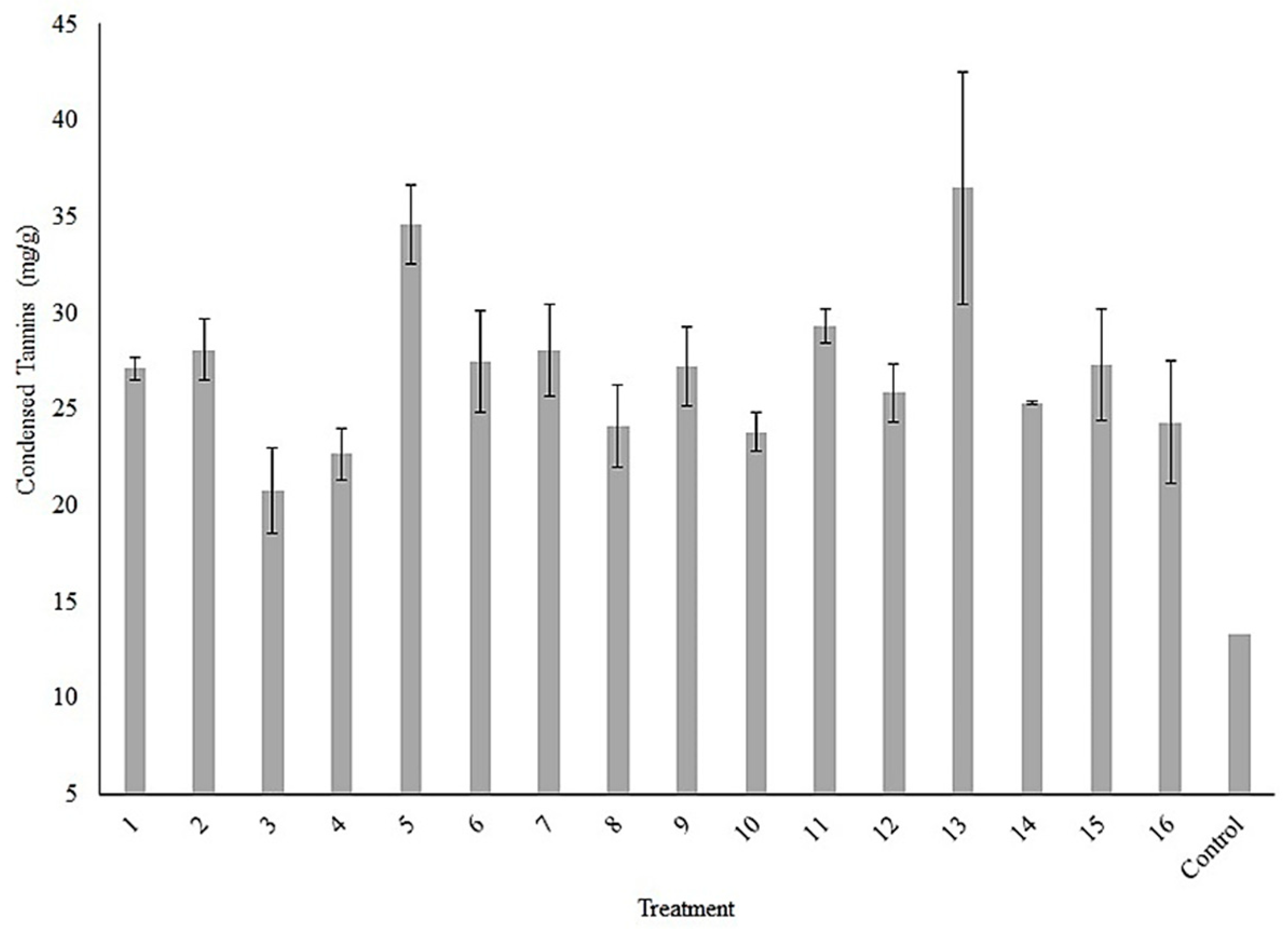
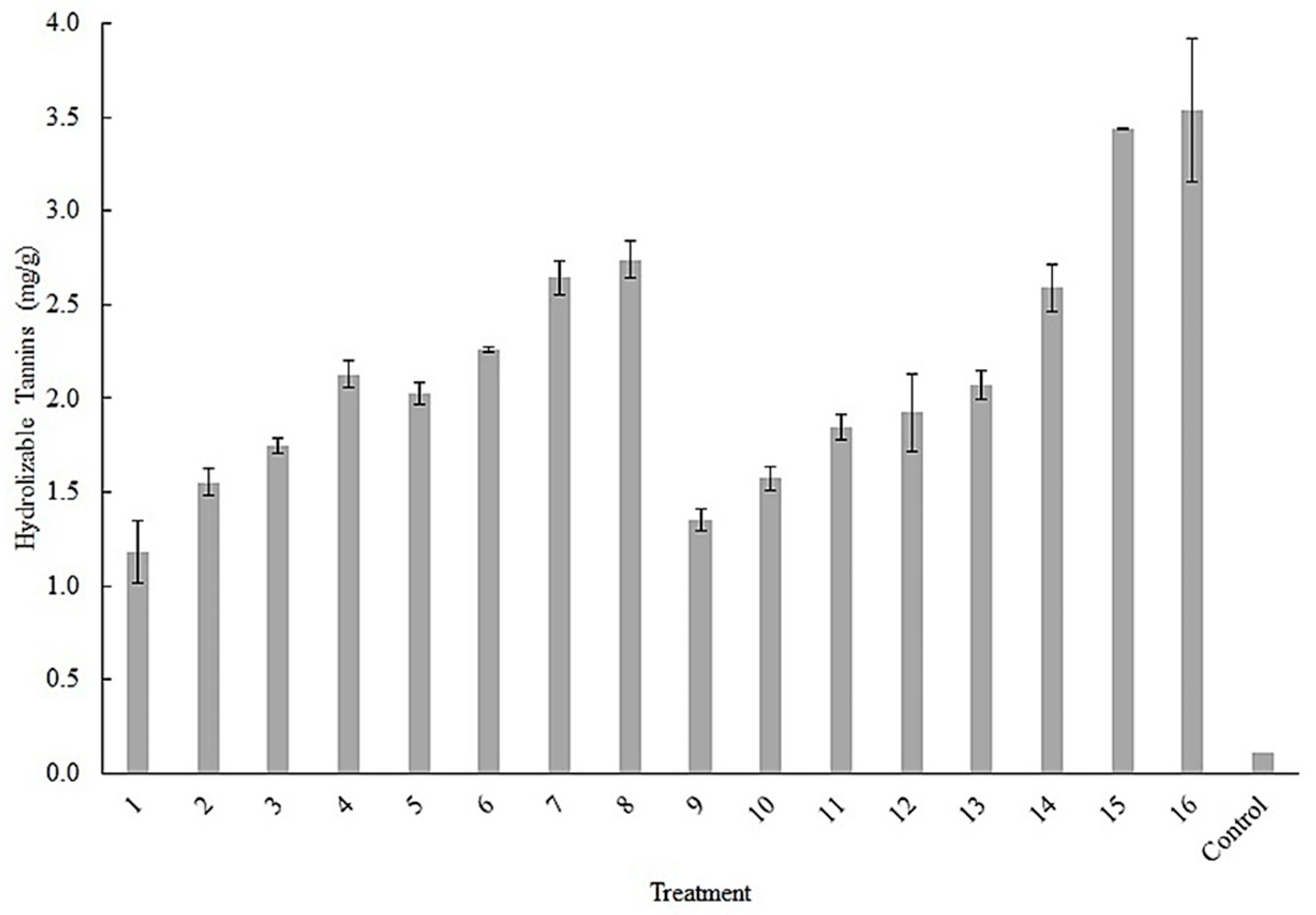
For the experimental design, the condensed and hydrolyzable tannins accumulation were evaluated. The 16 treatments were analyzed separately for each response factor, whose results are shown in Figure 2. The treatment 13 obtained the highest value for condensed tannins (43 mg/g). The results for the accumulation of hydrolyzable tannins are shown in Figure 3, being treatment 16, the one that exhibited the highest value (3.80 mg/g). All data were analyzed in the statistical package STATISTICA ver. 7.0. and Infostat software ver.2020, with a p < 0.5.
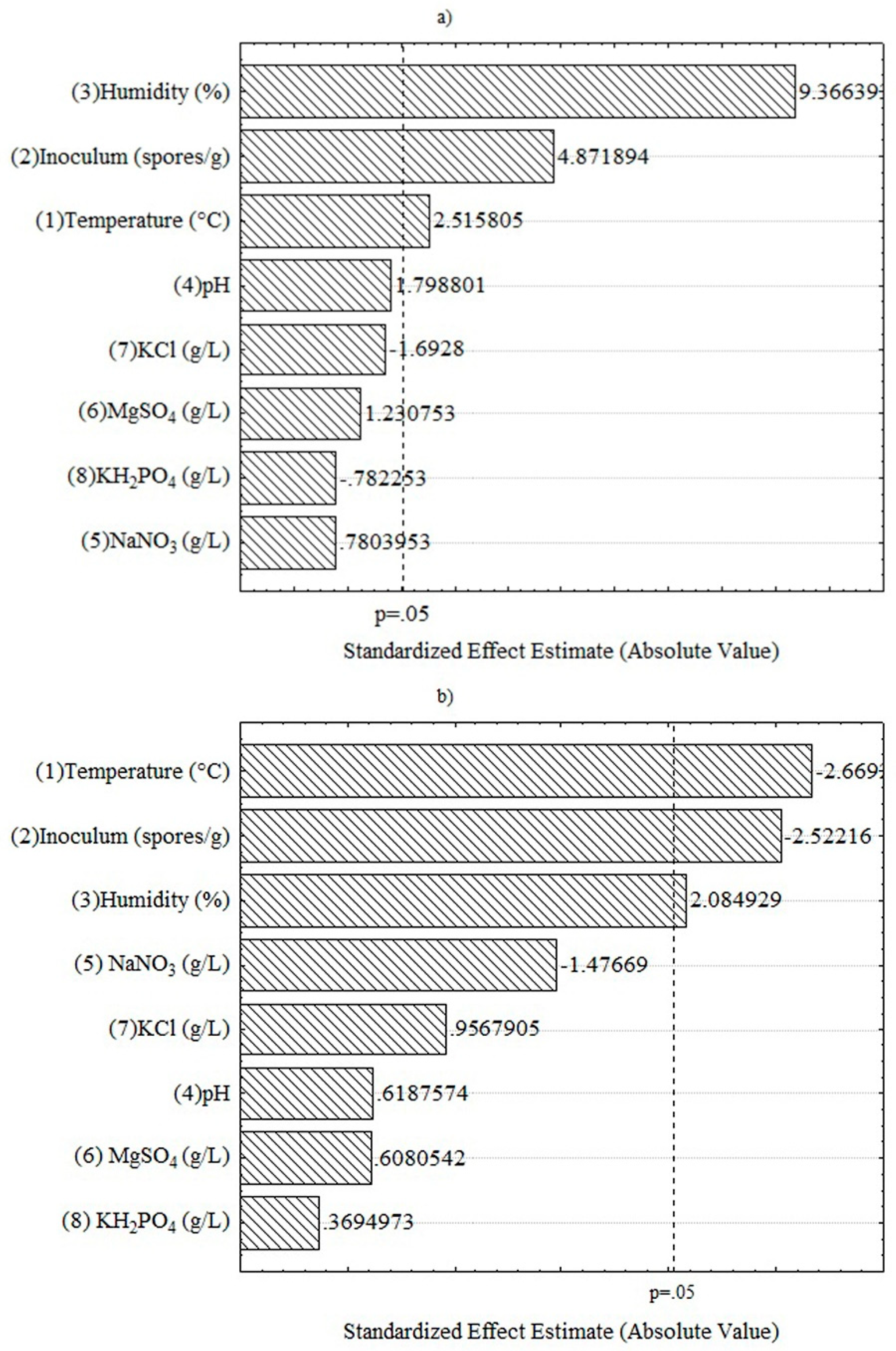
The factors that showed a significant effect on the tannins accumulation in fermentation process for both cases were temperature, humidity, and inoculum as are shown in Figure 4. In the Figure 4a are shown the estimated effect on hydrolyzable tannins, the factors of humidity (%), inoculum (spores/g) and temperature (°C) shown a positive effect in the fermentation process. The rest of factors no affect the hydrolyzable tannins accumulation in fermentation process. In the Figure 4b are shown the estimated effect on condensed tannins accumulation, the factors of temperature (°C) and inoculum (spores/g) and shown a negative effect and the humidity (%) shown a positive effect in the fermentation process. The rest of factors no affect the condensed tannins accumulation in fermentation process. For both response factors, no significant effect was observed in the salts of the Czapek-Dox medium; this may be due to the nutritional value of the raw material, since its use has been reported to increase the nutritional value and to be rich in beneficial compounds [50,51].

3.4. Antioxidant Activity of the Fermentation Extracts

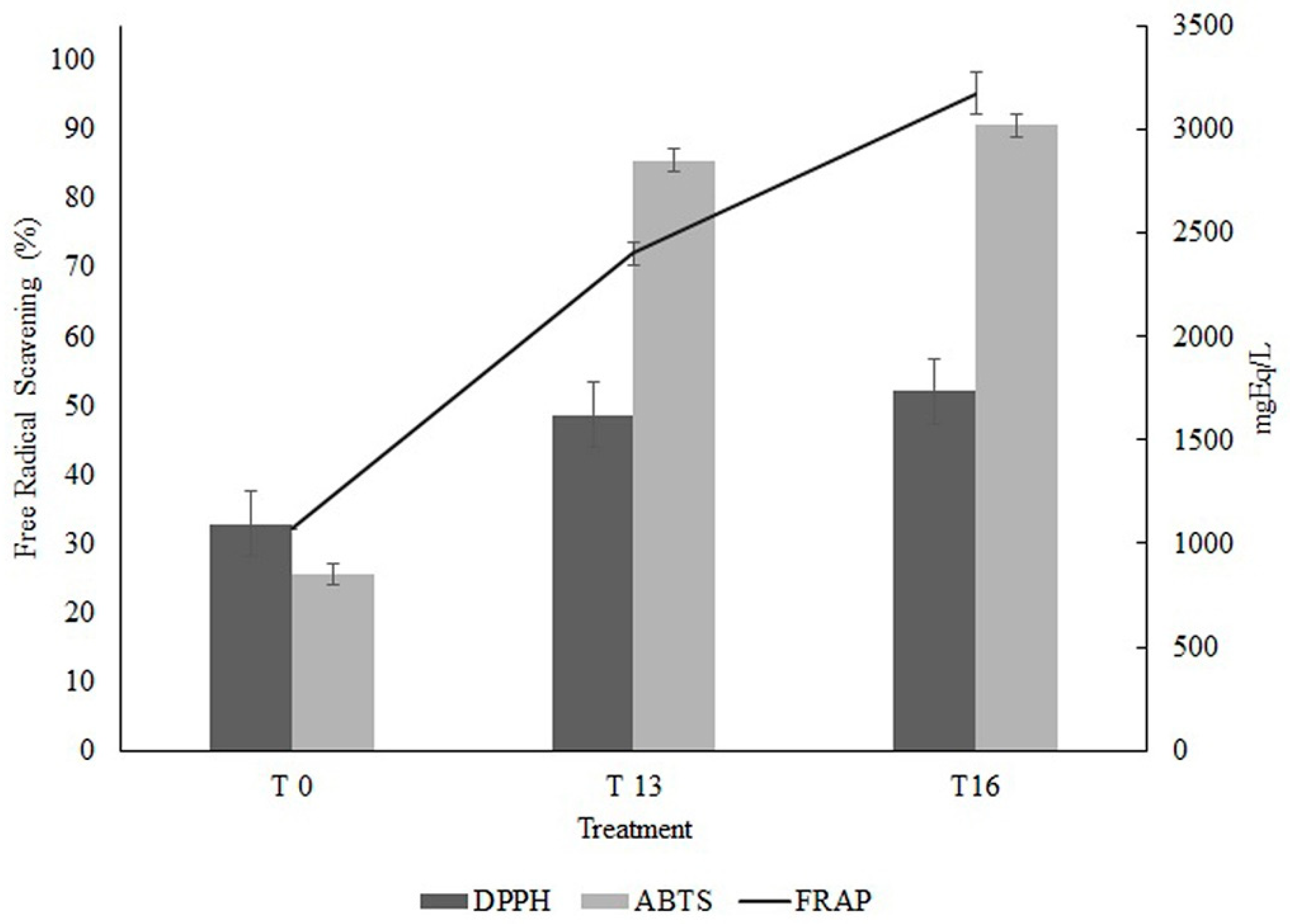
The results of the antioxidant activity performed by three different methods are shown in Figure 5, where we observed a similar behavior, since the sample of unfermented extract showed the lowest values of antioxidant activity, while the activity in samples of fermentation extracts increased considerably. For ABTS free radical scavenging percentage (FRS %), the results were values higher than 80 %, and in the case of the DPPH assay, this value was increasing until reaching values of approximately 50%. The results are similar to what was obtained by Ali et al., (2022) [52], who obtained values higher than 50% using a known concentration of tannins obtained from prickly pear peel by conventional means, while it is emphasized that the values found in this work are given by the crude fermentation extract.
For the FRAP test, the results were expressed in mgEq/L of Fe+2 and, as in the case of the two previous tests, this antioxidant activity increased in all the tests. Treatment 16 showed the highest antioxidant activity. The antioxidant activity of OFI flowers has also been reported previously, which in comparison with the present work, it showed in some cases a similar behavior to those obtained in this work, being the results expressed in µmol Trolox equivalent (µmol TEq)/g dry sample. For the case of the DPPH test, the results were 246.4 ± 7.0 and 251.0 ± 9. 3 µmolTE/gDW for treatment 13 and 16 respectively. The results reported by Brahmi et al., (2022) [12] who performed ultrasound-assisted extraction obtained 4078 µmolTE/gDW for DPPH, and 80.8 ± 4.1 µmolTE/gDW in ABTS, while for the ABTS assay of the present work 345.8 ± 2.5 and 373. 6 ± 4.0 µmolTE/gDW for treatment 13 and 16. The results may be due to the differences in the extracts and molecules obtained in both works, since in the present work the extracts were fermented, while in the compared work, compounds were extracted following another method.

3.5. Antimicrobial and Antifungal Activity of the Fermentation Extracts

For the pathogenic activity against E. coli, the inhibition halo formed on filter paper discs soaked with 10 µL of fermentation extract was observed, being analyzed against a blank (without extract) and a control (absolute ethanol), to rule out that the inhibition was given by agents other than the fermentation extracts. In the tests of the blank and the control, no inhibition halo was observed, while for the fermentation extract belonging to treatment 13, a halo of 9.8 mm was observed. Treatment 16 formed an inhibition halo of 13.2 mm, furthermore, the treatment with the highest inhibition was treatment 13, this may be due to the compounds present in it. Aruwa et al., 2019 [53] reports an inhibition halo using OFI extracts against E. coli of 12.2 mm using known concentrations, while in the present work, the crude extract from fermentation was used, they also report that their extracts, as in the present work, are in ethanolic medium, showing the highest inhibition halo. This behavior can be attributed to the compounds present, since they also report compounds such as caffeic acid 4-O-glucoside, Isorhamnetin, Isorhamnetin 3-O-glucoside, p-Coumaric acid 4-O-glucoside, which were also identified in the present work in the different fermentation treatments. Also, a significant amount of work on catechin obtained from different sources, mostly residues [54], have shown pathogenic activity against E. coli.
The inhibition test against phytopathogenic fungi was also carried out successfully, performing the simple diffusion test, an inhibition halo of 30 % was obtained for Alternaria sp. for treatment 13, while for treatment 16 a 49 % inhibition in the growth of this fungus was obtained, these results, compared with the control without extract, that did not present any inhibition halo, likewise it was compared with the control with ethanol to discard that the inhibition is given by the solvent of our extracts, which also did not present an inhibition halo. Also, the test against Botrytis sp. gave favorable results for the extracts, since in the case of treatment 13, an inhibition halo of 22 % was determined, and for treatment 16, an inhibition halo of 32 %, all of this compared against the control without extract and the ethanol control, which did not present an inhibition halo.
OFI extracts have already been evaluated for their effect against fungi. Alqurashi et al., (2022) [55] reported that OFI seed extracts show activity against the growth of Saccharomyces cerevisiae sp. but no effect on Aspergillus niger sp. growth, this corresponds to that reported also by Brahmi et al., (2020) [56] who tested oily extracts of OFI against Aspergillus niger 939 N, Aspergillus flavus NRRL 3251, Mucor sp NRRL 1829, Aspergillus ochraceus NRRL 3174, Aspergillus parasiticus CB 5 and Candida albicans ATCC 10, with unsatisfactory results. Brahmi mentions in that work that the antifungal activity is affected by the presence of polyphenols such as tannins and flavonoids, which in our extracts are present, being the reason why our extracts inhibited the growth of phytopathogenic fungi. Amel et al., (2013) [57] also reports activity against Candida albicans sp. using OFI fruit extracts, again attributing this activity to the polyphenolic compounds present in the extracts.

3.6. HPLC-MS Analysis

The results of the HPLC-RF-MS test are shown in Table 2, where it is possible to appreciate the differences in the compounds found in the unfermented sample and the different treatments, likewise, an increase in the number of compounds found in the treatments can be observed in comparison with the sample without the fermenter. The compounds found in the unfermented sample have been reported previously in works with OFI, such is the case for rhamnetin and isorhamnetin, which have been reported in the works of El-Hawary et al., (2020) and Mena et al., (2018) [58,59], as well as molecules that in their structure contain these compounds, i.e., condensed tannins. Also, these authors have reported molecules such as quercetin, but they are not the only ones, in works such as Aruwa et al., (2019); Benayad et al., (2014); di Bella et al (2022) [4,53,60,61], extracted compounds from different methods such as solvent extraction and temperature from different parts of OFI such as cladodes, prickly pear and flowers so our unfermented sample also presents these compounds. As for the fermented sample, more compounds are found, of which have already been reported as catechin in OFI [62,63]. Molecules have also been reported that can give as a by-product the molecules reported here. On the other hand, molecules such as 3,4-DHPEA-AC, pinocembrin, pterostilbene, scopoletin, sinensetin, which have been reported in other plants but not in OFI previously, were found, these molecules report varied uses, such as antioxidant, anticancer, antiviral, and antimicrobial activity, since being phenolic compounds they present these properties. So, the results show that this type of fermentation produces molecules that can be derived from other molecules of larger size and molecular weight, and these derived molecules present biological activities, being so that fermentation extracts present these same properties [64,68].

4. Conclusion

The humidity (%), inoculum (spores/g) and temperature (°C) affect the release of hydrolyzable and condensed tannins. The treatments 13 and 16 were the best to accumulate condensed (43 mg/g) and hydrolyzable tannins (3.8 mg/g) respectively. Besides, the fermented extracts showed a higher antioxidant activity compared than the unfermented peel extracts, as well as a high inhibition versus E. coli, Alternaria sp. and Botrytis spp. In the fermentation extracts, up to 14 compounds of phenolic origin were identified. The use of solid-state fermentation processes facilitates the accumulation of bioactive molecules such as tannins that can be applied in different industrial sectors.

Declaration of Competing Interest

The authors declare that they have no known competing financial interests or personal relationships that could have appeared to influence the work reported in this paper.

Acknowledgments

The authors extend our sincere thanks to CONAHCYT for scholarship support (1150316 CVU) in the research project. Thanks, the Autonomous University of Coahuila for the facilities in which the different tests reported in this article were conducted.

References

  1. Amaral, D.T.; Bonatelli, I.A.S.; Romeiro-Brito, M.; Moraes, E.M.; Franco, F.F. Spatial Patterns of Evolutionary Diversity in Cactaceae Show Low Ecological Representation within Protected Areas. Biol Conserv 2022, 273, 109677. [Google Scholar] [CrossRef]
  2. de Araújo, F.F.; de Paulo Farias, D.; Neri-Numa, I.A.; Pastore, G.M. Underutilized Plants of the Cactaceae Family: Nutritional Aspects and Technological Applications. Food Chem 2021, 362, 130196. [Google Scholar] [CrossRef] [PubMed]
  3. Pérez-Cadena, R.; Espinosa-Solares, T.; Medina-Moreno, S.A.; Martínez, A.; Lizardi-Jiménez, M.A.; Téllez-Jurado, A. Effect of the Age of Opuntia ficus-indica Cladodes on the Release of Simple Sugars. Biocatal Agric Biotechnol 2021, 33, 102010. [Google Scholar] [CrossRef]
  4. di Bella, G.; Vecchio, G. lo; Albergamo, A.; Nava, V.; Bartolomeo, G.; Macrì, A.; Bacchetta, L.; lo Turco, V.; Potortì, A.G. Chemical Characterization of Sicilian Dried Nopal [Opuntia ficus-indica (L.) Mill.]. J Food Composit Anal 2022, 106, 104307. [Google Scholar] [CrossRef]
  5. Feugang, J.M.; Konarski, P.; Zou, D.; Stintzing, F.C.; Zou, C. Nutritional and Medicinal Use of Cactus Pear (Opuntia Spp.) Cladodes and Fruits. Front Biosci 2006, 2574–2589. [Google Scholar] [CrossRef]
  6. Vargas-Rodríguez, L.; Arroyo-Figueroa, G.; Herrera-Méndez, C.; Pérez-Nieto, A.; García-Vieyra, M.I.; Rodríguez-Núñez, J.R. Physical Properties of Mucilage Prickly Pear. Acta Univ 2016, 26. [Google Scholar] [CrossRef]
  7. Agostini-Costa, T. da S. Genetic and Environment Effects on Bioactive Compounds of Opuntia Cacti – A Review. J Food Composit Anal 2022, 109, 104514. [Google Scholar] [CrossRef]
  8. Yang, L.; Lu, M.; Carl, S.; Mayer, J.A.; Cushman, J.C.; Tian, E.; Lin, H. Biomass Characterization of Agave and Opuntia as Potential Biofuel Feedstocks. Biomass Bioenergy 2015, 76, 43–53. [Google Scholar] [CrossRef]
  9. Kuti, J.O. Antioxidant Compounds from Four Opuntia Cactus Pear Fruit Varieties. Food Chem 2004, 85, 527–533. [Google Scholar] [CrossRef]
  10. Ginestra, G.; Parker, M.L.; Bennett, R.N.; Robertson, J.; Mandalari, G.; Narbad, A.; lo Curto, R.B.; Bisignano, G.; Faulds, C.B.; Waldron, K.W. Anatomical, Chemical, and Biochemical Characterization of Cladodes from Prickly Pear [Opuntia ficus-indica (L.) Mill.]. J Agric Food Chem 2009, 57, 10323–10330. [Google Scholar] [CrossRef]
  11. Blando, F.; Russo, R.; Negro, C.; de Bellis, L.; Frassinetti, S. Antimicrobial and Antibiofilm Activity against Staphylococcus aureus of Opuntia ficus-indica (L.) Mill. Cladode Polyphenolic Extracts. Antioxidants 2019, 8, 117. [Google Scholar] [CrossRef] [PubMed]
  12. Brahmi, F.; Blando, F.; Sellami, R.; Mehdi, S.; de Bellis, L.; Negro, C.; Haddadi-Guemghar, H.; Madani, K.; Makhlouf-Boulekbache, L. Optimization of the Conditions for Ultrasound-Assisted Extraction of Phenolic Compounds from Opuntia ficus-indica [L.] Mill. Flowers and Comparison with Conventional Procedures. Ind Crops Prod 2022, 184, 114977. [Google Scholar] [CrossRef]
  13. Bakar, B.; Çakmak, M.; Ibrahim, M.S.; Özer, D.; Saydam, S.; Karatas, F. Investigation of Amounts of Vitamins, Lycopene, and Elements in the Fruits of Opuntia ficus-indica Subjected to Different Pretreatments. Biol Trace Elem Res 2020, 198, 315–323. [Google Scholar] [CrossRef] [PubMed]
  14. Ouerghemmi, I.; Harbeoui, H.; Aidi-Wannes, W.; Bettaieb-Rebey, I.; Hammami, M.; Marzouk, B.; Saidani-Tounsi, M. Phytochemical Composition and Antioxidant Activity of Tunisian Cactus Pear (Opuntia ficus indica L.) Flower. J Food Biochem 2017, 41, e12390. [Google Scholar] [CrossRef]
  15. Messina, C.M.; Renda, G.; La Barbera, L.; Santulli, A. By-Products of Farmed European Sea Bass (Dicentrarchus labrax L.) as a Potential Source of n-3 PUFA. Biologia (Bratisl) 2013, 68, 288–293. [Google Scholar] [CrossRef]
  16. Messina, C.M.; Arena, R.; Morghese, M.; Santulli, A.; Liguori, G.; Inglese, P. Seasonal Characterization of Nutritional and Antioxidant Properties of Opuntia ficus-indica [(L.) Mill.] Mucilage. Food Hydrocoll 2021, 111, 106398. [Google Scholar] [CrossRef]
  17. Pizzi, A. Tannins Medical / Pharmacological and Related Applications: A Critical Review. Sustain Chem Pharm 2021, 22, 100481. [Google Scholar] [CrossRef]
  18. Rajasekaran, S.; Rajasekar, N.; Sivanantham, A. Therapeutic Potential of Plant-Derived Tannins in Non-Malignant Respiratory Diseases. J Nutr Biochem 2021, 94, 108632. [Google Scholar] [CrossRef]
  19. de Hoyos-Martínez, P.L.; Merle, J.; Labidi, J.; Charrier – El Bouhtoury, F. Tannins Extraction: A Key Point for Their Valorization and Cleaner Production. J Clean Prod 2019, 206, 1138–1155. [Google Scholar] [CrossRef]
  20. Manan, M.A.; Webb, C. Modern Microbial Solid State Fermentation Technology for Future Biorefineries for the Production of Added-Value Products. Biofuel Res J 2017, 4, 730–740. [Google Scholar] [CrossRef]
  21. Mansor, A.; Ramli, M.S.; Abdul Rashid, N.Y.; Samat, N.; Lani, M.N.; Sharifudin, S.A.; Raseetha, S. Evaluation of Selected Agri-Industrial Residues as Potential Substrates for Enhanced Tannase Production via Solid-State Fermentation. Biocatal Agric Biotechnol 2019, 20, 101216. [Google Scholar] [CrossRef]
  22. Torres-León, C.; Ramírez-Guzmán, N.; Ascacio-Valdés, J.; Serna-Cock, L.; dos Santos Correia, M.T.; Contreras-Esquivel, J.C.; Aguilar, C.N. Solid-State Fermentation with Aspergillus niger to Enhance the Phenolic Contents and Antioxidative Activity of Mexican Mango Seed: A Promising Source of Natural Antioxidants. LWT 2019, 112, 108236. [Google Scholar] [CrossRef]
  23. Olukomaiya, O.O.; Adiamo, O.Q.; Fernando, W.C.; Mereddy, R.; Li, X.; Sultanbawa, Y. Effect of Solid-State Fermentation on Proximate Composition, Anti-Nutritional Factor, Microbiological and Functional Properties of Lupin Flour. Food Chem 2020, 315, 126238. [Google Scholar] [CrossRef] [PubMed]
  24. Hu, C.C.; Liu, L.Y.; Yang, S.S. Protein Enrichment, Cellulase Production and in Vitro Digestion Improvement of Pangolagrass with Solid State Fermentation. J Microbiol Immunol Infect 2012, 45, 7–14. [Google Scholar] [CrossRef]
  25. Yafetto, L. Application of Solid-State Fermentation by Microbial Biotechnology for Bioprocessing of Agro-Industrial Wastes from 1970 to 2020: A Review and Bibliometric Analysis. Heliyon 2022, 8, e09173. [Google Scholar] [CrossRef]
  26. Munishamanna, K.B.; Suresha, K.B.; Veena, R.; Subramanya, S. Solid State Fermentation of Mango Peel and Mango Seed Waste by Different Yeasts and Bacteria for Nutritional Improvement. Int J Food Ferment Technol 2017, 7, 111. [Google Scholar] [CrossRef]
  27. Farinas, C.S. Advances in Cultivation Strategies of Aspergillus for Production of Enzymes Involved in the Saccharification of Lignocellulosic Feedstocks. In New and Future Developments in Microbial Biotechnology and Bioengineering: Aspergillus System Properties and Applications; Eds.; Elsevier: Chennai, India, 2016; pp. 141–154. ISBN 978-0-444-63505-1. [Google Scholar]
  28. Cano y Postigo, L.O.; Jacobo-Velázquez, D.A.; Guajardo-Flores, D.; Garcia Amezquita, L.E.; García-Cayuela, T. Solid-State Fermentation for Enhancing the Nutraceutical Content of Agrifood by-Products: Recent Advances and Its Industrial Feasibility. Food Biosci 2021, 41, 100926. [Google Scholar] [CrossRef]
  29. Cerda-Cejudo, N.D.; Buenrostro-Figueroa, J.J.; Sepúlveda, L.; Torres-Leon, C.; Chávez-González, M.L.; Ascacio-Valdés, J.A.; Aguilar, C.N. Recovery of Ellagic Acid from Mexican Rambutan Peel by Solid-State Fermentation-Assisted Extraction. Food Bioprod Process 2022, 134, 86–94. [Google Scholar] [CrossRef]
  30. Sepúlveda, L.; Wong-Paz, J.E.; Buenrostro-Figueroa, J.; Ascacio-Valdés, J.A.; Aguilera-Carbó, A.; Aguilar, C.N. Solid State Fermentation of Pomegranate Husk: Recovery of Ellagic Acid by SEC and Identification of Ellagitannins by HPLC/ESI/MS. Food Biosci 2018, 22, 99–104. [Google Scholar] [CrossRef]
  31. Araújo, L.F.; Medeiros, A.N.; Neto, A.P.; Oliveira, L.S.C.; da Silva, F.L.H. Protein Enrichment of Cactus Pear (Opuntia ficus - indica Mill) Using Saccharomyces cerevisiae in Solid-State Fermentation. Braz Arch Biol Technol 2005, 48. [Google Scholar] [CrossRef]
  32. Herrera, E.; Murillo, M.; Berumen, L.; Soto-Cruz, N.; Paez-Lerma, J. Protein enrichment of Opuntia ficus-indica using Kluyveromyces marxianus in solid-state fermentation. Cienc Inv Agr 2017, 44. [Google Scholar] [CrossRef]
  33. Ali, S.K.; Mahmoud, S.M.; El-Masry, S.S.; Alkhalifah, D.H.M.; Hozzein, W.N.; Aboel-Ainin, M.A. Phytochemical Screening and Characterization of the Antioxidant, Anti-Proliferative and Antibacterial Effects of Different Extracts of Opuntia ficus-indica Peel. J King Saud Univ Sci 2022, 34, 102216. [Google Scholar] [CrossRef]
  34. Kejla, L.; Schulzke, T.; Šimáček, P.; Auersvald, M. Anthrone Method Combined with Adsorption of Interferents as a New Approach towards Reliable Quantification of Total Carbohydrate Content in Pyrolysis Bio-Oils. J Anal Appl Pyrolysis 2023, 173, 106066. [Google Scholar] [CrossRef]
  35. Prasertsung, I.; Chutinate, P.; Watthanaphanit, A.; Saito, N.; Damrongsakkul, S. Conversion of Cellulose into Reducing Sugar by Solution Plasma Process (SPP). Carbohydr Polym 2017, 172, 230–236. [Google Scholar] [CrossRef]
  36. Gu, L.B.; Zhang, G.J.; Du, L.; Du, J.; Qi, K.; Zhu, X.L.; Zhang, X.Y.; Jiang, Z.H. Comparative Study on the Extraction of Xanthoceras sorbifolia Bunge (Yellow Horn) Seed Oil Using Subcritical n-Butane, Supercritical CO2, and the Soxhlet Method. LWT 2019, 111, 548–554. [Google Scholar] [CrossRef]
  37. Deepachandi, B.; Weerasinghe, S.; Andrahennadi, T.P.; Karunaweera, N.D.; Wickramarachchi, N.; Soysa, P.; Siriwardana, Y. Quantification of Soluble or Insoluble Fractions of Leishmania Parasite Proteins in Microvolume Applications: A Simplification to Standard Lowry Assay. Int J Anal Chem 2020, 1–8. [Google Scholar] [CrossRef]
  38. Ruiz, H.A.; Rodríguez-Jasso, R.M.; Rodríguez, R.; Contreras-Esquivel, J.C.; Aguilar, C.N. Pectinase Production from Lemon Peel Pomace as Support and Carbon Source in Solid-State Fermentation Column-Tray Bioreactor. Biochem Eng J 2012, 65, 90–95. [Google Scholar] [CrossRef]
  39. Mitchell, D.A.; von Meien, O.F.; Krieger, N.; Dalsenter, F.D.H. A Review of Recent Developments in Modeling of Microbial Growth Kinetics and Intraparticle Phenomena in Solid-State Fermentation. Biochem Eng J 2004, 17, 15–26. [Google Scholar] [CrossRef]
  40. Palacios, C.E.; Nagai, A.; Torres, P.; Rodrigues, J.A.; Salatino, A. Contents of Tannins of Cultivars of Sorghum Cultivated in Brazil, as Determined by Four Quantification Methods. Food Chem 2021, 337, 127970. [Google Scholar] [CrossRef]
  41. Sawczuk, R.; Karpinska, J.; Filipowska, D.; Bajguz, A.; Hryniewicka, M. Evaluation of Total Phenols Content, Anti-DPPH Activity and the Content of Selected Antioxidants in the Honeybee Drone Brood Homogenate. Food Chem 2022, 368, 130745. [Google Scholar] [CrossRef]
  42. Sepúlveda, L.; Romaní, A.; Aguilar, C.N.; Teixeira, J. Valorization of Pineapple Waste for the Extraction of Bioactive Compounds and Glycosides Using Autohydrolysis. Innov Food Sci Emerg Technol 2018, 47, 38–45. [Google Scholar] [CrossRef]
  43. Sik, B.; Ajtony, Z.; Lakatos, E.; Székelyhidi, R. The Effects of Extraction Conditions on the Antioxidant Activities, Total Polyphenol and Monomer Anthocyanin Contents of Six Edible Fruits Growing Wild in Hungary. Heliyon 2022, 8, e12048. [Google Scholar] [CrossRef] [PubMed]
  44. Wang, Z.; Zhu, J.; Li, W.; Li, R.; Wang, X.; Qiao, H.; Sun, Q.; Zhang, H. Antibacterial Mechanism of the Polysaccharide Produced by Chaetomium globosum CGMCC 6882 against Staphylococcus aureus. Int J Biol Macromol 2020, 159, 231–235. [Google Scholar] [CrossRef]
  45. Aqueveque, P.; Céspedes, C.L.; Becerra, J.; Aranda, M.; Sterner, O. Antifungal Activities of Secondary Metabolites Isolated from Liquid Fermentations of Stereum hirsutum (Sh134-11) against Botrytis cinerea (Grey Mould Agent). Food Chem Toxicol 2017, 109, 1048–1054. [Google Scholar] [CrossRef] [PubMed]
  46. dos Santos, T.C.; Reis, N.D.S.; Silva, T.P.; Machado, F.P.P.; Bonomo, R.C.F.; Franco, M. Prickly Palm Cactus Husk as a Raw Material for Production of Ligninolytic Enzymes by Aspergillus niger. Food Sci Biotechnol 2016, 25, 205–211. [Google Scholar] [CrossRef] [PubMed]
  47. Moßhammer, M.R.; Stintzing, F.C.; Carle, R. Cactus Pear Fruits (Opuntia Spp.): A Review of Processing Technologies and Current Uses. J Prof Assoc Cactus 2006, 8, 1–25. [Google Scholar] [CrossRef]
  48. Kuloyo, O.O.; du Preez, J.C.; García-Aparicio, M.P.; Kilian, S.G.; Steyn, L.; Görgens, J. Opuntia ficus-indica Cladodes as Feedstock for Ethanol Production by Kluyveromyces marxianus and Saccharomyces cerevisiae. World J Microbiol Biotechnol 2014, 30, 3173–3183. [Google Scholar] [CrossRef]
  49. Torres-León, C.; Ramírez-Guzmán, N.; Ascacio-Valdés, J.; Serna-Cock, L.; dos Santos Correia, M.T.; Contreras-Esquivel, J.C.; Aguilar, C.N. Solid-State Fermentation with Aspergillus niger to Enhance the Phenolic Contents and Antioxidative Activity of Mexican Mango Seed: A Promising Source of Natural Antioxidants. LWT 2019, 112, 108236. [Google Scholar] [CrossRef]
  50. Barba, F.J.; Putnik, P.; Bursać Kovačević, D.; Poojary, M.M.; Roohinejad, S.; Lorenzo, J.M.; Koubaa, M. Impact of Conventional and Non-Conventional Processing on Prickly Pear (Opuntia Spp.) and Their Derived Products: From Preservation of Beverages to Valorization of by-Products. Trends Food Sci Technol 2017, 67, 260–270. [Google Scholar] [CrossRef]
  51. Bouazizi, S.; Montevecchi, G.; Antonelli, A.; Hamdi, M. Effects of Prickly Pear (Opuntia ficus-indica L.) Peel Flour as an Innovative Ingredient in Biscuits Formulation. LWT 2020, 124, 109155. [Google Scholar] [CrossRef]
  52. Ali, S.K.; Mahmoud, S.M.; El-Masry, S.S.; Alkhalifah, D.H.M.; Hozzein, W.N.; Aboel-Ainin, M.A. Phytochemical Screening and Characterization of the Antioxidant, Anti-Proliferative and Antibacterial Effects of Different Extracts of Opuntia ficus-indica Peel. J King Saud Univ Sci 2022, 34, 102216. [Google Scholar] [CrossRef]
  53. Aruwa, C.E.; Amoo, S.O.; Kudanga, T. Extractable and Macromolecular Antioxidants of Opuntia ficus-indica Cladodes: Phytochemical Profiling, Antioxidant and Antibacterial Activities. S African J Bot 2019, 125, 402–410. [Google Scholar] [CrossRef]
  54. Ma, Y.; Ding, S.; Fei, Y.; Liu, G.; Jang, H.; Fang, J. Antimicrobial Activity of Anthocyanins and Catechins against Foodborne Pathogens Escherichia coli and Salmonella. Food Control 2019, 106, 106712. [Google Scholar] [CrossRef]
  55. Alqurashi, A.S.; Al Masoudi, L.M.; Hamdi, H.; Abu Zaid, A. Chemical Composition and Antioxidant, Antiviral, Antifungal, Antibacterial and Anticancer Potentials of Opuntia ficus-indica Seed Oil. Molecules 2022, 27, 5453. [Google Scholar] [CrossRef]
  56. Brahmi, F.; Haddad, S.; Bouamara, K.; Yalaoui-Guellal, D.; Prost-Camus, E.; de Barros, J.P.P.; Prost, M.; Atanasov, A.G.; Madani, K.; Boulekbache-Makhlouf, L.; Lizard, G. Comparison of Chemical Composition and Biological Activities of Algerian Seed Oils of Pistacia lentiscus L., Opuntia ficus indica (L.) Mill. and Argania spinosa L. Skeels. Ind Crops Prod 2020, 151, 112456. [Google Scholar] [CrossRef]
  57. Amel, R.; Hanen, F.; Ferid, L.; Riadh, K.; Chedly, A.; Aly, R. Upshot of the Ripening Time on Biological Activities, Phenol Content and Fatty Acid Composition of Tunisian Opuntia ficus-indica Fruit. Afr J Biotechnol 2013, 12, 5875–5885. [Google Scholar] [CrossRef]
  58. El-Hawary, S.S.; Sobeh, M.; Badr, W.K.; Abdelfattah, M.A.O.; Ali, Z.Y.; El-Tantawy, M.E.; Rabeh, M.A.; Wink, M. HPLC-PDA-MS/MS Profiling of Secondary Metabolites from Opuntia ficus-indica Cladode, Peel and Fruit Pulp Extracts and Their Antioxidant, Neuroprotective Effect in Rats with Aluminum Chloride Induced Neurotoxicity. Saudi J Biol Sci 2020, 27, 2829–2838. [Google Scholar] [CrossRef]
  59. Mena, P.; Tassotti, M.; Andreu, L.; Nuncio-Jáuregui, N.; Legua, P.; del Rio, D.; Hernández, F. Phytochemical Characterization of Different Prickly Pear (Opuntia ficus-indica (L.) Mill.) Cultivars and Botanical Parts: UHPLC-ESI-MSn Metabolomics Profiles and Their Chemometric Analysis. Food Res Int 2018, 108, 301–308. [Google Scholar] [CrossRef]
  60. Benayad, Z.; Martínez-Villaluenga, C.; Frias, J.; Gómez-Cordoves, C.; Es-Safi, N.E. Phenolic Composition, Antioxidant and Anti-Inflammatory Activities of Extracts from Moroccan Opuntia ficus-indica Flowers Obtained by Different Extraction Methods. Ind Crops Prod 2014, 62, 412–420. [Google Scholar] [CrossRef]
  61. Aruwa, C.E.; Amoo, S.; Kudanga, T. Phenolic Compound Profile and Biological Activities of Southern African Opuntia ficus-indica Fruit Pulp and Peels. LWT 2019, 111, 337–344. [Google Scholar] [CrossRef]
  62. Guevara-Figueroa, T.; Jiménez-Islas, H.; Reyes-Escogido, M.L.; Mortensen, A.G.; Laursen, B.B.; Lin, L. W.; de León-Rodríguez, A.; Fomsgaard, I.S.; Barba de la Rosa, A.P. Proximate Composition, Phenolic Acids, and Flavonoids Characterization of Commercial and Wild Nopal (Opuntia Spp.). J Food Compost Anal 2010, 23, 525–532. [Google Scholar] [CrossRef]
  63. de Albuquerque, J.G.; Escalona-Buendía, H.B.; de Magalhães, C.A.M.T.; dos Santos, L.M.; de Souza, A.J.; da Silva, V.M.A. Ultrasound Treatment for Improving the Bioactive Compounds and Quality Properties of a Brazilian Nopal (Opuntia ficus-indica) Beverage during Shelf-Life. LWT 2021, 149, 111814. [Google Scholar] [CrossRef]
  64. Antika, L.D.; Tasfiyati, A.N.; Hikmat, H.; Septama, A.W. Scopoletin: A Review of Its Source, Biosynthesis, Methods of Extraction, and Pharmacological Activities. Z Naturforsch C J Biosci 2022, 77, 303–316. [Google Scholar] [CrossRef]
  65. Elbatreek, M.H.; Mahdi, I.; Ouchari, W.; Mahmoud, M.F.; Sobeh, M. Current Advances on the Therapeutic Potential of Pinocembrin: An Updated Review. Biomed Pharmacother 2023, 157, 114032. [Google Scholar] [CrossRef]
  66. Amzad Hossain, M.; Ismail, Z. Quantification and Enrichment of Sinensetin in the Leaves of Orthosiphon stamineus. Arab J Chem 2016, 9, S1338–S1341. [Google Scholar] [CrossRef]
  67. Xu, D.; Qiao, F.; Xi, P.; Lin, Z.; Jiang, Z.; Romanazzi, G.; Gao, L. Efficacy of Pterostilbene Suppression of Postharvest Gray Mold in Table Grapes and Potential Mechanisms. Postharvest Biol Technol 2022, 183, 111745. [Google Scholar] [CrossRef]
  68. Paiva-Martins, F.; Fernandes, J.; Santos, V.; Silva, L.; Borges, F.; Rocha, S.; Belo, L.; Santos-Silva, A. Powerful Protective Role of 3,4-Dihydroxyphenylethanol−Elenolic Acid Dialdehyde against Erythrocyte Oxidative-Induced Hemolysis. J Agric Food Chem 2009, 58, 135–140. [Google Scholar] [CrossRef]
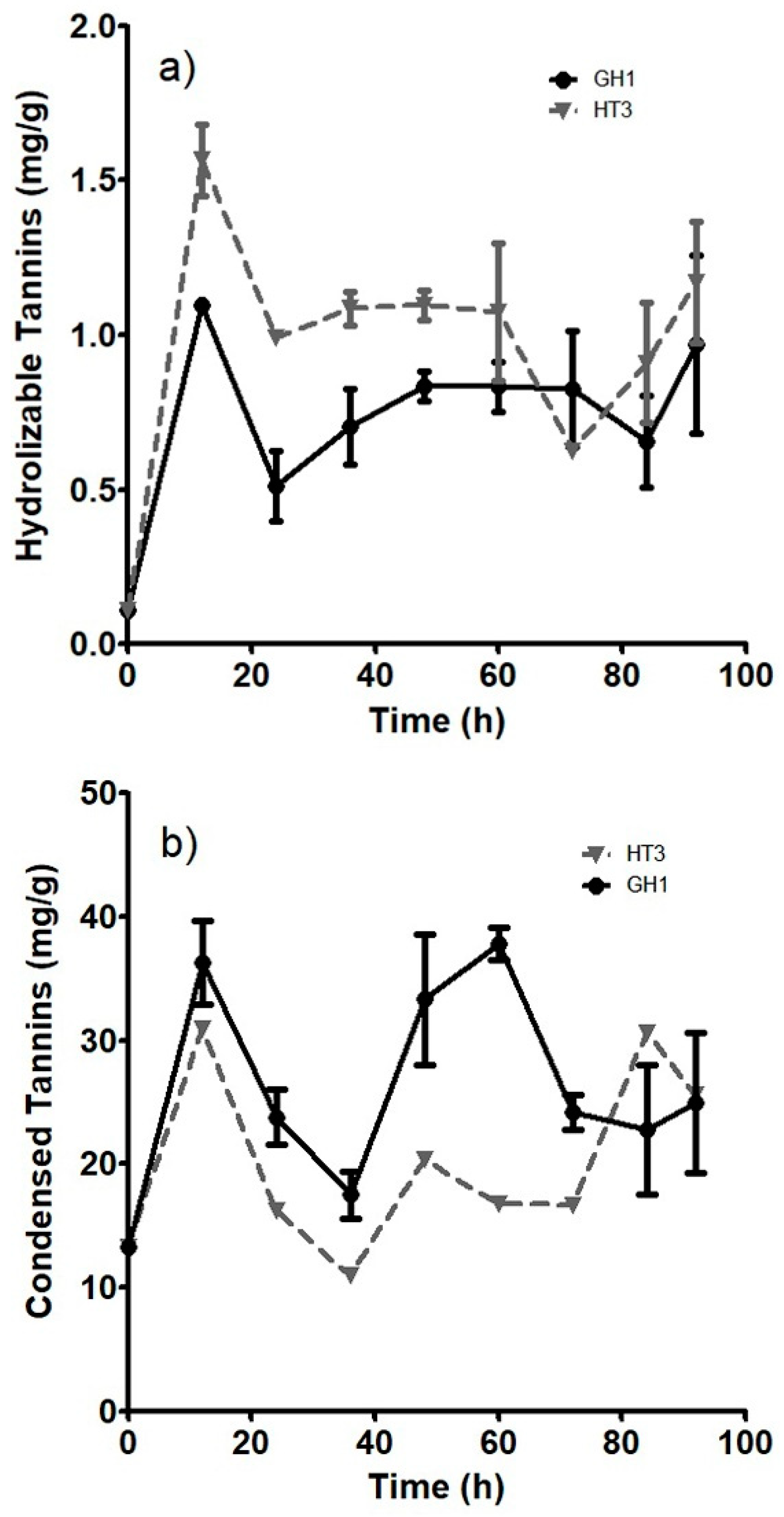
Figure 1. Kinetics of tannin accumulation. a) Hydrolyzable tannins, b) Condensed tannins. ● Represents the accumulation performed by A. niger GH1, ▼ Represents the accumulation by A. niger HT3.
Preprints 84991 g001
Figure 2. Condensed tannins accumulation of treatments of the experimental design.
Preprints 84991 g002
Figure 3. Hydrolyzable tannins accumulation of treatments of the experimental design.
Preprints 84991 g003
Figure 4. Pareto charts of factors that more influenced accumulation of bioactive compounds. a) Hydrolyzable tannins. b) Condensed tannins.
Preprints 84991 g004
Figure 5. Antioxidant activity of the best fermentation extracts.
Preprints 84991 g005
Table 1. Box Hunter & Hunter design for the evaluation of fermentation process conditions, levels, and factors.
Treatment Temperature(°C) Inoculum (spores/g) Humidity (%) pH NaNO3 (g/L) MgSO4 (g/L) KCl (g/L) KH2PO4 (g/L)
1 -1 -1 -1 -1 -1 -1 -1 -1
2 1 -1 -1 -1 -1 1 1 1
3 -1 1 -1 -1 1 -1 1 1
4 1 1 -1 -1 1 1 -1 -1
5 -1 -1 1 -1 1 1 1 -1
6 1 -1 1 -1 1 -1 -1 1
7 -1 1 1 -1 -1 1 -1 1
8 1 1 1 -1 -1 -1 1 -1
9 -1 -1 -1 1 1 1 -1 1
10 1 -1 -1 1 1 -1 1 -1
11 -1 1 -1 1 -1 1 1 -1
12 1 1 -1 1 -1 -1 -1 1
13 -1 -1 1 1 -1 -1 1 1
14 1 -1 1 1 -1 1 -1 -1
15 -1 1 1 1 1 -1 -1 -1
16 1 1 1 1 1 1 1 1
Factors Levels
+1 -1
Temperature (°C) 30 25
Inoculum (spores/g) 1x10⁷ 1x10⁶
Humidity (%) 70 60
pH 7 6
NaNO3 (g/L) 6 3
MgSO4 (g/L) 0.52 0.26
KCl (g/L) 0.52 0.26
KH2PO4 (g/L) 1.52 0.52
Table 2. HPLC-MS results for best treatments against unfermented sample.
Putative compound Unfermented sample Treatment 13 Treatment 16
(+)-Catechin - - +
3,4-DHPEA-AC - + +
Apigenin 7-O-diglucuronide - + +
Caffeic acid 4-O-glucoside + - -
Cyanidin + - -
Dihydroquercetin - + -
Feruloyl glucose - - +
Isorhamnetin + + +
Isorhamnetin 3-O-glucoside - + -
p-Coumaric acid 4-O-glucoside - + +
p-Coumaroyl glycolic acid - - +
Pinocembrin - + +
Pterostilbene - + +
Quercetin + - -
Rhamnetin + + +
Scopoletin - + +
Sinensetin - + +
Disclaimer/Publisher’s Note: The statements, opinions and data contained in all publications are solely those of the individual author(s) and contributor(s) and not of MDPI and/or the editor(s). MDPI and/or the editor(s) disclaim responsibility for any injury to people or property resulting from any ideas, methods, instructions or products referred to in the content.
Copyright: This open access article is published under a Creative Commons CC BY 4.0 license, which permit the free download, distribution, and reuse, provided that the author and preprint are cited in any reuse.
Prerpints.org logo

Preprints.org is a free preprint server supported by MDPI in Basel, Switzerland.

Subscribe

© 2024 MDPI (Basel, Switzerland) unless otherwise stated