Preprint
Article

Study on Physiological and Biochemical Index of Blood and Vitreous Humor in the Celestial Goldfish (Carassius auratus)

This version is not peer-reviewed.

Submitted:

27 September 2023

Posted:

27 September 2023

You are already at the latest version

Abstract
The aim of this study was to examine and understand the physiological and biochemical components of the blood and vitreous humor of goldfish (Carassius auratus). Blood was drawn from the arterial (static) line and vitreous humour was extracted using a syringe from healthy goldfish. An automatic haematology analyzer was used to detect 17 physiological indicators, including white blood cells and red blood cells. At the same time, 20 biochemical indexes, including albumin, calcium, and glutamic pyruvic transaminase, were found in the automatic biochemical analyzer. Experiments were also conducted on the effect of blood and vitreous fluid collection on the subsequent survival rate of the celestial goldfish. The findings demonstrated that there were values for 17 physiological indicators in the blood of the goldfish, including red blood cells (2.19 × 10¹²), white blood cells (62.21 × 10⁹), hemoglobin (138.25g/L), and no eosinophils or basophils. However, no data for 17 physiological indications of vitreous humor were discovered. The quantities of total protein, glutamic oxaloacetic transaminase, and potassium in the vitreous humor were substantially lower than those in the serum (P < 0.01) according to the results of 20 biochemical indexes in the serum and vitreous humor of goldfish. Alkaline phosphatase, chlorine, and creatinine between vitreous humor and serum did not differ significantly(P > 0.05). The levels of glutamic pyruvic transaminase and γ-glutamyl transpeptide in vitreous humor were significantly higher than those in serum (P < 0.01). This experiment provides basic datas for the healthy culture of goldfish and the perfection of hematology in goldfish, and the results will be convenient for the further study of development and formation of celestial eye traits.
Keywords: 
Subject: 
Biology and Life Sciences  -   Animal Science, Veterinary Science and Zoology

1. Introduction

The scientific name for the goldfish is Carassius auratus (Linnaeus, 1758), and it is a variety of the crucian carp family (Sparidae, Carassius, Car-assiusauratu) [1]. The valuable species of goldfish known as "the sky-watching dragon" or "sky-fazing" (eyes toward sky) is called the heavenly goldfish. The eyeball is like a dragon's eye, and part of it protrudes beyond the eye socket. When swimming in the water, the pupils are bright and more powerful than the dragon's eye. The beauty of the fish is that the eyes are always facing the sky, and it seems to be looking at you from any angle. This goldfish was a favorite in the imperial palace during the Qing dynasty since its name originally meant "looking upward to the Son of Heaven"[2]. So far, few studies have been done on celestial goldfish, much of which has focused on their genetics and body color [3,4,5]. Some reports have also suggested that celestial goldfish have a degenerated retina and weak vision [6,7], making them suitable as model animals for the study of human eye diseases [8]. Blood is an crucial component of the fish organism and immune system [9], and blood indicators have historically been emphasized by production practitioners and theorists, and are frequently used to assess the health, nutrition, and environmental adaptation status of fish [10], as well as water quality monitoring [11]. According to the available reports, however, there are no research on the physiological and biochemical components of the blood and vitreous humor of goldfish. The objective of this study was to measure and analyze a few physiological and biochemical indicators of goldfish blood and vitreous humor in order to provide a basic reference for the healthy culture and eyeball development of goldfish as well as to provide data for enhancing the comparative study of goldfish hematology.

2. Methods

2.1. Experimental Materials

Celestial goldfish came from Xiaotangshan Aquatic breeding Base of Beijing Institute of Fisheries Science. According to the breeding methods reported by Li Rongni et al. [4], the goldfish at the age of 16 months, were cultured in a cement pond deep in 35cm water at the age of 16 months, with a body length of about 12cm. In September, 45 individuals with eyeballs showing upward at a standard 90 degrees from horizontal were selected, as shown in Figure 1.

2.2. Methods

2.2.1. Preparation of EDTAK2 and Heparin Sodium Anticoagulant Solution

Weigh 3g EDTAK2 with an electronic balance, add distilled water to 200ml, mix well to make 15g/L EDTAK2 solution for use.

2.2.2. Effect of Three Methods of Pretreatment Syringe on Blood Sampling of Tail Vein of Goldfish and Preparation of Anticoagulant

Wrap the fish with clean medical gauze to secure it during blood collection, and the entire blood collection procedure must be completed quickly. To minimize harm to the fish body, the fish is quickly returned to the pool and iodine wound disinfection following blood collection to ensure that the fish are still alive. To test which pretreatment method is best for blood collection from the goldfish tail vein, the following three methods were applied to the syringe:
Syringe pretreatment A: Blood was drawn from the subscale vein of the tail of living goldfish using a moistened 1 ml syringe with 1% heparin sodium to see if it could be drawn smoothly and if it agglutinated in the syringe.
Syringe pretreatment B: Blood was drawn directly into the sublateral scalar vein of the tail of a living goldfish with a 1 ml syringe, and it was observed whether the blood could be drawn smoothly and whether the blood in the syringe was clotted or not;
Syringe pretreatment C: A 1ml syringe was moistened with EDTAK2 solution prepared in step 1.2.1, and blood was drawn from a vein under the caudal lateral scale of a live goldfish, and whether the blood in the syringe was agglutinated or not.
The method with the smoothest blood draw and non-clotting blood among the above three syringe pre-treatment methods was chosen for blood collection from goldfish in the formal test. After the blood draw was completed, the blood was slowly injected into a centrifuge tube to which 0.01 ml of EDTAK2 anticoagulant (prepared in step 1.2.1) had been added beforehand, mixed well to make anticoagulated blood, and placed in a refrigerator at 4°C for storage until use.

2.2.3. Preparating of Serum from Celestial Goldfish

Choose the pretreatment method used to draw the smoothest and non-clotting blood in step 2.2.2. The blood was slowly fed into the centrifuge tube without the use of an anticoagulant after the moving (static) pulse of the live goldfish tails were drawn. Without using an anticoagulant, the blood was spun at 8000 revolutions per minute for 10 minutes before the upper serum was absorbed and the tubes were stored in the refrigerator at 4 °C.

2.2.4. Collection of Vitreous Humor in the Celestial Goldfish

The goldfish were fixed with only their eyes visible, wrapped in sterile medical moist gauze. Select a 2ml syringe, insert the needle into the goldfish eyeball's posterior portion, slowly remove the vitreous fluid, remove the syringe needle, and slowly inject the vitreous fluid into the centrifuge tube with 0.01ml EDTAK2 anticoagulant (preparation step 1.2.1); then, store the centrifuge tube at 4 °C in the refrigerator until needed.
The vitreous humor was absorbed and stored at 4 °C in the refrigerator after being extracted using the aforementioned method. At the same time, withdraw the syringe needle and slowly inject it into the centrifuge tube without using an anticoagulant.
When pumping vitreous humor, always pay attention to position the needle away from the eyeball's blood vessels to prevent blood from mixing and to lessen damage to the fish. At the same time to grasp the depth of the needle into the eyeball, not too deep in order to avoid piercing the eyeball. After many experiments, during the actual operation of this trial, the needle was withdrawn after the vitreous humor was drawn, and the whole process did not have the phenomenon of mistakenly stabbing the blood vessels of the eyeball to the hemorrhage phenomenon of the eyeball. To ensure the survival of the goldfish, return them to the original location right away after pumping the vitreous humor.

2.2.5. Analysis of Physiological Indexes of Blood and Vitreous Humor of Goldfish

Taken three replicates each of whole blood with anticoagulant prepared in 1.2.2 and vitreous fluid with anticoagulant prepared in 1.2.4, and the blood routine indexes were measured using an automatic blood analyzer. White blood cells, red blood cells, hematocrit, mean corpuscular volume, average erythrocyte hemoglobin content, mean erythrocyte hemoglobin concentration, platelet count, erythrocyte distribution width (SD), neutrophils, lymphocytes, monocyte percentage, eosinophil count or percentage, basophil percentage, neutrophil percentage, lymphocyte percentage, and percentage of monocytes were the 17 indicators that were measured.

2.2.6. Analysis of Biochemical Indexes of Blood and Vitreous Humor of Goldfish

Both the upper vitreous humor and the goldfish serum that were made in 1.2.3 and 1.2.4 in triplicates had their respective biochemical indexes measured by a fully automatic biochemical analyzer. Total protein, albumin, alkaline phosphate plum, -glutamyl transpeptidase, total bile acid, glucose, urea, uric acid, creatinine, total cholesterol, triglyceride, high density lipoprotein cholesterol, low density lipoprotein cholesterol, potassium, sodium, chlorine, calcium, and cholinesterase were the 23 indexes that were measured.

2.2.7. Effect of Collecting Blood and Vitreous Humor on the Follow-Up Survival Rate of Goldfish

The following three groups of experiments were created to watch and compare the follow-up survival rate of goldfish collected blood and vitreous humor in order to examine the effect on the follow-up survival of goldfish after collecting blood and vitreous humor in this experiment.
In the first group, 15 goldfish after vitreous humor extraction in the experiment were randomly divided into three groups of five in each group. They were individually isolated and kept in the original location for two months, then the condition of the eyes was observed and the survival rate was counted.
The second group was formed by randomly dividing 15 goldfish into three groups after blood was drawn from their tail veins. After two months of isolation in the initial cement pond, their health was assessed, and the survival rate was calculated.
The third group consisted of 15 goldfish from the original location who were separated into three groups randomly as the control group. After fixing the fish with fresh, moist gauze for around 30 seconds (enough time to collect blood and vitreous humor), the gauze was taken off and the fish was returned to the original location. After two months of separate breeding, the health state and survival rate were assessed.

2.3. Calculations and Statistical Analysis

The data obtained from the test were statistically presented as mean±standard error (mean±SE) using Excel 2007 software and the t-test was used to analyse the significance of differences at a significance level of P<0.05 and a highly significant level of P<0.01.

3. Results and Discussion

3.1. Comparison of Methods of Collecting Blood from Arterial (Static) Pulse in the Tail of the Goldfish

After repeated tests, blood was drawn from the tail vein after moistening a 1 ml syringe with 1% sodium heparin most smoothly, and the probability of blood coagulation occurring during the blood draw was the smallest. The use of 15 g/L EDTAK2 solution to moisten 1 ml syringes or the use of 1 ml syringes to draw blood directly from goldfish has often resulted in blood clots clogging the needles, making it impossible to complete the blood collection. Therefore, 1% sodium heparin moistened 1 ml syringe was used in this experiment before tail vein blood collection.

3.2. Survival Statistics of the Celestial Goldfish after Blood and Vitreous Humor Extraction

After blood and vitreous humour were taken from the goldfish, they were cultured for another 2 months to observe the health status and survival rate. The survival rate was statistically analysed and compared with that of the uncollected goldfish, the difference was not significant. Table 1 displays the specific observation and statistical findings.

3.3. Analysis of Physiological Indexes of Blood and Vitreous Humor of Goldfish

Blood physiological indicators in celestial goldfish were detected by a fully automated haematology analyser with data results, but no data were available for the indicators measured in vitreous humor. The results showed that hemoglobin levels in celestial goldfish was 138.25g/L, 2.19×10¹²/L for red blood cells, and 62.21×10⁹/L for white blood cells. Table 2 displays the specific results.
Table 2-1. Result of blood and vitreous humor physiological indicators in the celestial goldfish.
sample WBC 109/L RBC
1012/L
HGB
g/L
HCT
%
MCV fl MCH Pg MCHC g/L PLT
109/L
RDW-CV
%
blood 62.21±8.54 2.19±0.09 138.25±14.74 41.70±1.58 187.10±1.44 75.75±1.16 410.50±2.05 6.00±0.94 21.70±0.95
vitreous humor No data display after test
Table 2-2. Result of blood and vitreous humor physiological indicators in the celestial goldfish.
sample NEU 109/L LYM
109/L
MON 109/L EOS 109/L BAS 109/L NEU% LYM% MON %
blood 1.37±0.10 59.53±14.32 2.05±0.19 0 0 2.05±0.03 94.75±0.26 2.50±0.28
vitreous humor No data display after test

3.4. Analysis of Biochemical Indexes of Blood and Vitreous Humor of the Goldfish

After analysis by a fully automated biochemical analyser, serum 20 biochemical components were also detected in the vitreous humor of the goldfish, including total protein, albumin, ghrelin, total bile acids, glucose, potassium, sodium, total cholesterol, triglycerides, HDL cholesterol, LDL cholesterol, 11 indicators of the concentration of the vitreous fluid were lower than the serum; alkaline phosphatase, total bile acids, Alkaline phosphatase, total bile acids, urea, chloride, calcium, creatinine, and cholinesterase, the concentrations of those were not significantly different between vitreous humor and serum; but glutamine transaminase and gamma-glutamyltransferase, the concentrations of them were significantly higher in vitreous humor than in serum. The results of biochemical indexes of blood and vitreous humor of the celestial goldfish are shown in Table 3.
Table 3-1. Result of blood and ocular humor biochemical indicators in the celestial goldfish.
sample ALT U/L AST U/L TP
g/L
ALB g/L ALP U/L GGT U/L TBAμmol/L GLU mmol/L UREμmol/L UA
μmol/L
serum 26.33±1.09 235.33±3.81 31.63±0.52 14.93±0.07 30.67±1.19 1.76±0.32 0.70±0.00 3.75±0.16 2.34±0.08 13.67±0.27
vitreous humor 87.00±4.03 114.00±1.09 2.05±0.05 2.50±0.03 26.00±0.54 14.89±0.47 0.40±0.11 0.70±0.02 1.86±0.02 -5.55±0.72
difference ** ** ** ** NS ** NS ** NS **
Table 3-2. Result of blood and ocular humor physiological indices in the celestial goldfish.
sample K
mmol/L
Na
mmol/L
Cl
mmol/L
Ca
mmol/L
CREA μmol/L CHOL
mmol/L
TG
mmol/L
HDL-C
mmol/L
LDL-C
mmol/L
CHE
mmol/L
serum 5.17±0.07 149.40±0.95 162.47±19.15 2.62±0.07 29.67±0.72 6.37±0.16 2.42±0.04 2.37±0.13 1.41±0.01 345.00±11.43
eyeball 2.24±0.05 132.85±0.08 151.20±15.05 2.61±0.05 30.00±2.05 1.13±0.05 0.10±0.02 0.39±0.02 0.17±0.00 323.00±8.50
difference ** ** NS NS NS ** ** ** ** NS
Note: ** indicates a very significant difference (P<0.01), * indicates a significant difference (P<0.05), NS indicates an insignificant difference (P>0.05).

4. Conclusions

4.1. Exploration of Blood Collection Methods for the Celestial Goldfish

The technique of taking fish blood from the arterial (static) pulse of the syringe is very popular and has the benefits of being quick and easy [12]. This approach was used in the study, although there were some issues in practice: Firstly, there is the inability to find blood vessels and repeated lancing, resulting in excessive bodily injury to the fish; Secondly, the needle slips out when the fish struggles, making it difficult to draw blood; Lastly, the use of syringes to draw blood is prone to clotting, making it difficult to carry out subsequent experiments. Before conducting this experiment, exploratory pre-experiments were conducted to address these issues: First, some experimental goldfish were dissected to examine the position and tissue structure of the tail arterial (static) pulse. It was discovered that the arterial (static) pulse of the goldfish tail were distributed in the muscles under the lateral line and on the tailbone, so the needle was chosen to be positioned obliquely below the lateral line scale of the goldfish tail stalk. Second, the medical gauze is moistened with the goldfish pond water before being wrapped around the fish to fasten it, particularly to hide its eyes so that only the tail stem and tail fin were visible. Wetting medical gauze and then wrapping the goldfish can help to reduce the stress and harm to them during the blood collection. Compared to tap water or distilled water, the osmotic pressure and oxygen content of the goldfish's pool water were more suitable for goldfish, allowing them to live in a more familiar and secure environment. With this method of stabilising the goldfish, the goldfish struggled the least and had the least effect on the blood sampling, which is in line with the suggestion of 'wrapping the fish with a wet towel during blood sampling, and only the needle penetrating into the fish body well' as mentioned by Zhao Haipeng[12]. Finally, to address the problem of coagulation that tends to occur with syringe blood draws, after repeated comparative experiments, it was found that 'the use of 1% sodium heparin to moisten a 1 ml syringe and then proffer blood in the tail vein' was the most effective. Additionally, anaesthetics are frequently used to keep fish from struggling, but considering the possible effects of anaesthetics on the fish's blood indicators, anaesthetics were not used in this study. The goldfish utilized in this experiment were medium-sized individual of about 12 cm, wrapped in medical wet gauze, and easy to control without anesthetics, so anaesthetics were not used throughout the experiment.
Anticoagulants are frequently used by researchers to treat blood draws or blood storage containers in order to stop premature blood clotting. Anticoagulants commonly used in fish haematology studies include heparin, EDTA.Na2, EDTA.K2 and sodium citrate, etc. Different anticoagulants have different effects on the results of haematological indexes due to their different compositions and anticoagulant mechanisms[13]. According to Li Ping's studies, EDTA-K2 anticoagulants were the best for detecting blood cells [14]. In order to measure the physiological parameters of goldfish blood in this experiment, EDTA-K2 was also added as an anticoagulant to the blood collection tube.

4.2. Effect on Subsequent Survival of Goldfish after Extraction of Blood and Vitreous Humor

The celestial goldfish are ornamental fish with ornamental, economic and scientific value. [15]. Ensuring the survival of goldfish is positive for their subsequent research, conservation or economic value. In this study, celestial goldfish were subjected to blood collection and then cultured for 2 months to observe their health and survival rate. It was found that the difference in survival rate was not significant when compared to untreated celestial goldfish. It also shows that the method of collecting blood from celestial goldfish in this study did not affect their subsequent survival. In the diagnosis and treatment of human diseases, blood collection is not life-threatening when controlled within appropriate limits. Therefore, when collecting blood from celestial goldfish, minimising irritation and damage to the fish and controlling the amount of blood collected will also ensure its subsequent survival.
In some Studies on human eye diseases , aqueous humor of patients had be directly collected by syringe extraction [16], and vitreous humor was also studied in this study using syringe extraction. Surgical removal of the vitreous body is sometimes used as a treatment for human eye-related diseases, but it is also prone to infectious endophthalmitis after surgery[17]. In this study, we also found those fish’ eyes were intraocular congestion and turbidity, after extracting the vitreous humor from the celestial goldfish, who were reared for an additional two months. And those fish ‘eyes maybe had some intraocular infection and inflammation . At the same time, it was discovered that the survival rate difference between the two groups was not statistically significant , when compared to normal celestial goldfish. This revealed that celestial goldfish can survive after extracted vitreous humor. The Experimental results would provid assurance for the subsequent study of celestial goldfish.

4.3. Comparison of the Contents of Erythrocyte and Hemoglobin in the Celestial Goldfish

Red blood cells are the main components of fish peripheral blood cells and play an important role in the body [18]. The red blood cell number of the celestial goldfish was (2.19×10¹²/L), which was close to the goldfish measured by Watson LJ et al (2. 03×1012/L) [19]. Celestial goldfish belongs to the Sparidae, Carassius, Car-assius auratus [1], and the number of red blood cells are similar to Xiangjiang wild carp (2.06×10¹²/L) [20] and carp (2.01×10¹²/L) [21], but there were differences from carp (1.41×10¹²/L) [22], furong crucian carp (1.47×10¹²/L) [23], crucian carp (1. 27×1012/L) [24] and koi (2.75-5.75× 10¹¹/L) [25], which may be due to the species and the environment affecting the red blood cell count of the fish. By comparing with carps, it can be determined that healthy celestial goldfish have a red blood cell count order of 1012/L.
The main function of hemoglobin is to transport and store oxygen,and it also plays a part in controlling the level of carbon monoxide, regulating pH and immune response[26]. HCT implicates in blood transporting oxygen [27]. The hemoglobin content of celestial goldfish measured in this study was 138.25 g/L, which was higher than that of common carp (88.7g/L) [22] and furong crucian carp hemoglobin(71.0g/L) [23]. The HCT of the goldfish was 41.70%, which was similar to crucian carp (39.92%)[24]. Goldfish has wide adaptability and has the ability to survive under low dissolved oxygen, which may be closely related to its own oxygen carrying capacity of hemoglobin and the ability of erythrocyte specific volume to utilize oxygen.

4.4. Analysis of the Number and Composition of White Blood Cells in the Celestial Goldfish

White blood cells are the main immune defense tissue of fish, and sometimes the changes of them in peripheral blood are used to evaluate the immune system [28]. The number of white blood cells of celestial goldfish was 62.21 × 10⁹ /L, which was slightly higher than that of goldfish (21.99 × 10⁹ / L) [19] and carp (20.95 × 10⁹ / L) [22], but All three have the same order of magnitude (109 / L). While the white blood cells of koi carp were 2.37-372 × 1011 / L [25]and that of Furong carps were 7.45 × 10¹¹ / L[23]. The order of magnitude of these two were both 1011 /L, which were much higher than the celestial goldfish. It may be that different fish species, different environments of the same fish species, different measurement methods and so on can lead to differences in white blood cells [12].
The types of white blood cells in fish mainly include neutrophils, monocytes, lymphocytes, eosinophils, basophils and so on [12]. Studies have shown that neutrophils in fish are important non-specific immune cells in fish, which participate in the inflammatory response of the body [29]. In this study, the number of neutrophils of the celestial goldfish was 1.37×10⁹/L, which was close to that of goldfish (1.132×10⁹/L) [19]. Monocytes also have a non-specific immune function, capturing their own senescent cells and foreign material for phagocytosis and digestion[30]. In this study, the monocytes of the celestial goldfish were 2.05×10⁹/L. Lymphocytes are the most common white blood cells, accounting for the highest proportion [12]. In this test, the number of lymphocytes of the celestial goldfish was 59.53 × 10⁹/L, which was slightly higher than that of goldfish (20.32×10⁹/L) [19]. Eosinophils are rarely observed in the peripheral blood of fish, and eosinophils and basophils can be observed in the peripheral blood of only a few fish. Some fish have neither basophils nor eosinophils [31]. The eosinophils and basophils of goldfish were 0.499 ×10⁹/L and 0.04×10⁹/L determined by WatsonLJ et al. [19], but no eosinophils and basophils were detected in the peripheral blood of goldfish in our study, which is consistent with previous findings. Eosinophils and basophils were not found in the blood of various ploidy crucian carp according to Liu Qiao et al. [32], and basophils were not discovered in the peripheral blood of crucian carp according to Zhu Hongwen et al. [33]. Eosinophils and basophils were not discovered in three varieties of Pu'an crucian carp by Zhou Xianjun et al. [34]. Eosinophils and basophils were not discovered by Zhao Haipeng [12] in the blood of several economic fish in middle and upper reaches of Yangtze river. Eosinophils and basophils in the blood of fish are mainly found in tissues such as the head, kidney, spleen, and liver, and are less abundant in peripheral blood [12].It may be due to the fact that the blood of celestial goldfish tested was peripheral blood and its eosinophils and basophils were not detected due to their low or almost absence.

4.5. Analysis of the Characteristics of Blood Biochemical Indexes in the Celestial Goldfish

The stability of different inorganic components in blood is crucial for maintaining the homeostasis of the fish, and Na+ and Cl-, which account for more than 95% of the blood's cations and anions, respectively, are crucial for maintaining plasma osmolality and the acid-base balance [35]. Hisao Ozaki [27] generalized the range of serum inorganic constituents of scleractinian fishes, and the concentrations of potassium, sodium, Cl , and calcium in the blood of celestial goldfish measured in the present study were within the range of the generalized standard values.
The blood's organic components include triglycerides, protein, cholesterol, creatinine, and blood sugar. The level of blood glucose reflects the metabolic level of the fish[36]. Triglycerides are the main substances in the metabolism of fish lipids, and cholesterol is the raw material for the synthesis of some hormones and enzymes, all of which are essential for normal life activities in fish [37]. Creatinine implicates in muscle activity [38]. The blood glucose of the celestial goldfish was 3.75mg/L and lower than that of grass carp (5.60-19.05mg/L) , abalone(6.60-26.61 mg/L)[12] and Furong carp [23]. The blood triglyceride (2.42mg/L) and total cholesterol (6.37mg/L) of goldfish were higher than those of Furong carp[23]. In this study, the celestial goldfish’s blood creatinine was lower than grass carp and bighead carp [39] and was close to long-snout catfish [39]. Goldfish is an ornamental fish, it is easier to obtain bait, because of less activity, so the content of blood glucose and creatinine is low, the consumption of energy is also low, the stored fat is more, the total cholesterol and triglyceride content is higher.
Blood proteins are important in determining health, nutrition and disease in goldfish[40]. According to Yukio Ozaki [27], the majority of bony fish had serum protein concentrations between 30 and 50 g/L. The total blood protein concentration of goldfish in this study, 31.62 g/L, was comparable to that of allogenic silver crucian carp [41]. The measured HDL and LDL content of blood of celestial goldfish was closer to that of furong crucian carp measured by Cheng Xiaofei et al [23].
Alkaline phosphatase, glutamic pyruvic transaminase, glutamic oxaloacetic transaminase, cholinesterase, and other enzymes are also included in the blood's organic components. Alkaline phosphatase (ALP), glutamic pyruvic transaminase (ALT), and glutamic oxaloacetic transaminase (AST) are not only found in blood but also in tissues such the liver, heart, Gill, kidney, and muscle in fish [42]. The celestial goldfish's blood ALP and AST levels were comparable to those of Furong carp and grass goldfish, respectively [23,43]. The level of ALT was between that of Furong carp[23] and grass goldfish[43].
The data for this experiment were obtained from the healthy celestial goldfish, whose biochemical indicators can be used as basic blood index parameters for goldfish.

4.6. Biochemical Components in Vitreous Humor of the Celestial Goldfish

Human eyeballs contain 80% vitreous humor, which is made up of 99% water and the remaining 1% of proteins, inorganic salts, carbohydrates, lactic acid, urea, vitamin C, amino acids, lipids, and other substances. Hyaluronic acid, proteoglycan, collagen, and non-collagen protein are the structural macromolecules of vitreous [44]. There are only a few vitreous cells in the vitreous, and they are mostly found at the base and in the vitreous cortex [44]. In our investigation, the vitreous humor was taken from the middle of the goldfish's vitreous body.,so no cells were found,
Chen CH et al [45] summarized that most of the serum components can be present within the vitreous fluid, but in lower concentrations than serum. The concentrations of 11 indexes in the celestial goldfish vitreous humor were lower than those in the serum, and this results were consistent with the findings of ChenCH et al. [45], but there was no appreciable difference in the concentrations of ALP, total bile acid, urea, chlorine, calcium, creatinine, and cholinesterase between in the vitreous humor and the serum. In addition, levels of glutamic pyruvic transaminase and -glutamyl transpeptidase in serum were substantially lower than those in vitreous humor. The synthesis of alanine was associated to glutamic pyruvic transaminase [46], while the transmembrane transport of amino acids and the control of glutathione (GSH) level were related to γ-glutamyl transpeptidase [47]. These two might be connected to the aberrant metabolism in the celestial goldfish’s vitreous humor.
In the vitreous humor, there was a small amount of albumin, which could be utilized as a gauge for the health of the blood-ocular barrier [48]. According to David [48], the albumin concentration in the vitreous humor of the telescope goldfish increased with the expansion of the eye in larger fish. .and the largest group of telescope goldfish had an albumin content in vitreous humor of 2.41g/L, which was only marginally higher than our celestial goldfish (2.50g/L). Both celestial and telescope goldfish had suffered from visual function deficit, but the eye diseases were different between them. Celestial goldfish and goldfish with dragon's eyes both have abnormal functioning of the eyes, however, the development of celestial goldfish is accompanied by varying degrees of retinal degeneration [6], resulting in narrow vision and low light; Although the telescope goldfish 's eyeballs were protruding as well, its myopia was severe, and there was no retinal degeneration [49]. By comparative analysis, the albumin concentration in the vitreous humor of our celestial goldfish was higher than that of David’s telescope goldfish [48], so it is speculated that probably celestial goldfish have a more severe degree of change in the condition of the membranes associated with the blood-retinal barrier than goldfish with dragon's eyes.
The celestial goldfish had lower levels of glucose, creatinine, and urea in their vitreous humor than the dead people [48]. It may be due to the fact that with the death of cells associated with the blood-eye barrier after the death, the membrane permeability increases and the relevant substances within the blood can enter the vitreous humour, so the vitreous humour of the celestial goldfish taken in this case was sampled in vivo. Additionally, there was no discernible difference of the contents of urea and creatinine between in vitreous humor and blood of the celestial, while the contents of urea and creatinine in the vitreous humor of cattle [50] and humans [48] were lower than their respective blood. It may be due to the altered permeability of the blood-retinal barrier to urea and creatinine in the eye of celestial goldfish.
The content of Ca2+ in vitreous humor of the celestial goldfish was similar to of humans (2.45 mmol/L) [51]. There was no discernible difference between the contents of Ca2+ and Cl-, in vitreous humor and blood, whereas the contents of Ca2+ and Cl-, in human vitreous humor were different from those in blood [48]. The ion transport and metabolism pathway in the celestial goldfish’s vitreous humor might be different from that of human vitreous body.
1) The difference in subsequent survival rates after blood and ocular fluids were extracted from the celestial goldfish was not significant when compared to the non-experimental goldfish. (P>0.05).
2) Tn the tested celestial goldfish blood, the red blood cells were 2.19×10¹²/L, the white blood cells were 62.21×10⁹/L, the hemoglobin was 138.25g/L, and eosinophils and basophils were not detected.
3) A total of 20 biochemical indexes of celestial goldfish‘s blood and vitreous humor were determined, of which 11 indexes had lower concentrations in vitreous humor than serum, including alkaline phosphatase, total bile acid, urea, chlorine, calcium, creatinine, cholinesterase ; 7 indicators were not significantly different in vitreous humor from those in serum; The concentrations of alanine aminotransferase and γ-glutamyl transpeptidase in vitreous humor were significantly higher than in serum.
The measured data were all from the constitutionally healthy celestial goldfish, and the ranges of physiological and biochemical indicators could be used as the basic blood index parameters of the celestial goldfish. Through the study of the physiological and biochemical indicators of celestial goldfish blood and vitreous humor, it could provide basic reference materials for better understanding of its biological characteristics and disease prevention and control, as well as the mechanism of celestial goldfish's unique eye formation.

Author Contributions

Conceptualization and supervision, X.Z.; data analysis, R.L., W.L. and Y.S.; writing—original draft preparation, R.C.; writing—review and editing, R.L. All authors have read and agreed to the published version of the manuscript.

Funding

This research was funded by Beijing Natural Science Foundation for Young Science (6234045); Self-support project of Beijing Academy of Agriculture and Forestry Sciences (Beijing characteristic and ornamental new fish species cultivation research); Key Laboratory of Ministry of Agriculture and Rural Affairs--National Engineering Research Center for Freshwater Fisheries (Beijing) (PT2023-19) and National Freshwater Aquatic Germplasm Resource Bank (FGRC: 18537).

Institutional Review Board Statement

All animal handling and manipulation procedures were based on the standards of the Chinese Council on Animal Care.

Informed Consent Statement

Informed consent was obtained from all the subjects involved in the study.

Data Availability Statement

The authors confirm that the data supporting the findings of this study are available within the article.
Conflicts of interest: The authors declare no conflict of interest.

References

  1. Wang, C. Studies on the Karyotype of Goldfish(Carassius auratus)I. A Comparative Study of the Chromosomes in Crucian and Red Dragon-eye Goldfish[J]. Journal of Genetics&Genomics. 1982, 9, 238–242. [Google Scholar]
  2. Wang, H. Atlas of Chinese Goldfish[M]. Beijing: Culture and art publishing house, 2000.
  3. Li, R.; Sun, Y.; Cui, R.; Zhang, X. Comprehensive Transcriptome Analysis of Different Skin Colors to Evaluate Genes Related to the Production of Pigment in Celestial Goldfish[J]. Biology 2022, 12, 1–12. [Google Scholar] [CrossRef] [PubMed]
  4. Li, R.; Sun, Y.; Tian, Z.; et al. Effects of genetic and environmental factors on the celestial eye in Carassius auratus [J]. Freshwater Fisheries 2022, 52, 66–73. [Google Scholar]
  5. Li, R.; Wang, S.; Tian, Z.; et al. Comparative transcriptome analysis of body color change in red celestial goldfish Carassius auratus [J]. Journal of Dalian Ocean University 2022, 37, 191–201. [Google Scholar]
  6. Sakaue, H.; Negi, A.; Matsumura, M.; et al. The developmental changes of ERGs on spontaneous retinal degeneration of Celestial goldfish[J]. Advances in Ophthalmology 1988, 70, 97–101. [Google Scholar] [CrossRef]
  7. Matsumura, M.; Ohkuma, M.; Honda, Y. Retinal degeneration in celestial goldfish. Developmental study [J]. Ophthalmic Research 1982, 14, 344–353. [Google Scholar] [CrossRef]
  8. Yoshihiro, O.; Tetsuo, K. Goldfish: an old and new model system to study vertebrate development, evolution and human disease[J]. Journal of Biochemistry 2019, 165, 209–218. [Google Scholar]
  9. Zhou, Y.; Xing, Y.; Feng, Q. Reserch advance in haemocytes of fisher[J]. Journal of Hainan University:Natural Science Ed 2003, 171–176.
  10. Bahmani, M.; Oryan, S.; Pourkazemi, M.; et al. Effects of ecophysiological stress on cellular immunity system of Persian sturgeon Acipenser persicus[R]. Presented at 14th Iranian congress of physiology and pharmacology 1999, 16–20. [Google Scholar]
  11. Ren, P.; Zhang, Y.; Ren, G.; et al. Changes in morphology and quantity of peripheral blood cells in Carassius auratus collected from polluted water area [J]. Chinese Journal of Zoology 2008, 43, 37–42. [Google Scholar]
  12. Zhao, H. Studies on haematology of several economic fish in middle and upper reaches of Yangtze river[D]. Southwest university 2008. [Google Scholar]
  13. Ning, D.; Li, X.; Wu, K.; et al. Influence of anticoagulant selection on whole blood cell analysis [J]. Acta Medicinae Sinica 2004, 17, 550–551. [Google Scholar]
  14. Ping, L. The value of heparin, sodium citrate and EDTA-K2 in blood cell analysis [J]. Medical Laboratory Science and Clinics 2021, 32, 63–65. [Google Scholar]
  15. Yoshihiro, O.; Tetsuo, K. Goldfish: an old and new model system to study vertebrate development, evolution and human disease[J]. Journal of Biochemistry 2019, 165, 209–218. [Google Scholar]
  16. Zhang, T.; Li, K.; Li, L.; et al. Proteome Characterization of Primary Angle-Closure Glaucoma Aqueous Humor [J]. Journal of Chengdu Medical College 2023, 18, 180–186. [Google Scholar]
  17. Huang, S.; Lu, S.; Lin, M.; et al. Factors Associated with Postvitrectomy Endophthalmitis [J]. Chinese Journal of Nosocomiology 2009, 401–403. [Google Scholar]
  18. Gao, Z.; Wang, W. Research progress on peripheral blood red blood cells of fish [J]. Reservoir Fisheries 2008, 28, 1–3. [Google Scholar]
  19. Watson, L.J.; Shechmeister, I.L.; Jackson, L.L. The hematology of goldfish Carassius auratus[J]. Cytologia 1963, 28, 118–130. [Google Scholar] [CrossRef]
  20. Wang, Y.; Liu, S.; Wang, G.; et al. Comparative Hematological Studies in Cyprinus carpio Xiangyunnensis and Cyprinus carpio Xiangjiangnensis [J]. Journal of Natural Science of Hunan Normal University 1988, 1, 71–75. [Google Scholar]
  21. Mi, R. Determination of hematological indexes of grass carp, carp and abalone [J]. Freshwater Fisheries 1982, 8, 10–16. [Google Scholar]
  22. Chng, C.; Ming, Q. Study on the blood physiobiochemic and hemorheologic properties of caprio in Weishan Lake [J]. Jiangsu Agricultural Sciences 2005, 5, 95–97. [Google Scholar]
  23. Cheng, X.; Jiang, G.; Xiang, J.; et al. Effects of dietary fishmeal repiacement with meat and bone meal on the growth performance, blood physiological and biochemical indices,muscle chemical composition and texture characteristics in juvenile furong crucian carp(fueong♀×red crucian carp) [J]. Acta Hydrobiologica Sinica 2022, 44, 85–94. [Google Scholar]
  24. Lin, G. Study on Carassius auratus blood [J]. Current Zoology 1979, 25, 210–219. [Google Scholar]
  25. Wang, D.; Deng, S.; Zou, L.; et al. Studied on the hematological indices of different Cyprinus carpio Koi [J]. Journal of Aquaculture 2016, 37, 1–5. [Google Scholar] [CrossRef]
  26. Yu, L.; Yang, D.; Liu, H.; et al. Correlation between Hemoglobin and Asphyxiation Point in Twelve Species of Freshwater Fish [J]. Chinese Journal of Zoology 2017, 52, 478–484. [Google Scholar]
  27. Hisao Oizaki(Translated by Xu Xuelong et al). Fish hematology and circulatory physiology[M]. Shanghai: Shanghai Scientific and Technical Publishers 1982, 6–96.
  28. Tierney, K.B.; Farrell, A.P.; Kennedy, C.J. The differential leucocyte landscape of four teleosts: juvenile Oncorhynchus kisutch, Clupea pallasi, Culaea inconstans and Pimephales promelas[J]. Journal of Fish Biology 2004, 65, 906–919. [Google Scholar] [CrossRef]
  29. Chen, F. Hematopathological studies in proliferative kidney disease of red grouper [J]. Tropic Oceanology 1997, 16, 49–53. [Google Scholar]
  30. Zhang, Y.; Sun, B.; Nie, P. Immune tissues and cells of fish: a review[J]. Acta Hydrobiologica Sinica 2000, 24, 648–654. [Google Scholar]
  31. Chen, G.; Zhou, H.; Ye, F.; et al. A hematological study and observation on development of blood cells in American red fish(Sciaenops ocellatus) [J]. Journal of Tropical Oceanography 2006, 25, 59–66. [Google Scholar]
  32. Liu, Q.; Wang, Y.; Liu, S.; et al. Comparison of blood and blood cells in crucian carp with different ploidy [J]. Advances in Natural Science 2004, 14, 1111–1117. [Google Scholar]
  33. Zhu, H.; Wang, H.; Qin, G. Studies on the blood cell morphology of crucian carp (Carassius auratus L.) [J]. Zoological Research 1985, 6, 147–153. [Google Scholar]
  34. Zhou, X.; Ano, M.; Huang, L.; et al. Hematologic Studies of Three Types of Carassius auratus [J]. Guizhou Agricultural Sciences 2012, 40, 133–135. [Google Scholar]
  35. Zhao, H.; Zhao, H.; Jin, L.; et al. Microscopic structures of peripheral hematocytes in sinilabeo rendahli [J]. Fisheries Science 2005, 24, 24–27. [Google Scholar]
  36. Chen, X. The Fishes Blood [J]. Journal of Chongqing Teachers College 2000, 19, 70–73. [Google Scholar]
  37. Cheng, C. The study of blood physiological and biochemeical parameters of Monopterus albus[D]. Nanchang: Jiangxi Agricultural University 2014.
  38. Han, N.; Shi, C. The Application of Blood Indexes in Ichthyological Research[J]. Journal of Anhui Agricultural Sciences 2010, 38, 18877–18878. [Google Scholar]
  39. Dong, S.; Miao, J.; Zhao, K. Study on Physiological and Biochemical Index of Blood in Leiocassis longirostri[J]. Hubei Agricultural Sciences 2016, 55, 3690–3693. [Google Scholar]
  40. Qian, Y.; Chen, H.; Qian, Y.; et al. ffects of starvation on hematological and blood biochemical indices in cultured Lateolabrax japonicus [J]. Journal of Fishery Sciences of China 2002, 2, 133–137. [Google Scholar]
  41. Gu, L.; Hou, Y.; Ding, B.; et al. Effects of Several Plant Extracts on Growth Performance and Blood Biochemical Indices in Carassius auratus gibelio [J]. Freshwater Fisheries 2008, 28, 23–26. [Google Scholar]
  42. Asadi, F.; Masoudifard, M.; Vdjhi, A.; et al. Serum biochemical parameters of Acipenser persicus[J]. Fish Physiol Biochem 2006, 32, 43–47. [Google Scholar] [CrossRef]
  43. Rahimikia, E. Analysis of antioxidants and serum biochemical responses in goldfish under nickel exposure by sub-chronic test[J]. Journal of Applied Animal Research 2017, 45, 320–325. [Google Scholar] [CrossRef]
  44. Fan, H. Study on the relationship between postmortem change of chemical composition in Vitreous humor and time of death [D]. Sichuan University 2005. [Google Scholar]
  45. Chen, C.H.; Chen, S.C. Studies on soluble proteins of vitreous in experimental animals. Exp Eye Res 1981, 32, 381. [Google Scholar] [CrossRef]
  46. Li, Y.; Wu, P.; Wang, F.; et al. Effects of methoxyfenozide on transaminase and alanine aminotransferase in the silkworm[J]. Guangdong Sericulture 2019, 53, 1–4. [Google Scholar]
  47. Liu, R. Research progress of gamma-glutamyltranspeptidase activity in mammals [J]. Shanghai Journal of Preventive Medicine 2002, 14, 24–25. [Google Scholar]
  48. Yew, D.T.; Lai, H.W.; Zhou, L.; Lam, K.W. Chromatographic identification of a biochemical alteration in the aqueous humour of megalophthalmic Black Moor goldfish[J]. Journal of Chromatography B: Biomedical Sciences and Applications 2001, 751, 349–355. [Google Scholar] [CrossRef]
  49. Easter, S.S.; Hitchcock, P.F. The myopic eye of the Black Moor goldfish. Vision Research 1986, 26, 1831–1833. [Google Scholar] [CrossRef]
  50. Lane, V.M.; Lincoln, S.D. Changes in urea nitrogen and creatinine concentrations in the vitreous humour of cattle after death[J]. Am J Vet Res 1985, 46, 1550–1552. [Google Scholar]
  51. Coe, J.I. postmortem chemistries of human Vitreous humour[J]. AmJ C1in Pathol 1969, 51, 741–750. [Google Scholar] [CrossRef] [PubMed]
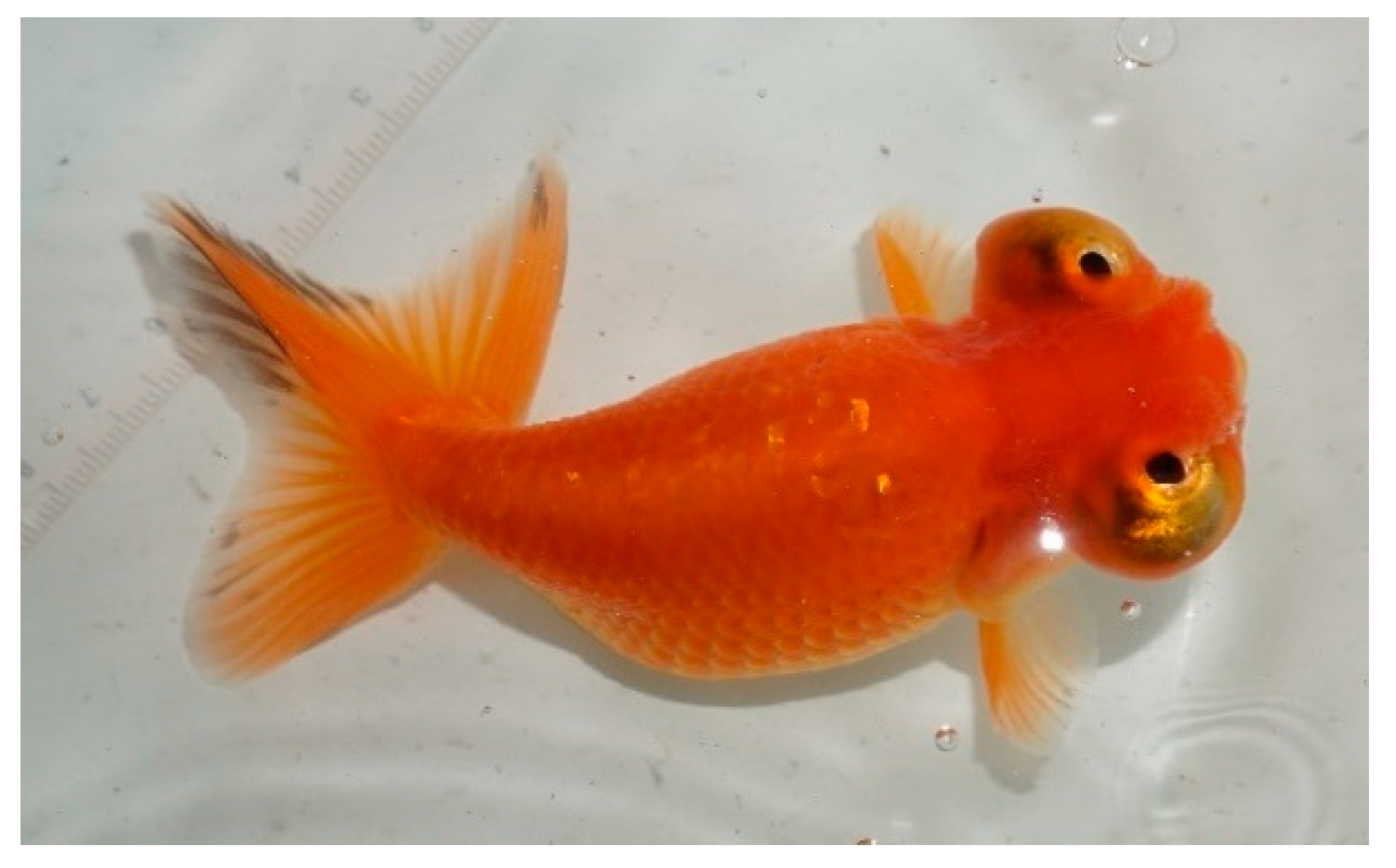
Figure 1. the celestial goldfish (cultured and photographed by Institute of Fishery Sciences, Beijing Academy of Agriculture and Forestry Sciences).
Preprints 86282 g001
Table 1. Statistics on the survival rate of goldfish in three groups.
Groups Survival Rate% Symptom Description
celestial goldfish group for blood collection 80±5.44a Recovered in about 1 month
celestial goldfish group for vitreous humor collection 90±5.44a Congestion in the eyeballs within 1 month, blurring of the eyeballs after 2 months
Untreated celestial goldfish group 90±5.44a normal
Disclaimer/Publisher’s Note: The statements, opinions and data contained in all publications are solely those of the individual author(s) and contributor(s) and not of MDPI and/or the editor(s). MDPI and/or the editor(s) disclaim responsibility for any injury to people or property resulting from any ideas, methods, instructions or products referred to in the content.
Copyright: This open access article is published under a Creative Commons CC BY 4.0 license, which permit the free download, distribution, and reuse, provided that the author and preprint are cited in any reuse.
Alerts
Prerpints.org logo

Preprints.org is a free preprint server supported by MDPI in Basel, Switzerland.

Subscribe

© 2025 MDPI (Basel, Switzerland) unless otherwise stated