Preprint
Case Report

Treatment of Three Ferrets Diagnosed with Ferret Systemic Coronaviral Disease Using the Nucleoside Analogue GS-441524

Altmetrics

Downloads

414

Views

514

Comments

0

Submitted:

25 September 2023

Posted:

28 September 2023

You are already at the latest version

Alerts
Abstract
Ferret Systemic Coronaviral Disease (FSCD) is a systemic disease, caused by ferret systemic coronavirus, that is considered lethal in most of the ferrets that are affected by it. To our knowledge, no treatment has been shown effective against FSCD, and most of the ferrets are euthanized or die after development of clinical disease. GS-441524 has been shown effective for successfully treating cats with Feline Infectious Peritonitis (FIP), a disease that shares similarities with FSCD. However, to our knowledge treatment with GS-441524 has not been reported for treatment of FSCD in ferrets. Here we describe three cases of ferrets diagnosed with FSCD successfully cured utilizing oral GS-441524. FSCD may be effectively treated following similar protocols utilized for feline infectious peritonitis in cats
Keywords: 
Subject: Medicine and Pharmacology  -   Veterinary Medicine

1. Introduction

Ferret systemic coronavirus disease (FSCD) is an aggressive disease resembling feline infectious peritonitis (FIP), the feline disease caused by a mutation of the feline coronavirus (FCoV) [1–3]. Differently than other coronavirus diseases that typically present mild symptoms that resolve themselves with time [4–6], FSCD has been shown to be lethal in the majority of cases, similarly to FIP in cats [7–13].
Ferrets with FSCD present a variety of non-specific symptoms, most commonly weight loss, lethargy, loss of appetite, vomiting, dehydration, diarrhea, sneezing, nasal discharge, difficulties breathing and palpable abdominal mass [9–14]. Few reports have also described neurological issues, such as seizures and ataxia [3,8,15]. Clinically, anemia and thrombocytopenia are key features of this disease, accompanied by a remarkable hyperproteinemia with low albumin and high globulin levels, as observed in FIP [3,8,12]. The histopathology of FSCV is characterized by granulomatous formation mainly in the mesenteric, lymph nodes, spleen, and kidneys, resembling the dry form of FIP in cats [16,17] although, ascites was also reported in a few cases [18].
While FIP is normally reported to progress quickly [19,20] the reported cases of FSCD seem to point to a slower progression [7,14]. Supportive care with corticosteroids and antibiotics has shown to prolong the ferret’s life for several months, however death is inevitable with eventual progression of the disease [13,14].
Two recent studies used ferrets in a SARS-CoV-2 infection model and demonstrated the efficacy of two antiviral drugs against the virus: the nucleoside analogues GS-441524 and EIDD-2801 [21,22]. Furthermore, a group of protease inhibitors (GC376) that has been tested against FIP in vitro and in vivo [23,24] has shown to be effective against FSCV in vitro, evidencing promising data for the treatment of this disease [25].The nucleoside analogue GS-441524 has been shown effective for treating cats with FIP with high success rate [26]. Because of the similarities between FSCD and FIP, GS-441524 could be used to treat ferrets affected by FSCV.
Here, we describe three cases of ferrets diagnosed with FSCD and successfully treated with oral GS-441524.

3. Materials and Methods

Owners of the three ferrets joined the Facebook group FIP Warriors 5.0 and were connected to the authors for data collection. The diagnosis of FSCD was confirmed by the veterinarians who examined the ferrets and reported in the medical records. The diagnosis was made evaluating the clinical signs, blood chemistry, complete blood counts and, in some cases, with abdominal ultrasound and biopsy or a positive ferret coronavirus PCR. The three ferrets received unlicensed GS-441524 similarly to what was as described in a previous article on cats treated for FIP [26].
A sample of the GS-441524 utilized by the three ferrets was analyzed with HPLC by SGS Health Sciences, to confirm the composition and purity (>99%). The ferrets received oral GS-441524 in size 4 gelatin capsules at 12-20mg/kg twice a day for 12-17 weeks. The information was collected through daily follow up with the owners during the first week, and weekly for the following weeks of treatment to ensure the proper dosing and monitor for weight changes, food intake and clinical signs. Ferrets were examined every 3-6 weeks by their veterinarians and blood chemistry and complete blood counts (CBC) were performed to monitor the treatment outcome and possible side effects associated with the treatment.

3. Results

3.1. Case 1

3.1.1. Subsubsection

An 8-month-old male ferret presented to the clinic lethargic with diarrhea, loss of appetite and weight loss. The blood chemistry and CBC showed low total protein levels with low albumin (1.5 g/dL) and normal globulins, indicating a low A/G ratio (0.5). The analysis also showed low HGB and HCT but normal white blood cells count (Table 1). A palpable mass was discovered in the abdominal region. A biopsy of the abdominal mass was collected as well as the mesenteric lymph node due to its enlargement. The histopathology analysis revealed the lymph node was distorted by pyogranulomatous inflammation with no detectable microorganisms present (Figure 1). Pyogranulomatous lymphadenitis in the ferret was described as a concern for FSCD. The veterinarian performed surgery to remove the mass and administered antibiotics and dexamethasone.
Six months later, the ferret presented signs of depression and a palpable mass was felt in his abdomen once again. Treatment with 12mg/kg GS-441524 sub-cutaneous injection was started. After 2 days of injections, the ferret was weighing 1.51 kg and the dosage was adjusted to 12mg/kg GS-441524 oral capsules every 12h. Dexamethasone was interrupted. After 10 days of treatment the ferret showed restored energy levels, the abdominal mass was not palpable anymore, and the abdomen was softer and normal looking. His appetite had also improved.
A little over 3 weeks of treatment, the ferret’s weight fluctuated between 1.47-1.52 kg and the blood chemistry showed improved values: albumin was normal (3.7 g/dL), globulins were elevated (5.1 g/dL), and A/G ratio increased to 0.7. A CBC showed increased HCT and HGB, as well as increased lymphocytes. Slightly increased BUN (38 mg/dL) and creatinine (1.0 mg/dL) were noted.
This sentence seems a little long, maybe something like:
At week 14 of treatment, the CBC was unremarkable. The blood chemistry showed an increased BUN and creatinine but the values were consistent with previous testing and remained stable. The protein levels were within the normal limit with A/G ratio of 1.2. With these promising results, the treatment was terminated.
Five weeks after the end of treatment, the ferret’s blood chemistry showed increased BUN at 42 mg/dL and creatinine at 1.3 mg/dL, however no measures were taken at this point. One year after treatment was terminated, the ferret continues to show good energy and appetite with no return of any clinical signs of FSCD.

3.2. Case 2

A 3-year-old female ferret weighing 0.68 kg presented with history of lethargy, loss of appetite, trouble breathing and difficulty swallowing water and food; no gastrointestinal or neurological symptoms were observed. The veterinarian prescribed 1.5 mg/kg prednisolone twice daily, as well as 1.8 mg/kg amoxicillin and metoclopramide injections. After two days of the initial symptoms, the CBC showed low HCT (32.2%). The blood chemistry showed normal albumin but high globulins (5.5 g/dL), with an A/G ratio of 0.5, suggesting a diagnosis of FSCD. Treatment with oral capsules of GS-441524 at 12 mg/kg every 12 hours was then started. At the second day of treatment the ferret was still presenting respiratory symptoms, coughing and wheezing, but had increased energy and appetite. The respiratory issues persisted for another few days, subsiding on the sixth day of treatment. Two weeks after initiating the treatment, the ferret’s weight was 0.74 kg and appeared clinically normal to the owner. At this point the blood chemistry showed decreased globulins resulting in A/G ratio of 0.6 (Table 2). Prednisolone was then tapered off over a 2-week span. At 12 weeks of treatment, the ferret weighed 0.81 kg and blood chemistry showed normal albumin, decreased globulins (3.7 g/dL), with an A/G ratio of 0.8. Because globulins were still higher than the reference range and the HCT decreased, the GS-441524 dosage was increase to 20 mg/kg and treatment was continued for another 3 weeks. At 17 weeks of treatment, HCT was normal but the globulins remained elevated. Treatment was then interrupted, and the observation phase was initiated. Seven months post treatment the ferret was found to have abdominal effusion. The veterinarian prescribed furosemide and the effusion resolved. Blood chemistry and CBC were not supportive of a relapse of FSCD and the ferret is currently undergoing further testing to identify the cause. At the time of writing the ferret did not exhibit any clinical signs of the disease.

3.3. Case 3

A 2.5-year-old female ferret presented to the clinic with ataxia and lethargy. The owner reported bruxism and ptyalism. A CBC showed elevated WBC (12.82K/ul), neutrophils (5.46K/ul) and monocytes (1.36K/ul ref 0.18-0.9K/ul). Blood chemistry showed elevated total protein 10.9g/dl and globulins (8.3g/dl) with A/G ratio 0.3. The ferret was administered fluids IV and cerenia. The following days the ferret was unresponsive and presented with a fever of 105 °F. The ferret was hospitalized and administered IV fluids, maropitant 2mg/kg sq SID, clavamox 15mg/kg PO BID, omeprazole 1mg/kg PO SID, buprenorphine 0.05mg/kg IM q6 and gabapentin 10mg/kg PO Q8. Clinical signs slightly improved. However, the fever persisted. An abdominal ultrasound revealed enlarged spleen and multiple enlarged, rounded mesenteric lymph nodes, medial iliac lymph nodes, and hypogastric lymph nodes, as well as a 2.88cm mass in the right cranial abdomen that contacts the pancreas. A diarrhea PCR panel resulted positive for ferret enteric coronavirus, supportive of FSCD with possible neurological involvement. Treatment with GS-441524 in injectable form was initiated at a dosage of 30mg/kg BID due to incorrect calculation made by the rescuer helping the owner. The following day the owner reported the ferret being more alert and no longer in pain. Over the course of 4 days the dosage was reduced to 15mg/kg and treatment was continued with oral GS-441524 in capsules. The ferret improved clinically and appeared normal to the owner after 2 weeks of treatment. Weight was increased to 0.93 kg, from 0.84 kg at the time treatment was initiated. At 4 weeks of treatment, the spleen was normal in size and blood chemistry revealed reduced globulins (6.5g/dl) with A/G 0.4. A CBC showed that WBC was still elevated (13.8k/ul) with elevated lymphocytes (Table 3). At 9 weeks of treatment, the ferret continued to do well and weighed 1.03 kg. The CBC showed WBC was still elevated (9.8k/ul) with elevated lymphocytes (7.74k/ul) and low neutrophils (1.27k/ul). Globulin was in the normal range (3.7g/dl ) with A/G 0.9. After 12 weeks of treatment blood chemistry and CBC were unremarkable, besides increased BUN, and the ferret was clinically normal. Treatment was then discontinued, and the observation phase initiated. Seven months after the treatment was terminated, the ferret remains healthy with no signs of recurrence of the disease.

4. Discussion

FSCD is considered lethal for most of the ferrets affected by clinical disease. No treatment has been reported to successfully cure ferrets with FSCD. Here we reported the case of three ferrets diagnosed with FSCD by their veterinarians. With no other current treatment, prednisolone and antibiotics were chosen to first mitigate the symptoms. However, with the experimental treatment using oral capsules of GS-441524, clinical signs and blood chemistry and CBC rapidly improved. After about 12 weeks of treatment, similarly to the treatment for FIP in cats [27] the ferrets were cured. The elevated BUN and creatinine may be due to damage caused by the disease although a side effect of the treatment cannot be excluded at this time. GS-441524 has not been reported to affect the kidney functionality in cats and increased kidney values occasionally reported during treatment are usually transient. However, more data is needed to determine whether the kidney functionality of the ferret was impacted by the disease or the treatment.

5. Conclusions

Oral treatment with GS-441524 has been shown effective in 3 ferrets diagnosed with FSCD with a treatment course without complications. However, a field trial is needed to determine the lowest effective dosage, treatment duration and toxicity in ferrets.

Author Contributions

J.P. and F.S. contributed to the data collection and writing of the manuscript. A.J.N. contributed to the data interpretation and manuscript editing. All authors have read and agreed to the published version of the manuscript.

Funding

This research received no external funding.

Institutional Review Board Statement

Not applicable.

Informed Consent Statement

A written informed consent must be obtained from the owner of the animals (or an authorized agent for the owner).

Data Availability Statement

The data presented in this study are available on request from the corresponding author.

Acknowledgments

Authors would like to thank the owners of the ferrets and FIP Warriors for supporting the owners involved in the study and connecting them to the authors.

Conflicts of Interest

The authors declare no conflict of interest.

References

  1. Martinez, J.; Reinacher, M.; Perpiñán, D.; Ramis, A. Identification of group 1 coronavirus antigen in multisystemic granulomatous lesions in ferrets (Mustela putorius furo). J. Comp. Pathol. 2008, 138, 54–58. [Google Scholar] [CrossRef] [PubMed]
  2. Martinez, J.; Ramis, A.J.; Reinacher, M.; Perpiñán, D. Detection of feline infectious peritonitis virus-like antigen in ferrets. Vet. Rec. 2006, 158, 523. [Google Scholar] [CrossRef]
  3. Garner, M.M.; Ramsell, K.; Morera, N.; Juan-Sallés, C.; Jiménez, J.; Ardiaca, M.; Montesinos, A.; Teifke, J.P.; Löhr, C.V.; Evermann, J.F.; et al. Clinicopathologic Features of a Systemic Coronavirus-Associated Disease Resembling Feline Infectious Peritonitis in the Domestic Ferret (Mustela putorius). Vet. Pathol. 2008, 45, 236–246. [Google Scholar] [CrossRef]
  4. Williams, B.H.; Kiupel, M.; West, K.H.; Raymond, J.T.; Grant, C.K.; Glickman, L.T. Coronavirus-associated epizootic catarrhal enteritis in ferrets. J. Am. Vet. Med. Assoc. 2000, 217, 4–526. [Google Scholar] [CrossRef] [PubMed]
  5. Wise, A.G.; Kiupel, M.; Maes, R.K. Molecular characterization of a novel coronavirus associated with epizootic catarrhal enteritis (ECE) in ferrets. Virol. 2006, 349, 164–174. [Google Scholar] [CrossRef] [PubMed]
  6. Wise, A.G. , Kiupel, M.; Garner, M.M.; Clark, A.K.; Maes, R.K. Comparative sequence analysis of the distal one-third of the genomes of a systemic and an enteric ferret coronavirus. Virus Res. 2010, 149, 42–50. [Google Scholar] [CrossRef]
  7. Autieri, C.R. , Miller, C.L.; Scott, K.E.; Kilgore, A.; Papscoe, V.A.; Garner, M.M.; Haupt, J.L.; Bakthavatchalu, V.; Muthupalani, S.; Fox, J.G. Systemic coronaviral disease in 5 ferrets. Comp. Med. 2015, 64, 6–508. [Google Scholar]
  8. Gnirs, K.; Quinton, J.F.; Dally, C.; Nicolier, A.; Ruel, Y. Cerebral pyogranuloma associated with systemic coronavirus infection in a ferret. J. Small Anim. Pract. 2016, 57, 36–39. [Google Scholar] [CrossRef]
  9. Lescano, J.; Quevedo, M.; Gonzales-Viera, O.; Luna, L.; Keel, M.K.; Gregory, F. First case of systemic coronavirus infection in a domestic ferret (Mustela putorius furo) in Peru. Transbound. Emerg. Dis. 2015, 62, 581–585. [Google Scholar] [CrossRef] [PubMed]
  10. Lindemann, D.M.; Eshar, D.; Schumacher, L.L.; Almes, K.M.; Rankin, A.J. Pyogranulomatous panophthalmitis with systemic coronavirus disease in a domestic ferret (Mustela putorius furo). Vet. Ophthalmol. 2016, 19, 2–167. [Google Scholar] [CrossRef]
  11. Michimae, Y. , Mikami, S.; Okimoto, K.; Toyosawa, K.; Matsumoto, I; Kouchi, M.; Koujitani, T.; Inoue, T.; Seki, T. The first case of feline infectious peritonitis-like pyogranuloma in a ferret infected by coronavirus in Japan. J. Toxicol. Pathol. 2010, 23, 99–101. [Google Scholar] [CrossRef] [PubMed]
  12. Shigemoto, J.; Muraoka, Y.; Wise, A.G.; Kiupel, M.; Maes, R.K.; Torisu, S. Two cases of systemic coronavirus-associated disease resembling feline infectious peritonitis in domestic ferrets in Japan. J. Exot. Pet Med. 2014, 23, 196–200. [Google Scholar] [CrossRef] [PubMed]
  13. Tarbert, D.K.; Bolin, L.L.; Stout, A.E.; Schaefer, D.M.W.; Ruby, R.E.; Fernandez, J. R-R.; Duhamel, G.E.; Whittaker, G.R.; de Matos, R. Persistent infection and pancytopenia associated with ferret systemic coronaviral disease in a domestic ferret. J. Vet. Diagn. Invest. 2020, 32, 4–616. [Google Scholar] [CrossRef]
  14. Wills, S.E. , Beaufrère, H.H.; Brisson,B. A.; Fraser, R.S.; Smith, D.A. Pancreatitis and systemic coronavirus infection in a ferret (Mustela putorius furo). Comp. Med. 2018, 68, 3–208. [Google Scholar] [CrossRef]
  15. Murray, J.; Kiupel, M.; Maes, R.K. Ferret coronavirus-associated diseases. Vet. Clin. North Am. Exot. Anim. Pract. 2010, 13, 543–560. [Google Scholar] [CrossRef] [PubMed]
  16. Pedersen, N.C. A review of feline infectious peritonitis virus infection: 1963–2008. J. Feline Med. Surg. 2009, 11, 225–258. [Google Scholar] [CrossRef] [PubMed]
  17. Haake, C.; Sarah Cook, S.; Pusterla, N.; Murphy, B. Coronavirus Infections in Companion Animals: Virology, Epidemiology, Clinical and Pathologic Features. Viruses 2020, 12(9), 1023–1045. [Google Scholar] [CrossRef]
  18. Graham, E.; Lamm, C.; Denk, D.; Stidworthy, M.- F.; Carrasco, D.C.; Kubiak, M. Systemic coronavirus-associated disease resembling feline infectious peritonitis in ferrets in the UK. Vet Rec. 2012, 171, 8–200. [Google Scholar] [CrossRef]
  19. Fischer, Y.; Ritz, S.; Weber, K.; Sauter-Louis, C.; Hartmann, K. Randomized, placebo-controlled study of the effect of propentofylline on survival time and quality of life of cats with Feline Infectious Peritonitis. J. Vet. Intern. Med. 2011, 25, 1270–1276. [Google Scholar] [CrossRef]
  20. Ritz, S.; Egberink, H.; Hartmann, K. Effect of Feline Interferon-Omega on the survival time and quality of life of cats with Feline Infectious Peritonitis. J. Vet. Intern. Med. 2007, 21, 1193–1197. [Google Scholar] [CrossRef]
  21. Cox, R.M.; Wolf, J.D.; Lieber, C.M.; Sourimant, J.; Lin, M.J.; Babusis, D.; DuPont, V.; Chan, J.; Barrett, K.T.; Lye, D.; et al. Oral prodrug of remdesivir parent GS-441524 is efficacious against SARS-CoV-2 in ferrets. Nat. Commun. 2021, 12, 6415–6426. [Google Scholar] [CrossRef] [PubMed]
  22. Cox, R.M.; Wolf, J.D.; Plemper, R.K. Therapeutically administered ribonucleoside analogue MK-4482/EIDD-2801 blocks SARS-CoV-2 transmission in ferrets. Nat. Microbiol. 2021, 6, 11–18. [Google Scholar] [CrossRef]
  23. Cook, S.E.; Vogel, H.; Castillo, D.; Olsen, M.; Pedersen, N.; Murphy, B. Investigation of monotherapy and combined anticoronaviral therapies against feline coronavirus serotype II in vitro. J. Fel. Med. Surg. 2021, 1–11. [Google Scholar] [CrossRef]
  24. Pedersen, N.C.; Kim, Y.; Liu, H.; Galasiti Kankanamalage, A.C.; Eckstrand, C.; Groutas, W.C.; Bannasch, M.; Meadows, J.M.; Chang, K-O. Efficacy of a 3C-like protease inhibitor in treating various forms of acquired feline infectious peritonitis. J. Feline. Med. Surg. 2018, 20, 378–392. [Google Scholar] [CrossRef] [PubMed]
  25. Perera, K.D.; Galasiti Kankanamalage, A.C.; Rathnayake, A.D.; Honeyfield, A.; Groutas, W.; Chang, K.-O.; Kim, Y. Protease inhibitors broadly effective against feline, ferret and mink coronaviruses. Antiviral Res. 2018, 160, 79–86. [Google Scholar] [CrossRef] [PubMed]
  26. Jones, S.; Novicoff, W.; Nadeau, J.; Evans, S. Unlicensed GS-441524-Like Antiviral Therapy Can Be Effective for at-Home Treatment of Feline Infectious Peritonitis. Animals. 2021, 11, 2257–2271. [Google Scholar] [CrossRef] [PubMed]
  27. Pedersen, N.C.; Perron, M.; Bannasch, M.; Montgomery, E.; Murakami, E.; Liepnieks, M.; Liu, H. Efficacy and safety of the nucleoside analog GS-441524 for treatment of cats with naturally occurring feline infectious peritonitis J. Feline. Med. Surg. 2019, 21, 271–281. [Google Scholar] [CrossRef] [PubMed]
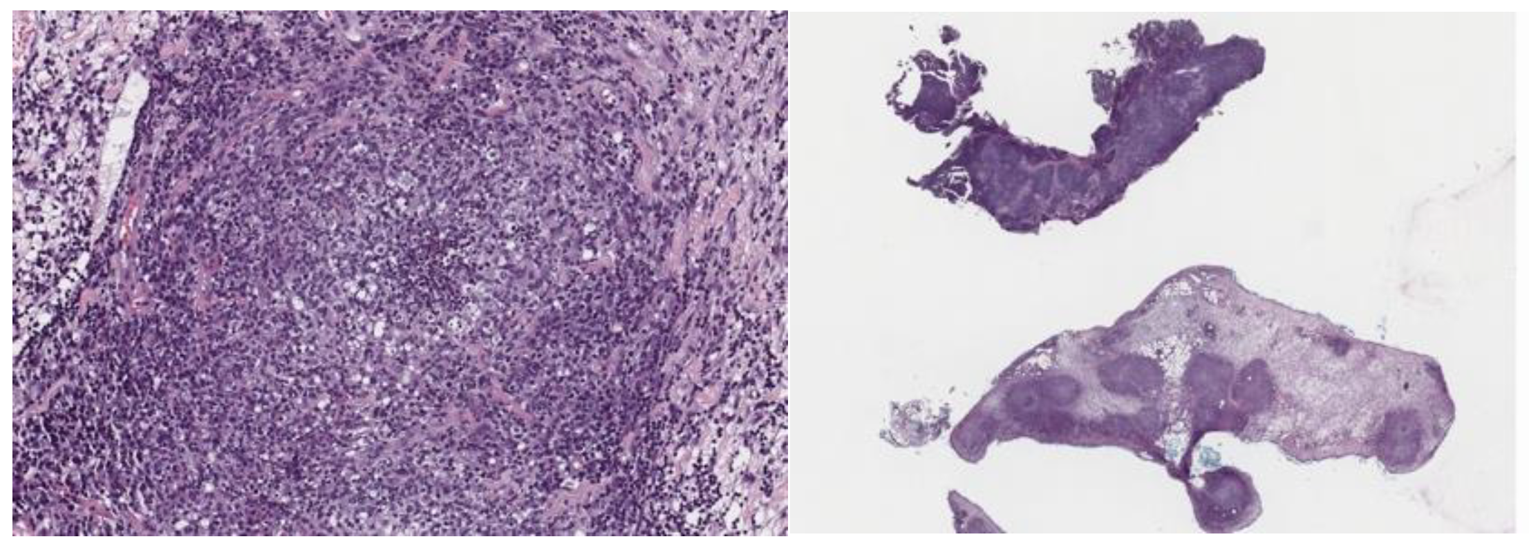
Figure 1. Histopathology images of two samples obtained from the mesenteric lymph nodes biopsied.
Preprints 86135 g001
Table 1. Summary of the blood chemistry and CBC obtained throughout treatment and the observations phase for case 1. Value that are below the reference range are indicated with L=low, and values above the reference range with H=high.
Tests (ref. range) Unit Diagnosis1 Week 31 Week 141 1 month post treatment 1
RBC (6.5-11.0) M/μL 7.9 12.9 H 11.3 H 10.2
HCT (43-55) % 37 L 63 H 57 H 55
HGB (15.0-19.0) g/dL 12 L 20.5 H 19.5 H 17.1
WBC (2.5-8.0) K/μL
Neutrophils (1.37-4.74) K/μL
4.3
2.75 (64%)
8.5 H
2.13 (25%)
6.8
1.97 (29%)
4.5
1.62 (36%)
Lymphocytes (0.87-3.36) K/μL 1.3 (31%) 5.70 (67%) H 4.42 (65%) H 2.43 (54%)
Total protein (5.5-7.6) g/dL 4.6 L 8.8 H 7.0 6.3
Albumin (2.4-4.5) g/dL 1.5 L 3.7 3.8 3.6
Globulin (2.9-4.9) g/dL 3.1 5.1 H 3.2 2.7 L
A/G ratio (0.8-2) 0.5 L 0.7 1.2 1.3
ALT (10-280) IU//L 51 170 288 H 186
ALP (15-45) IU/L
TBIL (0.0-1.0) mg/dL
86 H
0.1
16
0.1
20
0.1
21
0.2
Creatinine (0.2-0.8) mg/dL
BUN (10-33) mg/dL
0.3
11
1.0 H
38 H
1.0 H
38 H
1.3 H
42 H
1 All the tests were performed by ANTECH.
Table 2. Summary of the blood chemistry and CBC obtained throughout treatment and the observations phase for case 2. Value that are below the reference range are indicated with L=low, and values above the reference range with H=high.
Tests (ref. range) Unit Diagnosis1 Week 21 Week 121 Week 171 7 months after treatment1
RBC (6.35-11.20) M/μL 7.18 8.26 7.79 9.40 9.75
HCT (37-55) % 32.8 L 40.7 36.2 L 41.8 44.6
HGB (11.0-17.0) g/dL 11.5 13 12.5 14.7 15.5
WBC (2.0-10.0) K/μL
Neutrophils (0.62-3.30) K/μL
8.7
3.82 (43.7%)
16.7 H
10.72 (64.2%) H
6.5
2.15 (33.2%)
6.4
2.32 (36.4%)
5.5
3.28 (59.7%)
Lymphocytes (1.00-8.00) K/μL 3.73 (42.7%) 4.88 (29.3%) 3.59 (55.5%) 3.30 (51.8%) 1.79 (32.6%)
Total protein (5.2-7.3) g/dL 8.2 H 7.5 H 6.8 6.9 5.7
Albumin (2.6-3.8) g/dL 2.7 2.7 3.1 3.2 2.5 L
Globulin (1.8-3.1) g/dL 5.5 H 4.8 H 3.7 H 3.7 H 3.3 H
A/G ratio 0.5 0.6 0.8 0.9 0.8
ALT (82-289) U//L 135 68 63 L 75 L 102
ALP (9-84) U/L
TBIL (0.1-1.0) mg/dL
<10
0.3
32
0.3
29
0.2
21
0.4
20
0.2
Creatinine (0.4-0.9) mg/dL
BUN (10-45) mg/dL
0.3
17
0.3
16
0.6
36
0.6
33
0.6
29
1 All tests were performed by IDEXX.
Table 3. Summary of the blood chemistry and CBC obtained throughout treatment and the observations phase for case 3. Value that are below the reference range are indicated with L=low, and values above the reference range with H=high.
Tests (ref. range2) Unit Diagnosis (ref. range)1 Week 42 Week 92 Week 122 3 months after treatment2
RBC (6.5-11.0) M/μL 11.45 H (6.35-11.20) NA 12.7 H NA NA
HCT (43-55) % 40.2 (37.0-55.0) 51 62 H 58 H 53
HGB (15.0-19.0) g/dL 14.3 (11.0-17.0) NA 17.1 NA NA
WBC (2.5-8.0) K/μL
Neutrophils (1.37-4.74) K/μL
12.8 (2-10) H
5.46 (0.62-3.30)(42.6%) H
13.8 H
4.28 (31%)
9.8 H
1.27 (13%) L
7.5
1.87 (24%)
3.6
0.83 (23%)
Lymphocytes (0.87-3.36) K/μL 5.91 (1.00-8.00)(46.1%) 8.69 (63%) 7.74 (79%) H 5.93 (76%) H 2.66 (74%)
Total protein (5.2-7.3) g/dL 10.9 (5.2-7.3) H 9.0 H 7.1 7.4 6.8
Albumin (2.4-4.5) g/dL 2.6 (2.6-3.8) 2.5 3.4 4.2 3.5
Globulin (2.9-4.9) g/dL 8.3 (1.8-3.1) H 6.5 H 3.7 3.2 3.3
A/G ratio 0.3 0.4 0.9 1.3 1.1
ALT (10-280) U//L 65 (82-289) L 60 50 88 63
ALP (15-45) U/L
TBIL (0.0-1.0) mg/dL
43 (9-84)
0.4
45
0.0
40
0.1
33
0.1
29
0.1
Creatinine (0.2-0.8) mg/dL
BUN (10-33) mg/dL
0.6 (0.4-0.9)
13 (10-45)
NA
35 H
0.8
32
0.8
40 H
NA
35 H
1 Tests were performed with in house IDEXX. 2 Tests performed by ANTECH.
Disclaimer/Publisher’s Note: The statements, opinions and data contained in all publications are solely those of the individual author(s) and contributor(s) and not of MDPI and/or the editor(s). MDPI and/or the editor(s) disclaim responsibility for any injury to people or property resulting from any ideas, methods, instructions or products referred to in the content.
Copyright: This open access article is published under a Creative Commons CC BY 4.0 license, which permit the free download, distribution, and reuse, provided that the author and preprint are cited in any reuse.
Prerpints.org logo

Preprints.org is a free preprint server supported by MDPI in Basel, Switzerland.

Subscribe

© 2024 MDPI (Basel, Switzerland) unless otherwise stated