Preprint
Review

Association between Inflamatory Responses and Mitochondrial Dynamics in Salmonella Infection

Altmetrics

Downloads

115

Views

66

Comments

0

This version is not peer-reviewed

Submitted:

30 September 2023

Posted:

01 October 2023

You are already at the latest version

Alerts
Abstract
Salmonella is the intracellular pathogen and etiological known for the cause of asymptomatic carriage, gastroenteritis, systemic disease (typhoid fever), in severe cases death may also occur. Altered function of mitochondria due to Salmonella infection impacts the immune response. Mitochondria which are recognized as the “powerhouse” of the cells are also know to play central role in the immune metabolism. Mitochondrial dynamics such as fusion, fission, signaling, transport and mitophagy influences the immune system and also maintains the cellular integrity. Mitochondrial DNA and Reactive oxygen species formation also elicits the inflammatory responses. This review discusses the immune metabolism and effect of altered mitochondrial dynamics in case of Salmonella infection.
Keywords: 
Subject: Biology and Life Sciences  -   Immunology and Microbiology

INTRODUCTION

Salmonella is a gram negative, anaerobic, facultative bacterium which affects many species. S. typhi affects humans and higher primates and S. typhimurium affects rodents, cattel’s and mammals. S. typhi and S. typhimurium have 11% difference in their genome, which are otherwise 99% similar in their sequences of house-keeping genes. (Grari et al., 2021).
Milder cases are not easily diagnosed thus reported. Major changes in non-typhoidal Salmonellosis occurred in last century due to the emergence of food borne human infections caused by Salmonella entrica enteritis by muti-drug resistant (MDR) strains of Salmonella enterica typhimurium. MDR drugs comprises of fluroquinolones, 3rd gen cephalosporins (Hurley et al., 2014).
S. typhimurium conceals a factor which is its virulence factor that leads to the production of a free electron during respiration process and this free electron produced helps in the anaerobic respiration. Due to diminution of the native gut microbe increases the chances of growth of the Salmonella to form its colony. Some nutrients such as ethanolamine that is already present in the gut of an individual S. typhimurium takes its advantages and colonise in the gut during the infection where it outcompetes other microbes of the gut which has the same ecological niche as that of Salmonella (Hurley et al., 2014). It forms a structure known as salmonella containing vacuole (SCV) which helps in its survival the self- established niche in the host environment as it arrests the endosomal pathway in its late endosome stage. Challenges encountered by Salmonella during its life cycle includes environmental stress, inorganic acid stress in stomach, intra cellular stress, nutritional stress, organic acid stress in intestine, intestinal barriers, etc. (Grari et al., 2021). Salmonella has multifaceted ways in which it affects the host as it favours to proliferate in the non-restrictive habitat of macrophages. B-cells, T-cells, monocytes, dendritic cells, granulocytes and gut epithelial cells are affected after the entry of the bacterium into the host body. Organic nitro stative stresses are one of the distinguished immune approaches encountered by Salmonella. Various symptoms such as gastroenteritis, systemic disease in typhoid fever were observed during the infection also in many cases pain is developed in joints, irritation in eyes, and painful urination is observed along with chronic arthritis. (Takaya et al., 2020). Faster growth rates of bacterium salmonella and viability of the genome constructed using recombinant DNA technology makes it an exemplary pathogen in the study of host-pathogen reciprocity. (Garai et al., 2012)

BACKGROUND

Mitochondrial Interplay with Immune System
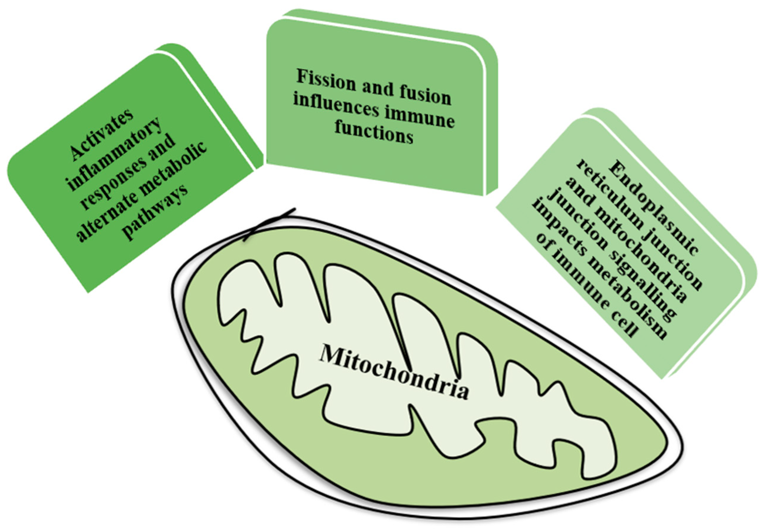
Figure 1. Mitochondrial dynamics.
Preprints 86595 g001
The changes in the number of mitochondrial number and structural modifications are known as mitochondrial dynamics. Mitochondrial signalling plays an important role in the activation of innate immune response pathogen associated molecular patterns (PAMPs) and damage associated molecular patterns (DAMPs) activates the pro-inflammatory cytokines by the formation of mitochondrial ROS. (Khan et al., 2020). Mitochondrial dynamics includes fusion, fission of mitochondria, movement or mitochondrial transport. (Xie et al., 2020)
Mitochondrial fusion includes mitochondrial inner membrane and mitochondrial outer membrane and is assisted by mitofusin1 and 2 (MFN1 & 2) and exalted by misato 1 (MSTO1). Mitochondrial fission is regulated by dynamic-related protein 1 which are inducted by coupler proteins.
Mitochondrial transport occurs alongside filaments of actin and microtubules present in the cytoplasm of the cell. Direct interaction of cytoskeleton proteins and mitochondrial outer membrane (MOM) proteins does not occur during the transportation. Hence, proteins like myosin, kinesin, and dynein associates with MOM and emerge to form a multiplex and disintegrated structures of mitochondria in the cytoplasm of cell are more easily transported as compared to lengthened and coalesced structures this implies that process of fission plays an important role in the mitochondrial transport. Further this ensures the pertinent distribution of mitochondria which is crucial for the process of division of cell and in the attainment of energy requirements of cell. (Xie et al., 2020)
Mitochondrial signaling is shown to be important in activating innate immune response PAMPs and DAMPs activates pro-inflammatory cytokines by generating RLRs (RIG-1) like receptor mitochondrial ROS. ROS production contributes to mitochondrial damage in a range of pathologies and is also important in redox signaling from the organelles to the rest of cells.
H2O2 liberated from the dismutation of superoxide (O2-) mitochondria contains its own superoxide dimutase (SOD). Production of ROS can lead to oxidative damage to then mitochondrial proteins H2O2 efflux from mitochondria can be measured by a non-fluorescent substrate in conjugation with horse radish peroxidase (HRP).
2 main modes of operation by isolated mitochondria that leads to extensive H2O2 efflux
  • Increased NADH/NAD+ ratio in matrix
  • Highly rescued CoQ pool in conjugation with a maximal Δp and no ATP synthesis.
(H2O2 efflux from mitochondria is negligible compared to 1 and 2).
Mitochondrial operation is when the mitochondria are working normally (ATP production) or using Δp for other functions such as thermogenesis. ROS production by complex 1 was in sub-mitochondrial particle (SMPs) where reduction of the CoQ pool and generation by a large Δp succinate leads to uncoupler-sensitive H2O2 production. Mitochondrial disruption leads to oxidative damage rises the possibility that allows the better understanding of mtROS production which will lead to then rational design of therapies to minimize the mitochondrial oxidative damage (Wang et al., 2018)
Mitochondrial autophagy also known as mitophagy is a form of autophagy in which damaged mitochondria are removed and this process is regulated by enzyme PTEN-induced putative kinase 1 (PINK1) and ubiquitin ligase parkin. Mitophagy has the potential in the treatment of inflammatory diseases with excessive reactive oxygen species (ROS) and mitochondrial dysfunction. Decline in mitophagy in T-cells escalates the process of apoptosis and ROS formation. A protein known as autophagy related protein 7 (Atg-7) is needed for the formation of autophagosomes structures. As found by scientist Pua et al. T-cells with inadequacy of protein Atg-7 has elevated mitochondrial content, boosted production of ROS and utterance of pro-apoptotic proteins such as Bak, cytochrome c and AIF. (Angajala et al., 2018)

Molecular patterns associated mitochondrial damage

Mitochondrial DAMPs (mt DAMPs) are the molecules liberated from mitochondrial DAMPs during the cell’s death into the extracellular area and comprise of not only proteins but also lipids or DNA and tend to elicit an immune response. Other examples of mitochondrial DAMPs are nuclear DNA, high-mobility group box 1 (HMGB1), or heat-shock proteins, calreticulin, mitochondrial DNA, N-formyl peptides, cardiolipin, etc. When these mitochondrial DAMPs come in contact with immune cells such as macrophages, neutrophils initiate innate immune response and when comes in contact with dendritic cells initiates adaptive immune response. When these mt DAMPs are discharged into the circulation from impaired areas promotes adherence of activated neutrophils and vascular endothelial cells and transmigration of immune cells into the organs located at distant sites which leads to the secretion of pro-inflammatory proteinases and cytokines causing to elicit an immune response in the organ. (Nakahira et al., 2015). Salmonella invades both phagocytic and non-phagocytic cells including mononuclear phagocytic cells which are present in lymphoid follicles, live and spleen. Epithelial cells and phagocytic cells such as dendritic cells and macrophages identify specific pathogens-associated molecular patterns and DMAPs present in bacteria. PPRs which are NOD-like receptors, TLR comprise early components of the immune system that function to detect invading pathogens through PAMPs and DAMPs and signal to recruit and activate phagocytic cells such as neutrophils and macrophages (Hurley et al., 2014)

Metabolic repurposing of mitochondria to drive inflammatory macrophages

LPS plays an important role in the inset of sepsis during systemic infection of observed by its role in macrophages. Clinical activation by bacterial LPS or IFN- γ leads to the alteration in secretory profile of the cells through production of NO. Alteration action by 1L-4, IL-10, or IL-13 leads to the production of polyamines and proline including proliferation and collagen production respectively (Page et al., 2022).
Salmonella typhimurium enhances glycolysis and reduces serine synthesis in infected macrophages (Jiang et al., 2021). Mitochondrial succinate present within macrophages might show opposing effects on the secretion of pro- and anti-inflammatory pathways by enhancing hypoxia-inducible factor 1α (HIF-1α) activation and inhibiting protyl hydroxylase (PHD) function or as a result of oxidation by mitochondrial succinate dehydrogenase (SDH). Succinate robustly boosts the lipopolysaccharide induces HIF-1α activator dimethyloxalylglycine (DMGO). Increased mitochondrial oxidation of succinate via SDH and an elevation of potential of membrane of mitochondria leads to the mitochondrial ROS production. Inhibitor of succinate oxidation, dimethyl malonate (DMM) promotes anti-inflammatory response. Cell permeable molecule (CPM) dimethyl malonate is rapidly hydrolysed within the cell to generate malonate, which is a potent inhibitor of SDH. (Mills et al., 2016). Survival and proliferation within SCV result in the inhibition of poly-phagosome fusion which leads to cytokine secretion (IL-12, IL-18) (Yoshimoto et al., 1997). Salmonella typhimurium enhances glycolysis and reduces serine synthesis in infected macrophages.
Infection of epithelial cells of lower intestines and macrophages by Salmonella. IL-1, IL-16, TNF- α results in cytokine secretion due to which phagocytosis occurs (Gutiérrez et al., 2021).
Basolateral receding into epithelial cells. Apoptosis of macrophage and secretion of cytokine IL-1 β (Lopez-Castejon et al., 2011).

Immune cells and their mitochondrial metabolism
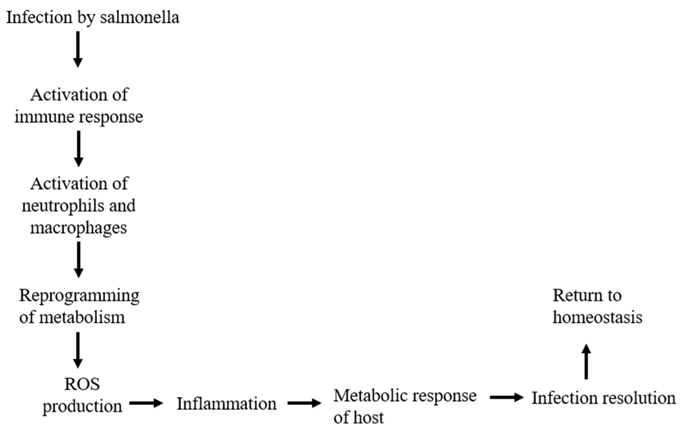
Figure 2. Flow chart of immune metabolism during Salmonella infection.
Preprints 86595 g002
Glycolysis is then important for the T-cell survival and function. It is critical for the gain of function in activated T-cell and particularly for the secretion of IFN- γ. Alteration in glycolysis reported inn disease associated with T-cell disfunction. Highly glycolytic T-cells are described in human inflammatory and infectious diseases (Dimeloe et al., 2017). pkfAB-encoded phosphofructokinase limits the steps in glycolysis (Zuo et al., 2021).
Mitochondria fuse and divide, forming interconnected networks of filamentous organelles or isolated fragmented units. Cells with highly effective oxidative phosphorylation (OXPHOS) have mitochondria with tight cristae (Hoitzing et al., 2015).
Inflammation and phagocytosis of bacteria by neutrophils and macrophages and recruitment of T and B-cells. In systemic Salmonellosis typhoid fever salmonella may target specific types of host cells, such as DC and/or macrophages that favors dissemination through lymphatics and blood stream to MLNs and to deeper tissues. This then leads to transport to spleen, bone marrow, liver and gall bladder. Nramp1 gene encodes for an ion transport and is expressed primarily in macrophages and DC. SPI-1 dependent translocation of bacterial invasion factors into host epithelial cells enables S. typhimurium to penetrate small intestine and peyer’s patches. Initial step of S. typhimurium dissemination from the gastrointestinal tract to host systemic sites like MLNs, spleen, liver and bone marrow (Kaur et al., 2012).

CONCLUSION

Salmonella typhi virulence factors lead to a broad clinical spectrum which fluctuates sever systemic disease to asymptomatic carriage which sometimes often remain undiagnosed. Non-typhoidal Salmonellsis have emerged from infections which are foodborne and are currently treated with antibiotics due to which it is currently developing MDR. Salmonella has the ability to invade both non-phagocytic cells and phagocytic cells and recognition via PAMPs and DAMPs, dendritic cells, macrophages, PPRs, TLRs elicits the immune response for example by secretion of various cytokines such as IL-12, IL-8, IL-16, etc.
Shift of mitochondrial function from normal ATP production to maintaining the immune metabolism by increasing the glycolysis, making OXPHOS highly efficient, and production of ROS.
Genes like Nramp1, which are primarily expressed in macrophages and DCs plays a crucial role in the transport of ions and susceptibility of the infection from Salmonella.
Conclusively, infection of Salmonella typhi is a multiplex interplay of bacteria and immune responses of the host. Understanding the convolutions of interactions such as mitochondrial interplay, glycolysis, and various signaling pathways involved in immune response deliver various insights that can be used for future research and development so as to develop various therapeutic approaches.
Table 1. Salmonella and immune metabolism.
Characteristic Immune metabolism associated with Salmonella Reference
Growth of Salmonella Competes with the host nutrients such as Fe (iron) which is necessary for the growth of Salmonella within host. Khan et al., 2014
Inflammation Inflammation leads to the formation of inflammasome and which releases various pro-inflammatory cytokines. Eckmann et al., 2001
Infection response Host cells seizes Fe availability. Parrow et al., 2013
Reprogramming of metabolism Switch from glucose metabolism to fatty acid oxidation occurs during the course of infection. Ganeshan & Chawla (2014)
Recruitment of immune cells Immune response generated leads to the recruitment of various immune cells such as macrophages, dendritic cells, etc. in which chemotaxis play a major role. Guak & Krawczyk (2020)
Infection resolution After the resolution of infection homeostasis is established again and a switch to OXPHOS occurs so as to repair metabolic damage. Ganeshan & Chawla (2014)
Table 2. Salmonella infection and mitochondrial dynamics.
Characteristic Mitochondrial dynamics associated with Salmonella infection Reference
Targeting mitochondria Salmonella targets various effector protein to translocate into the mitochondria. Layton et al., 2005
Fragmentation of mitochondria Salmonella can lead to the fusion and fission of mitochondria which leads to altered mitochondrial dynamics. Tiku et al., 2020
Energy production Energy is produced through OXPHOS which affects the function of mitochondria thus exerts influence on the host cells. Ramond et al., 2019
Production of ROS Salmonella infection leads to the production of ROS which is one of the crucial productions during infection. Rhen M. 2019
Inflammatory response Mitochondrial damage during the infection of Salmonella leads to the secretion of mt DNA and various proteins which elicits the immune response. Missiroli et al., 2020
Host defense Mitochondrial dynamics plays the defense such the induction of mitophagy which leads to the elimination of damaged mitochondria. Kubli et al., 2012

References

  1. Garai, P., Gnanadhas, D. P., & Chakravortty, D. (2012). Salmonella enterica serovars Typhimurium and Typhi as model organisms: revealing paradigm of host-pathogen interactions. Virulence, 3(4), 377–388. [CrossRef]
  2. Takaya, A., Yamamoto, T., & Tokoyoda, K. (2020). Humoral Immunity vs. Salmonella. Frontiers in immunology, 10, 3155. [CrossRef]
  3. Hurley, D., McCusker, M. P., Fanning, S., & Martins, M. (2014). Salmonella-host interactions - modulation of the host innate immune system. Frontiers in immunology, 5, 481. [CrossRef]
  4. Khan, S., Raj, D., Jaiswal, K., & Lahiri, A. (2020). Modulation of host mitochondrial dynamics during bacterial infection. Mitochondrion, 53, 140–149. [CrossRef]
  5. Xie, J. H., Li, Y. Y., & Jin, J. (2020). The essential functions of mitochondrial dynamics in immune cells. Cellular & molecular immunology, 17(7), 712–721. [CrossRef]
  6. Wang, Y., Branicky, R., Noë, A., & Hekimi, S. (2018). Superoxide dismutases: Dual roles in controlling ROS damage and regulating ROS signaling. The Journal of cell biology, 217(6), 1915–1928. [CrossRef]
  7. Pua, H. H., Guo, J., Komatsu, M., & He, Y. W. (2009). Autophagy is essential for mitochondrial clearance in mature T lymphocytes. Journal of immunology (Baltimore, Md. : 1950), 182(7), 4046–4055. [CrossRef]
  8. Angajala, A., Lim, S., Phillips, J. B., Kim, J. H., Yates, C., You, Z., & Tan, M. (2018). Diverse Roles of Mitochondria in Immune Responses: Novel Insights Into Immuno-Metabolism. Frontiers in immunology, 9, 1605. [CrossRef]
  9. Nakahira, K., Hisata, S., & Choi, A. M. (2015). The Roles of Mitochondrial Damage-Associated Molecular Patterns in Diseases. Antioxidants & redox signaling, 23(17), 1329–1350. [CrossRef]
  10. Page, M. J., Kell, D. B., & Pretorius, E. (2022). The Role of Lipopolysaccharide-Induced Cell Signalling in Chronic Inflammation. Chronic stress (Thousand Oaks, Calif.), 6, 24705470221076390. [CrossRef]
  11. Jiang, L., Wang, P., Song, X., Zhang, H., Ma, S., Wang, J., Li, W., Lv, R., Liu, X., Ma, S., Yan, J., Zhou, H., Huang, D., Cheng, Z., Yang, C., Feng, L., & Wang, L. (2021). Salmonella Typhimurium reprograms macrophage metabolism via T3SS effector SopE2 to promote intracellular replication and virulence. Nature communications, 12(1), 879. [CrossRef]
  12. Mills, E. L., Kelly, B., Logan, A., Costa, A. S. H., Varma, M., Bryant, C. E., Tourlomousis, P., Däbritz, J. H. M., Gottlieb, E., Latorre, I., Corr, S. C., McManus, G., Ryan, D., Jacobs, H. T., Szibor, M., Xavier, R. J., Braun, T., Frezza, C., Murphy, M. P., & O'Neill, L. A. (2016). Succinate Dehydrogenase Supports Metabolic Repurposing of Mitochondria to Drive Inflammatory Macrophages. Cell, 167(2), 457–470.e13. [CrossRef]
  13. Yoshimoto, T., Okamura, H., Tagawa, Y. I., Iwakura, Y., & Nakanishi, K. (1997). Interleukin 18 together with interleukin 12 inhibits IgE production by induction of interferon-gamma production from activated B cells. Proceedings of the National Academy of Sciences of the United States of America, 94(8), 3948–3953. [CrossRef]
  14. Gutiérrez, S., Fischer, J., Ganesan, R., Hos, N. J., Cildir, G., Wolke, M., Pessia, A., Frommolt, P., Desiderio, V., Velagapudi, V., & Robinson, N. (2021). Salmonella Typhimurium impairs glycolysis-mediated acidification of phagosomes to evade macrophage defense. PLoS pathogens, 17(9), e1009943. [CrossRef]
  15. Lopez-Castejon, G., & Brough, D. (2011). Understanding the mechanism of IL-1β secretion. Cytokine & growth factor reviews, 22(4), 189–195. [CrossRef]
  16. Dimeloe, S., Burgener, A. V., Grählert, J., & Hess, C. (2017). T-cell metabolism governing activation, proliferation and differentiation; a modular view. Immunology, 150(1), 35–44. [CrossRef]
  17. Zuo, J., Tang, J., Lu, M., Zhou, Z., Li, Y., Tian, H., Liu, E., Gao, B., Liu, T., & Shao, P. (2021). Glycolysis Rate-Limiting Enzymes: Novel Potential Regulators of Rheumatoid Arthritis Pathogenesis. Frontiers in immunology, 12, 779787. [CrossRef]
  18. Hoitzing, H., Johnston, I. G., & Jones, N. S. (2015). What is the function of mitochondrial networks? A theoretical assessment of hypotheses and proposal for future research. BioEssays : news and reviews in molecular, cellular and developmental biology, 37(6), 687–700. [CrossRef]
  19. Kaur, J., & Jain, S. K. (2012). Role of antigens and virulence factors of Salmonella enterica serovar Typhi in its pathogenesis. Microbiological research, 167(4), 199–210. [CrossRef]
  20. Khan C. M. (2014). The Dynamic Interactions between Salmonella and the Microbiota, within the Challenging Niche of the Gastrointestinal Tract. International scholarly research notices, 2014, 846049. [CrossRef]
  21. Eckmann, L., & Kagnoff, M. F. (2001). Cytokines in host defense against Salmonella. Microbes and Infection, 3(14-15), 1191-1200.
  22. Parrow, N. L., Fleming, R. E., & Minnick, M. F. (2013). Sequestration and scavenging of iron in infection. Infection and immunity, 81(10), 3503–3514. [CrossRef]
  23. Ganeshan, K., & Chawla, A. (2014). Metabolic regulation of immune responses. Annual review of immunology, 32, 609–634. [CrossRef]
  24. Guak, H., & Krawczyk, C. M. (2020). Implications of cellular metabolism for immune cell migration. Immunology, 161(3), 200–208. [CrossRef]
  25. Layton, A. N., Brown, P. J., & Galyov, E. E. (2005). The Salmonella translocated effector SopA is targeted to the mitochondria of infected cells. Journal of bacteriology, 187(10), 3565–3571. [CrossRef]
  26. Tiku, V., Tan, M. W., & Dikic, I. (2020). Mitochondrial Functions in Infection and Immunity. Trends in cell biology, 30(4), 263–275.
  27. Ramond, E., Jamet, A., Coureuil, M., & Charbit, A. (2019). Pivotal Role of Mitochondria in Macrophage Response to Bacterial Pathogens. Frontiers in immunology, 10, 2461. [CrossRef]
  28. Rhen M. (2019). Salmonella and Reactive Oxygen Species: A Love-Hate Relationship. Journal of innate immunity, 11(3), 216–226. [CrossRef]
  29. Missiroli, S., Genovese, I., Perrone, M., Vezzani, B., Vitto, V. A. M., & Giorgi, C. (2020). The Role of Mitochondria in Inflammation: From Cancer to Neurodegenerative Disorders. Journal of clinical medicine, 9(3), 740. [CrossRef]
  30. Kubli, D. A., & Gustafsson, Å. B. (2012). Mitochondria and mitophagy: the yin and yang of cell death control. Circulation research, 111(9), 1208–1221. [CrossRef]
  31. Thapa SB, Pandey RP, Bashyal P, Yamaguchi T, Sohng JK. Cascade biocatalysis systems for bioactive naringenin glucosides and quercetin rhamnoside production from sucrose. Appl Microbiol Biotechnol. 2019 Oct;103(19):7953-7969. [CrossRef]
  32. Pandey RP, Bashyal P, Parajuli P, Yamaguchi T, Sohng JK. Two Trifunctional Leloir Glycosyltransferases as Biocatalysts for Natural Products Glycodiversification. Org Lett. 2019 Oct 4;21(19):8058-8064. [CrossRef]
  33. Kyu-Min Kim, Jin-Soo Park, HaeRi Choi, Min-Seon Kim, Joo-Hyun Seo, Ramesh Prasad Pandey, Jin Woo Kim, Chang-Gu Hyun & Seung-Young Kim (2018) Biosynthesis of novel daidzein derivatives using Bacillus amyloliquefaciens whole cells, Biocatalysis and Biotransformation, 36:6, 469-475. [CrossRef]
  34. Parajuli P, Pandey RP, Trang NTH, Oh TJ, Sohng JK. Expanded acceptor substrates flexibility study of flavonol 7-O-rhamnosyltransferase, AtUGT89C1 from Arabidopsis thaliana. Carbohydr Res. 2015 Dec 11;418:13-19. [CrossRef]
  35. Shin JY, Pandey RP, Jung HY, Chu LL, Park YI, Sohng JK. In vitro single-vessel enzymatic synthesis of novel Resvera-A glucosides. Carbohydr Res. 2016 Apr 7;424:8-14. [CrossRef]
  36. Pandey, R., and M. M. Agarwal. "INFLUENCE OF FERTILITY LEVELS, VARIETIES AND TRANSPLANTING TIME ON RICE (ORYZA-SATIVA)." Indian Journal of Agronomy 36.4 (1991): 459-463.
Disclaimer/Publisher’s Note: The statements, opinions and data contained in all publications are solely those of the individual author(s) and contributor(s) and not of MDPI and/or the editor(s). MDPI and/or the editor(s) disclaim responsibility for any injury to people or property resulting from any ideas, methods, instructions or products referred to in the content.
Copyright: This open access article is published under a Creative Commons CC BY 4.0 license, which permit the free download, distribution, and reuse, provided that the author and preprint are cited in any reuse.
Prerpints.org logo

Preprints.org is a free preprint server supported by MDPI in Basel, Switzerland.

Subscribe

© 2024 MDPI (Basel, Switzerland) unless otherwise stated