Preprint
Article

Cu(Ⅱ) Coordination Polymers Containing Mixed Ligands with Different Flexibility: Structural Diversity and Iodine Adsorption

Altmetrics

Downloads

109

Views

74

Comments

0

A peer-reviewed article of this preprint also exists.

Submitted:

04 December 2023

Posted:

05 December 2023

You are already at the latest version

Alerts
Abstract
Reactions of N‚N'-bis(3-methylpyridyl)oxalamide (L1), N,N’-bis(3-methylpyridyl)adipoamide (L2) and N,N’-bis(3-methylpyridyl)sebacoamide (L3) with tricarboxylic acids and Cu(II) salts afforded {[Cu(L1)(1,3,5-HBTC)]·H2O}n (1,3,5-H3BTC = 1,3,5-benzenetricarboxylic acid), 1, {[Cu1.5(L2)1.5(1,3,5-BTC)(H2O)2]·6.5H2O}n, 2, [Cu(L2)0.5(1,3,5-HBTB)]n (1,3,5-H3BTB = 1,3,5-tri(4-carboxyphenyl)benzene), 3, [Cu4(L3)(OH)2(1,3,5-BTC)2]n, 4, {[Cu3(L3)2(1,3,5-BTB)2]·2.5MeOH·2H2O}n, 5, and {[Cu3(L3)2(1,3,5-BTB)2 ]·DMF·2H2O}n, 6, which have been structurally characterized by using single crystal X-ray crystallography. Complexes 1 – 4 form a 2D layer with the {44.62}-sql topology, a 2D layer with the (4.62)2(42.62.82)-bex topology, a 3-fold interpenetrated 3D net with the (412.63)-pcu topology and a 3D framework with the (410.632.83)(42.6)2(43.63) topology, respectively, whereas 5 and 6 are 3D frameworks with the (63)2(64.82)(68.85.102) topology. Complex 5 shows a better iodine-adsorption factor of 290.0 mg g-1 at 60 oC for 360 minutes than the other ones, revealing that the flexibility of the spacer ligand governs the structural diversity and the adsorption capacity.
Keywords: 
Subject: Chemistry and Materials Science  -   Inorganic and Nuclear Chemistry

1. Introduction

Coordination polymers (CPs) have shown crucial applications in many different areas due to their diverse structures and variable functions [1,2,3,4,5,6,7]. CPs can be constructed by the coordination of the designable spacer ligands to the metal ions, and through the self-assembly process, one- (1D), two- (2D) or three-dimensional (3D) network structures can be prepared.
The –(CH2)n– group of the bis-pyridyl-bis-amide (bpba) possesses suitable flexibility that may adopt the coordination environment of different metal ions, whereas the two amide groups play important roles as abundant potential hydrogen bond sites, affording CPs with remarkable topologies [8]. On the other hand, polycarboxylate ligands that show distinct coordination modes involving chelating and bridging are also important in the organization of CPs in a mixed system [9]. Benzene-1,3,5-tricarboxylic acid (1,3,5-H3BTC) is a planar molecule with C3-symmetry that may give anions of the types, BTC3- and HBTC2-, and intriguing structural types have been found in the bpba-based CPs supported by these anions [10,11,12]. Extension of 1,3,5-H3BTC to the larger 1,3,5-tri(4-carboxyphenyl)benzene (1,3,5-H3BTB) may thus afford CPs with different structural topology.
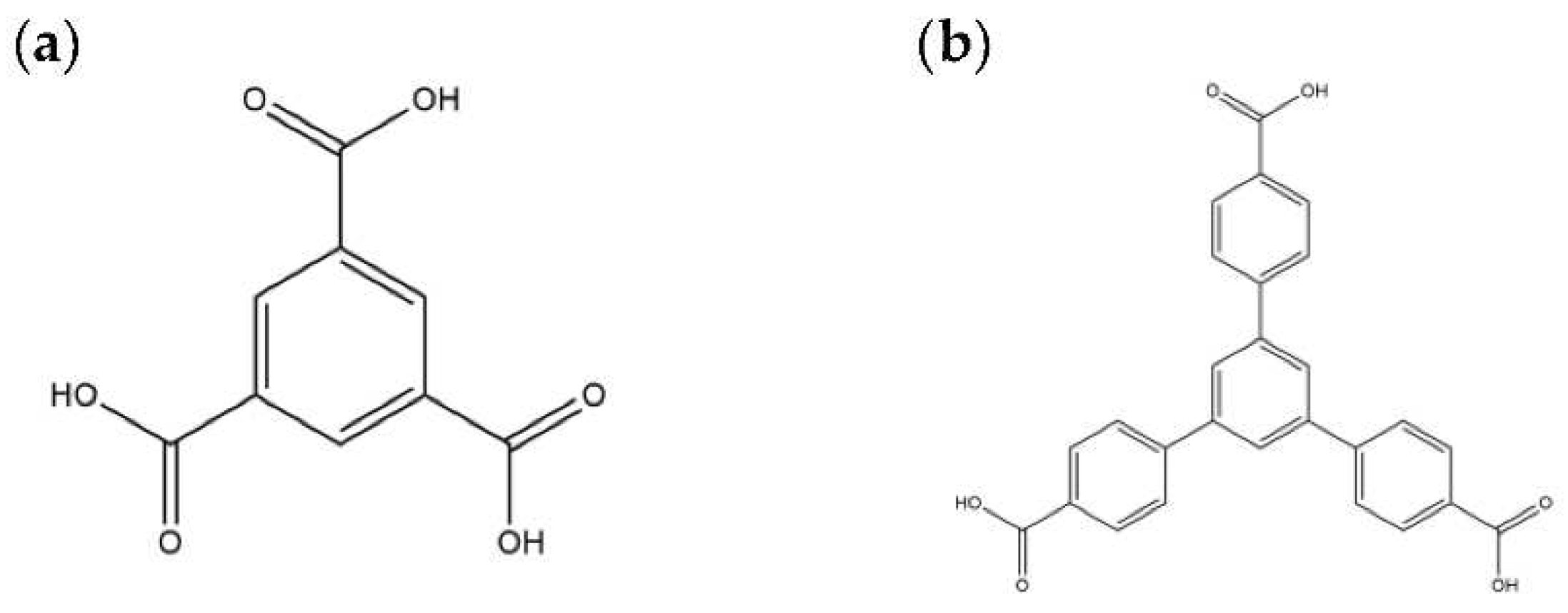
We are dedicated to illuminate the factors that may direct the structural diversity and govern the adsorption property of the flexible bpba-based CPs by exploring the variations in ligand conformation and coordination mode of the spacer ligands. In this study, flexible N‚N'-bis(3-pyridylmethyl)oxalamide (L1), N,N’-bis(3-methylpyridyl)adipoamide (L2) and N,N’-bis(3-methylpyridyl)sebacoamide (L3), Figure 1, were reacted with the Cu(II) metal salts and the tricarboxylic acids, 1,3,5-H3BTC, and 1,3,5-H3BTB, Figure 2, to yield {[Cu(L1)(1,3,5-HBTC)]·H2O}n (1,3,5-H3BTC = 1,3,5- benzenetricarboxylic acid), 1, {[Cu1.5(L2)1.5(1,3,5-BTC)(H2O)2]·6.5H2O}n, 2, [Cu(L2)0.5(1,3,5-HBTB)]n (1,3,5-H3BTB = 1,3,5-tri(4-carboxyphenyl)benzene), 3, [Cu4(L3)(OH)2(1,3,5-BTC)2]n, 4, {[Cu3(L3)2(1,3,5-BTB)2]·2.5MeOH·2H2O}n, 5, {[Cu3(L3)2(1,3,5-BTB)2]·DMF·2H2O}n, 6. The synthesis and structures of 1 – 6 as well as their iodine adsorptions form the subject of this report.

2. Results and Discussion

2.1. Structure of 1

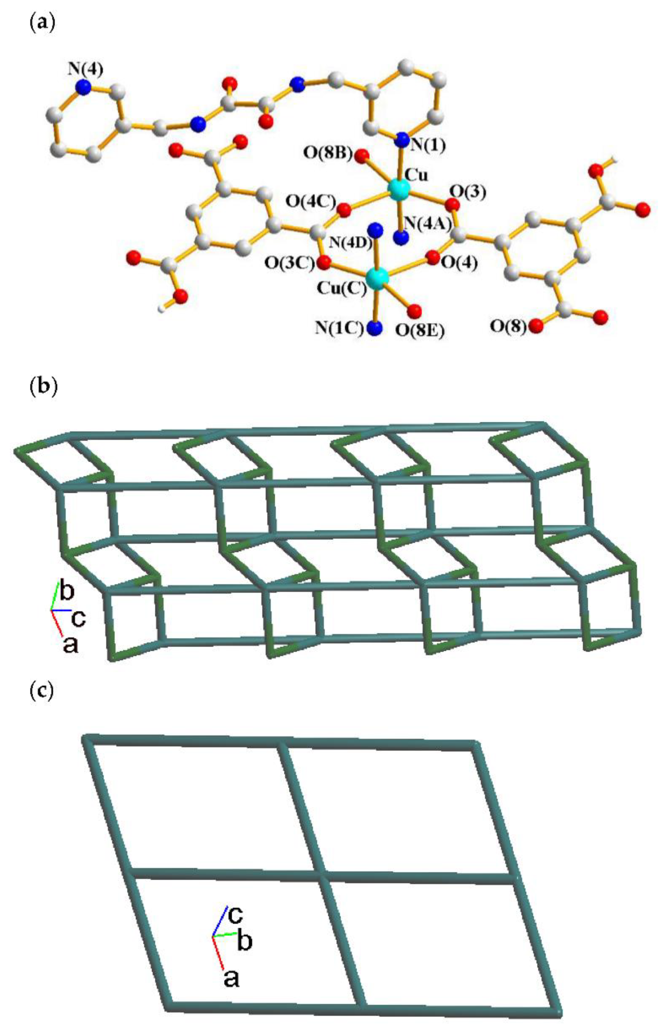
A single-crystal X-ray diffraction analysis shows that complex 1 crystallizes in the triclinic space group Pī. There are one Cu(II) cation, one L1 ligand, one 1,3,5-HBTC2- ligand and one cocrystallized water molecule in the asymmetric unit. The Cu(II) atom is coordinated by two nitrogen atoms from L1 ligands [Cu-N = 2.001(3) - 2.007(3) Å] and three oxygen atoms from three 1,3,5-HBTC2- ligands [Cu-O = 1.966(3) - 2.237(3) Å], resulting in a trigonal bipyramidal geometry, and a dicopper unit bridged by the 1,3,5-HBTC2- ligands, Figure 3a. The Cu(II) ions are linked together by 1,3,5-HBTC2- and L1 ligands to afford a 2D structure. If the 1,3,5-HBTC2- ligands are considered as 3-connected nodes and the Cu(II) ions as 5-connected nodes, the structure of 1 can be simplified as 3,5-connected binodal 2D net with the (42⋅67⋅8)(42⋅6)-3,5L2 topology (standard representation), Figure 3b.determined by using ToposPro program [13]. If the dinuclear Cu(II) units are defined as 4-connected nodes, the structure can be simplified as a 4-connected net with the (44⋅62)-sql topology (cluster representation), Figure 3c.

2.2. Structure of 2

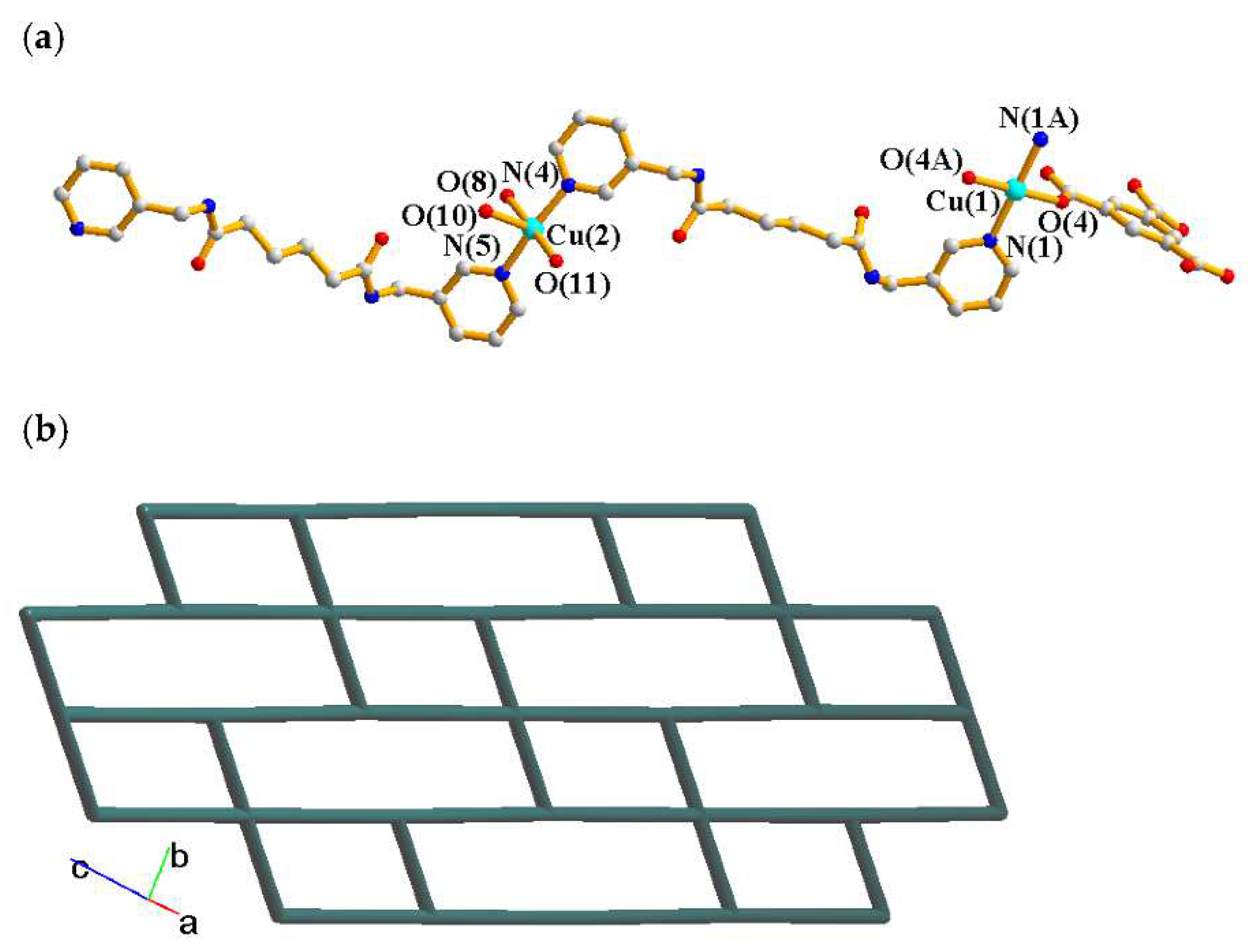
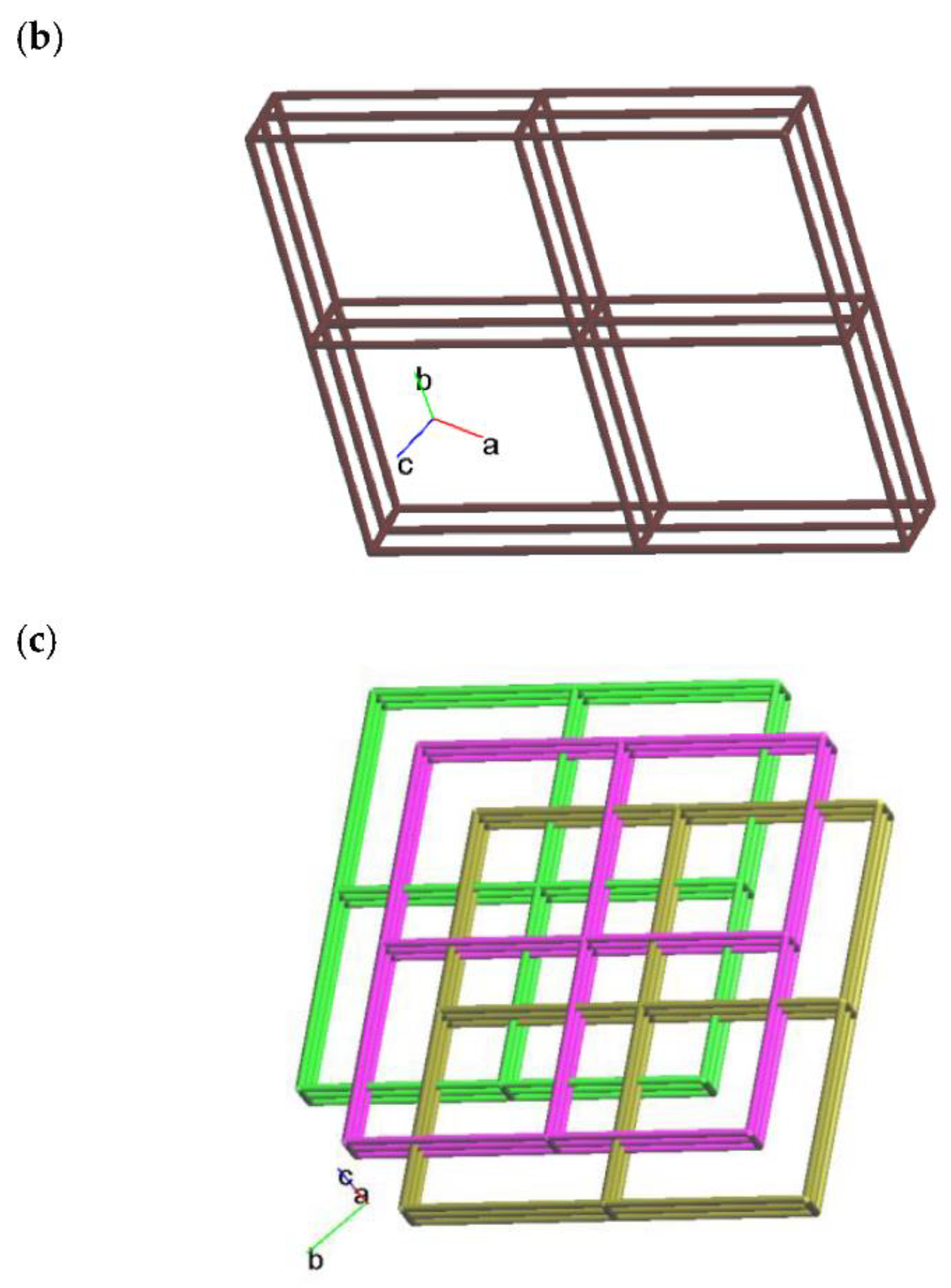
Crystals of complex 2 conform to the triclinic space group Pī and each asymmetric unit consists of two Cu(II) cations, one and a half of an L2 ligands, one 1,3,5-BTC3- ligand, two coordinated water and six and a half of a cocrystallized water molecules. The Cu(1) and Cu(2) metal centers are four- and five-coordinated, respectively, Figure 4a. While the Cu(1) atom is coordinated by two nitrogen atoms from the L2 ligand [Cu-N = 2.051(2) Å] and two oxygen atoms from two 1,3,5-BTC3- ligands [Cu-O = 1.960(2) Å], resulting in a distorted square geometry, the Cu(2) atom is coordinated by two nitrogen atoms from two L2 ligands [Cu-N = 2.000(3) - 2.006(3) Å], one oxygen atom from the 1,3,5-BTC3- ligand [Cu-O = 1.978(2) Å] and two oxygen atoms from two coordinated water molecules [Cu-O = 2.023(2) – 2.200(2) Å], giving a square pyramidal geometry. The Cu(II) ions are linked together by the 1,3,5-BTC3- and L2 ligands to afford a 2D layer. If the Cu(1) cations are defined as 4-connected nodes and Cu(2) cations as 3-connected nodes, the structure can be simplified as a 3,4-connected 2D net with the (4.62)2(42.62.82)-bex topology, Figure 4b.

2.3. Structure of 3

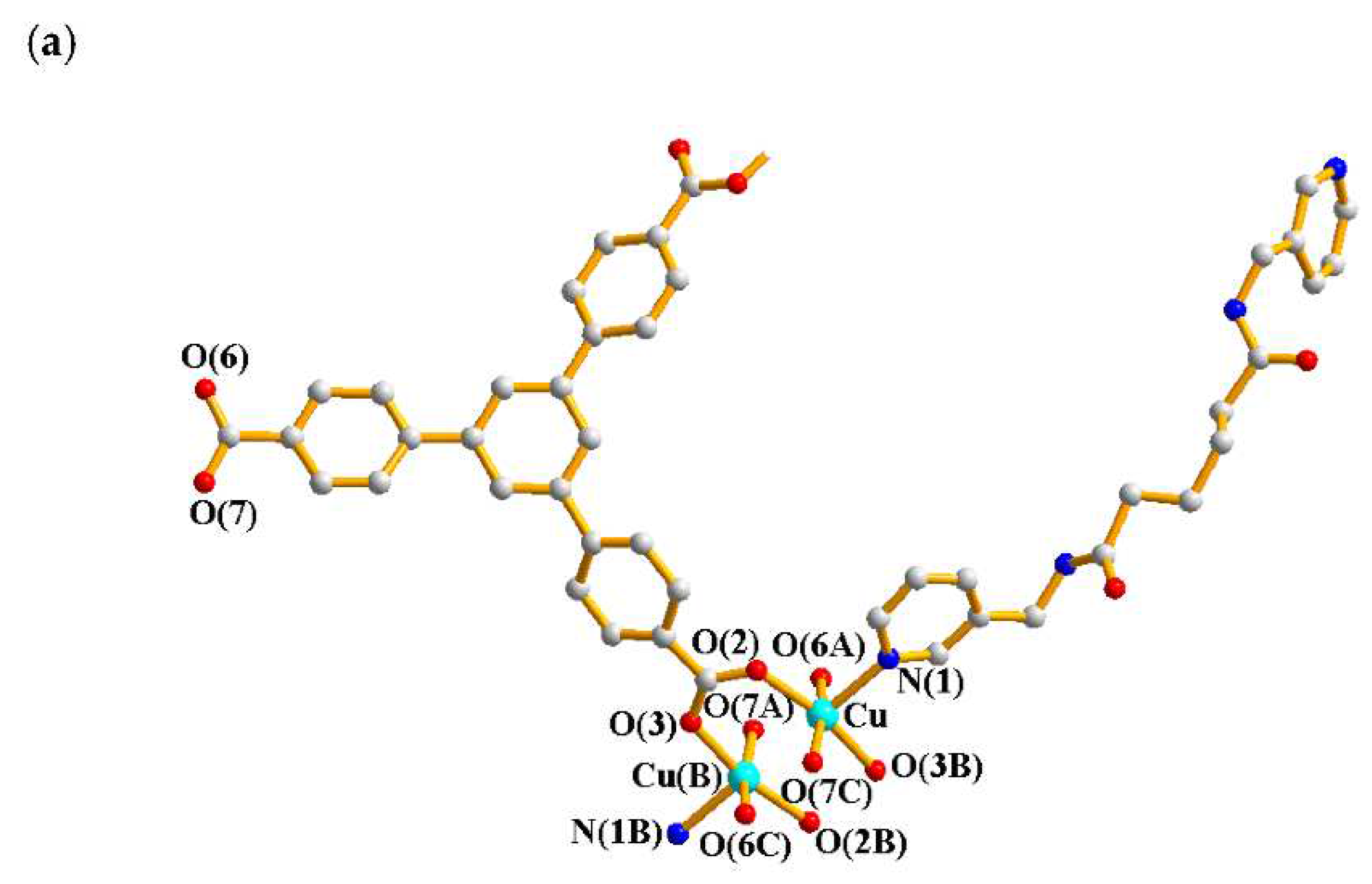
Complex 3 crystallizes in the monoclinic space group C2/c and the asymmetric unit comprises one Cu(II) cation, a half of an L2 ligand and one 1,3,5-HBTB2- ligand. The Cu(II) cation is coordinated by one nitrogen atom from the L2 ligand [Cu-N = 2.170(2) Å] and four oxygen atoms from four 1,3,5-HBTB2- ligands [Cu-O = 1.9623(19) – 1.9763(18) Å], resulting in a distorted square pyramidal geometry, Figure 5a. Two Cu(II) cations are bridged by the 1,3,5-HBTB2- ligand to form a dinuclear unit with a Cu---Cu distance of 2.6516(6) Å that is shorter than the sum of two van der Waals radius of Cu (2.8 Å), suggesting the presence of weak intermolecular forces. The Cu(II) ions are linked together by 1,3,5-HBTB2- and L2 ligands to afford a 3D structure. If the dinuclear Cu(II) units are defined as 6-connected nodes, the structure can be simplified as a 6-connected net with the (412⋅63)-pcu topology, Figure 5b. The 3D nets penetrate into the neighbors to form a 3-fold 3D interpenetration structure, Figure 5c, demonstrating that the combination of the flexible L2 and 1,3,5-HBTB2- may lead to the formation of the entangled CP [14].

2.4. Structure of 4

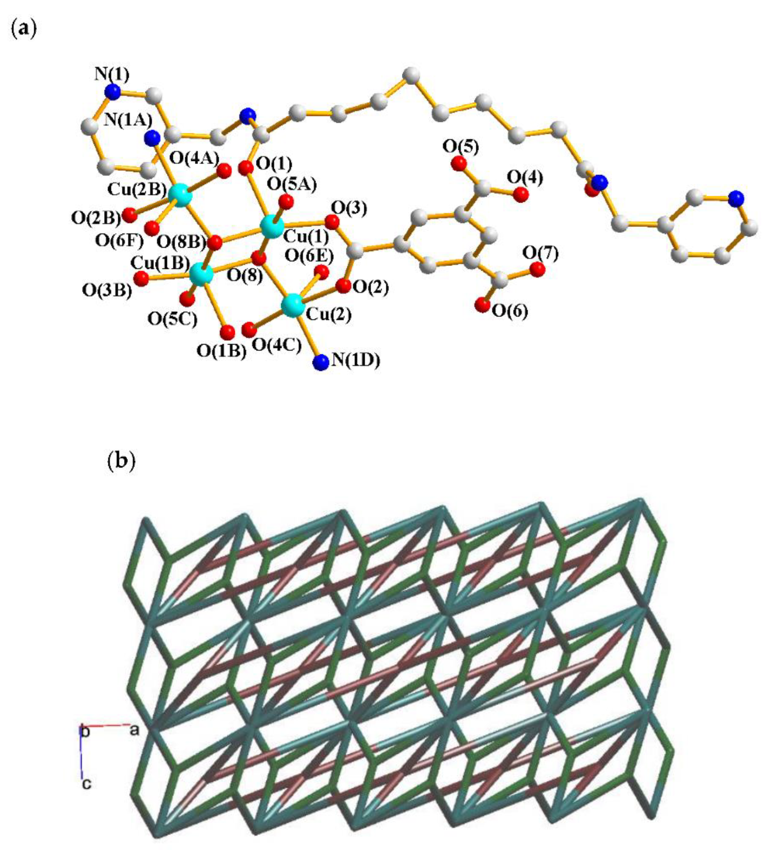
Crystals of complex 4 conform to the monoclinic space group C2/c. The asymmetric unit consists of two Cu(II) cations, a half of an L3 ligand, one 1,3,5-BTC3- ligand and one hydroxide ion. Figure 6a depicts the coordination environments about the Cu(II) ions. The Cu(1) and Cu(1B) atoms are symmetry-related by the inversion center and each atom is coordinated by one oxygen atom from the L3 ligand [Cu-O = 2.2720(16) Å], two oxygen atoms from two 1,3,5-BTC3- ligands [Cu-O = 1.9377(14) - 19434(14) Å] and two oxygen atoms from two hydroxy groups [Cu-O = 1.9551(13) - 1.9620(13) Å], forming the square pyramidal geometry. On the other hand, each of the Cu(2) and Cu(2B) atoms is coordinated by one nitrogen atom from the L3 ligand [Cu-N = 2.0164(17) Å], three oxygen atoms from three 1,3,5-BTC3- ligands [Cu-O = 1.9473(13) - 2.2909(15) Å] and one oxygen atom from the hydroxy group [Cu-O = 1.9482(13) Å], resulting in a distorted pentagonal bipyramidal geometry. The Cu(II) ions are linked together by 1,3,5-BTC3- and L3 ligands to afford a 3D structure. If the tetranuclear Cu(II) units are defined as 10-connected nodes, 1,3,5-BTC3- ligands as 3-connected nodes and L3 as 4-connected nodes, the structure can be simplified as a 3,4,10-connected trinodal net with the point symbol of (410⋅632⋅83)(42⋅6)2(43⋅63) topology, Figure 6b.

2.5. Structure of 5 and 6

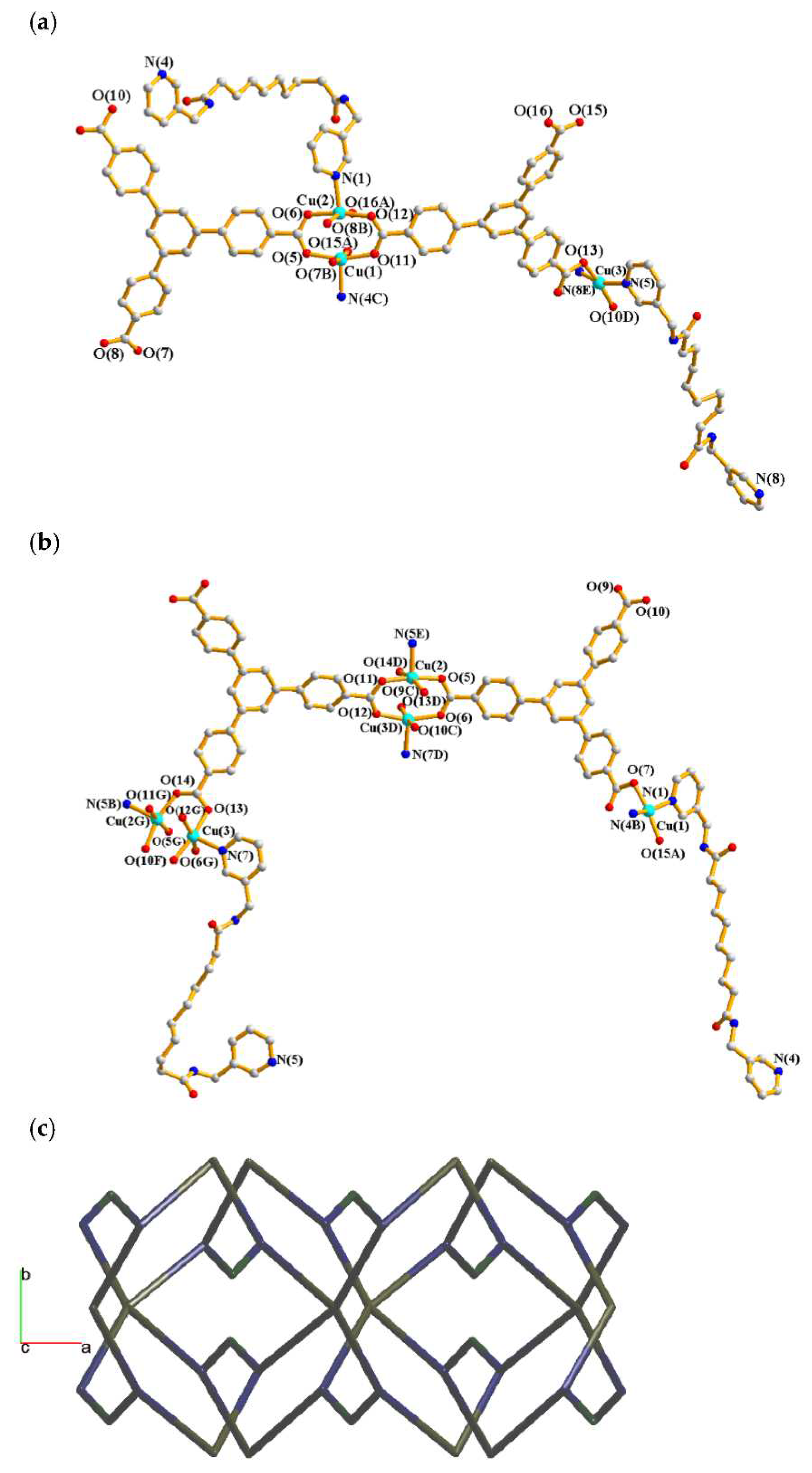
Complexes 5 and 6 crystallize in the orthorhombic space group Pna21. Each of the asymmetric units of 5 and 6 comprise three Cu(II) cations, two L3 ligands and two 1,3,5-BTB3- ligands, with additional two and a half of a cocrystallized methanol molecule and two cocrystallized water molecules in 5, and two cocrystallized DMF molecules and two cocrystallized water molecules in 6, respectively. Figure 7a shows the coordination environments about the Cu(II) ions in 5. While the Cu(1) atom is coordinated by one nitrogen atom from the L3 ligand [Cu(1)-N = 2.164(6) Å] and four oxygen atoms from four 1,3,5-BTB3- ligands [Cu-O = 1.940(4) – 2.005(4) Å], the Cu(2) atom is coordinated by one nitrogen atom from L3 ligand [Cu(2)-N = 2.189(6) Å] and four oxygen atoms from four 1,3,5-BTB3- ligands [Cu-O = 1.933(5) – 2.189(6) Å], resulting in square pyramidal geometries of both of the Cu(1) and Cu(2) atoms. The Cu(1) and Cu(2) atoms are bridged by the 1,3,5-BTB3- ligands to form a dinuclear unit with a Cu---Cu distance of 2.6843(9) Å, indicating the presence of weak intermolecular forces. The Cu(3) atom is coordinated by two nitrogen atoms from two L3 ligand [Cu-N = 2.054(7) and 2.079(7) Å] and two oxygen atoms from two 1,3,5-BTB3- ligands [Cu-O = 1.913(6) and 1.917(5) Å], displaying a distorted square planar geometry.
Figure 7b shows the coordination environments about the Cu(II) ions in 6. The Cu(1) atom is coordinated by two nitrogen atoms from L3 ligand [Cu-N = 2.027(6) and 2.034(6) Å] and two oxygen atoms from two 1,3,5-BTB3- ligands [Cu-O = 1.918(5) and 1.941(5) Å], resulting in a distorted square planar geometry. Each of the Cu(2) and Cu(3) atoms is coordinated by one nitrogen atom from L3 ligand [Cu(2)-N = 2.183(5) Å; Cu(3)-N = 2.181(6) Å] and four oxygen atoms from four 1,3,5-BTB3- ligands [Cu(2)-O = 1.929(4) – 1.987(4) Å; Cu(3)-O = 1.929(4) – 2.016(4) Å], resulting in square pyramidal geometries for Cu(2) and Cu(3). The Cu---Cu distance of 2.6922(9) Å between Cu(2) and Cu(3) is longer that in complex 5, indicating that the Cu(II)---Cu(II) interaction is subject to the nature of the cocrystallized solvent molecules. The Cu(II) ions in 5 and 6 are linked together by 1,3,5-BTB3- and L3 ligands to afford 3D structures. If the dinuclear Cu(II) units are defined as 6-connected nodes, the mononuclear cations as 4-connected nodes and 1,3,5-BTB3- as 3-connected nodes, while the L3 ligands as linkers, the structures of 5 and 6 can be simplified as 3,4,6-connected 3D nets with the point symbol of (63)2(64⋅82)(68⋅85⋅102), Figure 7c.

2.6. Ligand Conformations and Coordination Modes

The ligand conformations of the bpba ligands have been proposed based on the torsion angles (θ) of their methylene carbon atoms [0 θ 90o, gauche (G) and 90 < θ 180o, anti (A)]. On the other hand, cis and trans are given if the two C=O groups are in the same and opposite directions, respectively. Three orientation, syn-syn, syn-anti and anti-anti, are also defined based on the relative position of pyridyl nitrogen and amide oxygen atoms [8]. Accordingly, the ligand conformations of L1L3 in 16 are listed in Table 1. It is also noted that while the bpba ligands in 1, 2, 3, 5 and 6 bridge two Cu(II) ions through the two pyridyl nitrogen atoms, that in 4 bridge four Cu(II) ions through two pyridyl nitrogen and two amide oxygen atoms. Noticeably, although complexes 5 and 6 adopt the same structural type, the ligand conformations of the L3 ligands are significantly different, presumably due to the difference in the cocrystallized solvents. Moreover, the tricarboxylate ligands in 16 bridge two to five Cu(II) ions through various coordination modes, which are also listed in Table 1.

2.7. Powder X-ray Analysis

In order to check the phase purity of the products, powder X-ray diffraction (PXRD) experiments have been carried out for all complexes. As shown in Figures S7–S12, the peak positions of the experimental and simulated PXRD patterns are in agreement with each other, which demonstrate that the crystal structures are truly representative of the bulk materials. The differences in intensity may be owing to the preferred orientation of the powder samples.

2.8. Thermal Properties

Thermal gravimetric analysis (TGA) was carried out to examine the thermal decomposition of 30 to 800 or 900 ℃. The sample were heated in nitrogen gas at a pressure of 1 atm with heating rate of 10 ℃ min-1 and finished at 800 ℃ or 900 ℃, Table 2, Figures S13–S18, indicating that complexes 16 display two-step weight loss involving loss of solvent and loss of organic ligands on heating.

2.9. Iodine Adsorption

Radioactive iodine such as 129I represents one of the most critical nuclear wastes, which is harmful to human health and has to be captured and disposed of effectively. [15,16,17] On the other hand, CPs possessing porous structures may facilitate iodine adsorption through noncovalent interactions involving iodine and various sorption sites. Iodine adsorption experiments were thus carried out for complexes 1 - 6 to evaluate the degree of adsorption of iodine vapor at 25 and 60 oC within time interval of 30, 60, 120, 180 and 360 minutes, respectively. For each experiment, 10 mg of the complex was placed in a 4 mL sample bottle inside a 20 mL sample bottle containing 100 mg of iodine, which was then sealed and kept in the oven. Each experiment was repeated three times and the results averaged. It can be found that the colors of the complexes are different at different temperatures and time intervals, Figures S19–S30.
Tables S1–S12 summarize the I2-adsorption of 1 - 6, followed by the plots displaying iodine vapor adsorption rates. With the increase of temperature from 25 to 60 oC, the absorption rate of iodine also has a good increase for each complex, giving a best adsorption factor of 290.0 mg g-1 at 60 oC for 360 minutes for 5. In order to confirm whether the structures of the iodine-adsorbed complexes remain unchanged, their powder X-ray diffraction (PXRD) patterns were measured. As shown in Figures S31–S42, most of the experimental patterns are consistent with the theoretical ones, but 2 at 60 oC for 30 and 60 minutes, and 5 and 6 at 60 oC for 60 minutes display some changes.
The ability of the CPs to adapt iodine molecules to the voids of the network structures may govern the iodine adsorption capacity [18,19,20,21]. The solvent accessible volumes calculated by using the PLATON program [22] for 16 were 1.5, 17.3, 34.4, 2.9, 11.8 and 10.4 %, respectively, of the unit cell volume, indicating that complex 3 that displays the 3-fold interpenetrated 3D net with the (412⋅63)-pcu topology may accommodate more iodine than the other complexes. However, the best adsorption factor of 290.0 mg g-1 at 60 oC for 360 minutes was observed for 5, demonstrating the important role of the flexibility of the neutral spacer ligands, L1, L2, and L3, in determining the iodine adsorption capacities of 16. The 3D framework of 5 containing the flexible L3 may be more susceptible to the changes of the ligand conformation upon the attack of the iodine molecules and thus more appropriate to accommodate the iodine molecules, which can probably be verified by the subtle change of the PXRD pattern of 5, Figure S40, upon iodine adsorption at 60 oC. On the other hand, the framework of the entangled 3 comprising L2 is not vulnerable to the iodine attack, thus allowing less iodine adsorption. The different performances in iodine adsorption between 5 and 6 are presumably due to the different cocrystallized solvents.

2.10. Energy Dispersive X-ray (EDX) Analysis

EDX analyses were performed for complexes 16 after iodine adsorption to investigate their iodine uptakes, Figures S43–S48. Three positions of the iodine-adsorbed sample of complexes 1 – 6 were selected for each measurement and the amount of iodine is different for each position, indicating the inhomogeneous distribution of iodine in the iodine-adsorbed sample.

2.11. Gas Adsorption

Low-pressure N2 adsorption and desorption measurements were performed at 77 K for complexes 16, which were heated at 120 ℃ for 24 hours to obtain fully activated samples before the measurements. Figures S49–S54 demonstrate that the complexes 1 and 36 remain stable upon the removal of the solvent molecules, while the structure of 2 has changed. The Brunauer–Emmet–Teller (BET) surface areas are 7.49, 13.60, 12.82, 12.60, 11.73, 7.98 m²/g and the N2 uptake capacities are 6.93, 18.33, 12.70, 11.28, 10.83 and 8.12 cm³/g, respectively, for 1 – 6, Figures S55–S60, indicating larger surface area and N2 uptake for 2 upon desolvation. Moreover, pore-size distribution curves show that the pore sizes are 2.9, 3.4, 3.4, 3.7, 3.4 and 3.4 nm respectively, for 16, Figures S61–S66.
As demonstrated by the experiments, the BET surface area and N2 uptakes of 16 derived from the low-pressure N2 adsorption and desorption measurements are not closely related to their iodine adsorption capacities. Therefore, the iodine adsorption capacity may also depend on the characteristics of the CPs and their surface features.

3. Materials and Methods

3.1. Materials

The reagent Cu(CH3COO)2·H2O was purchased from Showa, 1,3,5- benzenetricarboxylic acid (1,3,5-H3BTC) from Alfa Aesar and 1,3,5-tri(4-carboxyphenyl)benzene (1,3,5-H3BTB) from Alfa Aesar. The ligands N‚N'-bis(3-methylpyridyl)oxalamide (L1), N‚N'-bis(3-methylpyridyl)adipoamide (L2) and N‚N'-bis(3-methylpyridyl)sebacoamide (L3) were prepared according to published procedures [23,24,25].

3.2. Preparations

3.3.1.{[. Cu(L1)(1,3,5-HBTC)]·H2O}n, 1

A mixture of Cu(CH3COO)2·H2O (0.020 g, 0.10 mmol), L1 (0.027 g, 0.10 mmol) and 1,3,5-H3BTC (0.021 g, 0.10 mmol) in 10 mL of H2O was sealed in a 23 mL Teflon-lined steel autoclave, which was heated under autogenous pressure to 120 ℃ for two days, and then cooled down to room temperature for two days. Blue crystals suitable for single-crystal X-ray diffraction were obtained. Yield: 0.081 g (72 %). Anal. Calcd for C23H20CuN4O9 (MW = 599.97): C, 49.33; N, 10.00; H, 3.60 %. Found: C, 49.39; N, 9.85; H, 3.48 %. FT-IR (cm-1): 3313(s), 2357(m), 1621(s), 1519(m), 1430(m), 1351(s), 1099(m), 860(w), 756(m), 609(w), 512(w).

3.2.2.{[. Cu1.5(L2)1.5(1,3,5-BTC)(H2O)2]·6.5H2O}n, 2

Complex 2 was prepared by following the similar procedures for 1, except that Cu(CH3COO)2·H2O (0.020 g, 0.10 mmol), L2 (0.033 g, 0.10 mmol) and 1,3,5-H3BTC (0.021 g, 0.10 mmol) in 10 mL of NaOH (0.01 M) aqueous solution were used, which was heated at 100 ℃. Blue crystals were obtained. Yield: 0.054 g (86 %). Anal. Calcd for C36H53Cu1.5N6O17.50 (MW = 945.15): C, 45.75; N, 8.89; H, 5.65 %. Found: C, 46.19; N, 8.69; H, 5.62 %. FT-IR (cm-1): 3054(m), 2359(m), 1606(s), 1427(m), 1352(s), 1196(m), 1058(w), 811(w), 710 (m).

3.3.3.[. Cu(L2)0.5(1,3,5-HBTB)]n, 3

Complex 3 was prepared by following the similar procedures for 1, except that Cu(CH3COO)2·H2O (0.020 g, 0.10 mmol), L2 (0.033 g, 0.10 mmol) and 1,3,5-H3BTB (0.044 g, 0.10 mmol) in 7 mL of H2O and 3 mL of DMA were used. Green crystals were obtained. Yield: 0.032 g (42 %). Anal Calcd for C36H27CuN2O7 (MW = 663.13): C, 65.20; N, 4.22; H, 4.10 %. Anal Calcd for C36H27CuN2O7·0.5DMA·3H2O (MW = 760.72): C, 59.99; N, 4.60; H, 4.97 %. Found: C, 59.94; N, 4.84; H, 4.73 %. FT-IR (cm-1): 2360(m), 1604(s), 1390(s), 1176(w), 1015(m), 853(w), 774(s), 669(w).

3.3.4.[. Cu4(L3)(OH)2(1,3,5-BTC)2]n, 4

Complex 4 was prepared by following the similar procedures for 1, except that Cu(CH3COO)2·H2O (0.020 g, 0.10 mmol), L3 (0.038 g, 0.10 mmol) and 1,3,5-H3BTC (0.021 g, 0.10 mmol) in 10 mL of H2O were used. Blue crystals were obtained. Yield: 0.016 g (60 %). Anal. Calcd for C40H38Cu4N4O16 (MW = 1084.90): C, 44.28; N, 5.16; H, 3.53 %. Found: C, 44.49; N, 5.22; H, 3.87 %. FT-IR (cm-1): 3238 (m), 2363(m), 1583(s), 1442(m), 1353(s),1094(w), 761(m), 716(m), 586(w).

3.3.5.{[. Cu3(L3)2(1,3,5-BTB)2]·2.5MeOH·2H2O}n, 5, and {[Cu3(L3)2(1,3,5-BTB)2 ]·DMF·2H2O}n, 6

Complexes 5 and 6 were prepared by following the similar procedures for 1, but in different solvent systems. While complex 5 was prepared from a reaction mixture of Cu(CH3COO)2·H2O (0.020 g, 0.10 mmol), L3 (0.038 g, 0.10 mmol) and 1,3,5-H3BTB (0.044 g, 0.10 mmol) in 3 mL of H2O and 7 mL of MeOH, complex 6 was obtained in 7 mL of H2O and 3 mL of DMF. Green crystals were obtained for 5. Yield: 0.018 g (28 %). Anal. Calcd for C100.50H104Cu3N8O20.50 (MW = 1942.53): C, 62.14; N, 5.77; H, 5.40 %. Found: C, 61.93; N, 5.59; H, 4.86 %. FT-IR (cm-1): 3068(m), 2925(m), 2360(w), 1596(s), 1406(s), 1179(w), 1015(w), 854(w), 778(m), 700(w). Green crystals were obtained for 6. Yield: 0.036 g (56 %). Anal. Calcd for C101H101Cu3N9O19 (MW = 1935.52): C, 62.67; N, 6.51; H, 5.26 %. Anal. Calcd for 6 + 3 H2O, C101H107Cu3N9O22 (MW = 1989.52): C, 60.97; N, 6.34; H, 5.42 %. Found: C, 60.67; N, 6.33; H, 5.02 %. FT-IR (cm-1): 3065(m), 2357(w), 1599(s), 1393(s), 1177(w), 1015(m), 851(m), 775(s), 702(m), 668(w).
The IR spectra of complexes 16 are provided as Figures S1–S6 in the Supplementary Materials.

3.5. X-ray Crystallography

A Bruker AXS SMART APEX II CCD diffractometer, equipped with a graphite-monochromated MoKα radiation (0.71073 Å), was used to collect diffraction data for complexes 16. The diffraction data were then reduced by using standard methods [26], followed by empirical absorption corrections based on “multi-scan”. The positions of some of the heavier atoms were located by direct method or Patterson method and the remaining atoms were found in a series of alternating difference Fourier maps and least-square refinements. The hydrogen atoms except those of the water molecules were added by using the HADD command in SHELXTL 6.1012 [27] Due to the serious disordering, the solvent molecules in 3 were squeezed by using the PLATON program [22] and their diffraction data were reported without solvent contribution. Table 3 lists the crystal and structure refinement parameters for 16. CCDC no. 2311169-2311174 contain the supplementary crystallographic data for this paper. These data can be obtained free of charge via http://www.ccdc.cam.ac.uk/conts/retrieving.html or from the Cambridge Crystallographic Data Centre, 12 Union Road, Cambridge CB2 1EZ, UK; fax: +44 1223 336 033; e-mail: deposit@ccdc.cam.ac.uk; or at www: http://www.ccdc.cam.ac.uk .

4. Conclusions

Six new CPs supported by the mixed ligands with different flexibility have been synthesized. Complexes 14 form a 2D layer with the {44.62}-sql topology, a 2D layer with the (4.62)2(42.62.82)-bex topology, a 3-fold interpenetrated 3D net with the (412⋅63)-pcu topology and a 3D net with the (410⋅632⋅83)(42⋅6)2(43⋅63) topology, respectively, whereas 5 and 6 are 3D nets with the same (63)2(64⋅82)(68⋅85⋅102) topology, showing that the use of the extended 1,3,5-H3BTB afforded different structural types as compared with those derived from 1,3,5-H3BTC and combination of the flexible L2 with 1,3,5-H3BTB gave an entangled CP. Among the six CPs, complex 5 reveals the best iodine-adsorption capacity. This report offers an insight into understanding the roles of the flexibility of the bpba and tricarboxylate ligands in determining the structural diversity as well as the iodine adsorption capacity.

Supplementary Materials

The following supporting information can be downloaded at the website of this paper posted on Preprints.org.

Author Contributions

Investigation, S.-Y.L.; data curation, Y.-L.S. and W.-H.C.; review and supervision, M.G. and J.-D.C. All authors have read and agreed to the published version of the manuscript.

Funding

This research was funded by the National Science and Technology Council of the Republic of China: NSTC 112-2113-M-033-004.

Institutional Review Board Statement

Not applicable.

Informed Consent Statement

Not applicable.

Data Availability Statement

Data are contained within the article or Supplementary Material.

Acknowledgments

We are grateful to the National Science and Technology Council of the Republic of China for support.

Conflicts of Interest

The authors declare no conflict of interest.

References

  1. Tiekink, E.R.; Vittal, J.J. Frontiers in Crystal Engineering. 2006.
  2. Batten, S.R.; Neville, S.M.; Turner, D.R. Coordination Polymers Design, Analysis and Application; The Royal Society of Chemistry: London, 2009.
  3. Allendorf, M.D.; Bauer, C.A.; Bhakta, R.K.; Houk, R.J.T. Luminescent metal–organic frameworks. Chem. Soc. Rev. 2009, 38, 1330. [Google Scholar] [CrossRef] [PubMed]
  4. Shi, Y.X.; Zhang, W.H.; Abrahams, B.F.; Braunstein, P.; Lang, J.P. Fabrication of Photoactuators: Macroscopic Photomechanical Responses of Metal-Organic Frameworks to Irradiation by UV Light. Angew. Chem. Int. Ed. 2019, 58, 9453–9458. [Google Scholar] [CrossRef] [PubMed]
  5. Shi, Y.X.; Chen, H.H.; Zhang, W.H.; Day, G.S.; Lang, J.P.; Zhou, H.C. Photoinduced nonlinear contraction behavior in metalorganic frameworks. Chem. Eur. J. 2019, 25, 8543–8549. [Google Scholar] [CrossRef] [PubMed]
  6. Liu, D.; Lang, J.-P.; Abrahams, B.F. Highly Efficient Separation of a Solid Mixture of Naphthalene and Anthracene by a Reusable Porous Metal−Organic Framework through a Single-Crystal-to-Single Crystal Transformation. J. Am. Chem. Soc. 2011, 133, 11042. [Google Scholar] [CrossRef]
  7. Liu, C.-Y.; Chen, X.-R.; Chen, H.-X.; Niu, Z.; Hirao, H.; Braunstein, P.; Lang, J.-P. Ultrafast Luminescent Light-Up Guest Detection Based on the Lock of the Host Molecular Vibration. J. Am. Chem. Soc. 2020, 142, 6690–6697. [Google Scholar] [CrossRef]
  8. Kedar, B.T.; Chen, J.-D. Crystal engineering of coordination polymers containing flexible bis-pyridyl-bis-amide ligands. CrystEngComm. 2015, 17, 4611–4626. [Google Scholar]
  9. Mondal, S.; Dastidar, P. Mixed Ligand Coordination Polymers for Metallogelation and Iodine Adsorption. Cryst. Growth Des. 2019, 19, 470–478. [Google Scholar] [CrossRef]
  10. Chen, C.-J.; Chen, C.-L.; Lee, W.-T.; Hu, J.-H.; Chhetri, P.M.; Chen, J.-D. , Coordination polymers with a semi-rigid spacer ligand in a mixed system: Roles of benzene-1, 3, 5-tricarboxylate anions. J. Mol. Struct. 2021, 1244, 131252. [Google Scholar] [CrossRef]
  11. Hu, J.H.; Liu, Y.C.; Chen, J.-D. Cobalt (II) coordination polymers constructed from bis (N-pyrid-3-ylmethyl) adipoamide and polycarboxylic acids: Reversible structural transformation upon proton delivery and removal. CrystEngComm 2021, 23, 7471–7478. [Google Scholar] [CrossRef]
  12. Hu, J.H.; Liu, Y.C.; Liu, Y.H.; Chen, J.-D. Structural transformations in cobalt (ii) coordination polymers constructed from flexible N, N′-bis (3-pyridylmethyl) sebacoamide and benzene-1, 3, 5-tricarboxylic acid. CrystEngComm 2022, 24, 4120–4127. [Google Scholar] [CrossRef]
  13. Blatov, V.A.; Shevchenko, A.P.; Proserpio, D.M. Applied topological analysis of crystal structures with the program package ToposPro. Cryst. Growth Des. 2014, 14, 3576–3586. [Google Scholar] [CrossRef]
  14. Huang, W.-C.; Chen, W.-H.; Chen, C.-L.; Liao, T.-T.; Chen, Y.-W.; Chen, J.-D. Formation of entangled Co(II) coordination polymers based on bis-pyridyl-bis-amide and angular dicarboxylate ligands: A structural comparison. CrystEngComm 2023, 25, 5575–5587. [Google Scholar] [CrossRef]
  15. Safarifard, V.; Morsali, A. Influence of an amine group on the highly efficient reversible adsorption of iodine in two novel isoreticular interpenetrated pillared-layer microporous metal–organic frameworks. CrystEngComm. 2014, 16, 8660–8663. [Google Scholar] [CrossRef]
  16. Guo, B.; Li, F.; Wang, C.; Zhang, L.; Sun, D. A rare (3, 12)-connected zirconium metal–organic framework with efficient iodine adsorption capacity and pH sensing. J. Mater. Chem. A. 2019, 7, 13173–13179. [Google Scholar] [CrossRef]
  17. Arici, M.; Arici, T.A.; Demiral, H. , Taş, M.; Yeşilel, O.Z. A porous Zn (II)-coordination polymer based on a tetracarboxylic acid exhibiting selective CO 2 adsorption and iodine uptake. Dalton Trans. 2020, 49, 10824–10831. [Google Scholar] [CrossRef] [PubMed]
  18. Lee, W.T.; Liao, T.T.; Chen, J.-D. Nickel (II) Coordination Polymers Supported by Bis-pyridyl-bis-amide and Angular Dicarboxylate Ligands: Role of Ligand Flexibility in Iodine Adsorption. Int. J. Mol. Sci. 2022, 23, 3603. [Google Scholar] [CrossRef] [PubMed]
  19. Mohanambe, L.; Vasudevan, S. Insertion of iodine in a functionalized inorganic layered solid. Inorg. Chem. 2004, 43, 6421–6425. [Google Scholar] [CrossRef] [PubMed]
  20. Zhang, N.; Sun, L.X.; Bai, F.Y.; Xing, Y.H. Thorium–organic framework constructed with a semirigid triazine hexacarboxylic acid ligand: Unique structure with thorium oxide wheel clusters and iodine adsorption behavior. Inorg. Chem. 2020, 59, 3964–3973. [Google Scholar] [CrossRef] [PubMed]
  21. Liu, J.H.; Qi, Y.J.; Zhao, D.; Li, H.H.; Zheng, S.T. Heterometallic organic frameworks built from trinuclear indium and cuprous halide clusters: Ligand-oriented assemblies and iodine adsorption behavior. Inorg. Chem. 2018, 58, 516–523. [Google Scholar] [CrossRef]
  22. Spek, A.L. checkCIF validation ALERTS: What they mean and how to respond. Acta Crystallogr. 2020, E76, 1–11. [Google Scholar] [CrossRef]
  23. Liu, Y.F.; Hu, J.H.; Lee, W.T.; Yang, X.K.; Chen, J.-D. Structural transformations of cobalt (II) coordination polymers constructed from N, N′-di-3-pyridyladipoamide and tetracarboxylic acids: Disentanglement upon water coordination. Cryst. Growth Des. 2020, 20, 7211–7218. [Google Scholar] [CrossRef]
  24. Liao, T.-T.; Lin, S.-Y.; Chen, J.-D. Co (ii) coordination polymers supported by a benzenetetracarboxylate and bis-pyridyl-bis-amides with different flexibilities. CrystEngComm 2023, 25, 1723–1730. [Google Scholar] [CrossRef]
  25. Liu, Y.-H.; Liao, T.-T.; Lin, S.-Y.; Zhong, S.-Y.; Chen, T.-R.; Chen, J.-D. Cd (II) and Zn (II) coordination polymers constructed from bis-pyridyl-bis-amide and dicarboxylic or tetracarboxylic acid: Synthesis, structures and luminescent properties. Inorg. Chim. Acta. 2023, 556, 121641. [Google Scholar] [CrossRef]
  26. Bruker AXS. APEX2, V2008.6, SADABS V2008/1, SAINT V7.60A, SHELXTL V6.14; Bruker AXS Inc.: Madison, WI, USA, 2008. [Google Scholar]
  27. Sheldrick, G.M. Crystal structure refinement with SHELXL. Acta Crystallogr. 2015, C71, 3–8. [Google Scholar]
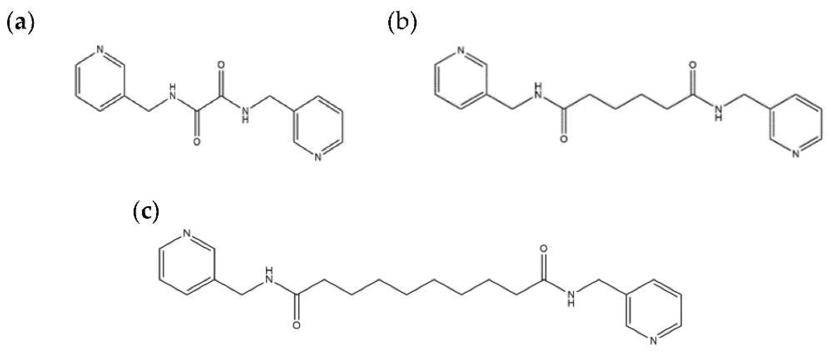
Figure 1. Structures of (a) L1, (b) L2 and (c) L3.
Preprints 92184 g001
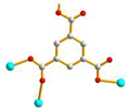
Figure 2. Structures of (a) 1,3,5-H3BTC and (b) 1,3,5-H3BTB.
Preprints 92184 g002
Figure 3. (a) Coordination environments of the Cu(II) ion in 1. Symmetry transformations used to generate equivalent atoms: (A) x-1, y-1, z -1; (B) x + 1, y, z; (C) -x + 1, -y + 2, -z + 1; (D) -x + 2, -y + 3, -z + 2; (E) -x, -y + 2, -z + 1. (b) A drawing showing the 2D net with the (42⋅67⋅8)(42⋅6)-3,5L2 topology. (c) A drawing showing the 2D net with the (44⋅62)-sql topology.
Preprints 92184 g003
Figure 4. (a) Coordination environment of Cu(II) ions in 2. Symmetry transformations used to generate equivalent atoms: (A) -x + 3, -y + 1, -z + 3. (b) A drawing showing the 2D net with the (4.62)2(42.62.82)-bex topology.
Preprints 92184 g004
Figure 5. (a) Coordination environment of Cu(II) ions in 3. Symmetry transformations used to generate equivalent atoms: (A) x + 1/2, -y + 1/2, z + 1/2; (B) -x + 1, -y + 1, -z + 2; (C) -x + 1/2, y + 1/2, -z + 3/2. (b) A drawing showing the structure with the pcu topology. (c) A drawing showing the 3-fold interpenetrated net.
Preprints 92184 g005aPreprints 92184 g005b
Figure 6. (a) Coordination environment of Cu(II) ions in 4. Symmetry transformations used to generate equivalent atoms: (A) -x + 2, -y + 2, -z + 1; (B) -x + 3/2, -y + 3/2, -z + 1; (C) x - 1/2, y - 1/2, z; (D) x + 1/2, y - 1/2, z; (E) -x + 2, y, -z + 1/2; (F) x – 1/2, -y + 3/2, z + 1/2. ). (b) A drawing showing the 3D framework with the (410⋅632⋅83)(42⋅6)2(43⋅63) topology.
Preprints 92184 g006
Figure 7. Coordination environments of Cu(II) ions in 5. Symmetry transformations used to generate equivalent atoms: (A) x + 1/2, -y - 1/2, z; (B) x - 1/2, -y + 1/2, z; (C) x, y, z + 1; (D) -x + 1/2, y - 1/2, z + 3/2; (E) x, y, z – 1. (b) Coordination environment of Cu(II) ion in 6. Symmetry transformations used to generate equivalent atoms: (A) -x + 5/2, y + 1/2, z + 3/2; (B) x, y, z – 1; (C) x - 1/2, -y + 3/2, z; (D) x + 1/2, -y + 1/2, z; (E) x + 1/2, -y + 1/2, z – 1; (F) x - 1, y - 1, z; (G) x - 1/2, -y + 1/2, z. (c) A drawing showing the 3,4,6-connected net with the point symbol (63)2(64⋅82)(68⋅85⋅102).
Preprints 92184 g007
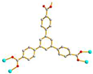
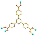
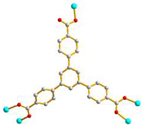
Table 1. Ligand conformations and bonding modes of complexes 1 - 6.
Ligand conformation Coordination mode
1 Preprints 92184 i001
trans syn-syn
Preprints 92184 i002
μ3-κO: κO’: κO’’
2 Preprints 92184 i003
AAA trans syn-syn
Preprints 92184 i004
μ2-κO:κO’
3 Preprints 92184 i005
AGA cis anti-anti
Preprints 92184 i006
μ4-κO: κO’: κO’’: κO’’’
4 Preprints 92184 i007
AAAAAAA cis anti-anti
Preprints 92184 i008
μ5-κO:κO’:O’’:O’’’:O’’’’
5 Preprints 92184 i009
GGAAAAA cis syn-syn
Preprints 92184 i010
μ5-κO:κO’:O’’:O’’’:O’’’’
6 Preprints 92184 i011
AAAAAAA trans anti-anti
Preprints 92184 i012
μ5-κO:κO’:O’’:O’’’:O’’’’
Table 2. Thermal properties of complexes 1 - 6.
Complex Weight loss of solvent
oC (calc/found), %
Weight loss of ligand
oC (calc/found), %
1 H2O
30 - 250 (3.21/4.38)
L1 + 1,3,5-HBTC2-
250 - 800 (85.14/85.15)
2 6.5 H2O
30 - 120 (16.19/14.38)
1.5 (L2) + 1,3,5-BTC3-
120 - 900 (73.64/70.97)
3 0.5 DMA + 3 H2O
30 - 250 (12.83/7.86)
0.5 (L2) + 1,3,5-HBTB2-
250 - 800 (78.75/81.97)
4 - L3 + 2 (1,3,5-BTC3-) + 2 (OH-)
270 - 800 (76.50/80.31)
5 2.5 MeOH + 2 H2O
30 - 270 (5.97/4.28)
2 (L3) + 2 (1,3,5-BTB3-)
270 - 800 (84.12/85.60)
6 DMF + H2O
30 - 250 (4.71/6.03)
2 (L3) + 2 (1,3,5-BTB3-)
250 - 800 (84.42/84.54)
Table 3. Crystal data for complexes 1 - 6.
Complex 1 2 3
Formula C23H20CuN4O9 C36H53Cu1.5N6O17.50 C36H27CuN2O7
Formula weight 599.97 945.15 663.13
Crystal system Triclinic Triclinic Monoclinic
Space group Pī Pī C2/c
a, Å 10.1482(9) 8.9062(2) 18.8531(6)
b, Å 11.1855(10) 11.2565(3) 25.8032(8)
c, Å 11.7055(11) 22.7418(5) 17.4503(6)
α, ° 111.285(3) 99.7718(14) 90
β, ° 97.429(3) 94.3827(15) 99.4362(19)
γ,° 108.425(3) 105.8728(14) 90
V, Å3 1128.97(18) 2142.95(9) 8374.2(5)
Z 2 2 8
Dcalc, Mg/m3 1.647 1.465 1.052
F(000) 574 989 2736
µ(Mo Kα), mm-1 1.032 0.831 0.561
Range(2θ) for data collection, deg 3.88 ≤ 2θ ≤ 51.99 3.66 ≤ 2θ ≤ 56.62 3.34 ≤ 2θ ≤ 56.62
Independent reflection 4415
[R(Int) = 0.0643]
10315
[R(Int) = 0.0539]
10067
[R(Int) = 0.0745]
Data / restraint / parameter 4415 / 0 / 338 10315 / 0 / 556 10067 / 0 / 432
quality-of-fit indicatorc 1.054 1.015 0.990
Final R indices
[I > 2σ(I)] a,b
R1 = 0.0555,
wR2 = 0.1419
R1 = 0.0550,
wR2 = 0.1228
R1 = 0.0536,
wR2 = 0.1164
R indices (all data) R1 = 0.0755,
wR2 = 0.1668
R1 = 0.1158,
wR2 = 0.1452
R1 = 0.1022,
wR2 = 0.1345
Formula C40H38Cu4N4O16 C100.50H104Cu3N8O20.50 C101H101Cu3N9O19
Formula weight 1084.90 1942.53 1935.52
Crystal system Monoclinic Orthorhombic Orthorhombic
Space group C2/c Pna21 Pna21
a, Å 16.5969(9) 20.9682(10) 22.0739(18)
b, Å 13.9067(4) 25.3489(11) 24.3392(18)
c, Å 17.6110(5) 18.1699(8) 17.9481(15)
α, ° 90 90 90
β, ° 90.2248(9) 90 90
γ,° 90 90 90
V, Å3 4064.73(19) 9657.7(8) 9642.8(13)
Z 4 4 4
Dcalc, Mg/m3 1.773 1.336 1.333
F(000) 2200 4056 4036
µ(Mo Kα), mm-1 2.145 0.728 0.728
Range (2θ) for data collection,deg 3.82 ≤ 2θ ≤ 56.59 2.75 ≤ 2θ ≤ 51.99 2.82 ≤ 2θ ≤ 56.63
Independent reflection 5049
[R(Int) = 0.0283]
18994
[R(Int) = 0.0510]
20288
[R(Int) = 0.0765]
Data / restraint / parameter 5049 / 0 / 311 18994 / 2119 / 1157 20288 / 1 / 1181
quality-of-fit indicatorc 1.085 1.026 1.002
Final R indices
[I > 2σ(I)] a,b
R1 = 0.0265,
wR2 = 0.0690
R1 = 0.0526,
wR2 = 0.1408
R1 = 0.0571,
wR2 = 0.1019
R indices (all data) R1 = 0.0323,
wR2 = 0.0747
R1 = 0.0664,
wR2 = 0.1503
R1 = 0.1364,
wR2 = 0.1244
aR1 = Fo Fc / Fo. bwR2 = [w(Fo2 Fc2)2 / w(Fo2)2]1/2. w = 1 / [σ2(Fo2) + (ap)2 + (bp)], p = [max(Fo2 or 0) + 2(Fc2)] / 3. a = 0.1039, b = 1.1215 for 1; a = 0.0650, b = 0 for 2; a = 0.0585, b = 0 for 3; a = 0.0330, b = 9.2346 for 4; a = 0.0856, b = 10.2363 for 5; a = 0.0496, b = 0 for 6. cquality-of-fit = [w(Fo2 Fc2)2 / Nobserved Nparameters )]1/2.
Disclaimer/Publisher’s Note: The statements, opinions and data contained in all publications are solely those of the individual author(s) and contributor(s) and not of MDPI and/or the editor(s). MDPI and/or the editor(s) disclaim responsibility for any injury to people or property resulting from any ideas, methods, instructions or products referred to in the content.
Copyright: This open access article is published under a Creative Commons CC BY 4.0 license, which permit the free download, distribution, and reuse, provided that the author and preprint are cited in any reuse.
Prerpints.org logo

Preprints.org is a free preprint server supported by MDPI in Basel, Switzerland.

Subscribe

© 2024 MDPI (Basel, Switzerland) unless otherwise stated