Preprint
Review

Advancements in Cold Atmospheric Plasma for Skin Disease Treatment and Skincare: A Brief Update

Altmetrics

Downloads

287

Views

121

Comments

0

This version is not peer-reviewed

Submitted:

12 December 2023

Posted:

13 December 2023

You are already at the latest version

Alerts
Abstract
Cold atmospheric plasma (CAP) has emerged as a promising therapeutic technology, distinguished by its safety, efficiency, and minimal side effects. With increasing applications in dermatology, CAP demonstrates antimicrobial efficacy against bacteria, viruses, and fungi, supports tissue proliferation and wound healing, and hinders the growth and migration of tumor cells. This review encompasses the concept, physical characteristics, and current research applications of CAP in dermatology. CAP, a gas cloud produced by ionizing gases, operates at room temperature, ensuring safety and efficacy in dermatological use. Its effects are mediated through active components like reactive oxygen and nitrogen species (RONS) and ultraviolet radiation. The extensive research covered in this paper explores CAP's applications in skin infections, immune-related diseases, and tumor diseases. As a biologically suitable technology, cold atmospheric pressure plasma finds widespread use in medicine, including medical device sterilization, dentistry, oncology, and dermatology. The direct and indirect forms of CAP applications, including plasma-activated liquids, complement each other. CAP holds promise as a dermatological treatment technique, but further exploration and research are needed. This article briefly explores CAP's diverse applications and potential utility within dermatology. The objective is to enhance the progress of plasma medicine by furnishing a concise overview of the applications of this groundbreaking technology in dermatological contexts.
Keywords: 
Subject: Medicine and Pharmacology  -   Medicine and Pharmacology

1. Introduction to Plasma Technology

In physics and chemistry, plasma is the fourth state of matter, distinct from solids, liquids, and gases. Defined as a hot, ionized gas consisting of positively charged ions and free electrons, plasma manifests under extreme heat and energy conditions [1]. There are several types of plasma, each distinguished by the specific conditions that give rise to its unique characteristics. In astrophysics, for instance, stellar plasma dominates the composition of stars, characterized by temperatures exceeding thousands of degrees Celsius. Terrestrial plasma, found in technologies like fluorescent lights and plasma TVs, emerges at comparatively lower temperatures but still involves a dynamic interplay of charged particles [2].
Mechanistically, the transition from a gaseous state to plasma occurs when energy input surpasses the ionization energy of the constituent atoms or molecules, liberating electrons and forming a sea of charged particles. The remarkable conductive properties of plasma and its ability to respond to electromagnetic fields find applications in various technological domains, ranging from fusion research in controlled nuclear reactions to the field of medicine with plasma sterilization techniques [1,2,3]. Plasma has been widely used in many life sciences, such as plasma medicine, agriculture, and disinfection against microbial bacteria and fungi [1,4]. Nonthermal plasma-based technology has recently gained attention for its diverse manifestations and potential advancements, especially in medical technology [1]. Table 1 concisely summarizes a range of biomedical applications linked to plasma.
This comprehensive review investigates the versatile applications of Cold Atmospheric Plasma (CAP) in dermatology, underscoring its safety, effectiveness, and minimal side effects [1]. Simultaneously, it explores CAP's distinctive features, its generation at room temperature, and essential elements such as reactive oxygen and nitrogen species (RONS) and ultraviolet radiation. Furthermore, the article examines CAP's efficacy in addressing skin infections, immune-related conditions, and tumors. This concise reference strives to advance the field of plasma medicine, fostering additional research into CAP-based interventions for dermatological treatments.

2. Plasma Medicine

This interdisciplinary field stands at the forefront of healthcare innovation, seamlessly integrating plasma physics, life sciences, and clinical medicine [1,2,3,4,11,12]. It explores the therapeutic potential of plasma, an ionized gas, across various medical domains. In disinfection, plasma exhibits unique capabilities that offer a novel approach to pathogen eradication [13]. In the pursuit of healing applications, low-temperature atmospheric pressure plasmas are harnessed to facilitate the controlled release of specific chemically reactive substances [1,2,3,14].
The expansive scope of plasma medicine extends to the forefront of cancer treatment research, where experimental investigations unfold at esteemed universities and research centers [15]. Moreover, its influence reaches into the intricate domain of neuroscience, seamlessly weaving together the disciplines of plasma physics, medicine, biology, plasma chemistry, and engineering to explore potential applications [1,2,3,4,16]. Within the therapeutic landscape, plasma medicine significantly emphasizes wound healing applications, contributing to the ever-evolving tapestry of medical advancements. In essence, the promise held by plasma medicine in reshaping healthcare is underscored by its innovative and diverse applications, creating a paradigm shift in the approach to medical treatments [1,2,3,4,11,12]. Plasma has demonstrated its effectiveness in various medical applications, such as wound healing, blood coagulation, pollutant degradation, material surface functionalization, cancer treatment, and dental procedures [1,2,3,14,17]. Monitoring its performance and real-time impact on the targeted therapy is crucial to tailor plasma for specific medical needs. This approach offers a novel perspective on developing clinical systems for plasma-based therapeutic interventions [17].

2.1. Medical Applications of Plasma

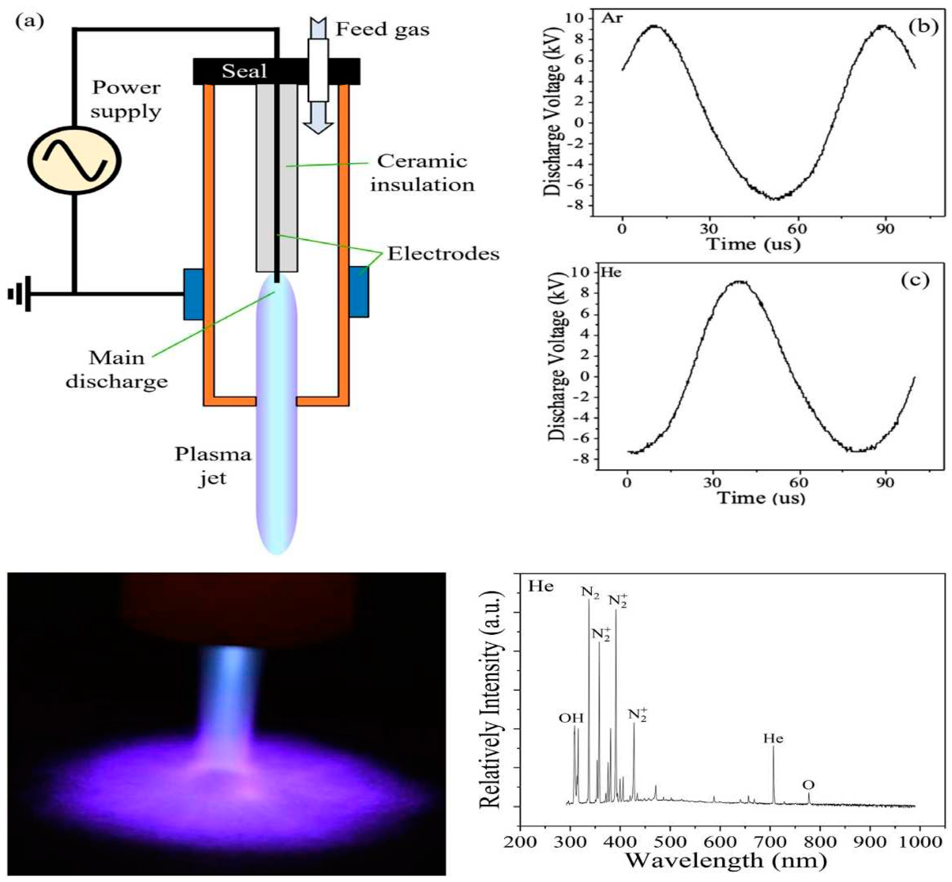
Medical applications of plasma involve using different types of plasma to achieve specific therapeutic goals. The studies conducted highlight seven distinct mechanisms through which plasma directly aids in wound healing [1,2,3,14]. This multidisciplinary field, plasma medicine, seamlessly integrates principles from plasma physics, medicine, biology, plasma chemistry, and engineering [1,17]. The direct application of physical plasma on or within the human body has proven effective for therapeutic purposes [18]. Researchers have delved into various applications, including wound healing and cancer treatment, expanding the scope of knowledge in this emerging field [15]. Figure 1 illustrates the cold plasma jet system, featuring a schematic diagram and discharge voltages for argon and helium feeding gases. Additionally, the figure depicts a helium-fed cold atmospheric plasma (He-fed CAP) treating a surface with plume spreading and the detection of RONS using optical emission spectroscopy (exposure: 5000 ms) [19].
Plasma medicine demonstrates its relevance in neuroscientific applications, emphasizing its interdisciplinary nature [17]. This therapeutic approach is not confined to a singular method but encompasses a spectrum of plasma types tailored for specific therapeutic goals [14]. The potential applications extend to disinfection, wound healing, and cancer treatment, marking plasma medicine as a promising avenue in clinical practice [20]. Table 2 presents a comprehensive overview of diverse medical applications within plasma medicine. Notably, most of the research in plasma medicine is conducted in vitro and animal models, highlighting the emerging nature of the field and its integration of plasma physics, life sciences, and clinical medicine [1,2,3,4,17]. Two primary forms of plasma find application within medical contexts.

2.1.1. Cold Atmospheric Plasma (CAP)

CAP is a non-thermal plasma that operates at or near room temperature. CAP has gained attention in medicine for its diverse applications, including wound healing, sterilization, and cancer treatment. The operational mechanism of non-thermal biocompatible plasma (NBP) may be associated with the collaborative effects of its constituents, including ultraviolet radiation and reactive species [36]. The unique property of CAP is its ability to generate various RONS without significantly increasing the temperature of the surrounding tissues. This makes CAP suitable for delicate medical procedures where maintaining a low-temperature environment is crucial. Studies have shown promising results in using CAP for wound disinfection and promoting tissue proliferation [5,36,37].

2.1.2. Thermal Plasma

Thermal plasma, in contrast, involves higher temperatures and is widely used in medical applications such as coagulation, cutting, and tissue ablation. In surgical procedures, thermal plasma is employed to precisely remove or modify tissues, offering a controlled and efficient approach [11,12,13,14,38]. The ability of thermal plasma to reach higher temperatures allows for a more intense and targeted application in surgical settings. Thermal plasma devices are commonly used in various medical specialties, including dermatology and surgery [11,12,13,14,38]. These types of plasma exhibit distinct characteristics that make them suitable for specific medical applications. Researchers continue to explore and refine these technologies to enhance their efficacy and broaden their medical utility.

3. CAP in Dermatology

CAP, which contains low-temperature heavy ions, is the focus of this study. CAP has gained increasing attention in clinical medicine, particularly dermatology [1,2,3]. Plasma is formed by ionizing gases under heat or a strong electromagnetic field, resulting in a cloud of charged particles, including electrons, ions, metastable species, ultraviolet light, visible light, electromagnetic fields, and reactive substances [1,2,3]. Named for its equal distribution of positive and negative charges, plasma exhibits diverse characteristics based on temperature, categorizing it into thermal plasma (around 10,000 K), warm plasma (3,000 to 5,000 K), and cold plasma (near room temperature) [39]. CAP, a type of non-equilibrium plasma, has gained attention, especially in dermatology, due to its ability to operate at room temperature safely and efficiently [1,2,3]. CAP influences organisms through active components such as chemical active particles RONS, ultraviolet radiation (UV), and charged particles (electrons, positive and negative ions) at the molecular level, making it suitable for skin treatment [18,40,41]. When applied to skin tissues, it should meet three characteristics: operation at atmospheric pressure below 40 ℃, generation of average electron energy for impact excitation without causing thermal damage to the skin [2,3]. Consequently, as research on plasma medicine deepens, the rapid development of this field expands its scope of study, with skin being an excellent site for plasma treatment [2,3]. CAP represents a distinct form of plasma characterized by operating at or proximal to room temperature and under atmospheric pressure conditions. Unlike conventional plasmas that typically function at elevated temperatures and low pressures, CAP offers a unique profile by operating at temperatures conducive to room conditions. This attribute renders it particularly applicable in diverse domains, including medical, material processing, and surface treatment, where concerns about excessive heat are pertinent. CAP's operation at atmospheric pressure is significant, obviating the need for vacuum systems and allowing for facile generation and sustenance in open-air environments. This accessibility enhances its practical utility across various settings.
The applications of CAP are broad and encompass medical aspects, such as wound healing, sterilization of medical apparatus, and potential applications in cancer treatment [17]. Additionally, CAP finds utility in material processing, where it contributes to enhanced adhesion and modification of surface properties, as well as in sterilization procedures, leveraging its capacity to generate reactive species effective against bacteria and microorganisms. Diverse methodologies are employed in CAP generation, including dielectric barrier discharges and atmospheric pressure glow discharges, selected based on the specific requisites of the intended application. The resultant plasma generates an array of reactive species, including ions, electrons, free radicals, and excited atoms and molecules, all of which play pivotal roles in the varied applications of CAP. Ongoing research continues to explore novel applications and refine existing technologies. The capability of CAP to operate at room temperature and atmospheric pressure broadens the horizons of plasma utilization, particularly in contexts where conventional high-temperature plasmas may pose limitations. Traditional plasmas are often generated at high temperatures and low pressures, such as in stars or industrial processes [42]. However, cold atmospheric pressure plasmas are created at temperatures close to or at room temperature, making them suitable for various applications, including medicine, materials processing, and surface treatment [15,16,17,18,43,44].

3.1. Physical Characteristics and Classification of CAP:

While thermal equilibrium plasma has shown significant potential in medical applications, patients often struggle with the discomfort caused by increased skin temperature. In contrast, CAP does not induce such side effects. CAP treatment of biological tissues is feasible, ensuring that the temperature of the treated skin remains between 37 and 38 ℃ [15,16,17,18]. This is attributed to the faster heating rate of electrons compared to ions in an electric field, resulting in plasma generation at room temperature or close to it [2]. Common discharge forms of CAP in laboratories include glow, spark, corona, dielectric barrier discharge (DBD), sliding arc discharge, microwave, radiofrequency, etc. [45,46]. CAP devices can be categorized into three types: direct plasma source, indirect plasma source, and mixed plasma source. Direct plasma sources use the human body (skin or internal tissues) as an electrode. In contrast, indirect plasma sources involve exciting plasma between two electrodes, with the active components acting on the target tissue. Mixed plasma sources combine the advantages of both types, typically achieved by using a grid-like metal electrode with lower tissue resistance [3]. The main types of plasma sources for medical applications are indirect (Atmospheric Pressure Plasma Jet, APPJ) and direct DBD plasma sources [47]. Plasma can be generated from air, rare gases, or various mixed gases as carrier gases. After activation in multiple forms, discharge produces effective substances such as positive and negative ions, free electrons, neutral reactive oxygen/nitrogen species (ROS/RNS), free radicals, and molecular fragments, all of which can interconvert [4]. Plasma health applications have emerged as a thought model in recent years, representing interdisciplinary research in medicine, biology, physics, chemistry, and engineering [1,2,3,4,5], with research increasingly intensifying [48].

3.2. Direct and Indirect Plasma

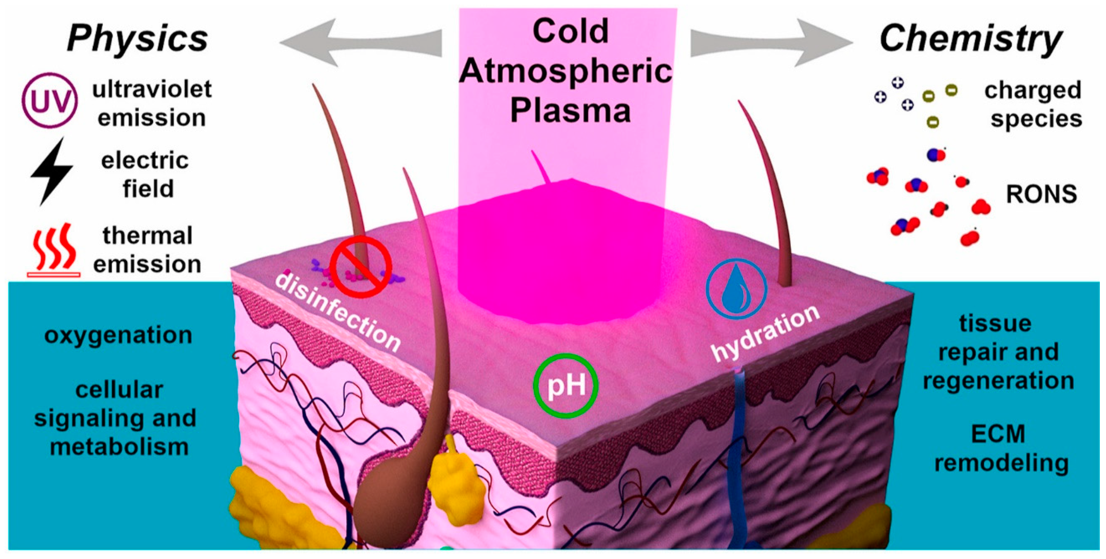
Applying cold atmospheric pressure plasma in medicine can be classified into direct and indirect forms. Direct applications include DBD and APPJ, both devices [47]. Indirect plasma applications primarily involve plasma-activated liquids (PAL) to exert their effects. Plasma-activated liquids result from the interaction between plasma and liquid surfaces and include plasma-activated water (PAW), plasma-activated media (PAM), plasma-activated solution (PAS), plasma-activated oil (PAO), and plasma-activated hydrogel (PAH) [49]. Even without additional chemical substances, gaseous plasma interacting with water produces unique chemical reactions and energy transfer, yielding plasma-activated water (PAW) with significant transient broad-spectrum biological activity [50], as depicted in Figure 2, RONS play crucial roles in the dermatological application of cold atmospheric pressure plasma [12,13]. The interaction between plasma and liquids can lead to various direct reactions at the gas-liquid interface and indirect cascade reactions in the liquid. This results in plasma-activated liquids containing a mixture of highly reactive species. Reactive nitrogen oxides (RONS) can be classified into long-lived and short-lived types [51]. Examples of short-lived species include hydroxyl radicals (OH-), singlet oxygen (1 O2), and superoxide anions (O2-), with lifetimes ranging from seconds to minutes [52]. In contrast, hydrogen peroxide (H2O2), nitrite (NO2-), nitrate (NO3-), nitrous acid (HNO2), and ozone (O3) belong to long-lived species [53]. Most RONS found in the liquid phase during the indirect action of cold atmospheric pressure plasma are long-lived [51,52,53]. The plasma device and working gas greatly influence the type and concentration of reactive species in PALs and the liquid used [47]. Given the presence of RONS in plasma-activated liquids, they also hold potential applications in the biomedical field [1,2,3,4,5,48].
The invention of plasma-activated liquids has liberated the plasma application from constraints related to the size, conditions, and high voltage of plasma sources. This development allows for producing large, uniform plasma areas, significantly reducing operational challenges in clinical practice, saving treatment time, and enhancing portability, safety, scope, and depth of effect compared to direct applications. Remarkably, plasma-activated hydrogels have demonstrated the capability to sustain the release of RONS [54].

3.3. Penetration and Mechanism of Action of Active Components of CAP in the Skin

The application of CAP in skin diseases includes direct treatment, plasma-activated water, and activation of matrix injection. However, the epidermis acts as a natural barrier, posing a challenge to the delivery of drugs or charged active ingredients into the skin [55]. In CAP, colliding gas molecules with high-energy electrons can generate original reactive substances such as O2 and N2 molecules, N, O, and H atoms in ground and excited states, and OH radicals. Further reactions between these original substances produce RONS, including OH, NO, NO2, HNO2, HNO3, and H2O2 [51,52,53]. The transport of active particles from CAP to the skin includes two pathways: transdermal plasma delivery (plasma puncture) and tissue penetration [55].
Transdermal plasma delivery occurs when the electric field of the plasma generates a voltage drop on the skin, with most of the voltage drop applied to the high-resistance stratum corneum. When the surface potential reaches the driving voltage, this voltage distribution leads to corneum breakdown [49,56]. The CAP air plasma directly applied to the skin surface generates an electric field of about 170 kV·cm⁃1 between the skin surface and the underlying layers, eventually penetrating the skin and forming holes [14]. When CAP acts on the skin, it generates rapid but mild heat, which breaks down the lipid structure of the stratum corneum, thereby increasing skin permeability without damaging deeper tissues and improving transdermal delivery [57]. Simultaneously, CAP perforation promotes the transfer of NO generated on the skin surface to the skin tissue, ultimately leading to increased vasodilation, blood flow, and tissue oxygenation [19].
The penetration of active particles from CAP into tissues includes penetration through tissue moisture and direct interaction with skin tissues [58]. The RONS produced by CAP must pass through three barriers before entering cells: the plasma-tissue fluid barrier, tissue fluid-tissue barrier, and tissue-cell barrier [14]. CAP affects liquids by changing liquid surface dents and flow patterns, making the liquid surface charged, causing convection, etc., which promotes the entry of active ions into the liquid phase [59]. The direct effect of CAP on skin tissues has been mainly studied through gelatin and agarose models. The results show that CAP jets can penetrate 150 μm gelatin directly and detect plasma effects up to 4 mm in agarose models [19]. In addition, the active components generated by CAP can create nano-sized pores in the phospholipid bilayer and disrupt the phospholipid bilayer structure, facilitating the entry of RONS into cells [14,60].

4. CAP for Therapeutic Purposes in Dermatology

In contemporary dermatology, CAP has gained recognition for its diverse applications, demonstrating potential benefits in treating various skin conditions. Studies by Bernhardt et al. highlight CAP's capacity to positively affect atopic eczema, itch, and pain, suggesting a promising therapeutic avenue [2]. Furthermore, a systematic review underscores the effectiveness of CAP in providing therapy for various skin diseases [13,22,23]. The emerging potential of CAP for dermo-cosmetic treatments, including aging prevention, has been acknowledged [19].
This evolving landscape underscores the multifaceted role of CAP in dermatology, with ongoing research exploring intricate mechanisms and expanding clinical applications [2,13,19,22,23]. Recent investigations contribute to a nuanced understanding of CAP's impact on skin health. Additionally, CAP has demonstrated promising applications in dermatology, showing efficacy in inducing proapoptotic effects more efficiently in tumor cells compared to benign counterparts, highlighting its potential for targeted therapy in skin-related conditions [1,25,26,27,28]. The versatility of CAP in dermatological applications encompasses decontamination/sterilization, use in dental medicine, and enhancement of skin therapy [17,21,24,35]. Recent studies further suggest that CAP can ameliorate various skin diseases, showcasing its therapeutic potential in dermatology [1,2,3,17]. Its emerging role in skin therapy presents a novel approach with potential benefits in clinical dermatology, supported by achievements and ongoing research [1,2,3,17].
Plasma, formed by ionizing gas under heat or a strong electromagnetic field, constitutes a cloud of charged particles, including electrons, ions, metastable species, ultraviolet light, visible light, electromagnetic fields, and reactive substances [1,2,3]. Due to its equal distribution of positive and negative charges, plasma represents the fourth state of matter alongside solids, liquids, and gases. Plasmas can be categorized into thermal, warm, and cold plasma based on gas temperature. CAP, a non-equilibrium plasma applicable in medical contexts, occurs at atmospheric pressure with significantly higher electron temperatures than gas temperatures yet remains tolerable for the human body [5].
Despite the diverse components generated within the plasma, reactive nitrogen oxides are key active agents. Plasma exhibits broad-spectrum antimicrobial action, cell proliferation, tissue regeneration stimulation at low doses, and induction of programmed cell death (apoptosis) at high doses [62]. Since the 20th century, researchers have utilized plasma to eradicate bacteria, leading to the emergence of plasma medicine. Currently, plasma medicine finds widespread application and exploration in medical device sterilization, dentistry, oncology, dermatology, and other fields [1,2,3,17].

4.1. CAP's Suppressive Effect on Microbial Infection

4.1.1. Infectious Skin Diseases

The application of CAP has garnered significant attention due to its remarkable oxidative capabilities mediated by RONS. Various studies have highlighted the potent antimicrobial properties of CAP and plasma-activated liquids against a spectrum of microorganisms, including bacteria, fungi, viruses, and parasites [63,64]. Compared to traditional antibiotics, CAP offers distinct advantages, such as a reduced likelihood of inducing drug resistance and minimal toxicity [65]. Notably, CAP's efficacy extends to inhibiting microbial infections on diverse surfaces, encompassing non-living surfaces, biofilms, and bacteria and fungi within contaminated or infected tissues [66].
Maisch et al. conducted a comprehensive investigation into the inhibitory effects of CAP on Gram-positive (methicillin-resistant Staphylococcus aureus, methicillin-sensitive Staphylococcus aureus) and Gram-negative bacteria (Escherichia coli) [67]. Their study involved inoculating bacteria on the skin surface of six-month-old pigs, followed by CAP treatment. The results indicated that a six-minute CAP treatment achieved a sterilization rate of 99.9% for Staphylococcus aureus and Escherichia coli. In contrast, a four-minute treatment was effective against methicillin-resistant Staphylococcus aureus. Importantly, no discernible morphological changes in tissues and cells indicated CAP's safety [67].
Further insights into CAP's inhibitory mechanisms were proposed by Mai-Prochnow et al. [868. They suggested that CAP components, including reactive oxygen and nitrogen, act directly on cell walls or membranes. Reactive oxygen triggers lipid peroxidation reactions, leading to deactivation of iron-dependent dehydrases, destruction of monocyte cell iron proteins, and DNA damage, ultimately causing cell deactivation [68]. Dezest et al. [69] explored CAP's inhibitory effect on Escherichia coli, revealing that CAP induced bacterial death by altering the bacterial structure and initiating oxidative stress reactions. The study emphasized complex interactions, including electric field and ion effects [69].
CAP's broad-spectrum antimicrobial mechanism, disrupting biofilm matrices and inactivating bacterial quorum sensing signals, presents a challenge for bacteria in developing resistance [70,71]. In a study of diabetic foot infections, CAP significantly reduced bacterial loads in chronic diabetic foot ulcers, leading to improved wound healing [72]. The CAP-treated group exhibited a lower incidence of systemic inflammatory responses and required fewer surgical interventions, highlighting the potential applications of CAP in treating chronic diabetic foot infections [73]. These findings collectively underscore the promising role of CAP in combating infectious diseases with unique advantages over conventional treatments.

4.1.2. Effects of CAP on Bacteria

Extensive in vitro and in vivo research has unequivocally demonstrated atmospheric pressure cold plasma's direct inhibitory and bactericidal effects on bacteria and their biofilms [63,66,74]. In both settings, applying atmospheric pressure cold plasma has proven effective in inhibiting and destroying bacteria and disrupting biofilm structures [75]. Notably, plasma-activated liquids (PALs) have emerged as a promising avenue for bacterial eradication, with studies showcasing their efficacy against common strains such as Escherichia coli, Staphylococcus aureus, and Neisseria gonorrhoeae [76,77]. The mechanism behind this antimicrobial action involves the generation of RONS in Plasma-Activated Water (PAW), impacting the oxidative-reduction status of antioxidants, penetrating bacterial cell membranes, disrupting cell structures, and ultimately leading to bacterial death [78,79]. Furthermore, investigations by Liu et al. [80] underscore the potential of plasma-activated saline in modulating inflammatory factors and genes, altering the morphological structure of pathogens, and enhancing the host's immune response against Neisseria gonorrhoeae, thereby illustrating its therapeutic promise in addressing infections associated with biofilms.
Biofilms, intricate communities of microbial cells (bacteria and fungi) adhered to various surfaces, present a formidable challenge for traditional antibiotics, disinfectants, and standard immune clearance methods [81]. Their complex structures contribute to chronic and persistent infections, as these conventional approaches often prove limited efficacy against biofilms. The dynamic and prolonged presence of RONS (RONS) generated by Plasma-Activated Water (PAW) plays a pivotal role in disrupting biofilm structures, rendering bacteria more susceptible to elimination [82,83]. This innovative approach capitalizes on the unique properties of PAW to address the challenges posed by biofilms, offering a promising avenue for treating biofilm-associated infections. The interdisciplinary nature of this research, spanning microbiology, chemistry, and immunology, underscores the multifaceted potential of atmospheric pressure cold plasma and plasma-activated liquids in revolutionizing our approach to combating bacterial infections and biofilm-related complications.

4.1.3. Effects of CAP on Fungi

Various fungi, encompassing dermatophytes, Candida, molds, and spore-forming fungi, have the potential to induce skin infections, with nail fungal infections, particularly onychomycosis, posing a clinical challenge due to the dense structure of the nail plate, leading to low drug concentrations and extended treatment durations. Exploratory research has showcased the efficacy of plasma in combating the causative agents of nail fungal infections across pathogen models, detached nail models, and clinical trials [1,84,85]. White Candida albicans and Trichophyton mentagrophytes treated with atmospheric pressure plasma jet (APPJ) displayed inhibited growth, reduced adhesion, and decreased infectivity in a nail model [86,87]. Furthermore, findings for other dermatophytes, such as Epidermophyton floccosum and verrucous Trichophyton, affirmed that CAP can impede the growth and infection of skin fungi [88,89]. In a pilot study involving 19 participants with nail fungal infections, the overall clinical cure rate was 53.8%, and the mycological cure rate was 15.4%, with high patient satisfaction and no significant or prolonged adverse reactions, establishing atmospheric pressure cold plasma as a safe method for treating nail fungal infections [90]. In a cohort of 40 patients with nail fungal infections, the integration of atmospheric pressure cold plasma therapy with nail plate ablation and refreshment (NPAR) or antifungal drug treatment resulted in clinical cure rates exceeding 70%, and mycological cure rates increased to 85.7% [90]. Cold plasma emerges as a promising alternative therapy for skin fungal infections, with longer plasma treatment times associated with more potent inhibition by plasma-activated hydrogel (PAH) compared to plasma-activated water (PAW) [91], offering a potential novel approach for future treatments of skin fungal diseases.

4.1.4. Effects of CAP on Viruses and Parasites

Reports suggest that atmospheric pressure cold plasma has some antiviral effects [92]. However, its current utilization in dermatology is relatively limited. Friedman et al. [93,94] reported positive outcomes in treating viral warts using cold plasma in two adults and five pediatric patients. Bunz et al. [95] observed cold plasma's lower but measurable antiviral effect on HSV-1. Further exploration is required to investigate the impact of other atmospheric pressure cold plasma devices and parameters on viruses. Furthermore, studies have shown the efficacy of plasma against parasites like Demodex mites and head lice. Malik et al. [96] conducted a half-face survey of three patients with rosacea, comparing bi-weekly cold plasma treatment with daily topical ivermectin. The proportion of Demodex mites in the follicles and the total number decreased significantly in the ivermectin and cold plasma treatment sites without adverse reactions. Bosch et al. [97] developed an atmospheric pressure cold plasma device resembling a comb (CAPComb) for treating head lice. The results indicated that a single application of a plasma comb on hair could cause death to lice and eggs. The study concluded the safety of the plasma comb based on ozone concentration, UV emission rate, and leakage current.

4.2. CAP Promotes Tissue Proliferation and Wound Healing

The effectiveness of CAP in wound healing is evident, showcasing its abilities to foster antiseptic properties and pro-angiogenic effects. Furthermore, CAP exhibits notable efficacy in promoting tissue proliferation, thereby contributing significantly to the overall process of wound healing [98,99]. Researchers, including Hasse et al., explored the impact of CAP on skin wounds. In their study, tissue samples with a 5mm diameter were extracted from normal human skin and subjected to CAP irradiation [100]. The results revealed that a 1 to 3-minute CAP exposure stimulated cell proliferation, promoting epidermal repair and facilitating wound healing. Interestingly, partial cell apoptosis was observed after 3 to 5 minutes of CAP exposure. Another study by Arndt et al. [101] investigated the influence of CAP on gene expression in keratinocytes, the cells responsible for skin barrier formation. Using MicroPlaster-β to generate CAP, they treated wounds and found that CAP induced the expression of genes coding for interleukin (IL)-8, transforming growth factor (TGF)-β1, and TGF-β2. Both in vitro and in vivo experiments demonstrated that keratinocytes' proliferation, migration, and apoptosis processes were not significantly affected during wound healing. The experiments highlighted the time-dependent nature of CAP in promoting wound healing. Within specific timeframes and doses, CAP could stimulate the expression of cell factors related to wound healing without compromising the activity of keratinocytes. This underscores CAP's potential to facilitate wound healing effectively. The results showed that CAP treatment significantly accelerated wound healing compared to the control group. Specifically, CAP increased re-epithelialization, fibroblast proliferation, and collagen synthesis while reducing the inflammatory response. CAP has a promoting effect on cell proliferation, migration, and angiogenesis [101]. Researchers found that CAP significantly promoted cell proliferation and the release of growth factors and chemokines, such as vascular endothelial growth factor (VEGF) and fibroblast growth factor (FGF), by activating fibroblasts. Vascular endothelial growth factor is crucial for angiogenesis and wound healing, while fibroblast growth is essential for tissue repair and regeneration [102]. The promoting effect of CAP on wound healing has also been confirmed in a study on human skin wounds. Rat skin wounds were treated with CAP, and the results showed increased angiogenesis, granulation tissue formation, and collagen synthesis [20]. These findings suggest that CAP can accelerate wound healing by promoting cell proliferation, angiogenesis, and the release of growth factors. It stimulates skin cell proliferation and migration [103,104]. While a meta-analysis suggested limited clinical benefit for chronic wounds, smaller studies have associated CAP with positive outcomes, including improved wound infection and healing [105]. Moreover, atmospheric pressure cold plasma can facilitate wound healing through other mechanisms. In a randomized clinical trial by Stratmann [106], cold plasma therapy accelerated the healing of chronic wounds. However, there was no significant difference in microbial load between atmospheric pressure cold plasma and placebo groups. Even before this clinical trial, atmospheric pressure cold plasma therapy had been proven beneficial for chronic wounds, not solely due to its antibacterial effects [105,106]. Nevertheless, basic research indicates CAP's ability to eliminate refractory skin bacteria in vitro and positively impact wound healing in rat models [107]. Considering the significant role of CAP in treating bacterial infections, it can promote wound healing by significantly reducing bacterial load in the treated wounds, ensuring safety [105,106].
Moreover, CAP can facilitate wound healing through other mechanisms. Over the past decade, a series of in vitro studies have indicated that plasma-triggered wound healing depends on stimulating cell proliferation and survival [53,54], synthesis of Extracellular Matrix (ECM) proteins [108,109,110], changes in adhesion proteins and cytoskeletal structures [11,12,13,14,15,16,17,18,19,20,21,22,23,24,25,26,27,28,29,30,31,32,33,34,35,36,37,38,39,40,41,42,43,44,45,46,47,48,49,50,51,52,53,54,55,56,57,58,59,60,61,62,63,64,65,66,67,68,69,70,71,72,73,74,75,76,77,78,79,80,81,82,83,84,85,86,87,88,89,90,91,92,93,94,95,96,97,98,99,100,101,102,103,104,105,106,107,108,109,110,111,112,113], and apoptosis of cells [114,115,116]. Additionally, some studies suggest that atmospheric pressure cold plasma treatment can induce genes involved in wound healing [117] and facilitate oxidative-reductive regulation of known critical targets for wound healing [118,119,120,121]. To ensure a more uniform treatment of wounds, many scholars have further investigated the mechanisms and applications of plasma-activated liquids in wound treatment, as depicted in Figure 3. They found that Plasma-Activated Water (PAW) could accelerate wound healing by reducing bacterial counts in treated wounds [122]. In their study, Lee et al. [123] cultured keratinocytes with Plasma-Activated Media (PAM) and treated a mouse model with PAW. PAM treatment promoted the transformation and migration of keratinocytes, attributed to changes in the expression of integrin-dependent focal adhesion molecules and Matrix Metalloproteinases (MMPs). The study also identified increased intracellular Reactive Oxygen Species (ROS) levels as a driving force for cell migration, regulated by NADPH oxidase and changes in mitochondrial membrane potential. In in vivo experiments using a full-thickness acute skin wound model in mice, PAW treatment contributed to an increased re-epithelialization rate, emphasizing the activation of potential intracellular ROS-dependent signaling molecules. Zou et al. [124] produced plasma-activated oil by treating olive oil with cold plasma. PAO not only killed bacteria on the wound but also promoted the release of growth factors such as CD34 and Vascular Endothelial Growth Factor (VEGF), resulting in a 28.5% faster wound healing rate than the control group. Applying plasma-activated liquid in a Fibroin-Silk Fibroin (SF) composite hydrogel showed that Plasma-Activated Hydrogel (PAH) significantly enhanced the healing of radiation-induced wounds. Cell and tissue responses to PAH promoted the regenerative process of wounds, while atmospheric pressure cold plasma also improved the mechanical and chemical properties of SF gel [125].

4.3. CAP for the Treatment of Inflammatory Skin Diseases

CAP regulates the formation of stratum corneum cells through the reactive oxygen pathway [126]. Stratum corneum cells play a crucial role in maintaining skin barrier function. Research indicates that CAP modulates the redox balance, inducing alterations in the structure and function of stratum corneum cells [127]. Clinical studies have demonstrated that CAP can improve the symptoms by reducing skin inflammation and restoring the normal differentiation of stratum corneum cells [85]. The redox-sensitive transcription factor nuclear factor erythroid 2-related factor 2 (NRF2) plays a key role in mediating the effects of CAP on stratum corneum cells. CAP activates NRF2, leading to the upregulation of antioxidant enzymes and the restoration of redox homeostasis in stratum corneum cells [128]. The modulation of NRF2 by CAP may represent a novel therapeutic approach for skin diseases characterized by oxidative stress and inflammation [126,127,128,129]. CAP exerts its effects by regulating cell viability, proliferation, migration, and inflammatory responses, offering a novel approach to managing Inflammatory skin disorders [126,127,128,129].

4.3.1. Psoriasis

In a recent study by Gan et al. (2019) [130], the therapeutic effects of CAP on psoriasis were investigated through cellular and animal experimentation. To simulate a psoriatic cell model, lipopolysaccharide (LPS) or tumor necrosis factor-alpha (TNF-α) was introduced into human keratinocyte cells (HaCaT cells), and the cells were cultured with plasma-activated medium (PAM). Subsequently, an atmospheric pressure plasma jet (APPJ) was directly applied to a mouse model of psoriasis induced by imiquimod. Results showed that PAM increased reactive oxygen species (ROS) in HaCaT cells, leading to cell apoptosis. Interestingly, imiquimod-induced psoriasis-like dermatitis exhibited gradual improvement following APPJ treatment, demonstrating reduced epidermal hyperplasia and thinning of the epidermal layer. The ROS and reactive nitrogen species (RNS) generated by cold plasma inhibited excessive proliferation of keratinocytes, promoting apoptosis. Moreover, it suppressed the proliferation of T lymphocytes and enhanced the expression of programmed death-ligand 1 (PD-L1), which negatively correlates with T lymphocyte proliferation [131]. Furthermore, ROS and RNS directly regulated the release of inflammatory factors such as IL-6, IL-12, and VEGF at the site of skin lesions, contributing to the alleviation of psoriasis [132]. Case reports also indicated satisfactory outcomes in treating inverse psoriasis and recalcitrant palmoplantar psoriasis using atmospheric pressure cold plasma [133,134]. Kim et al. (2023) developed atmospheric pressure cold plasma patches for treating psoriatic skin lesions. These patches produced ROS and RNS on flexible polymer film surfaces using surface dielectric barrier discharge. The patches alleviated psoriasis symptoms by inducing the opening of calcium ion channels in keratinocytes and mitigating the effects of the electric field [135].

4.3.2. Atopic Dermatitis

Researchers have explored the therapeutic effects of atmospheric pressure cold plasma on different mouse models of atopic dermatitis (AD), demonstrating positive outcomes in alleviating skin inflammation, endoplasmic reticulum stress, and oxidative stress [136,137]. Scholars have explored the positive effects of treating AD mice models with non-thermal atmospheric plasma; cold plasma reduced skin cell apoptosis in a mouse model induced with dinitrofluorobenzene (DNFB). It alleviated skin inflammation, endoplasmic reticulum (ER) stress, and oxidative stress mediated by plasma-induced MANF expression [138].
Furthermore, localized treatment with cold plasma in a mouse model induced by house dust mite extract decreased the severity of dermatitis, transepidermal water loss (TEWL), and serum IgE levels [139]. In another dust mite AD model, plasma inhibited the increase in epidermal thickness recruitment of mast cells and eosinophils and reduced the expression of AD-associated cytokines and chemokines [140]. Choi et al. demonstrated the positive effects of cold plasma on keratinocytes stimulated with pro-inflammatory cytokines and in AD model mice induced by 2,4-dinitrochlorobenzene (DNCB). The study suggested that non-thermal plasma could improve AD by modulating NF-κB activity [141].
In summary, plasma treatment can inhibit the production and release of inflammatory factors. IgE reduces the number of inflammatory cells, thus mitigating the severity of dermatitis and protecting the skin barrier. Clinical studies on mild to moderate atopic dermatitis patients with symmetrical skin lesions revealed improvements in skin lesions and itching through treatment with non-thermal atmospheric plasma. Microbiome analysis further indicated a significant reduction in the proportion of Staphylococcus aureus in the treated group [142].

4.3.3. Vitiligo

As of the most recent advancements in dermatology, there have been notable updates in the application of CAP for the treatment of Vitiligo. This innovative therapeutic approach harnesses the power of low-temperature plasma to stimulate repigmentation in areas affected by Vitiligo, a chronic skin disorder characterized by depigmented patches. Research studies and clinical trials have shown promising results, highlighting the potential of CAP in enhancing melanocyte activity and promoting the restoration of normal pigmentation [143]. Zhai et al. [144] conducted a study on the efficacy and safety of PAH (Cold Atmospheric Plasma) in vitiligo, utilizing mouse models and patients with active focal vitiligo. Skin biopsies revealed that local treatment of vitiligo-like lesions on the dorsal skin of mice with cold atmospheric plasma restored melanin distribution. This treatment led to a reduction in T cells and dendritic cell infiltration and a decrease in the release of inflammatory factors. The study observed an enhancement in the expression of the transcription factor NRF2 and a reduction in inducible nitric oxide synthase activity, thereby increasing cellular resistance to oxidative stress and excessive immune responses. In this randomized controlled trial, atmospheric pressure cold plasma treatment achieved partial and complete pigmentation in 80% and 20% of vitiligo skin lesions. Notably, during the treatment and follow-up periods, there were no occurrences of pigmentation or other adverse events in the surrounding areas [144].

4.4. CAP Suppresses Tumor Cell Proliferation and Migration

The research on the use of plasma in treating tumors has rapidly expanded from cellular studies to animal models [145,146]. Among various potential anti-tumor mechanisms, apoptosis induced by cold atmospheric plasma under atmospheric pressure has been extensively studied [147,148]. This process involves the activation of different signaling pathways, including MAPK and PI3K/Akt pathways, leading to cell death [149]. Other possible mechanisms include induction of autophagy [150,151], pyroptosis [152], ferroptosis [153,154], and regulation of cancer cell metabolism [155] and cause cell cycle arrest, DNA damage, and mitochondrial dysfunction [156].
Studies indicate exceptional responsiveness of melanoma cells to cold plasma, with melanoma cells exhibiting higher sensitivity to reactive oxygen and nitrogen species (RONS) induced by cold plasma compared to normal skin cells [157]. This heightened sensitivity suggests cold plasma's potential applicability and safety in anticancer treatments [157]. Researchers investigated the inhibitory effect of CAP on melanoma cells in vitro. The study found that CAP could induce apoptosis in melanoma cells, significantly increasing intracellular ROS levels [158]. CAP treatment also caused DNA damage, cell cycle arrest, and reduced cell migration. Furthermore, CAP inhibited the growth of melanoma xenografts in mice, demonstrating its potential as a novel therapy for melanoma. Another study investigated varying induction of apoptosis or senescence in melanoma tumor cells in response to different doses of a novel cold atmospheric plasma (CAP) device, revealing a previously undocumented mechanism of senescence critical for the therapeutic potential of CAP [159]. Many studies indicate melanoma cells react remarkably to cold plasma [160,161,162,163]. When normal skin and melanoma cells are treated with cold plasma, melanoma cells show higher sensitivity to the RONS induced by cold plasma, making cold plasma applicable and safe in cancer therapy [164].
The skin tumor includes basal cell carcinoma (BCC) and squamous cell carcinoma (SCC), and there is also a significant effect of using atmospheric pressure cold plasma indirectly [165,166,167]. In a study on skin squamous cell carcinoma, CAP inhibited tumor cell proliferation and induced apoptosis through the generation of ROS [165,166,167]. Additionally, CAP reduced squamous cell carcinoma cells' invasive and migratory abilities, suggesting its potential as an adjuvant therapy for skin cancer. CAP-induced apoptosis in tumor cells is thought to be related to the selective activation of pro-apoptotic pathways while sparing normal cells. The increased sensitivity of tumor cells to CAP-induced apoptosis may be attributed to their higher intracellular ROS levels, making them more susceptible to additional oxidative stress [165,166,167]. The anti-tumor effects of CAP have also been demonstrated in other types of cancer, including breast cancer, lung cancer, and pancreatic cancer [168,169].
Wang et al. studied the effects of Plasma-Activated Medium (PAM) on squamous cell carcinoma cells and its mechanisms [170]. They chose the A431 epidermal cancer cell line and cultured A431 cells with PAM for 2 hours. The results showed that PAM increased intracellular reactive oxygen species (ROS) levels in a dose/time-dependent manner and reduced A431 cell proliferation. Furthermore, PAM induced apoptosis in A431 cells. Another group of researchers prepared PAM using atmospheric pressure cold plasma irradiation with DMEM and PBS. In vitro, treatment of TE354T cells with PAM showed that PAM induced apoptosis signals in basal cell carcinoma cells, and this effect was associated with the activation of the MAPK signaling pathway [171]. However, due to the penetration ability of atmospheric pressure cold plasma ranging from nanometers to millimeters [172], it isn't easy to penetrate deep into tissues. PAM has strong flowability, making it difficult to control its range of action inside the body, and it can be diluted. Plasma-activated hydrogel (PAH) can help overcome some clinical limitations and provide a convenient carrier to combine cold plasma with existing drugs, enhancing treatment response and contributing to the clinical translation of cold plasma. A review offers practical considerations and prospects for the development of PAH by integrating expertise in biomaterials and plasma medicine [173]. Zhang et al.'s study indicates that plasma-activated thermosensitive biological hydrogels can eliminate residual tumor tissue after surgical resection, inhibit in situ recurrence, and exhibit no apparent systemic toxicity [174]. The biological hydrogel has excellent storage capacity, can slowly release plasma-generated ROS, and demonstrates sound ROS-mediated anticancer effects in vitro. The research results indicate that this novel plasma-activated biological hydrogel is an effective therapeutic agent for postoperative local cancer treatment.

4.5. Cosmetic and Skincare Applications of CAP

Microplasma radiofrequency technology, acting through thermal effects and high-energy generation, has been clinically used to treat atrophic scars and seborrheic keratosis [175,176,177]. Meanwhile, atmospheric pressure cold plasma at room temperature also finds applications in beauty [178]. Stretch marks or striae are common skin lesions, typically caused by rapid changes in body weight, hormonal imbalance, or excessive use of steroids [179,180,181]. In Suwanchinda's clinical study, 23 patients with striae distensae were divided into two halves. One side received biweekly CAP treatment for five sessions, followed by a 30-day follow-up, while the other half remained untreated [182]. In a separate hypertrophic scars clinical trial conducted by Suwanchinda, objective assessment demonstrated noteworthy enhancement in color, melanin, hemoglobin, and texture on the treated side compared to the untreated side, except for volume. Patient satisfaction levels were 72.2% moderate, 11.1% significant, 11.1% slight, and 5.6% reported no change. Notably, only one patient reported a small scab as an adverse effect. Following atmospheric pressure cold plasma treatment, diverse subjective and objective indicators exhibited significant improvement [183].
Moreover, the ROS in plasma alters the connective network, promotes tissue oxygenation, oxidizes sebaceous lipids, and limits the penetration of the model drug curcumin. This suggests that plasma may provide a new, sensitive tool for regulating the skin barrier, offering a novel treatment for sensitive and barrier-damaged skin [18,110,152]. In a study, researchers designed a portable atmospheric pressure cold plasma device for skin regeneration and studied its effects on rat skin when combined with vitamin C ointment. Mechanical measurements showed positive effects of atmospheric pressure cold plasma on treated tissues compared to the control group. Additionally, changes in collagen levels and epidermal thickness were examined histologically. The results showed increased collagen levels after plasma treatment alone, and the skin response was accelerated when both were used. After applying high-voltage cold plasma, the thickness of the epidermis increased, indicating improved skin elasticity. This research suggests that using a portable plasma device combined with vitamin C ointment positively affects skin beauty [184].

4.6. CAP's Role in Treating Immune-Mediated Skin Diseases

Immune skin diseases are diseases caused by the imbalance of immune regulation, affecting the body's immune response [185]. Among them, psoriasis, atopic dermatitis, and vitiligo are highly prevalent and representative immune skin diseases. CAP has been reported to have a specific therapeutic effect on these diseases in laboratory and clinical settings. Researchers have conducted a series of studies to explore the therapeutic effects of CAP on psoriasis. The results showed that CAP jets alleviated imiquimod-induced psoriasis-like dermatitis in mice, and in vitro experiments showed that CAP upregulated ROS levels in HaCaT cells and induced apoptosis [186]. In addition, our developed CAP jet array enhanced the transdermal delivery of drugs, and its combination with topically applied Tripterygium glycosides significantly reduced imiquimod-induced psoriasis-like dermatitis in mice [187]. The active components RONS in CAP inhibit the excessive proliferation of keratinocytes and promote their apoptosis. They can also inhibit T lymphocyte proliferation by upregulating the expression of PDGL1 protein [188].
In addition, ROS and Reactive Nitrogen Species (RNS) can directly stimulate the release of inflammatory factors such as IL-6, IL-12, and VEGF within skin lesions, thereby alleviating psoriasis [189]. Clinical experiments have also reported the effectiveness of CAP in the treatment of psoriasis: a cold plasma patch used for treating psoriatic skin lesions blocks discharge-induced ROS and Reactive Nitrogen Species (RNS) on the surface of flexible polymer films, thereby regulating calcium channels in keratinocytes to alleviate psoriatic skin lesions [190]. Reports show that handheld dielectric barrier discharge (DBD) plasma devices have achieved good clinical results in treating two cases of inverse psoriasis and one of palmoplantar psoriasis [191,192].
CAP has shown positive effects in different models of atopic dermatitis (AD) in mice. In a mouse model induced by 2,4-dinitrofluorobenzene (DNFB), CAP reduced skin cell apoptosis and alleviated skin inflammation, endoplasmic reticulum stress, and oxidative stress, and this anti-inflammatory effect was mediated by MANF expression [193]. In a mouse model induced by house dust mite extract (DFE), local treatment with CAP reduced the severity of dermatitis, transepidermal water loss (TEWL), and serum IgE levels [194]. CAP inhibited the epidermal thickness and the recruitment of mast cells and eosinophils in the AD mouse model induced by house dust mite (HDM) while reducing the expression of AD-related cytokines and chemokines [195]. CAP inhibited the release of inflammatory factors, the number of mast cells and eosinophils in the AD mouse model induced by 2,4-dinitrochlorobenzene (DNCB), and significantly suppressed AD-related immune reactions compared to the use of 1% hydrocortisone ointment alone [196]. Clinical studies have also demonstrated the therapeutic potential of CAP for AD. In a clinical study involving 22 patients with mild to moderate AD, CAP improved AD skin lesions and itching. Microbiome analysis showed a significant reduction in the proportion of Staphylococcus aureus in the CAP treatment group [197].
Plasma-activated hydrogel (PAH) has shown promising efficacy and safety in mouse models of vitiligo and active localized vitiligo patients. Skin biopsies showed that local treatment with PAH increased melanin distribution in depigmented lesions on the backs of mice reduced T-cell and dendritic cell infiltration, and decreased the release of inflammatory factors. CAP upregulated the expression of the transcription factor NRF2 and weakened nitric oxide synthase activity to enhance cell resistance to oxidative stress and excessive immune responses. In a randomized controlled trial, CAP treatment partially and completely pigmented 80% and 20% of vitiligo lesions, respectively, with no excessive pigmentation or other adverse events during treatment and follow-up [198]. Table 3 delineates the various applications of cold atmospheric plasma in dermatology.

5. Safety and Tolerance of CAP Applications

Studies conducted by Kos et al. [199] used an Atmospheric Pressure Plasma Jet (APPJ) generator for skin wound models in mice. The results indicated that CAP treatment was safe and effective within a particular treatment time and dosage. However, prolonged CAP treatment could cause direct skin damage. Additionally, as helium gas release increased, the skin's surface temperature irradiated with CAP gradually increased, accompanied by an increase in the crucial substance RONS [199]. Daeschlein et al. [8] studied three different sources of plasma. They treated the fingers of healthy male volunteers with CAP for 3 to 240 seconds per session for three consecutive days, monitoring skin humidity. The results showed no plasma treatments damaged the skin barrier or caused skin dryness [200]. In a prospective randomized controlled study, Isbary et al. [9] treated 24 patients with chronic skin infections with CAP for two minutes each day. The results showed a significant reduction in wound bacterial proliferation after plasma treatment, regardless of the bacterial species, and all patients tolerated the treatment well without side effects [201]. These studies suggest that CAP can achieve effective therapeutic results within a specific time and dosage without causing damage to normal tissues and structures. As explored in these studies, CAP treatment demonstrated safety and effectiveness within particular time and dosage parameters [199,200,201]. However, prolonged treatment may lead to skin damage, and helium gas release during CAP irradiation can elevate skin temperature and RONS levels. CAP does not harm the skin barrier or induce dryness, as observed in trials involving healthy volunteers and patients with chronic skin infections [202,203].

6. Challenges and Solutions in CAP Applications for Skin Disease Treatment and Skincare:

CAP has emerged as a promising modality for treating various skin diseases and addressing skincare concerns. However, the field encounters several challenges that impede its seamless integration into clinical practice. This discussion delves into critical challenges associated with CAP applications, offering insightful solutions and recommendations to propel the field forward. One significant challenge is more standardization across CAP protocols for skin disease treatment. The absence of standardized guidelines impedes reproducibility and effective assessment of CAP efficacy [204,205]. It is imperative to establish comprehensive treatment guidelines and parameters to address this, accounting for device variations and treatment conditions. This step is crucial for fostering consistency and reliability in CAP applications.
Safety concerns constitute another pivotal challenge requiring meticulous attention. A thorough examination of potential risks and adverse effects is paramount for broader clinical adoption [199,200,201,202,203]. Comprehensive safety analyses must be conducted, identifying potential risks and proposing effective mitigation measures to ensure the safe application of CAP. This thorough examination contributes to establishing CAP as a safe and reliable therapeutic option. Variable patient response challenges personalized medicine approaches in CAP treatment. To address this, implementing customized treatment plans that consider individual patient factors, such as skin type, medical history, and genetic predispositions, is essential. This approach ensures tailored interventions that optimize the effectiveness of CAP treatment [204,205].
Controlled trials are imperative to optimize CAP treatment duration. These trials should identify the most effective treatment durations for specific skin diseases while minimizing potential discomfort. This approach balances treatment effectiveness and patient comfort, improving patient adherence. Nevertheless, the limited understanding of CAP's precise mechanisms of action poses a hurdle to optimization [204,205]. Therefore, substantial investment in comprehensive research is necessary to elucidate the molecular and cellular mechanisms underlying CAP's therapeutic effects in skin disease treatment. This knowledge is fundamental for refining and enhancing the efficacy of CAP applications. Integration challenges with standard skincare and dermatological therapies call for exploring synergistic effects [1,2,3,4,5,6]. Investigating the combined impact of CAP with traditional therapies and optimizing combination treatment strategies is crucial for achieving enhanced efficacy in skincare and dermatology. Addressing the potentially high cost of CAP devices and treatments is paramount for ensuring accessibility [205]. Research and development efforts should focus on creating cost-effective CAP devices and treatment modalities without compromising efficacy. This initiative aims to democratize access to CAP therapy, making it available to a broader spectrum of patients. Critical considerations include understanding the long-term effects of repeated CAP treatments and ensuring sustainability [202,203]. Conducting longitudinal studies is essential for assessing CAP's enduring impacts and establishing sustainable therapy practices. This knowledge is vital for shaping the future trajectory of CAP applications in dermatology and skincare. Table 4 provides an overview of the challenges and corresponding solutions in applying CAP for treating skin diseases and skincare. Addressing CAP application challenges requires a multifaceted approach encompassing standardization, safety analysis, personalized medicine, optimization strategies, mechanistic understanding, integration with existing therapies, cost considerations, and long-term sustainability. By navigating these challenges with precision and diligence, the field of CAP for skin disease treatment and skincare can advance with confidence and efficacy.

7. Conclusion and Future Prospects of CAP in Dermatology

Despite the promising results of CAP in dermatology, several challenges must be addressed before its widespread clinical application. Standardizing treatment protocols, encompassing optimal duration, frequency, and dosage, is crucial for ensuring CAP's safety and efficacy. Further research is needed to understand CAP's underlying mechanisms in dermatological conditions and identify potential treatment response biomarkers. Long-term safety studies are necessary to assess the potential side effects and risks of repeated CAP treatments. Developing portable and user-friendly CAP devices will also facilitate implementation in various clinical settings.
In conclusion, CAP is a promising therapeutic modality in dermatology, demonstrating applications from antimicrobial treatment to wound healing and cancer therapy [4]. Its unique physical characteristics, including low temperature and the ability to generate RONS, make it a safe and versatile tool for various dermatological conditions. As research advances, CAP holds the potential to become a valuable addition to the armamentarium of dermatological treatments, offering new options for patients with diverse skin disorders.

Author Contributions

Conceptualization, wrote the manuscript, methodology, final revision, A.T.K.; Resources, A.T.K. and Z.O.; Data curation, A.T.K and L.X.M; Software, L.X.M and Z.O.; Supervision, funding acquisition, A.T.K and L.X.M.; Revise the manuscript, A.T.K and L.X.M., All authors have read and agreed to the published version of the manuscript.

Funding

This work was supported by Department of Dermatology, Southern University of Science and Technology Hospital, Shenzhen, China.

Institutional Review Board Statement

Not applicable.

Informed Consent Statement

Not applicable.

Data Availability Statement

The datasets used and/or analyzed during this study are available from the corresponding author on reasonable request.

Conflicts of Interest

The authors declare no conflict of interest.

References

  1. Gan L, Jiang J, Duan JW, Wu XJZ, Zhang S, Duan XR, Song JQ, Chen HX. Cold atmospheric plasma applications in dermatology: A systematic review. J Biophotonics. 2021 Mar;14(3):e202000415. Epub 2020 Dec 21. PMID: 33231354. [CrossRef]
  2. Braný, D., Dvorská, D., Halašová, E., & Škovierová, H. (2020). Cold Atmospheric Plasma: A Powerful Tool for Modern Medicine. International journal of molecular sciences, 21(8), 2932. [CrossRef]
  3. Laroussi, M. (2020). Cold plasma in medicine and healthcare: The new frontier in low-temperature plasma applications. Frontiers in Physics, 8, 74. [CrossRef]
  4. Chen, Z., Garcia, G., Arumugaswami, V., & Wirz, R. E. (2020). Cold atmospheric plasma for SARS-CoV-2 inactivation. Physics of Fluids, 32(11).
  5. Chen, Z., Chen, G., Obenchain, R., Zhang, R., Bai, F., Fang, T., ... & Gu, Z. (2022). Cold atmospheric plasma delivery for biomedical applications. Materials Today, 54, 153-188. [CrossRef]
  6. Duarte, S., & Panariello, B. H. D. (2020). Comprehensive biomedical applications of low temperature plasmas. Archives of biochemistry and biophysics, 693, 108560. [CrossRef]
  7. Bekeschus, S., von Woedtke, T., Emmert, S., & Schmidt, A. (2021). Medical gas plasma-stimulated wound healing: Evidence and mechanisms. Redox biology, 46, 102116. [CrossRef]
  8. Khanikar, R. R., & Bailung, H. (2022). Cold atmospheric pressure plasma technology for biomedical application. Plasma science and technology.
  9. Khalaf, A. T., Sun, Y., Wang, F., Sheng, M., Li, Y., & Liu, X. (2020). Photodynamic therapy using HMME for port-wine stains: clinical effectiveness and sonographic appearance. BioMed Research International, 2020. [CrossRef]
  10. Khanikar, R. R., Bailung, H., & Sankaranarayanan, K. (2022). Review of the cold atmospheric plasma technology application in food, disinfection, and textiles: A way forward for achieving circular economy. Frontiers in Physics, 10, 942952. [CrossRef]
  11. Von Woedtke, T., Reuter, S., Masur, K., & Weltmann, K. D. (2013). Plasmas for medicine. Physics Reports, 530(4), 291-320.
  12. Laroussi, M. Plasma Medicine: A Brief Introduction. Plasma 2018, 1, 47-60. [CrossRef]
  13. Sakudo, A., Yagyu, Y., & Onodera, T. (2019). Disinfection and sterilization using plasma technology: Fundamentals and future perspectives for biological applications. International journal of molecular sciences, 20(20), 5216. [CrossRef]
  14. Liu, D., Szili, E. J., & Ostrikov, K. K. (2020). Plasma medicine: Opportunities for nanotechnology in a digital age. Plasma processes and polymers (Print), 17(10), 2000097. [CrossRef]
  15. Alizadeh, E., & Ptasińska, S. (2021). Recent advances in plasma-based cancer treatments: approaching clinical translation through an intracellular view. Biophysica, 1(1), 48-72. [CrossRef]
  16. Ercan, U. K., Özdemir, G. D., Özdemir, M. A., & Güren, O. (2023). Plasma medicine: The era of artificial intelligence. Plasma Processes and Polymers, e2300066. [CrossRef]
  17. Choi, E. H., Uhm, H. S., & Kaushik, N. K. (2021). Plasma bioscience and its application to medicine. AAPPS Bulletin, 31, 1-38. [CrossRef]
  18. Privat-Maldonado, A., Schmidt, A., Lin, A., Weltmann, K. D., Wende, K., Bogaerts, A., & Bekeschus, S. (2019). ROS from physical plasmas: Redox chemistry for biomedical therapy. Oxidative medicine and cellular longevity, 2019. [CrossRef]
  19. Busco, G., Robert, E., Chettouh-Hammas, N., Pouvesle, J. M., & Grillon, C. (2020). The emerging potential of cold atmospheric plasma in skin biology. Free Radical Biology and Medicine, 161, 290-304. [CrossRef]
  20. Koga-Ito, C. Y., Kostov, K. G., Miranda, F. S., Milhan, N. V. M., Azevedo Neto, N. F., Nascimento, F., & Pessoa, R. S. (2023). Cold Atmospheric Plasma as a Therapeutic Tool in Medicine and Dentistry. Plasma Chemistry and Plasma Processing, 1-37. [CrossRef]
  21. von Woedtke, T., Emmert, S., Metelmann, H. R., Rupf, S., & Weltmann, K. D. (2020). Perspectives on cold atmospheric plasma (CAP) applications in medicine. Physics of Plasmas, 27(7). [CrossRef]
  22. Bharti, B., Li, H., Ren, Z., Zhu, R., & Zhu, Z. (2022). Recent advances in sterilization and disinfection technology: A review. Chemosphere, 136404. [CrossRef]
  23. Deng, L. Z., Mujumdar, A. S., Pan, Z., Vidyarthi, S. K., Xu, J., Zielinska, M., & Xiao, H. W. (2020). Emerging chemical and physical disinfection technologies of fruits and vegetables: a comprehensive review. Critical reviews in food science and nutrition, 60(15), 2481-2508. [CrossRef]
  24. von Woedtke, T., Schmidt, A., Bekeschus, S., Wende, K., & Weltmann, K. D. (2019). Plasma medicine: A field of applied redox biology. In vivo, 33(4), 1011-1026. [CrossRef]
  25. Azzariti, A., Iacobazzi, R. M., Di Fonte, R., Porcelli, L., Gristina, R., Favia, P., ... & Sardella, E. (2019). Plasma-activated medium triggers cell death and the presentation of immune activating danger signals in melanoma and pancreatic cancer cells. Scientific reports, 9(1), 4099. [CrossRef]
  26. Mitra, S., Nguyen, L. N., Akter, M., Park, G., Choi, E. H., & Kaushik, N. K. (2019). Impact of ROS generated by chemical, physical, and plasma techniques on cancer attenuation. Cancers, 11(7), 1030. [CrossRef]
  27. Tanaka, H., Bekeschus, S., Yan, D., Hori, M., Keidar, M., & Laroussi, M. (2021). Plasma-treated solutions (PTS) in cancer therapy. Cancers, 13(7), 1737. [CrossRef]
  28. Canady, J., Murthy, S. R., Zhuang, T., Gitelis, S., Nissan, A., Ly, L., ... & Basadonna, G. (2023). The first cold atmospheric plasma phase I clinical trial for the treatment of advanced solid tumors: A novel treatment arm for cancer. Cancers, 15(14), 3688. [CrossRef]
  29. Zhao, L. X., Zhao, H. X., Chen, H., Hu, C., Zhang, Y., & Li, H. P. (2023). Characteristics of Portable Air Floating-Electrode Dielectric-Barrier-Discharge Plasmas Used for Biomedicine. Plasma Chemistry and Plasma Processing, 1-19. [CrossRef]
  30. Milhan, N. V. M., Chiappim, W., Sampaio, A. D. G., Vegian, M. R. D. C., Pessoa, R. S., & Koga-Ito, C. Y. (2022). Applications of plasma-activated water in dentistry: A review. International Journal of Molecular Sciences, 23(8), 4131. [CrossRef]
  31. Nguyen, L., Lu, P., Boehm, D., Bourke, P., Gilmore, B. F., Hickok, N. J., & Freeman, T. A. (2018). Cold atmospheric plasma is a viable solution for treating orthopedic infection: a review. Biological chemistry, 400(1), 77-86. [CrossRef]
  32. Moszczyńska, J., Roszek, K., & Wiśniewski, M. (2023). Non-thermal plasma application in medicine—focus on reactive species involvement. International Journal of Molecular Sciences, 24(16), 12667. [CrossRef]
  33. Bhatt, P., Kumar, V., Subramaniyan, V., Nagarajan, K., Sekar, M., Chinni, S. V., & Ramachawolran, G. (2023). Plasma modification techniques for natural polymer-based drug delivery systems. Pharmaceutics, 15(8), 2066. [CrossRef]
  34. Thanigaivel, S., Priya, A. K., Balakrishnan, D., Dutta, K., Rajendran, S., & Soto-Moscoso, M. (2022). Insight on recent development in metallic biomaterials: Strategies involving synthesis, types and surface modification for advanced therapeutic and biomedical applications. Biochemical Engineering Journal, 187, 108522. [CrossRef]
  35. Kolimi, P., Narala, S., Nyavanandi, D., Youssef, A. A. A., & Dudhipala, N. (2022). Innovative treatment strategies to accelerate wound healing: trajectory and recent advancements. Cells, 11(15), 2439. [CrossRef]
  36. Haertel, B., Von Woedtke, T., Weltmann, K. D., & Lindequist, U. (2014). Non-thermal atmospheric-pressure plasma possible application in wound healing. Biomolecules & therapeutics, 22(6), 477. [CrossRef]
  37. Joseph, A., Rane, R., & Vaid, A. (2021). Atmospheric Pressure Plasma Therapy for Wound Healing and Disinfection: A Review. Wound Healing Research: Current Trends and Future Directions, 621-641.
  38. Gibson, P., & Suslov, N. (2012). The design of the PlasmaJet® thermal plasma system and its application in surgery. Plasma Medicine, 2(1-3). [CrossRef]
  39. Tang, L., & Huang, H. (2005). Biomass gasification using capacitively coupled RF plasma technology. Fuel, 84(16), 2055-2063. [CrossRef]
  40. Von Woedtke, T., Laroussi, M., & Gherardi, M. (2022). Foundations of plasmas for medical applications. Plasma Sources Science and Technology, 31(5), 054002. [CrossRef]
  41. Heinlin, J., Isbary, G., Stolz, W., Morfill, G., Landthaler, M., Shimizu, T., ... & Karrer, S. (2011). Plasma applications in medicine with a special focus on dermatology. Journal of the European Academy of Dermatology and Venereology, 25(1), 1-11. [CrossRef]
  42. Charles, C. (2014). Grand challenges in low-temperature plasma physics. Frontiers in Physics, 2, 39. [CrossRef]
  43. Bernhardt, T., Semmler, M. L., Schäfer, M., Bekeschus, S., Emmert, S., & Boeckmann, L. (2019). Plasma medicine: Applications of cold atmospheric pressure plasma in dermatology. Oxidative medicine and cellular longevity, 2019. [CrossRef]
  44. Ghadirian, F., Abbasi, H., Bavi, O., & Naeimabadi, A. (2023). How living cells are affected during the cold atmospheric pressure plasma treatment. Free Radical Biology and Medicine. [CrossRef]
  45. Soliman, H. M. (2014). Design and Experimental Investigations of Electrical Breakdown in a Plasma Jet Device and Applications (Doctoral dissertation, Benha University).
  46. Ngo, A. D. (2023). Investigation of Cold Atmospheric Plasma for Wound Care (Doctoral dissertation, Oklahoma State University).
  47. Seong, M. J., Ha, Y. J., Park, G. H., Kim, S. J., Joh, H. M., & Chung, T. H. (2023). Effects of operational parameters on plasma characteristics and liquid treatment of a DBD-based unipolar microsecond-pulsed helium atmospheric pressure plasma jet. Physics of Plasmas, 30(11). [CrossRef]
  48. Mumtaz, S., Khan, R., Rana, J. N., Javed, R., Iqbal, M., Choi, E. H., & Han, I. (2023). Review on the Biomedical and Environmental Applications of Nonthermal Plasma. Catalysts, 13(4), 685. [CrossRef]
  49. Tsoukou, E. (2021). Understanding the Potential of Plasma Activated Liquids for Biomedical Applications.
  50. Zhou, R., Zhou, R., Wang, P., Xian, Y., Mai-Prochnow, A., Lu, X., ... & Bazaka, K. (2020). Plasma-activated water: Generation, origin of reactive species and biological applications. Journal of Physics D: Applied Physics, 53(30), 303001. [CrossRef]
  51. Al-Shehri, S. S. (2021). Reactive oxygen and nitrogen species and innate immune response. Biochimie, 181, 52-64. [CrossRef]
  52. Edge, R., & Truscott, T. G. (2021). The reactive oxygen species singlet oxygen, hydroxy radicals, and the superoxide radical anion—examples of their roles in biology and medicine. Oxygen, 1(2), 77-95. [CrossRef]
  53. Jirešová, J., Scholtz, V., Julák, J., & Šerá, B. (2022). Comparison of the effect of plasma-activated water and artificially prepared plasma-activated water on wheat grain properties. Plants, 11(11), 1471. [CrossRef]
  54. Chen, J., Wang, Z., Sun, J., Zhou, R., Guo, L., Zhang, H., ... & Ostrikov, K. (2023). Plasma-Activated Hydrogels for Microbial Disinfection. Advanced Science, 10(14), 2207407. [CrossRef]
  55. Chen, Z., Chen, G., Obenchain, R., Zhang, R., Bai, F., Fang, T., ... & Gu, Z. (2022). Cold atmospheric plasma delivery for biomedical applications. Materials Today, 54, 153-188. [CrossRef]
  56. Banga, A. K., & Panus, P. C. (2017). lontophoretic devices: clinical applications and rehabilitation medicine. Critical Reviews™ in Physical and Rehabilitation Medicine, 29(1-4). [CrossRef]
  57. Wen, X., Xin, Y., Hamblin, M. R., & Jiang, X. (2021). Applications of cold atmospheric plasma for transdermal drug delivery: A review. Drug Delivery and Translational Research, 11, 741-747. [CrossRef]
  58. Chen, C., Liu, D. X., Liu, Z. C., Yang, A. J., Chen, H. L., Shama, G., & Kong, M. G. (2014). A model of plasma-biofilm and plasma-tissue interactions at ambient pressure. Plasma Chemistry and Plasma Processing, 34, 403-441. [CrossRef]
  59. Mai-Prochnow, A., Zhou, R., Zhang, T., Ostrikov, K., Mugunthan, S., Rice, S. A., & Cullen, P. J. (2021). Interactions of plasma-activated water with biofilms: Inactivation, dispersal effects and mechanisms of action. npj Biofilms and Microbiomes, 7(1), 11. [CrossRef]
  60. Jang, J. Y., Hong, Y. J., Lim, J., Choi, J. S., Choi, E. H., Kang, S., & Rhim, H. (2018). Cold atmospheric plasma (CAP), a novel physicochemical source, induces neural differentiation through cross-talk between the specific RONS cascade and Trk/Ras/ERK signaling pathway. Biomaterials, 156, 258-273. [CrossRef]
  61. Arndt, S., Unger, P., Wacker, E., Shimizu, T., Heinlin, J., Li, Y. F., ... & Karrer, S. (2013). Cold atmospheric plasma (CAP) changes gene expression of key molecules of the wound healing machinery and improves wound healing in vitro and in vivo. PloS one, 8(11), e79325. [CrossRef]
  62. Frescaline, N., Duchesne, C., Favier, M., Onifarasoaniaina, R., Guilbert, T., Uzan, G., ... & Lataillade, J. J. (2020). Physical plasma therapy accelerates wound re-epithelialisation and enhances extracellular matrix formation in cutaneous skin grafts. The Journal of Pathology, 252(4), 451-464. [CrossRef]
  63. Daeschlein, G. (2018). Antimicrobial activity of plasma. Comprehensive Clinical Plasma Medicine: Cold Physical Plasma for Medical Application, 113-125.
  64. Deepak, G. D. (2022). Review on recent advances in cold plasma technology. The European Physical Journal Applied Physics, 97, 39. [CrossRef]
  65. Murillo, D., Huergo, C., Gallego, B., Rodríguez, R., & Tornín, J. (2023). Exploring the Use of Cold Atmospheric Plasma to Overcome Drug Resistance in Cancer. Biomedicines, 11(1), 208. [CrossRef]
  66. Rao, Y., Shang, W., Yang, Y., Zhou, R., & Rao, X. (2020). Fighting mixed-species microbial biofilms with cold atmospheric plasma. Frontiers in Microbiology, 11, 1000. [CrossRef]
  67. Maisch, T., Shimizu, T., Mitra, A., Heinlin, J., Karrer, S., Li, Y. F., ... & Zimmermann, J. L. (2012). Contact-free cold atmospheric plasma treatment of Deinococcus radiodurans. Journal of industrial microbiology and biotechnology, 39(9), 1367-1375. [CrossRef]
  68. Mai-Prochnow, A., Alam, D., Zhou, R., Zhang, T., Ostrikov, K., & Cullen, P. J. (2021). Microbial decontamination of chicken using atmospheric plasma bubbles. Plasma Processes and Polymers, 18(1), 2000052. [CrossRef]
  69. Dezest, M., Bulteau, A. L., Quinton, D., Chavatte, L., Le Bechec, M., Cambus, J. P., ... & Cousty, S. (2017). Oxidative modification and electrochemical inactivation of Escherichia coli upon cold atmospheric pressure plasma exposure. PLoS One, 12(3), e0173618. [CrossRef]
  70. Ziuzina, D., Boehm, D., Patil, S., Cullen, P. J., & Bourke, P. (2015). Cold plasma inactivation of bacterial biofilms and reduction of quorum sensing regulated virulence factors. PLoS One, 10(9), e0138209. [CrossRef]
  71. Brun, P., Bernabè, G., Marchiori, C., Scarpa, M., Zuin, M., Cavazzana, R., ... & Martines, E. (2018). Antibacterial efficacy and mechanisms of action of low power atmospheric pressure cold plasma: membrane permeability, biofilm penetration and antimicrobial sensitization. Journal of applied microbiology, 125(2), 398-408. [CrossRef]
  72. Mirpour, S., Fathollah, S., Mansouri, P., Larijani, B., Ghoranneviss, M., Mohajeri Tehrani, M., & Amini, M. R. (2020). Cold atmospheric plasma as an effective method to treat diabetic foot ulcers: A randomized clinical trial. Scientific Reports, 10(1), 10440. [CrossRef]
  73. Hiller, J., Stratmann, B., Timm, J., Costea, T. C., & Tschoepe, D. (2022). Enhanced growth factor expression in chronic diabetic wounds treated by cold atmospheric plasma. Diabetic Medicine, 39(6), e14787. [CrossRef]
  74. Daeschlein, G., Napp, M., Lutze, S., Arnold, A., von Podewils, S., Guembel, D., & Jünger, M. (2015). Skin and wound decontamination of multidrug-resistant bacteria by cold atmospheric plasma coagulation. JDDG: Journal der Deutschen Dermatologischen Gesellschaft, 13(2), 143-149. [CrossRef]
  75. Gu, X., Huang, D., Chen, J., Li, X., Zhou, Y., Huang, M., ... & Yu, P. (2022). Bacterial inactivation and biofilm disruption through indigenous prophage activation using low-intensity cold atmospheric plasma. Environmental Science & Technology, 56(12), 8920-8931. [CrossRef]
  76. O'connor, N., Cahill, O., Daniels, S., Galvin, S., & Humphreys, H. (2014). Cold atmospheric pressure plasma and decontamination. Can it contribute to preventing hospital-acquired infections?. Journal of hospital infection, 88(2), 59-65. [CrossRef]
  77. Liu, J., Yang, C., Cheng, C., Zhang, C., Zhao, J., & Fu, C. (2021). In vitro antimicrobial effect and mechanism of action of plasma-activated liquid on planktonic Neisseria gonorrhoeae. Bioengineered, 12(1), 4605-4619. [CrossRef]
  78. Xu, H., Liu, D., Wang, W., Liu, Z., Guo, L., Rong, M., & Kong, M. G. (2018). Investigation on the RONS and bactericidal effects induced by He+ O2 cold plasma jets: In open air and in an airtight chamber. Physics of Plasmas, 25(11). [CrossRef]
  79. Das, S., Gajula, V. P., Mohapatra, S., Singh, G., & Kar, S. (2022). Role of cold atmospheric plasma in microbial inactivation and the factors affecting its efficacy. Health Sciences Review, 100037. [CrossRef]
  80. Yang, L., Niyazi, G., Qi, Y., Yao, Z., Huang, L., Wang, Z., ... & Liu, D. (2021). Plasma-activated saline promotes antibiotic treatment of systemic methicillin-resistant Staphylococcus aureus infection. Antibiotics, 10(8), 1018. [CrossRef]
  81. Cooper, R. A., Bjarnsholt, T., & Alhede, M. (2014). Biofilms in wounds: a review of present knowledge. Journal of wound care, 23(11), 570-582. [CrossRef]
  82. Shabani, H., Dezhpour, A., Jafari, S., Moghaddam, M. M., & Nilkar, M. (2023). Antimicrobial activity of cold atmospheric-pressure argon plasma combined with chicory (Cichorium intybus L.) extract against P. aeruginosa and E. coli biofilms. Scientific Reports, 13(1), 9441. [CrossRef]
  83. Guo, J., Wang, J., Xie, H., Jiang, J., Li, C., Li, W., ... & Lin, F. (2022). Inactivation effects of plasma-activated water on Fusarium graminearum. Food Control, 134, 108683. [CrossRef]
  84. Khalaf, A. T., Wei, Y., Wan, J., Zhu, J., Peng, Y., Abdul Kadir, S. Y., ... & Shi, Z. (2022). Bone tissue engineering through 3D bioprinting of bioceramic scaffolds: a review and update. Life, 12(6), 903. Zhai, S. Y., Kong, M. G., & Xia, Y. M. (2022). Cold atmospheric plasma ameliorates skin diseases involving reactive oxygen/nitrogen species-mediated functions. Frontiers in Immunology, 13, 868386.
  85. Sharma, N. K., Misra, S., Varun, V., & Pal, U. N. (2020). Experimental and simulation analysis of dielectric barrier discharge based pulsed cold atmospheric pressure plasma jet. Physics of Plasmas, 27(11). [CrossRef]
  86. Siadati, S., Pet’ková, M., Kenari, A. J., Kyzek, S., Gálová, E., & Zahoranová, A. (2020). Effect of a non-thermal atmospheric pressure plasma jet on four different yeasts. Journal of physics D: Applied physics, 54(2), 025204.
  87. Al-Ghanmi, A. R., Fuliful, F. K., Shukur, A. S., Alsultany, F. H., & Wadi, I. A. Cold Atmospheric Pressure Plasma Apparatus for Use in Treatment of Fungal Skin Infections.
  88. Fuliful, F. K., Shukur, A. S., Al-Ghanmi, A. R., & Wadi, I. (2022). Cold Atmospheric Pressure Plasma Apparatus for Use in Treatment of Fungal Skin Infections. Available at SSRN 4201870.
  89. Lux, J., Dobiáš, R., Kuklová, I., Litvik, R., Scholtz, V., Soušková, H., ... & Julák, J. (2020). Inactivation of dermatophytes causing onychomycosis and its therapy using non-thermal plasma. Journal of Fungi, 6(4), 214. [CrossRef]
  90. Liu, Z., Zheng, Y., Dang, J., Zhang, J., Dong, F., Wang, K., & Zhang, J. (2019). A novel antifungal plasma-activated hydrogel. ACS applied materials & interfaces, 11(26), 22941-22949. [CrossRef]
  91. Filipić, A., Gutierrez-Aguirre, I., Primc, G., Mozetič, M., & Dobnik, D. (2020). Cold plasma, a new hope in the field of virus inactivation. Trends in Biotechnology, 38(11), 1278-1291. [CrossRef]
  92. Friedman, P. C., Miller, V., Fridman, G., Lin, A., & Fridman, A. (2021). U.S. Patent Application No. 17/255,853.
  93. Friedman, P. C., Fridman, G., & Fridman, A. (2020). Using cold plasma to treat warts in children: A case series. Pediatric Dermatology, 37(4), 706-709. [CrossRef]
  94. Bunz, O., Mese, K., Funk, C., Wulf, M., Bailer, S. M., Piwowarczyk, A., & Ehrhardt, A. (2020). Cold atmospheric plasma as antiviral therapy–effect on human herpes simplex virus type 1. The Journal of general virology, 101(2), 208. [CrossRef]
  95. Malik, S., Gill, M., Fridman, G., Fridman, A., & Friedman, P. C. (2022). Cold atmospheric plasma reduces demodex count on the face comparably to topical ivermectin, as measured by reflectance confocal microscopy. [CrossRef]
  96. Ten Bosch, L., Habedank, B., Siebert, D., Mrotzek, J., & Viöl, W. (2019). Cold atmospheric pressure plasma comb—a physical approach for pediculosis treatment. International Journal of Environmental Research and Public Health, 16(1), 19. [CrossRef]
  97. Gao, J., Wang, L., Xia, C., Yang, X., Cao, Z., Zheng, L., ... & Cheng, C. (2019). Cold atmospheric plasma promotes different types of superficial skin erosion wounds healing. International Wound Journal, 16(5), 1103-1111. [CrossRef]
  98. Arndt, S., Unger, P., Berneburg, M., Bosserhoff, A. K., & Karrer, S. (2018). Cold atmospheric plasma (CAP) activates angiogenesis-related molecules in skin keratinocytes, fibroblasts and endothelial cells and improves wound angiogenesis in an autocrine and paracrine mode. Journal of dermatological science, 89(2), 181-190. [CrossRef]
  99. Hasse, S., Duong Tran, T., Hahn, O., Kindler, S., Metelmann, H. R., von Woedtke, T., & Masur, K. (2016). Induction of proliferation of basal epidermal keratinocytes by cold atmospheric-pressure plasma. Clinical and experimental dermatology, 41(2), 202-209. Hasse, S., Duong Tran, T., Hahn, O., Kindler, S., Metelmann, H. R., von Woedtke, T., & Masur, K. (2016). Induction of proliferation of basal epidermal keratinocytes by cold atmospheric-pressure plasma. Clinical and experimental dermatology, 41(2), 202-209. [CrossRef]
  100. Arndt, S., Landthaler, M., Zimmermann, J. L., Unger, P., Wacker, E., Shimizu, T., ... & Karrer, S. (2015). Effects of cold atmospheric plasma (CAP) on ß-defensins, inflammatory cytokines, and apoptosis-related molecules in keratinocytes in vitro and in vivo. PLoS one, 10(3), e0120041. [CrossRef]
  101. Tan, F., Rui, X., Xiang, X., Yu, Z., & Al-Rubeai, M. (2021). Multimodal treatment combining cold atmospheric plasma and acidic fibroblast growth factor for multi-tissue regeneration. The FASEB Journal, 35(5), e21442. [CrossRef]
  102. Bolgeo, T., Maconi, A., Gardalini, M., Gatti, D., Di Matteo, R., Lapidari, M., Longhitano, Y., Savioli, G., Piccioni, A., & Zanza, C. (2023). The Role of Cold Atmospheric Plasma in Wound Healing Processes in Critically Ill Patients. Journal of personalized medicine, 13(5), 736. [CrossRef]
  103. Dubey, S. K., Parab, S., Alexander, A., Agrawal, M., Achalla, V. P. K., Pal, U. N., ... & Kesharwani, P. (2022). Cold atmospheric plasma therapy in wound healing. Process Biochemistry, 112, 112-123. [CrossRef]
  104. Guo, J., Huang, Y., Xu, B., & Yang, J. (2022). Efficacy of Cold Atmospheric Plasma Therapy on Chronic Wounds: An Updated Systematic Review and Meta-Analysis of RCTs. Computational and mathematical methods in medicine, 2022, 5798857. [CrossRef]
  105. Stratmann, B., Costea, T. C., Nolte, C., Hiller, J., Schmidt, J., Reindel, J., ... & Tschoepe, D. (2020). Effect of cold atmospheric plasma therapy vs standard therapy placebo on wound healing in patients with diabetic foot ulcers: a randomized clinical trial. JAMA network open, 3(7), e2010411-e2010411.
  106. Li, M., Gao, J., Wang, L., Liu, J., Fu, C., Yang, X., ... & Yang, C. (2023). Basic research and clinical exploration of cold atmospheric plasma for skin wounds. Bioengineering & Translational Medicine, e10550.
  107. Schmidt, Kang, S. U., Choi, J. W., Chang, J. W., Kim, K. I., Kim, Y. S., Park, J. K., ... & Kim, C. H. (2017). N2 non-thermal atmospheric pressure plasma promotes wound healing in vitro and in vivo: Potential modulation of adhesion molecules and matrix metalloproteinase-9. Experimental dermatology, 26(2), 163-170. [CrossRef]
  108. A., Bekeschus, S., Wende, K., Vollmar, B., & von Woedtke, T. (2017). A cold plasma jet accelerates wound healing in a murine model of full-thickness skin wounds. Experimental dermatology, 26(2), 156-162.
  109. Schmidt, A., Liebelt, G., Nießner, F., von Woedtke, T., & Bekeschus, S. (2021). Gas plasma-spurred wound healing is accompanied by regulation of focal adhesion, matrix remodeling, and tissue oxygenation. Redox biology, 38, 101809. [CrossRef]
  110. Ma, Y., Ha, C. S., Hwang, S. W., Lee, H. J., Kim, G. C., Lee, K. W., & Song, K. (2014). Non-thermal atmospheric pressure plasma preferentially induces apoptosis in p53-mutated cancer cells by activating ROS stress-response pathways. PloS one, 9(4), e91947. [CrossRef]
  111. Laroussi, M., Mohades, S., & Barekzi, N. (2015). Killing adherent and nonadherent cancer cells with the plasma pencil. Biointerphases, 10(2). [CrossRef]
  112. Laroussi, M., Lu, X., & Keidar, M. (2017). Perspective: The physics, diagnostics, and applications of atmospheric pressure low temperature plasma sources used in plasma medicine. Journal of Applied Physics, 122(2). [CrossRef]
  113. Lertpatipanpong, P., Sillapachaiyaporn, C., Oh, G., Kang, Y. H., Hwang, C. Y., & Baek, S. J. (2023). Effect of cold atmospheric microwave plasma (CAMP) on wound healing in canine keratinocytes. Frontiers in Cell and Developmental Biology, 11, 1105692. [CrossRef]
  114. Barton, A., Wende, K., Bundscherer, L., Hasse, S., Schmidt, A., Bekeschus, S., ... & Masur, K. (2013). Nonthermal plasma increases expression of wound healing related genes in a keratinocyte cell line. Plasma Medicine, 3(1-2). [CrossRef]
  115. Dezest, M., Chavatte, L., Bourdens, M., Quinton, D., Camus, M., Garrigues, L., ... & Bulteau, A. L. (2017). Mechanistic insights into the impact of cold atmospheric pressure plasma on human epithelial cell lines. Scientific reports, 7(1), 41163. [CrossRef]
  116. Brun, P., Brun, P., Vono, M., Venier, P., Tarricone, E., Deligianni, V., ... & Leonardi, A. (2012). Disinfection of ocular cells and tissues by atmospheric-pressure cold plasma. PloS one, 7(3), e33245. [CrossRef]
  117. Okazaki, Y., Ito, N., Tanaka, H., Hori, M., & Toyokuni, S. (2022). Non-thermal plasma elicits ferrous chloride-catalyzed DMPO-OH. Free Radical Research, 56(9-10), 595-606. [CrossRef]
  118. Lee, H. R., Kang, S. U., Kim, H. J., Ji, E. J., Yun, J. H., Kim, S., ... & Kim, C. H. (2023). Liquid plasma as a treatment for cutaneous wound healing through regulation of redox metabolism. Cell Death & Disease, 14(2), 119. [CrossRef]
  119. Zou, X., Xu, M., Pan, S., Gan, L., Zhang, S., Chen, H., ... & Ostrikov, K. K. (2019). Plasma activated oil: Fast production, reactivity, stability, and wound healing application. ACS Biomaterials Science & Engineering, 5(3), 1611-1622.
  120. Lee, H. R., Lee, H. Y., Heo, J., Jang, J. Y., Shin, Y. S., & Kim, C. H. (2021). Liquid-type nonthermal atmospheric plasma enhanced regenerative potential of silk–fibrin composite gel in radiation-induced wound failure. Materials Science and Engineering: C, 128, 112304. [CrossRef]
  121. Xu, D., Wang, S., Li, B., Qi, M., Feng, R., Li, Q., ... & Kong, M. G. (2020). Effects of plasma-activated water on skin wound healing in mice. Microorganisms, 8(7), 1091. [CrossRef]
  122. Lee, H. R., Kang, S. U., Kim, H. J., Ji, E. J., Yun, J. H., Kim, S., ... & Kim, C. H. (2023). Liquid plasma as a treatment for cutaneous wound healing through regulation of redox metabolism. Cell Death & Disease, 14(2), 119. [CrossRef]
  123. Zou, X., Xu, M., Pan, S., Gan, L., Zhang, S., Chen, H., ... & Ostrikov, K. K. (2019). Plasma activated oil: Fast production, reactivity, stability, and wound healing application. ACS Biomaterials Science & Engineering, 5(3), 1611-1622.
  124. Lee, H. R., Lee, H. Y., Heo, J., Jang, J. Y., Shin, Y. S., & Kim, C. H. (2021). Liquid-type nonthermal atmospheric plasma enhanced regenerative potential of silk–fibrin composite gel in radiation-induced wound failure. Materials Science and Engineering: C, 128, 112304. [CrossRef]
  125. Boo Y. C. (2020). Natural Nrf2 Modulators for Skin Protection. Antioxidants (Basel, Switzerland), 9(9), 812. [CrossRef]
  126. Ishitsuka, Y., Ogawa, T., & Roop, D. (2020). The KEAP1/NRF2 Signaling Pathway in Keratinization. Antioxidants (Basel, Switzerland), 9(8), 751. [CrossRef]
  127. Zhai, S. Y., Kong, M. G., & Xia, Y. M. (2022). Cold atmospheric plasma ameliorates skin diseases involving reactive oxygen/nitrogen species-mediated functions. Frontiers in Immunology, 13, 868386. [CrossRef]
  128. Scharf, C., Eymann, C., Emicke, P., Bernhardt, J., Wilhelm, M., Görries, F., ... & Beule, A. (2019). Improved wound healing of airway epithelial cells is mediated by cold atmospheric plasma: A time course-related proteome analysis. Oxidative Medicine and Cellular Longevity, 2019. [CrossRef]
  129. Bourdens, M., Jeanson, Y., Taurand, M., Juin, N., Carrière, A., Clément, F., ... & Planat-Bénard, V. (2019). Short exposure to cold atmospheric plasma induces senescence in human skin fibroblasts and adipose mesenchymal stromal cells. Scientific reports, 9(1), 8671. [CrossRef]
  130. Gan, L., Duan, J., Zhang, S., Liu, X., Poorun, D., Liu, X., ... & Chen, H. (2019). Cold atmospheric plasma ameliorates imiquimod-induced psoriasiform dermatitis in mice by mediating antiproliferative effects. Free radical research, 53(3), 269-280. [CrossRef]
  131. Lee, Y. S., Lee, M. H., Kim, H. J., Won, H. R., & Kim, C. H. (2017). Non-thermal atmospheric plasma ameliorates imiquimod-induced psoriasis-like skin inflammation in mice through inhibition of immune responses and up-regulation of PD-L1 expression. Scientific reports, 7(1), 15564. [CrossRef]
  132. Zhong, S. Y., Dong, Y. Y., Liu, D. X., Xu, D. H., Xiao, S. X., Chen, H. L., & Kong, M. G. (2016). Surface air plasma-induced cell death and cytokine release of human keratinocytes in the context of psoriasis. British Journal of Dermatology, 174(3), 542-552. [CrossRef]
  133. Zheng, L., Gao, J., Cao, Y., Yang, X., Wang, N., Cheng, C., & Yang, C. (2020). Two case reports of inverse psoriasis treated with cold atmospheric plasma. Dermatologic Therapy, 33(6), e14257. [CrossRef]
  134. Gareri, C., Bennardo, L., & De Masi, G. (2020). Use of a new cold plasma tool for psoriasis treatment: A case report. SAGE Open Medical Case Reports, 8, 2050313X20922709. [CrossRef]
  135. Kim, N., Lee, S., Lee, S., Kang, J., Choi, Y. A., Park, J., ... & Kim, S. H. (2022). Portable Cold Atmospheric Plasma Patch-Mediated Skin Anti-Inflammatory Therapy. Advanced Science, 9(34), 2202800.
  136. Moon, I. J., Yun, M. R., Yoon, H. K., Lee, K. H., Choi, S. Y., Lee, W. J., ... & Won, C. H. (2021). Treatment of atopic dermatitis using non-thermal atmospheric plasma in an animal model. Scientific Reports, 11(1), 16091. [CrossRef]
  137. Bai, F., Ran, Y., Zhai, S., & Xia, Y. (2023). Cold Atmospheric Plasma: A Promising and Safe Therapeutic Strategy for Atopic Dermatitis. International Archives of Allergy and Immunology, 1-14. [CrossRef]
  138. Sun, T., Zhang, X., Hou, C., Yu, S., Zhang, Y., Yu, Z., ... & Ni, G. (2022). Cold Plasma Irradiation Attenuates Atopic Dermatitis via Enhancing HIF-1α-Induced MANF Transcription Expression. Frontiers in Immunology, 13, 941219. [CrossRef]
  139. Moon, I. J., Yun, M. R., Yoon, H. K., Lee, K. H., Choi, S. Y., Lee, W. J., ... & Won, C. H. (2021). Treatment of atopic dermatitis using non-thermal atmospheric plasma in an animal model. Scientific Reports, 11(1), 16091. [CrossRef]
  140. Lee, M. H., Lee, Y. S., Kim, H. J., Han, C. H., Kang, S. U., & Kim, C. H. (2019). Non-thermal plasma inhibits mast cell activation and ameliorates allergic skin inflammatory diseases in NC/Nga mice. Scientific Reports, 9(1), 13510. [CrossRef]
  141. Choi, J. H., Song, Y. S., Lee, H. J., Hong, J. W., & Kim, G. C. (2016). Inhibition of inflammatory reactions in 2, 4-Dinitrochlorobenzene induced Nc/Nga atopic dermatitis mice by non-thermal plasma. Scientific Reports, 6(1), 27376. [CrossRef]
  142. Kim, Y. J., Lim, D. J., Lee, M. Y., Lee, W. J., Chang, S. E., & Won, C. H. (2021). Prospective, comparative clinical pilot study of cold atmospheric plasma device in the treatment of atopic dermatitis. Scientific Reports, 11(1), 14461. [CrossRef]
  143. Ahmed, M. M., Montaser, S. A., Elhadry, A. A., & El-Aragi, G. M. (2022). Study of the Possible Cytogenetic and Immunological Effects of Cold Atmospheric Pressure Plasma Jet on Whole Blood Cultures of Vitiligo Patients. Plasma Medicine, 12(4). [CrossRef]
  144. Zhai S, Xu M, Li Q, Guo K, Chen H, Kong MG, Xia Y. Successful Treatment of Vitiligo with Cold Atmospheric Plasma‒Activated Hydrogel. J Invest Dermatol. 2021 Nov;141(11):2710-2719.e6. Epub 2021 May 21. PMID: 34029575. [CrossRef]
  145. Dubuc, A., Monsarrat, P., Virard, F., Merbahi, N., Sarrette, J. P., Laurencin-Dalicieux, S., & Cousty, S. (2018). Use of cold-atmospheric plasma in oncology: A concise systematic review. Therapeutic advances in medical oncology, 10, 1758835918786475. [CrossRef]
  146. Limanowski, R., Yan, D., Li, L., & Keidar, M. (2022). Preclinical Cold Atmospheric Plasma Cancer Treatment. Cancers, 14(14), 3461. [CrossRef]
  147. Förster, S., Niu, Y., Eggers, B., Nokhbehsaim, M., Kramer, F. J., Bekeschus, S., ... & Stope, M. B. (2023). Modulation of the Tumor-Associated Immuno-Environment by Non-Invasive Physical Plasma. Cancers, 15(4), 1073. [CrossRef]
  148. Zimmermann, T., Staebler, S., Taudte, R. V., Ünüvar, S., Grösch, S., Arndt, S., ... & Bosserhoff, A. K. (2023). Cold Atmospheric Plasma Triggers Apoptosis via the Unfolded Protein Response in Melanoma Cells. Cancers, 15(4), 1064. [CrossRef]
  149. Faramarzi, F., Zafari, P., Alimohammadi, M., Moonesi, M., Rafiei, A., & Bekeschus, S. (2021). Cold physical plasma in cancer therapy: mechanisms, signaling, and immunity. Oxidative medicine and cellular longevity, 2021. [CrossRef]
  150. Alimohammadi, M., Golpour, M., Sohbatzadeh, F., Hadavi, S., Bekeschus, S., Niaki, H. A., ... & Rafiei, A. (2020). Cold atmospheric plasma is a potent tool to improve chemotherapy in melanoma in vitro and in vivo. Biomolecules, 10(7), 1011. [CrossRef]
  151. Golpour, M., Alimohammadi, M., Sohbatzadeh, F., Fattahi, S., Bekeschus, S., & Rafiei, A. (2022). Cold atmospheric pressure plasma treatment combined with starvation increases autophagy and apoptosis in melanoma in vitro and in vivo. Experimental Dermatology, 31(7), 1016-1028. [CrossRef]
  152. Yang, X., Chen, G., Yu, K. N., Yang, M., Peng, S., Ma, J., ... & Han, W. (2020). Cold atmospheric plasma induces GSDME-dependent pyroptotic signaling pathway via ROS generation in tumor cells. Cell death & disease, 11(4), 295. [CrossRef]
  153. Jo, A., Bae, J. H., Yoon, Y. J., Chung, T. H., Lee, E. W., Kim, Y. H., ... & Chung, J. W. (2022). Plasma-activated medium induces ferroptosis by depleting FSP1 in human lung cancer cells. Cell Death & Disease, 13(3), 212. [CrossRef]
  154. Lin, A., Sahun, M., Biscop, E., Verswyvel, H., De Waele, J., De Backer, J., ... & Bogaerts, A. (2023). Acquired non-thermal plasma resistance mediates a shift towards aerobic glycolysis and ferroptotic cell death in melanoma. Drug Resistance Updates, 67, 100914. [CrossRef]
  155. Xu, D., Xu, Y., Ning, N., Cui, Q., Liu, Z., Wang, X., ... & Kong, M. G. (2018). Alteration of metabolite profiling by cold atmospheric plasma treatment in human myeloma cells. Cancer cell international, 18, 1-11. [CrossRef]
  156. Keidar, M., Yan, D., Beilis, I. I., Trink, B., & Sherman, J. H. (2018). Plasmas for treating cancer: Opportunities for adaptive and self-adaptive approaches. Trends in biotechnology, 36(6), 586-593. [CrossRef]
  157. Bengtson, C., & Bogaerts, A. (2020). On the Anti-Cancer Effect of Cold Atmospheric Plasma and the Possible Role of Catalase-Dependent Apoptotic Pathways. Cells, 9(10), 2330. [CrossRef]
  158. Yadav, D. K., Adhikari, M., Kumar, S., Ghimire, B., Han, I., Kim, M. H., & Choi, E. H. (2020). Cold atmospheric plasma generated reactive species aided inhibitory effects on human melanoma cells: An in vitro and in silico study. Scientific reports, 10(1), 3396. [CrossRef]
  159. Alimohammadi, M., Golpour, M., Sohbatzadeh, F., Hadavi, S., Bekeschus, S., Niaki, H. A., ... & Rafiei, A. (2020). Cold atmospheric plasma is a potent tool to improve chemotherapy in melanoma in vitro and in vivo. Biomolecules, 10(7), 1011. [CrossRef]
  160. Arndt, S., Wacker, E., Li, Y. F., Shimizu, T., Thomas, H. M., Morfill, G. E., ... & Bosserhoff, A. K. (2013). Cold atmospheric plasma, a new strategy to induce senescence in melanoma cells. Experimental dermatology, 22(4), 284-289. [CrossRef]
  161. Xia, J., Zeng, W., Xia, Y., Wang, B., Xu, D., Liu, D., ... & Dong, Y. (2019). Cold atmospheric plasma induces apoptosis of melanoma cells via Sestrin2-mediated nitric oxide synthase signaling. Journal of biophotonics, 12(1), e201800046. [CrossRef]
  162. Pefani-Antimisiari, K., Athanasopoulos, D. K., Marazioti, A., Sklias, K., Rodi, M., De Lastic, A. L., ... & Antimisiaris, S. G. (2021). Synergistic effect of cold atmospheric pressure plasma and free or liposomal doxorubicin on melanoma cells. Scientific Reports, 11(1), 14788. [CrossRef]
  163. Chen, G., Chen, Z., Wang, Z., Obenchain, R., Wen, D., Li, H., ... & Gu, Z. (2021). Portable air-fed cold atmospheric plasma device for postsurgical cancer treatment. Science advances, 7(36), eabg5686. [CrossRef]
  164. Kim, S. J., Seong, M. J., Mun, J. J., Bae, J. H., Joh, H. M., & Chung, T. H. (2022). Differential Sensitivity of Melanoma Cells and Their Non-Cancerous Counterpart to Cold Atmospheric Plasma-Induced Reactive Oxygen and Nitrogen Species. International Journal of Molecular Sciences, 23(22), 14092. [CrossRef]
  165. Pereira, S., Pinto, E., Ribeiro, P. A., & Sério, S. (2019). Study of a Cold Atmospheric Pressure Plasma jet device for indirect treatment of Squamous Cell Carcinoma. Clinical Plasma Medicine, 13, 9-14. [CrossRef]
  166. Saadati, F., Moritz, J., Berner, J., Freund, E., Miebach, L., Helfrich, I., ... & Bekeschus, S. (2021). Patient-derived human basal and cutaneous squamous cell carcinoma tissues display apoptosis and immunomodulation following gas plasma exposure with a certified argon jet. International Journal of Molecular Sciences, 22(21), 11446. [CrossRef]
  167. Pasqual-Melo, G., Nascimento, T., Sanches, L. J., Blegniski, F. P., Bianchi, J. K., Sagwal, S. K., ... & Bekeschus, S. (2020). Plasma treatment limits cutaneous squamous cell carcinoma development in vitro and in vivo. Cancers, 12(7), 1993. [CrossRef]
  168. Dai, X., & Zhu, K. (2023). Cold atmospheric plasma: Novel opportunities for tumor microenvironment targeting. Cancer Medicine, 12(6), 7189-7206. [CrossRef]
  169. Mihai, C. T., Mihaila, I., Pasare, M. A., Pintilie, R. M., Ciorpac, M., & Topala, I. (2022). Cold atmospheric plasma-activated media improve paclitaxel efficacy on breast cancer cells in a combined treatment model. Current Issues in Molecular Biology, 44(5), 1995-2014. [CrossRef]
  170. Wang, L., Yang, X., Yang, C., Gao, J., Zhao, Y., Cheng, C., ... & Liu, S. (2019). The inhibition effect of cold atmospheric plasma-activated media in cutaneous squamous carcinoma cells. Future Oncology, 15(5), 495-505. [CrossRef]
  171. Yang, X., Yang, C., Wang, L., Cao, Z., Wang, Y., Cheng, C., ... & Zhao, Y. (2020). Inhibition of basal cell carcinoma cells by cold atmospheric plasma activated solution and differential gene expression analysis. International Journal of Oncology, 56(5), 1262-1273. [CrossRef]
  172. Szili, E. J., Hong, S. H., Oh, J. S., Gaur, N., & Short, R. D. (2018). Tracking the penetration of plasma reactive species in tissue models. Trends in biotechnology, 36(6), 594-602. [CrossRef]
  173. Živanić, M., Espona-Noguera, A., Lin, A., & Canal, C. (2023). Current State of Cold Atmospheric Plasma and Cancer-Immunity Cycle: Therapeutic Relevance and Overcoming Clinical Limitations Using Hydrogels. Advanced Science, 10(8), 2205803. [CrossRef]
  174. Zhang, H., Xu, S., Zhang, J., Wang, Z., Liu, D., Guo, L., ... & Chu, P. K. (2021). Plasma-activated thermosensitive biogel as an exogenous ROS carrier for post-surgical treatment of cancer. Biomaterials, 276, 121057. [CrossRef]
  175. Tan, F., Wang, Y., Zhang, S., Shui, R., & Chen, J. (2022). Plasma dermatology: Skin therapy using cold atmospheric plasma. Frontiers in Oncology, 12, 918484. [CrossRef]
  176. Mohammad Reza Lotfi, Mohammadreza Khani, Elahe Razaghiha et al. Immunometric study and examination of physical and chemical characteristics of spark plasma, 09 June 2023, PREPRINT (Version 1) available at Research Square. [CrossRef]
  177. Lan, T., Xiao, Y., Tang, L., Hamblin, M. R., & Yin, R. (2018). Treatment of atrophic acne scarring with fractional micro-plasma radio-frequency in Chinese patients: A prospective study. Lasers in surgery and medicine, 50(8), 844-850. [CrossRef]
  178. Lotfi, M., Khani, M., & Shokri, B. (2023). A review of cold atmospheric plasma applications in dermatology and aesthetics. Plasma Medicine, 13(1). [CrossRef]
  179. Metelmann, H. R., Böttger, K., & von Woedtke, T. (2022). Cold Plasma Treatment and Aesthetic Medicine. In Textbook of Good Clinical Practice in Cold Plasma Therapy (pp. 229-243). Cham: Springer International Publishing.
  180. Braný, D., Dvorská, D., Halašová, E., & Škovierová, H. (2020). Cold atmospheric plasma: A powerful tool for modern medicine. International journal of molecular sciences, 21(8), 2932. [CrossRef]
  181. Emmert, S., Brehmer, F., Hänßle, H., Helmke, A., Mertens, N., Ahmed, R., ... & Viöl, W. (2013). Atmospheric pressure plasma in dermatology: Ulcus treatment and much more. Clinical Plasma Medicine, 1(1), 24-29. [CrossRef]
  182. Suwanchinda, A., & Nararatwanchai, T. (2022). The efficacy and safety of the innovative cold atmospheric-pressure plasma technology in the treatment of striae distensae: A randomized controlled trial. Journal of Cosmetic Dermatology, 21(12), 6805-6814. [CrossRef]
  183. Suwanchinda, A., & Nararatwanchai, T. (2022). Efficacy and safety of the innovative cold atmospheric-pressure plasma technology in the treatment of keloid: A randomized controlled trial. Journal of Cosmetic Dermatology, 21(12), 6788-6797. [CrossRef]
  184. Shakouri, R., Khani, M. R., Samsavar, S., Jezeh, M. A., Abdollahimajd, F., Hosseini, S. I., Dilmaghanian, A., Ghasemi, E., Alihoseini, M. R., & Shokri, B. (2021). In vivo study of the effects of a portable cold plasma device and vitamin C for skin rejuvenation. Scientific reports, 11(1), 21915. [CrossRef]
  185. Liu, Y., Wang, H., Taylor, M., Cook, C., Martínez-Berdeja, A., North, J. P., ... & Cheng, J. B. (2022). Classification of human chronic inflammatory skin disease based on single-cell immune profiling. Science immunology, 7(70), eabl9165. [CrossRef]
  186. Gan, L., Duan, J., Zhang, S., Liu, X., Poorun, D., Liu, X., ... & Chen, H. (2019). Cold atmospheric plasma ameliorates imiquimod-induced psoriasiform dermatitis in mice by mediating antiproliferative effects. Free radical research, 53(3), 269-280. [CrossRef]
  187. Zhang, S., Chen, B., Liu, D., & Chen, H. (2021). The Combination of Low-Temperature Plasma and Tripterygium wilfordii Hook F on Ameliorating Imiquimod-Induced Psoriasiform Dermatitis in Mice. Applied Sciences, 12(1), 356. [CrossRef]
  188. Lee, Y. S., Lee, M. H., Kim, H. J., Won, H. R., & Kim, C. H. (2017). Non-thermal atmospheric plasma ameliorates imiquimod-induced psoriasis-like skin inflammation in mice through inhibition of immune responses and up-regulation of PD-L1 expression. Scientific reports, 7(1), 15564. [CrossRef]
  189. Zhong, S. Y., Dong, Y. Y., Liu, D. X., Xu, D. H., Xiao, S. X., Chen, H. L., & Kong, M. G. (2016). Surface air plasma-induced cell death and cytokine release of human keratinocytes in the context of psoriasis. British Journal of Dermatology, 174(3), 542-552. [CrossRef]
  190. Kim, N., Lee, S., Lee, S., Kang, J., Choi, Y. A., Park, J., ... & Kim, S. H. (2022). Portable Cold Atmospheric Plasma Patch-Mediated Skin Anti-Inflammatory Therapy. Advanced Science, 9(34), 2202800.
  191. Zheng, L., Gao, J., Cao, Y., Yang, X., Wang, N., Cheng, C., & Yang, C. (2020). Two case reports of inverse psoriasis treated with cold atmospheric plasma. Dermatologic Therapy, 33(6), e14257. [CrossRef]
  192. Gareri, C., Bennardo, L., & De Masi, G. (2020). Use of a new cold plasma tool for psoriasis treatment: A case report. SAGE Open Medical Case Reports, 8, 2050313X20922709. [CrossRef]
  193. Sun, T., Zhang, X., Hou, C., Yu, S., Zhang, Y., Yu, Z., ... & Ni, G. (2022). Cold Plasma Irradiation Attenuates Atopic Dermatitis via Enhancing HIF-1α-Induced MANF Transcription Expression. Frontiers in Immunology, 13, 941219. [CrossRef]
  194. Moon, I. J., Yun, M. R., Yoon, H. K., Lee, K. H., Choi, S. Y., Lee, W. J., ... & Won, C. H. (2021). Treatment of atopic dermatitis using non-thermal atmospheric plasma in an animal model. Scientific Reports, 11(1), 16091. [CrossRef]
  195. Lee, M. H., Lee, Y. S., Kim, H. J., Han, C. H., Kang, S. U., & Kim, C. H. (2019). Non-thermal plasma inhibits mast cell activation and ameliorates allergic skin inflammatory diseases in NC/Nga mice. Scientific Reports, 9(1), 13510. [CrossRef]
  196. Choi, J. H., Song, Y. S., Lee, H. J., Hong, J. W., & Kim, G. C. (2016). Inhibition of inflammatory reactions in 2, 4-Dinitrochlorobenzene induced Nc/Nga atopic dermatitis mice by non-thermal plasma. Scientific Reports, 6(1), 27376. [CrossRef]
  197. Kim, Y. J., Lim, D. J., Lee, M. Y., Lee, W. J., Chang, S. E., & Won, C. H. (2021). Prospective, comparative clinical pilot study of cold atmospheric plasma device in the treatment of atopic dermatitis. Scientific Reports, 11(1), 14461. [CrossRef]
  198. Zhai, S., Xu, M., Li, Q., Guo, K., Chen, H., Kong, M. G., & Xia, Y. (2021). Successful treatment of vitiligo with cold atmospheric plasma‒activated hydrogel. Journal of Investigative Dermatology, 141(11), 2710-2719. [CrossRef]
  199. Kos, S., Blagus, T., Cemazar, M., Filipic, G., Sersa, G., & Cvelbar, U. (2017). Safety aspects of atmospheric pressure helium plasma jet operation on skin: In vivo study on mouse skin. PloS one, 12(4), e0174966. [CrossRef]
  200. Daeschlein, G., Scholz, S., Ahmed, R., von Woedtke, T., Haase, H., Niggemeier, M., ... & Juenger, M. (2012). Skin decontamination by low-temperature atmospheric pressure plasma jet and dielectric barrier discharge plasma. Journal of Hospital Infection, 81(3), 177-183. [CrossRef]
  201. Isbary, G., Heinlin, J., Shimizu, T., Zimmermann, J. L., Morfill, G., Schmidt, H. U., ... & Stolz, W. (2012). Successful and safe use of 2 min cold atmospheric argon plasma in chronic wounds: results of a randomized controlled trial. British Journal of Dermatology, 167(2), 404-410. [CrossRef]
  202. Tan, F., Wang, Y., Zhang, S., Shui, R., & Chen, J. (2022). Plasma dermatology: Skin therapy using cold atmospheric plasma. Frontiers in Oncology, 12, 918484. [CrossRef]
  203. Chen, Z., & Wirz, R. E. (2021). Cold atmospheric plasma (CAP) technology and applications. Morgan & Claypool Publishers. [CrossRef]
  204. Bonzanini, A. D., Shao, K., Stancampiano, A., Graves, D. B., & Mesbah, A. (2021). Perspectives on machine learning-assisted plasma medicine: Toward automated plasma treatment. IEEE Transactions on Radiation and Plasma Medical Sciences, 6(1), 16-32. [CrossRef]
  205. Chen, Z., Chen, G., Obenchain, R., Zhang, R., Bai, F., Fang, T., ... & Gu, Z. (2022). Cold atmospheric plasma delivery for biomedical applications. Materials Today, 54, 153-188. [CrossRef]
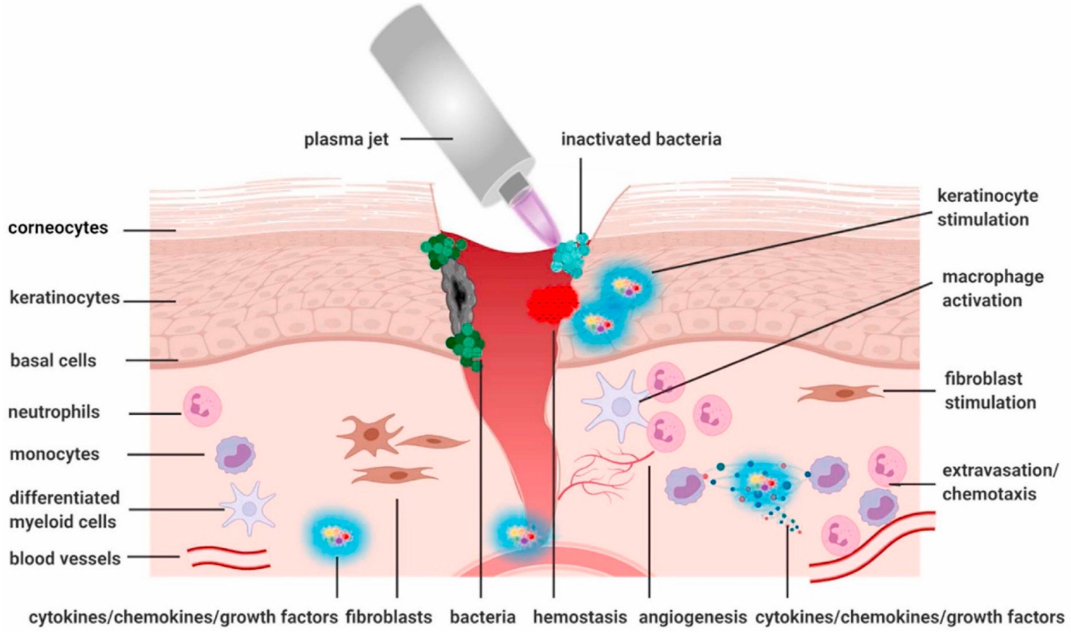
Figure 1. Schematic and discharge voltages of the cold plasma jet device: (a) the device diagram, (b) discharge voltage with argon, and (c) discharge voltage with helium. Additionally, the left side illustrates the He-fed plasma jet treating a surface with plume spreading. In contrast, the right side shows the He-fed plasma jet detected by the optical emission spectrum, containing reactive oxygen and nitrogen species (RONS). Reproduced with permission from Ref. [4]. Copyright 2022 AIP publishing.
Preprints 93115 g001
Figure 2. The formation, penetration, and characterization of CAPs entail complex interactions influenced by chemical and physical factors. CAPs interact intricately with treated targets, generating reactive oxygen and nitrogen species. (RONS) from atmospheric gases and water vapor. CAP exposure enriches aqueous media with species like hydrogen peroxide. Furthermore, CAP therapy influences the extracellular matrix (ECM), a crucial 3-dimensional network in skin health. This interaction may modulate skin inflammation and infection, while the ECM's role as a cell scaffold is essential for tissue development [61,62]. Factors influencing species quantity include plasma settings and distance from the target. Reproduced with permission from Ref. [19]. Copyright 2020 Elsevier.
Preprints 93115 g002
Figure 3. Gas plasma treatment effects on wound healing targets. Gas plasma treatment influences key cells (keratinocytes, fibroblasts, immune cells) in and around the wound bed, reducing microbial growth. It stimulates keratinocytes and fibroblasts, activates macrophages, and attracts neutrophils and lymphocytes toward the damaged tissue. Reproduced with permission from Ref. [7]. Copyright 2021 Elsevier.
Preprints 93115 g003
Table 1. Plasma and Its Multifaceted Biomedical Applications.
Application Description
Cold Atmospheric Plasma An emerging multi-agent technology and multi-modal therapy with diverse applications across various biomedical fields [5].
Oral Biofilm-Related Infections Plasma medicine effectively addresses oral biofilm-related infections, showcasing its efficacy in promoting oral health [6].
Therapeutic Use of Physical Plasma Involves the direct application of physical plasma on or in the human body for therapeutic purposes [7].
Plasma Technology for Biomedical Applications There is growing interest in utilizing plasmas for biomedical applications, particularly in plasma medicine, focusing on therapeutic advancements [8].
Biomedical Applications of Cold Atmospheric Plasma Encompasses sterilization, wound healing, blood coagulation, oral/dental disease treatment, cancer therapy, and immunotherapy [5,7,9].
Diverse Range of Applications Demonstrates various applications and advancements, offering examples of ideas and applications in the medical and biomedical domains [10].
Table 2. Various medical applications of plasma medicine.
Medical Applications Description
Therapeutic Purposes Direct application of Cold Atmospheric Plasma (CAP) for various medical treatments [20,21].
Disinfection Utilizes ionized gas (physical plasma) for disinfection, effectively inactivating various microorganisms, including viruses, resistant microbes, fungal cells, bacteria, spores, and biofilms created by microbes [13,22,23].
Healing Under study for its potential in healing, plasma medicine stimulates cell proliferation and angiogenesis with lower plasma treatment intensity, contributing to wound healing [17,21,24].
Cancer Treatment Exploration of plasma medicine for cancer treatment can inactivate cells and initiate cell death with higher plasma intensity [25,26,27,28].
Blood Coagulation Utilization of plasma for blood coagulation [28,29].
Dental Applications Application of ionized gas in plasma medicine for various dental purposes [20,30].
Sterilization of Implants and Surgical Instruments Plasma-generated active species are harnessed for sterilizing implants and surgical instruments [31,32].
Modifying Biomaterial Surface Properties Plasma medicine can modify biomaterial surface properties [33,34].
Treatment of Skin Diseases and Wounds Ongoing research on the potential of plasma medicine for treating skin diseases and wounds [17,21,24,35].
Multidisciplinary Research Intersection of scientific domains for diverse research in plasma medicine, combining plasma physics, life sciences, and clinical medicine [1,2,3,17].
Note: Most of the research in plasma medicine is conducted in vitro and animal models, highlighting the emerging nature of the field and its integration of plasma physics, life sciences, and clinical medicine [1,2,3,4,17].
Table 3. Cold Atmospheric Plasma Applications in Dermatology.
Application Description
1. CAP's Suppressive Effect on Microbial Skin Infection With advantages over traditional antibiotics, CAP effectively inhibits infections on diverse surfaces. Studies demonstrate its safety and efficacy in sterilizing bacteria, disrupting biofilms, and reducing bacterial loads in diabetic foot ulcers. CAP's broad-spectrum mechanism challenges bacterial resistance, making it promising for combating infectious diseases.
1.1 Effects of CAP on Bacteria Research confirms CAP's efficacy against bacteria and biofilms, including common strains like Escherichia coli. Plasma-activated liquids, especially Plasma-Activated Water (PAW), show promise by generating reactive species, disrupting biofilms, and modulating inflammation, offering a novel approach to address biofilm-related infections.
1.2 Effects of CAP on Fungi CAP has shown efficacy in treating nail fungal infections, inhibiting the growth of causative agents in pathogen models and clinical trials. A pilot study and a cohort of 40 patients demonstrated overall clinical cure rates exceeding 70%, establishing atmospheric pressure cold plasma as a safe and promising alternative therapy for skin fungal infections.
1.3 Effects of CAP on Viruses and Parasites CAP shows potential antiviral effects, with positive outcomes reported in treating viral warts and lower but measurable results on HSV-1. It also demonstrates efficacy against parasites like Demodex mites and head lice, suggesting its possible use in dermatology pending further exploration and studies.
2. CAP Promotes Tissue Proliferation and Wound Healing CAP is effective in wound healing by promoting antiseptic properties, pro-angiogenic effects, tissue proliferation, and the expression of growth factors and chemokines. CAP accelerates wound healing by reducing bacterial load, stimulating cell proliferation, and inducing gene expression, offering a promising approach for improved wound treatment, including the use of plasma-activated liquids like Plasma-Activated Water (PAW) and Plasma-Activated Hydrogel (PAH).
3. CAP for the Treatment of Inflammatory Skin Diseases CAP regulates the formation of stratum corneum cells by modulating the redox balance and activating the NRF2 pathway. Clinical studies show that CAP can alleviate skin inflammation, restore normal cell differentiation, and may represent a novel therapeutic approach for inflammatory skin disorders by regulating cell viability, proliferation, migration, and inflammatory responses.
3.1 Psoriasis CAP demonstrated therapeutic effects on psoriasis in cellular and animal studies, reducing epidermal hyperplasia and improving symptoms by inducing apoptosis and regulating reactive species. Atmospheric pressure cold plasma patches also showed promise in treating psoriatic skin lesions by mitigating electric field effects and inducing calcium ion channel opening in keratinocytes.
3.2 Atopic Dermatitis CAP has shown positive effects in alleviating atopic dermatitis (AD) in mouse models by reducing skin inflammation, oxidative stress, and dermatitis severity. Clinical studies on AD patients revealed improvements in skin lesions, itching, and a reduction in Staphylococcus aureus proportion after plasma treatment.
3.3 Vitiligo CAP) shows promise in treating Vitiligo by stimulating repigmentation in affected areas. Studies, including one by Zhai et al., demonstrate CAP's efficacy and safety in enhancing melanocyte activity, reducing inflammation, and achieving partial or complete pigmentation in vitiligo skin lesions without adverse events.
4. CAP Suppresses Tumor Cell Proliferation and Migration CAP demonstrated therapeutic efficacy in tumor treatment, inducing apoptosis in melanoma and skin cancer cells, with potential applications in various cancers. Plasma-activated hydrogel (PAH) is a carrier for CAP, enhancing treatment response, and a novel plasma-activated biological hydrogel demonstrates efficacy in eliminating residual tumor tissue without systemic toxicity after surgical resection.
5. Cosmetic and Skincare Applications of CAP Microplasma radiofrequency treats scars, while CAP benefits beauty. Suwanchinda's studies show improvements in striae and hypertrophic scars with high satisfaction. Plasma's reactive oxygen species offer a novel approach to skin barrier regulation. Combining cold plasma with vitamin C boosts collagen and skin elasticity, promoting skin beauty.
6. CAP's Role in Treating Immune-Mediated Skin Diseases CAP has demonstrated therapeutic effects on immune skin diseases. It showed positive results in reducing inflammation and oxidative stress and improving skin lesions. Additionally, CAP, delivered through a plasma-activated hydrogel, proved effective and safe in mouse models and vitiligo patients, enhancing melanin distribution and reducing inflammatory factors. Overall, CAP presents a promising and safe approach for managing immune skin diseases, supported by laboratory and clinical evidence.
Table 4. Challenges and Solutions in CAP for Skin Disease Treatment and Skincare.
Challenge Elaboration Possible Solutions References
1. Lack of Standardization The absence of standardized protocols in CAP applications for skin diseases hinders reproducibility and efficacy assessment. Establish standardized treatment guidelines and parameters for CAP applications, considering variations in devices and conditions. [2][6]
2. Safety Concerns Safety aspects, including potential risks and adverse effects, require thorough examination for wider clinical adoption. Conduct comprehensive safety analyses, identify potential risks, and propose mitigation measures to ensure the safe application of CAP. [1][5]
3. Variable Patient Response Variability in individual patient responses to CAP treatment challenges personalized medicine approaches. Implement personalized treatment plans by considering patient-specific factors, such as skin type, medical history, and genetic factors. [2][6]
4. Optimizing Treatment Duration Determining the optimal duration of CAP treatment to balance effectiveness and patient comfort is challenging. Conduct controlled trials to identify the most effective treatment durations for specific skin diseases while minimizing potential discomfort. [3][5]
5. Understanding Mechanisms of Action Limited understanding of the precise mechanisms underlying CAP's therapeutic effects impedes optimization. Invest in comprehensive research to elucidate CAP action's molecular and cellular mechanisms in skin disease treatment. [4][6]
6. Integration with Standard Therapies Integrating CAP into existing standard skincare and dermatological therapies poses integration challenges. Explore synergistic effects with traditional therapies and optimize combination treatment strategies for enhanced efficacy. [2][6]
7. Cost of Treatment The potential high cost of CAP devices and treatments may limit accessibility for some patients. Foster research and development to create cost-effective CAP devices and treatment modalities without compromising efficacy. [3][5]
8. Long-Term Effects and Sustainability Understanding the long-term effects of repeated CAP treatments and ensuring sustainability is crucial. Conduct longitudinal studies to assess the long-term effects and establish sustainable practices in CAP therapy. [1][6]
Disclaimer/Publisher’s Note: The statements, opinions and data contained in all publications are solely those of the individual author(s) and contributor(s) and not of MDPI and/or the editor(s). MDPI and/or the editor(s) disclaim responsibility for any injury to people or property resulting from any ideas, methods, instructions or products referred to in the content.
Copyright: This open access article is published under a Creative Commons CC BY 4.0 license, which permit the free download, distribution, and reuse, provided that the author and preprint are cited in any reuse.
Prerpints.org logo

Preprints.org is a free preprint server supported by MDPI in Basel, Switzerland.

Subscribe

© 2024 MDPI (Basel, Switzerland) unless otherwise stated