3. Experiment and discussion of results
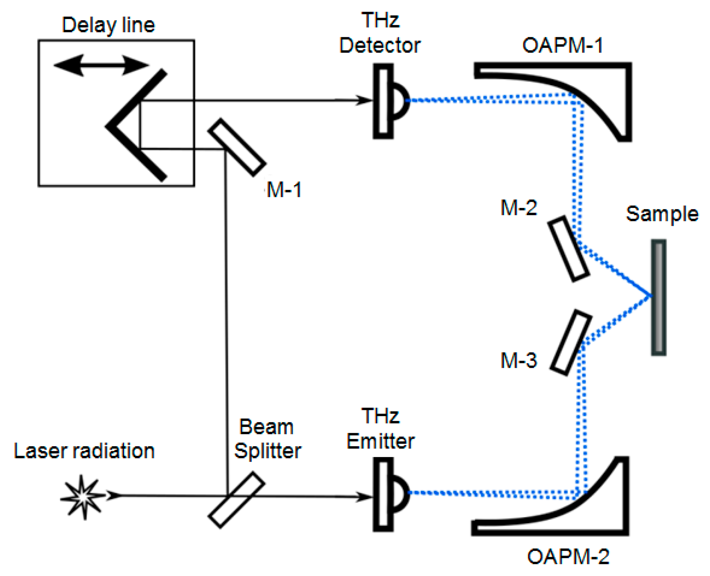
In order to investigate the polarization and spectral properties of a metamaterial formed by omega-shaped elements on a silicon substrate, THz radiation path of a pulse terahertz spectrometer was built accordingly to sketch represented in
Figure 2.
The sensitivity to polarization is a significant characteristic shown by photoconductive antennas employed in THz spectrometer sources and radiation receivers. This sensitivity enables the detector to function as a polarization analyzer. In this particular scenario, the orientation of the antennas is such that the electric field vector is aligned with the horizontal plane. This alignment corresponds to the plane of the schematic diagram in
Figure 2 and also agrees with the p-polarization of incidence radiation. To convert the alignment to s-polarization, it was made available to rotate the assembly, including mirrors 3-2 and 3-3, along with the sample holder. The radiation will be displaced off the plane. Here, the sample will be positioned beneath the mirrors. As a result, the plane of the metamaterial will be aligned in parallel to the polarization plane of the THz radiation. The polarization of the THz wave in this case is perpendicular to the plane of incidence. A more detailed description of the spectrometer set-up can be found in [
17].
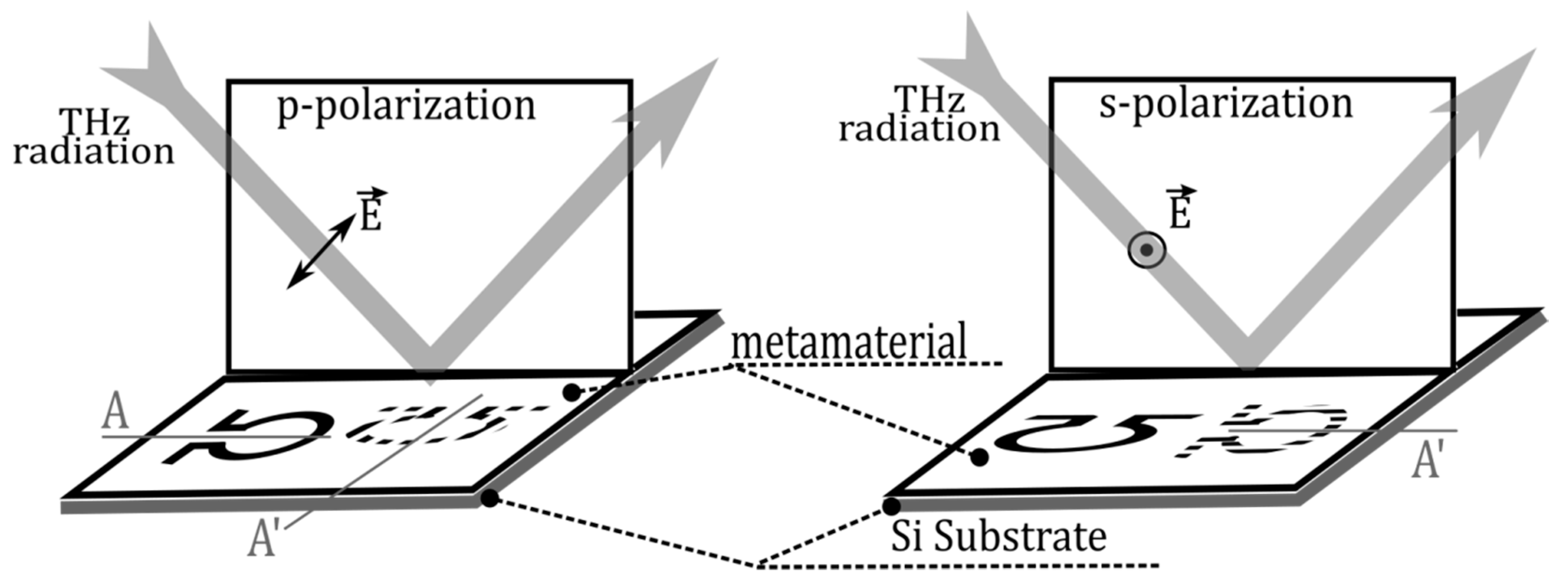
The study focused on examining changes in the spectral composition of THz radiation reflected from a metamaterial. The investigation specifically looked at how these changes were influenced by the orientation of the symmetry axis of the omega-shaped element in relation to the projection of the radiation polarization direction onto the plane of the metamaterial. The system of designations for rotation angles that has been adopted is explained in
Figure 3.
The standard representation of the metamaterial in
Figure 3 consists of a single omega element. The element represented in the schematic by a dashed line signifies the alteration in the orientation of its symmetry axis A as the metamaterial undergoes rotation around an axis perpendicular to its surface for π/2 to reach the position A’. The measurement of the rotation angle of the symmetry axis of the omega-shaped element is conducted with respect to the electric-field vector of the incident wave. Therefore, the zero value of the angle
corresponds to the position of the symmetry axis inside the plane of oscillations of the electric vector of THz radiation that is directed towards the sample. At an angle
of 90 degrees, the symmetry axis of the omega-shaped element is orthogonal to the electric vector of the incident wave, while the "arms" of the omega-shaped element are situated within the plane of oscillations of the electric vector of the incident THz radiation. The designations and nomenclature frequently employed for the components of the electric field vector of the wave are
and
. The numerical values within the notations of the dependencies depicted in the schematics presented below are representative of the rotation angles
expressed in degrees.
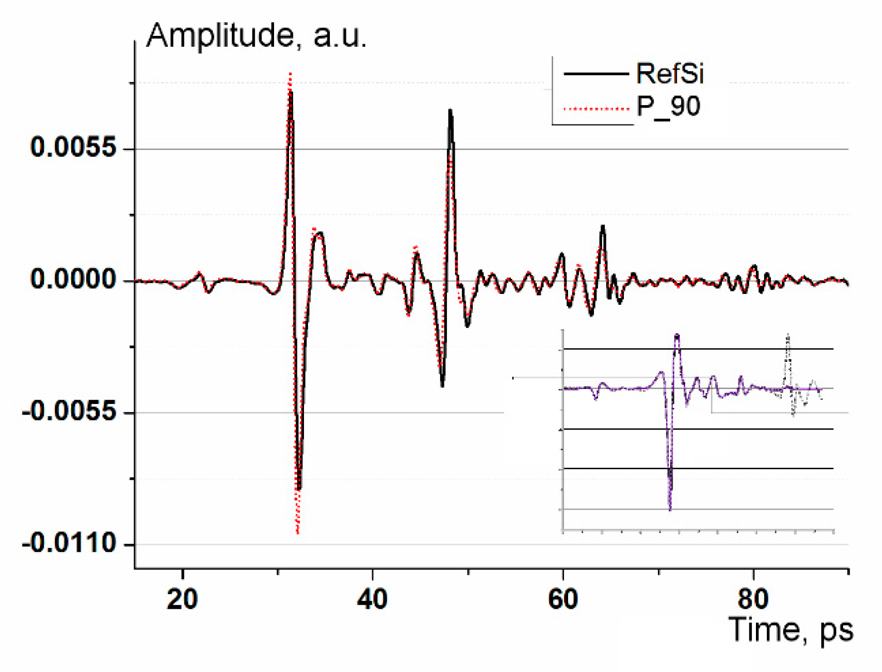
Time-domain spectrometers are conventionally used to perform a point-by-point measurement of the current in a PCA detector, induced by the electrical component of the THz pulse field. The current is proportional to the field strength and recorded as function of delay time. The waveform length was 140 ps, and the delay line scanning step was chosen to be ~0.075 ps. The indicated parameters correspond to a spectral resolution of ~0.008 THz and a frequency range of up to 7.5 THz after the Fourier transform. The excess range width is justified by the possibility to determine more accurately the position of the pulse on the time scale. Accordingly, visual control of the sample positioning is ensured during the waveform recording. Also, it is possible to estimate the average refractive index, as well as to choose parameters for data preprocessing before the Fourier transform. The selected waveform duration and corresponded spectral resolution are sufficient and meet the measurement requirements.
The high transparency and relatively large refractive index of the Si substrate, equal to an average of 3.5, cause the appearance of several echo pulses in the waveform. Echoes are produced by reflections in the plane-parallel silicon layer. As a result, the spectrum after the Fourier transform is distorted by the so-called Fabry-Perot effect [
18]. Moreover, components of the transmission spectrum are added to the reflection spectrum. The information about these components is carried by echo pulses passing twice through the metasurface layer.
Figure 4 demonstrates an example of the waveform and preprocessing results.
It is notable that the reflectivity of the matrix is higher than that of the Si substrate. Therefore, we used the reflection from the aluminum plate as a reference signal. The weak peak visible on the waveform at 23 ps is caused by the design of the emitter and is compensated in the Fourier transform by a symmetrical analogue that is formed in the detector antenna. To suppress the Fabry-Perot effects, we used the window function proposed in [
19].
This function is smooth and its derivatives are smooth as well. Compared to the common window functions, such as Hann, Hanning, etc., it introduces less attenuation near the peak due to a wider and flatter top. The result of applying the window function is presented in the inset of
Figure 4. Here, the solid line shows the result of applying the window function, while the suppressed echo signal is indicated by the dotted line. The resulting spectra retained a small residual modulation, which did not prevent the correct analysis of polarization-dependent spectral changes.
The reflection from a thin aluminum plate, that was fixed in the holder instead of the sample, was used as a reference signal.
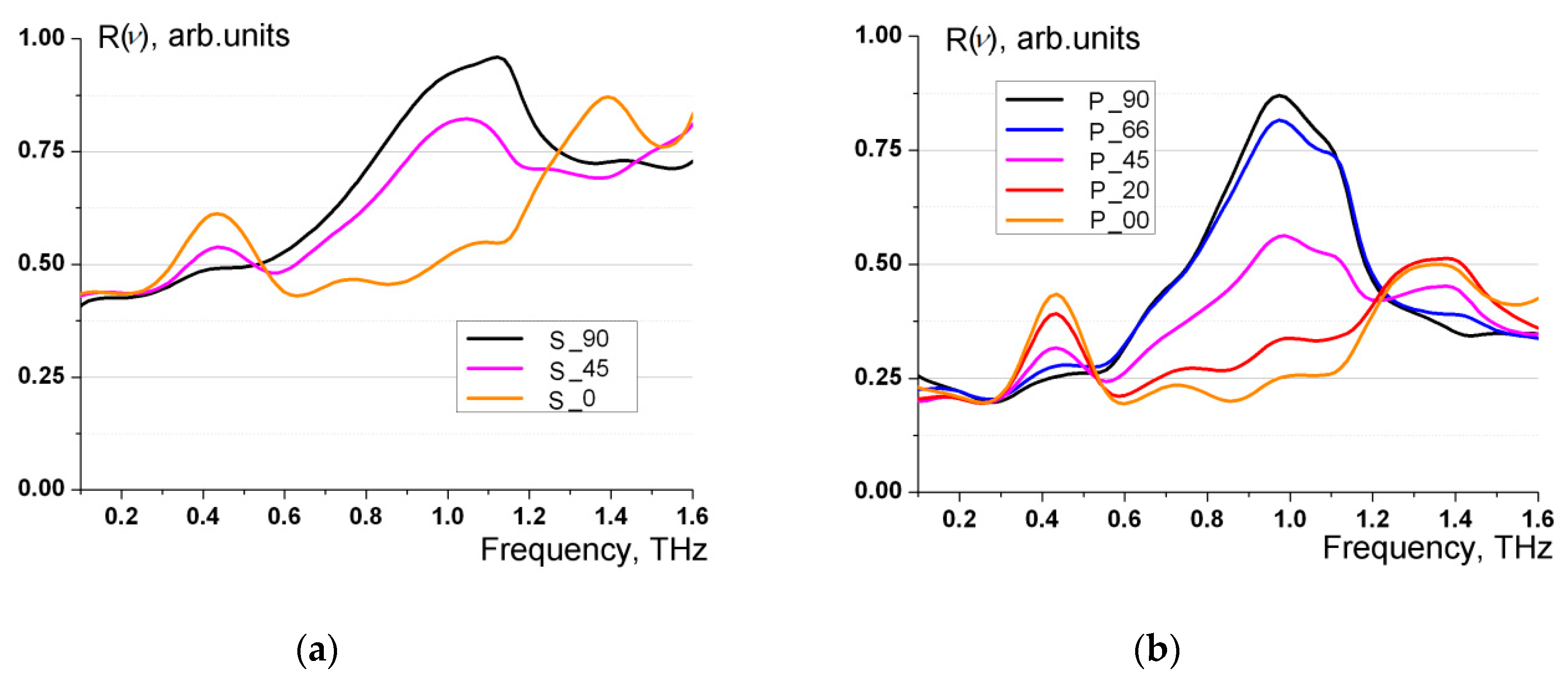
Figure 5 illustrates families of characteristic reflection spectra.
The acquired experimental outcomes are explicable in light of the theory of dipole radiation of electromagnetic waves [
20]. The dipole radiation theory is applicable because the dimensions of each omega-shaped element are considerably smaller than the wavelength of the electromagnetic field. In the dipole approximation, the electric field intensity of the wave emitted by an omega-shaped element has the following form
where
is the radius-vector from the center of the omega-element to the observation point;
is the vacuum permeability;
is the distance from the center of the omega-element to the observation point;
is the unit vector of the wave normal; с is the speed of light in vacuum;
is the dipole moment of the omega-shaped element,
is the magnetic moment of the omega-shaped element (two dots above the vectors denote the second derivative of these vector quantities).
According to equation (2), it can be deduced that an omega-shaped element exhibits efficient wave radiation when it experiences a substantial induction of electric dipole moment
and/or magnetic moment
. The metamaterial under investigation uses omega-shaped elements that possess pre-calculated parameters, which are characterized by the simultaneous excitation of both electric dipole moment and magnetic moment, with the absolute values of both moments satisfying the following relation with a certain degree of accuracy
Therefore, the omega-shaped element can be regarded as a bianisotropic particle exhibiting both electric and magnetic properties.
The omega-shaped element exhibits most effective excitation when its "arms" are aligned with the plane of oscillation of the electric field vector of the incident wave, specifically in the case of
. Here, the displacement of conduction electrons along the "arms" is accompanied by the appearance of electric current within the coil. Specifically, both the electric dipole moment and magnetic moment are induced simultaneously. The plots of reflection coefficients in
Figure 4 for these cases of effective activation of the omega-shaped element at
are denoted as s_90 and p_90. It can be deduced from
Figure 5 that the wave reflection coefficients attain their maximum values precisely in the cases indicated as s_90 and p_90. In the case of s_90, the "arms" of the omega-shaped element are parallel to the incident wave vector
. In the case involving p_90, the "arms" of the omega-shaped element are situated within the plane of oscillations of the incident wave vector
. Throughout the experiment, the angle
between the symmetry axis of the omega-shaped element and the plane of oscillation of the incident wave electric vector takes the values of 90, 66, 45, 20 and 0 degrees. It is possible to consider the component
of the electric vector of the incident wave parallel to the "arms" of the omega-shaped element, which is equal to
,
, where
is the incidence angle of the wave on the surface of the metamaterial. As the angle
decreases, there is a corresponding decrease in the component
, resulting in an ensuing decrease in the reflection coefficient.
Figure 5 illustrates precisely this behavior of reflection coefficients for incident waves with s- and p- polarizations.
Thus, the experimental findings relating to the reflection coefficients, which are displayed in
Figure 5, are consistent with the outcomes of the theoretical analysis.
Additionally, the frequency dependence of the reflection coefficients for s- and p- polarization of the incident wave, as depicted in
Figure 5, is noteworthy. The resonant nature of the metamaterial reflection spectra can be attributed to the enhanced excitation of each omega-shaped element at a certain frequency, which is related to the geometrical parameters of the omega-shaped element. The resonant excitation of the omega-shaped element occurs when its full length in the straightened state is approximately equal to half the wavelength of the electromagnetic field. Consequently, the resonant frequency satisfies the relation
Here,
is the full length of the omega-shaped element in the straightened state,
l is the omega-shaped element arm length,
is the coil radius of the omega-shaped element. The metamaterial under investigation uses omega-shaped elements with parameters
l =12.8 μm,
=19.8 μm. Therefore, based on equation (4), it can be concluded that the resonance frequency is
= 1.0 THz, which is approximately equal to the maximum frequency of the reflection coefficients in
Figure 5. These coefficients are obtained at the angles between the symmetry axis of the omega-shaped element and the plane of oscillation of the electric vector of the incident wave
. At these specific angles
, the omega-shaped elements are activated predominantly by the electric vector of the incident wave, resulting in the fulfilment of relation (4). When the angle between the symmetry axis of the omega-shaped element and the plane of oscillation of the electric vector of the incident wave assumes values of
, it can be observed that the omega-shaped elements are mostly excited by the magnetic field vector of the incident wave. In this instance, the electric current originates primarily in the coil of the omega-shaped element, with its arms not contributing, which results in another resonant frequency formula
By employing formula (4), the resonance frequency is determined to be
THz. The frequency obtained is roughly equivalent to the frequency of the second maximum of the reflection coefficients shown in
Figure 5.
Hence, the frequency dependence of reflection coefficients displayed in
Figure 5, including the occurrence of two main maxima, correlates with the findings of the theoretical study.
The incidence angle of radiation on the metamaterial is not accounted for in formulas (4) and (5). Experimental studies performed at normal incidence of THz waves on the metamaterial confirm the presence of two main maxima in the reflection spectra at the same frequency values and as well as notable polarization anisotropy in the reflection coefficients.
It is essential to consider a significant distinction between the reflected waves at s- and p-polarization of the incident wave, which arises from the different polarization of the reflected radiation. According to equation (2), when a wave with s-polarization is incident on a metamaterial, the resulting reflected radiation is likewise linearly polarized and comprises only an s-component. When the incident wave is p-polarized, the reflected radiation demonstrates an elliptical polarization that closely resembles circular polarization.
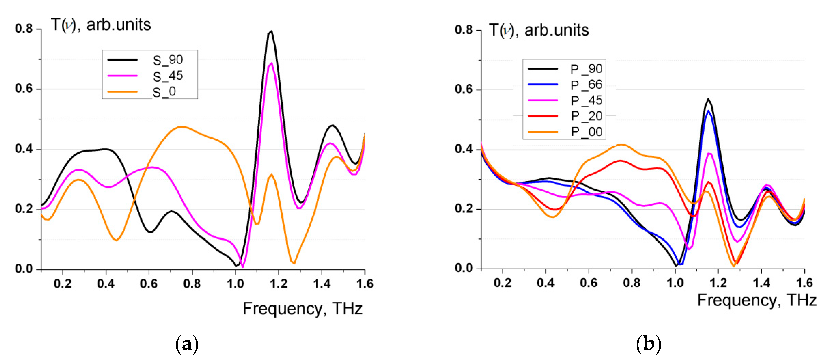
Additionally, an investigation into the transmission of THz radiation through the metamaterial was conducted.
Figure 6 illustrate families of characteristic transmission spectra.
When constructing the spectra, standard normalization to the wave intensity in the absence of metamaterial was performed. The transmission spectra
exhibit a resonant nature and demonstrate an almost total lack of the transmitted wave in close proximity to the frequency
for the cases s_90, s_45, p_90, p_66, as well as in the vicinity of the frequency
for the cases s_0, p_20, p_0. The analysis reveals that the spectra
are close to the frequency dependence
of the reflection coefficients presented in
Figure 5.
