Preprint
Review

Recent Computational Methods for Pathological Brain Detection

Altmetrics

Downloads

139

Views

34

Comments

0

This version is not peer-reviewed

Submitted:

16 December 2023

Posted:

26 December 2023

You are already at the latest version

Alerts
Abstract
Pathological Brain Detection (PBD) is a crucial field aimed at identifying and diagnosing structural or functional abnormalities in the brain related to neurological, neurodegenerative, or psychiatric disorders. This detection process typically employs medical imaging technologies such as MRI, CT, or PET scans, along with neurological evaluations, blood tests, and other diagnostic tools. Early detection is essential for effective treatment, improving prognosis, and enhancing the quality of life for affected individuals. Each common computational method provides unique insights into brain pathologies. Despite significant advancements in technology and methodology, PBD faces challenges such as variability in brain anatomy, the complexity of disorders, data quality, standardization issues, interpretability of models, and ethical concerns. Addressing these challenges necessitates collaboration among researchers, clinicians, and policymakers to develop robust and ethical methods for improving the detection and diagnosis of brain disorders.
Keywords: 
Subject: Computer Science and Mathematics  -   Artificial Intelligence and Machine Learning

1. Introduction

The brain is a complex and highly specialized organ that serves as the control center of the human body and many other animals [1,2]. It is part of the central nervous system and is located within the skull [3]. The primary functions of the brain include: (i) Processing Information: The brain receives and processes sensory information from the body's various systems [4], including sight, hearing, touch, taste, and smell [5,6]. It interprets this information to create our perception of the world around us. (ii) Motor Control: The brain controls voluntary and involuntary movements of the body [7]. It sends signals to muscles and other organs through the nervous system to enable movement, from simple actions like picking up an object to complex tasks like playing a musical instrument [8]. (iii) Homeostasis: The brain helps maintain the body's internal balance (homeostasis) by regulating functions like body temperature, blood pressure, and hormone production [9,10,11]. (iv) Autonomic Functions: It controls autonomic functions such as heart rate, breathing, digestion, and the circulatory system., etc [12,13].
A pathological brain [14,15,16] refers to a brain that exhibits abnormal structural or functional changes often associated with various neurological, neurodegenerative, or psychiatric disorders [17]. These alterations can result from a wide range of conditions, including tumors [18], infections [19], traumatic injuries [20], neurodegenerative diseases like Alzheimer's [21] or Parkinson's [22], and mental health disorders like depression [23] or schizophrenia [24]. Pathological changes can be observed through techniques such as brain imaging [24,25] (e.g., MRI or CT scans) or post-mortem examination, helping healthcare professionals diagnose and understand the underlying causes of cognitive, emotional, or physical impairments in individuals with such conditions. Accurate identification and characterization of pathological brain changes are crucial for appropriate treatment and management strategies.

2. Pathological Brain Detection

Pathological brain detection (PBD) [26,27] is the process of identifying and diagnosing abnormal structural or functional changes within the brain that are indicative of underlying neurological, neurodegenerative, or psychiatric disorders [28]. Figure 1 presents several samples of pathological brains [28]. This detection typically involves the use of various medical imaging techniques, such as MRI [29], CT [30], or PET scans [31], to visualize and analyze the brain's structure and activity.
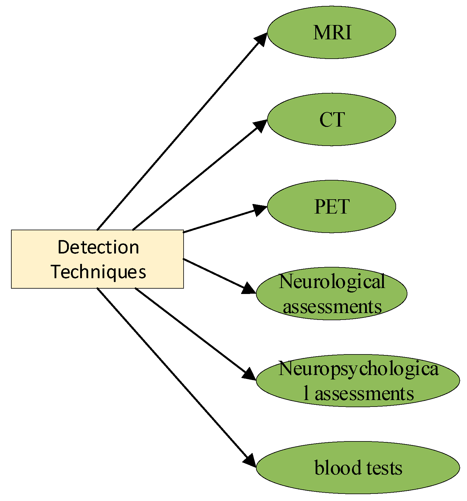
As shown in Figure 2, it can also encompass neurological and neuropsychological assessments, blood tests, and other diagnostic tools to evaluate cognitive, emotional, or physical impairments associated with these pathologies [32]. Accurate and timely detection of pathological brain changes is essential for early intervention, treatment planning [33], and improving the prognosis and quality of life for individuals affected by these conditions.
Early detection allows for the prompt initiation of appropriate treatments or interventions. In many neurological and psychiatric conditions, the effectiveness of treatment is significantly higher when administered in the early stages of the disease. Delayed intervention may result in irreversible damage, making it more challenging to manage or reverse the condition.
Some brain pathologies, such as neurodegenerative diseases (e.g., Alzheimer's [34], Parkinson's), can progress slowly over time. Detecting these changes early can help implement strategies to slow down or halt disease progression, potentially preserving cognitive [35] and motor functions for a longer period.
Timely intervention can enhance a patient's quality of life. For instance, in cases of brain tumors or vascular lesions, early detection and treatment can prevent the development of severe symptoms, including paralysis, seizures, or cognitive deficits, which can significantly impact a person's daily life.
Detecting brain pathologies at an earlier stage can lead to more cost-effective treatments. Late-stage interventions often require more extensive medical procedures, hospitalizations, and long-term care, all of which are associated with higher healthcare costs.

3. Common Computational Methods for PBD

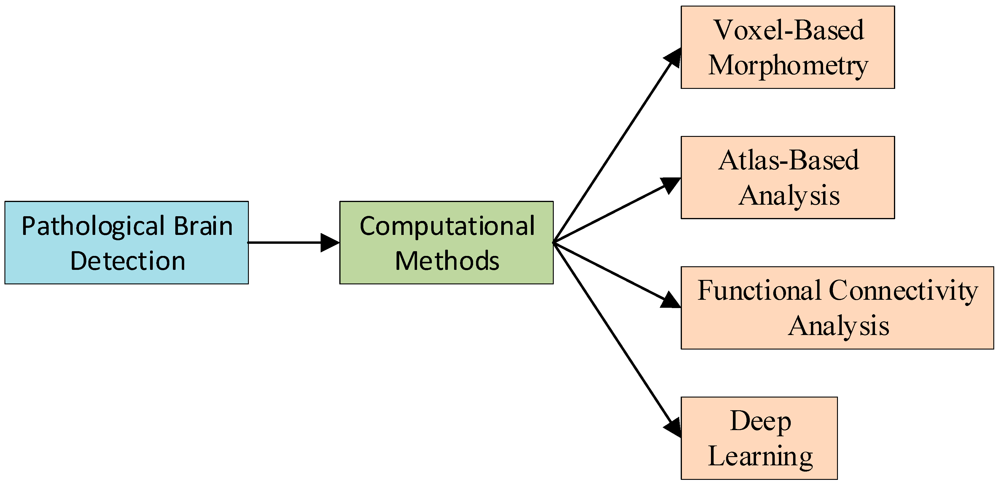
Voxel-Based Morphometry (VBM) [36] is a neuroimaging analysis technique that allows researchers to investigate focal differences in brain anatomy. Utilizing the principles of computational neuroanatomy, VBM involves the segmentation of brain images into different tissue types, followed by a voxel-wise comparison of these tissues across different populations [37]. This method is especially common in the study of diseases like Alzheimer's, where it aids in detecting and quantifying brain atrophy [38,39]. VBM's strength lies in its ability to identify subtle and distributed patterns of brain structure changes, making it an invaluable tool in the arsenal of computational methods for Pathological Brain Detection [40]. Figure 3 shows common computational methods for PBD.
Atlas-Based Analysis [41] serves as another pivotal computational approach, providing a detailed anatomical framework for interpreting brain images. By mapping new image data onto a standardized atlas, researchers can localize and quantify alterations in brain structure and function with greater accuracy. This is particularly useful for evaluating surgical risks and treatment plans for conditions like epilepsy or brain tumors [42]. The precision of Atlas-Based Analysis ensures that pathological regions are identified relative to established neuroanatomical benchmarks, enabling consistent and reproducible assessments across different studies and populations.
Functional Connectivity Analysis [43] delves into the realm of understanding the brain's functional organization by examining the temporal correlations between different brain regions. This approach has been instrumental in elucidating the impact of various pathologies on brain function, such as the disruption of connectivity patterns seen in multiple sclerosis or schizophrenia [44]. When combined with advanced statistical techniques, Functional Connectivity Analysis allows for the mapping of complex brain networks and the detection of network anomalies that underpin cognitive and behavioral deficits [45]. Meanwhile, Deep Learning [46,47], particularly through Convolutional Neural Networks, is revolutionizing PBD by providing robust tools for automated feature extraction and classification in neuroimaging. These models learn hierarchical representations of data, achieving state-of-the-art results in tasks like lesion detection, segmentation, and predictive modeling of brain disorders. The adaptability and power of Deep Learning algorithms have established them as a cornerstone of modern computational methods in neuroimaging, paving the way for breakthroughs in the rapid and accurate detection of pathological brains.

3.1. Voxel-Based Morphometry

Voxel-Based Morphometry (VBM) [48,49] is a neuroimaging technique used to detect and analyze structural changes in the brain, making it a valuable tool for identifying pathological brains. VBM involves comparing an individual's brain image to a standardized brain template or atlas. This comparison highlights deviations from the norm, which can be indicative of pathological changes. Differences in brain tissue volume, density, or shape can be detected and quantified [50,51,52].
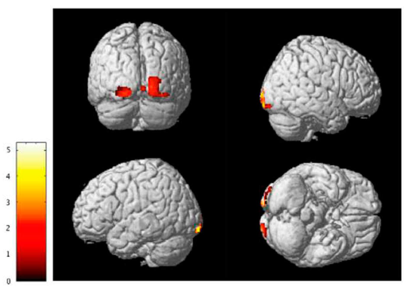
Figure 4 shows an example of VBM [53]. Continuing from the description of VBM, this technique's strength lies in its ability to systematically scrutinize the entire brain for structural variations, without the constraint of region-specific analysis. This whole-brain approach ensures that no potential area of pathology is overlooked, providing a comprehensive assessment of the brain's anatomical integrity. After the detection of atypical regions, VBM can be used to correlate these anatomical findings with clinical symptoms and outcomes. This association is critical for understanding the pathophysiological underpinnings of various neurological and psychiatric conditions. For instance, the correlation between gray matter reductions in specific cortical regions and cognitive deficits in dementia has been well documented using VBM [54]. Moreover, by employing longitudinal designs, VBM can track the efficacy of therapeutic interventions over time, allowing for an objective measure of disease progression or remission.
VBM's applications have also expanded into the research domain, informing our understanding of the structural brain changes associated with genetic factors, environmental influences, and even learning and adaptation processes. The versatility of VBM is further enhanced when combined with other imaging modalities, such as functional MRI [55,56], providing a more holistic view of the brain's alterations in structure and function. As imaging technology and analysis techniques continue to advance, VBM remains a vital and dynamic tool for neuroscientific research and clinical practice, aiding in the quest to elucidate and combat brain pathologies.

3.2. Atlas-Based Analysis

Atlas-Based Analysis [41,57] is effective in detecting pathological brains because it involves aligning individual brain images to a standardized brain atlas, allowing for precise comparison of brain structures across subjects. This method highlights deviations from the norm, making it sensitive to structural abnormalities indicative of pathological conditions such as tumors, atrophy, or lesions [58]. By utilizing a common reference, Atlas-Based Analysis can systematically and objectively identify changes in brain anatomy, aiding in the diagnosis and characterization of various neurological and neurodegenerative disorders. It provides a framework for localizing and quantifying pathological alterations throughout the brain, contributing to early detection and treatment planning.
In research contexts, Atlas-Based Analysis is instrumental in studying the progression of neurodegenerative diseases. By comparing brain structures over time against standardized atlases, researchers can track disease progression and assess the impact of various therapeutic interventions. This longitudinal analysis is invaluable in understanding the natural history of diseases and in the development of new treatments.
Finally, the application of Atlas-Based Analysis is expanding beyond diagnostics to surgical planning and navigation. In neurosurgery, for instance, it assists surgeons in accurately targeting pathological areas while minimizing damage to healthy tissue, thereby improving surgical outcomes and patient recovery.

3.3. Functional Connectivity Analysis

Functional Connectivity Analysis [59,60] is capable of detecting pathological brains because it assesses the patterns of communication and coordination between different brain regions. In healthy individuals, there's a typical functional connectivity pattern [59], but various neurological and psychiatric disorders can disrupt these patterns. Pathological conditions like schizophrenia, depression, or Alzheimer's disease often manifest as altered functional connectivity, reflecting changes in how different brain regions interact during cognitive or resting-state tasks. By analyzing functional MRI data and identifying disruptions in connectivity networks, Functional Connectivity Analysis can provide insights into the underlying neural mechanisms of these disorders, aiding in their detection, characterization, and understanding, and potentially informing treatment strategies [61].
Furthermore, FCA contributes to the field of brain network research. It helps in mapping the intricate networks of neural connections and understanding how disruptions in these networks relate to specific cognitive and behavioral symptoms. This understanding is crucial for developing targeted therapies aimed at restoring normal connectivity patterns in affected brain regions. In a broader context, FCA is being integrated with other neuroimaging techniques, such as structural MRI and PET scans, to provide a more comprehensive view of brain disorders. This multimodal approach enhances the accuracy of diagnoses, helps differentiate between similar conditions, and offers a more detailed understanding of brain pathology.
Lastly, advancements in data processing and analysis techniques are continually refining FCA, making it more sensitive and specific in detecting subtle changes in brain connectivity. These improvements are opening new avenues in personalized medicine, where treatments can be tailored based on individual connectivity patterns, leading to better patient outcomes in managing neurological and psychiatric disorders.

3.4. Deep Learning

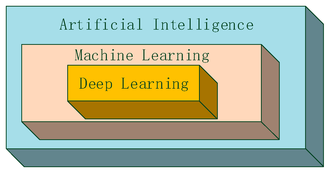
Deep learning [46,62] can effectively detect pathological brains due to its capacity to automatically learn intricate patterns and features from large-scale neuroimaging data. The relationship between deep learning, machine learning, and artificial intelligence is shown in Figure 5.
When applied to tasks such as image classification or segmentation using techniques like convolutional neural networks (CNNs) [63,64], deep learning models can identify subtle structural or functional abnormalities in brain scans. They excel at capturing complex relationships within the data, enabling the recognition of disease-specific patterns that may not be apparent through traditional methods [65].
By training on extensive datasets, deep learning algorithms become adept at distinguishing between healthy and pathological brain patterns, making them valuable tools for early detection [66], diagnosis [67], and even predicting disease progression [68], ultimately enhancing our understanding and management of various neurological and psychiatric disorders.
Deep learning significantly enhances pathological brain detection by tailoring diagnoses and treatments to individual neuroimaging profiles. Its ability to discern specific neurological disorder subtypes leads to more precise diagnoses. Additionally, in prognostics, deep learning predicts disease progression by analyzing longitudinal data, crucial for early intervention in conditions like Alzheimer's or Parkinson's.
This technology also aids in therapeutic development by identifying biomarkers and guiding drug research and clinical trials. It evaluates treatment effectiveness over time, offering a quantitative approach to monitor patient responses. While promising, challenges such as large dataset requirements, model transparency, and ethical data handling need addressing to fully leverage deep learning in this field.

4. Discussions

Current progress in pathological brain detection is marked by significant advancements in both technology and methodology. Machine learning [69] and deep learning approaches continue to gain prominence, allowing for more accurate and automated detection of brain abnormalities in neuroimaging data [70]. These methods are becoming increasingly capable of handling large datasets and identifying subtle structural and functional changes associated with neurological, neurodegenerative, and psychiatric disorders.
Moreover, there is a growing emphasis on multi-modal approaches that integrate different imaging techniques and data sources, enabling a more comprehensive understanding of brain pathology. Additionally, ongoing research focuses on early detection [71] and prediction of disease progression [72], paving the way for more personalized and timely interventions [73]. Overall, these developments hold promise for improving the diagnosis and management of pathological brain conditions.
Building on this momentum, the field of pathological brain detection is evolving to incorporate advanced analytical frameworks such as graph theory in the assessment of functional connectivity, which provides insights into the integrity of neural networks disrupted by pathological states. The application of graph theoretical approaches to neuroimaging data has revealed new biomarkers for diseases characterized by connectivity alterations, such as Autism Spectrum Disorders and schizophrenia [74].
Furthermore, the refinement of image acquisition techniques, like ultra-high-field MRI [75], offers unprecedented spatial resolution that captures detailed anatomical features of brain pathology. When these high-resolution images are processed using sophisticated computational algorithms, even the most subtle pathological changes can be identified, which were previously undetectable with standard resolution imaging.
In tandem with technological innovation, there is an increasing trend towards the open sharing of neuroimaging datasets and the development of standardized protocols for data analysis. This collaborative environment not only accelerates the pace of discovery but also ensures that findings are robust and replicable across diverse populations and settings. These collective efforts signify a transition towards a more collaborative and translational approach in brain research, which is essential for the development of targeted and effective therapies for brain disorders. The integration of computational methods with clinical expertise is key to the future of precision medicine, where treatment strategies can be tailored to the individual patient based on sophisticated diagnostic algorithms.

5. Conclusion

PBD faces several significant challenges. One major issue is the inherent variability in brain anatomy and function among individuals, making it challenging to establish clear diagnostic criteria and thresholds for detection. Additionally, neurological and psychiatric disorders often have complex and heterogeneous presentations, further complicating accurate detection. Data quality [76] and standardization [77] issues in neuroimaging, as well as the need for large, diverse datasets, are persistent challenges.
The interpretability of machine learning and deep learning models used in brain detection can also be a concern, as their decisions may lack transparency. Finally, ethical considerations surrounding data privacy, consent, and the responsible use of sensitive medical information pose important challenges in the field of pathological brain detection. Addressing these challenges requires ongoing collaboration between researchers, clinicians, and policymakers to develop robust and ethical approaches to improve detection and diagnosis of brain disorders.

Acknowledgments

We thank all the anonymous reviewers for their hard reviewing work.

References

  1. Fang, L.; Liu, Q.; Meyer, E.; Welle, A.; Huang, W.; Scheller, A.; Kirchhoff, F.; Bai, X. A subset of OPCs do not express Olig2 during development which can be increased in the adult by brain injuries and complex motor learning. Glia 2022, 71, 415–430. [Google Scholar] [CrossRef] [PubMed]
  2. Tanaka, M.; Diano, M.; Battaglia, S. Editorial: Insights into structural and functional organization of the brain: evidence from neuroimaging and non-invasive brain stimulation techniques. Front. Psychiatry 2023, 14, 1225755. [Google Scholar] [CrossRef]
  3. Perens, J.; Salinas, C.G.; Roostalu, U.; Skytte, J.L.; Gundlach, C.; Hecksher-Sørensen, J.; Dahl, A.B.; Dyrby, T.B. Multimodal 3D Mouse Brain Atlas Framework with the Skull-Derived Coordinate System. Neuroinformatics 2023, 21, 269–286. [Google Scholar] [CrossRef]
  4. Zhang, Y.; Dong, Z.; Wu, L.; Wang, S. A hybrid method for MRI brain image classification. Expert Syst. Appl. 2011, 38, 10049–10053. [Google Scholar] [CrossRef]
  5. Aguilar-Ruiz, J.S.; Bifet, A.; Gama, J. Data Stream Analytics. Analytics 2023, 2, 346–349. [Google Scholar] [CrossRef]
  6. Albustanji, R.N.; Elmanaseer, S.; Alkhatib, A.A.A. Robotics: Five Senses plus One—An Overview. Robotics 2023, 12, 68. [Google Scholar] [CrossRef]
  7. Chapman, A.D.; Selhorst, S.; LaComb, J.; LeDantec-Boswell, A.; Wohl, T.R.; Adhicary, S.; Nielsen, C.M. Endothelial Rbpj Is Required for Cerebellar Morphogenesis and Motor Control in the Early Postnatal Mouse Brain. Cerebellum 2022, 22, 613–627. [Google Scholar] [CrossRef] [PubMed]
  8. Zhang, Y.; Wu, L. An mr brain images classifier via principal component analysis and kernel support vector machine. Prog. Electromagn. Res. 2012, 130, 369–388. [Google Scholar] [CrossRef]
  9. Mikkelsen, A.C.D.; Thomsen, K.L.; Mookerjee, R.P.; Hadjihambi, A. The role of brain inflammation and abnormal brain oxygen homeostasis in the development of hepatic encephalopathy. Metab. Brain Dis. 2022, 38, 1707–1716. [Google Scholar] [CrossRef]
  10. Alonge, K.M.; Porte, D.; Schwartz, M.W. Distinct Roles for Brain and Pancreas in Basal and Postprandial Glucose Homeostasis. Diabetes 2023, 72, 547–556. [Google Scholar] [CrossRef]
  11. Ranjan, A.K.; Gulati, A. Controls of Central and Peripheral Blood Pressure and Hemorrhagic/Hypovolemic Shock. J. Clin. Med. 2023, 12, 1108. [Google Scholar] [CrossRef]
  12. Sorek, G.; Shaklai, S.; Gagnon, I.; Schneider, K.; Chevignard, M.; Stern, N.; Fadida, Y.; Kalderon, L.; Katz-Leurer, M. Impact of Subarachnoid Hemorrhage on the Cardiac Autonomic Function During Rehabilitation in Children After Severe Traumatic Brain Injury. Neurotrauma Rep. 2023, 4, 458–462. [Google Scholar] [CrossRef]
  13. Kirk, C.; Childs, C. Combat Sports as a Model for Measuring the Effects of Repeated Head Impacts on Autonomic Brain Function: A Brief Report of Pilot Data. Vision 2023, 7, 39. [Google Scholar] [CrossRef]
  14. Mut-Arbona, P.; Huang, L.; Baranyi, M.; Tod, P.; Iring, A.; Calzaferri, F.; Ríos, C.d.L.; Sperlágh, B. Dual Role of the P2X7 Receptor in Dendritic Outgrowth during Physiological and Pathological Brain Development. J. Neurosci. 2023, 43, 1125–1142. [Google Scholar] [CrossRef]
  15. Jin, M.; Cai, S.-Q. Mechanisms Underlying Brain Aging Under Normal and Pathological Conditions. Neurosci. Bull. 2022, 39, 303–314. [Google Scholar] [CrossRef]
  16. Parajuli, B.; Koizumi, S. Strategies for Manipulating Microglia to Determine Their Role in the Healthy and Diseased Brain. Neurochem. Res. 2022, 48, 1066–1076. [Google Scholar] [CrossRef]
  17. Rodkin, S.; Nwosu, C.; Sannikov, A.; Raevskaya, M.; Tushev, A.; Vasilieva, I.; Gasanov, M. The Role of Hydrogen Sulfide in Regulation of Cell Death following Neurotrauma and Related Neurodegenerative and Psychiatric Diseases. Int. J. Mol. Sci. 2023, 24, 10742. [Google Scholar] [CrossRef] [PubMed]
  18. Li, S.; Wang, C.; Chen, J.; Lan, Y.; Zhang, W.; Kang, Z.; Zheng, Y.; Zhang, R.; Yu, J.; Li, W. Signaling pathways in brain tumors and therapeutic interventions. Signal Transduct. Target. Ther. 2023, 8, 1–23. [Google Scholar] [CrossRef] [PubMed]
  19. Villadiego, J., et al., Full protection from sars-cov-2 brain infection and damage in susceptible transgenic mice conferred by mva-cov2-s vaccine candidate. Nature Neuroscience 2023, 26, 226–238. [CrossRef] [PubMed]
  20. Babiloni, A.H.; Baril, A.-A.; Charlebois-Plante, C.; Jodoin, M.; Sanchez, E.; De Baets, L.; Arbour, C.; Lavigne, G.J.; Gosselin, N.; De Beaumont, L. The Putative Role of Neuroinflammation in the Interaction between Traumatic Brain Injuries, Sleep, Pain and Other Neuropsychiatric Outcomes: A State-of-the-Art Review. J. Clin. Med. 2023, 12, 1793. [Google Scholar] [CrossRef] [PubMed]
  21. Johnson, N.H., et al., Inflammasome activation in traumatic brain injury and alzheimer's disease. Translational Research, 2023; 254, 1–12. [CrossRef]
  22. Alnaaim, S.A.; Al-Kuraishy, H.M.; Alexiou, A.; Papadakis, M.; Saad, H.M.; Batiha, G.E.-S. Role of Brain Liver X Receptor in Parkinson’s Disease: Hidden Treasure and Emerging Opportunities. Mol. Neurobiol. 2023, 61, 341–357. [Google Scholar] [CrossRef]
  23. Jahan, A.B.; Tanev, K. Neurobiological Mechanisms Of Depression Following Traumatic Brain Injury. Brain Inj. 2022, 37, 24–33. [Google Scholar] [CrossRef] [PubMed]
  24. Townsend, L.; Pillinger, T.; Selvaggi, P.; Veronese, M.; Turkheimer, F.; Howes, O. Brain glucose metabolism in schizophrenia: a systematic review and meta-analysis of 18FDG-PET studies in schizophrenia. Psychol. Med. 2022, 53, 4880–4897. [Google Scholar] [CrossRef] [PubMed]
  25. Constantinides, C.; Han, L.K.M.; Alloza, C.; Antonucci, L.A.; Arango, C.; Ayesa-Arriola, R.; Banaj, N.; Bertolino, A.; Borgwardt, S.; Bruggemann, J.; et al. Brain ageing in schizophrenia: evidence from 26 international cohorts via the ENIGMA Schizophrenia consortium. Mol. Psychiatry 2022, 28, 1201–1209. [Google Scholar] [CrossRef]
  26. Liu, F.-T.; Lu, J.-Y.; Li, X.-Y.; Liang, X.-N.; Jiao, F.-Y.; Ge, J.-J.; Wu, P.; Li, G.; Shen, B.; Wu, B.; et al. 18F-Florzolotau PET imaging captures the distribution patterns and regional vulnerability of tau pathology in progressive supranuclear palsy. Eur. J. Nucl. Med. 2023, 50, 1395–1405. [Google Scholar] [CrossRef]
  27. Hiono, T.; Kobayashi, D.; Kobayashi, A.; Suzuki, T.; Satake, Y.; Harada, R.; Matsuno, K.; Sashika, M.; Ban, H.; Kobayashi, M.; et al. Virological, pathological, and glycovirological investigations of an Ezo red fox and a tanuki naturally infected with H5N1 high pathogenicity avian influenza viruses in Hokkaido, Japan. Virology 2023, 578, 35–44. [Google Scholar] [CrossRef]
  28. Zhang, Y.; Wu, L.; Wang, S. Magnetic resonance brain image classification by an improved artificial bee colony algorithm. Prog. Electromagn. Res. 2011, 116, 65–79. [Google Scholar] [CrossRef]
  29. Billot, B.; Magdamo, C.; Cheng, Y.; Arnold, S.E.; Das, S.; Iglesias, J.E. Robust machine learning segmentation for large-scale analysis of heterogeneous clinical brain MRI datasets. Proc. Natl. Acad. Sci. 2023, 120. [Google Scholar] [CrossRef]
  30. Lagares, A.; Castaño-Leon, A.M.; Richard, M.; Tsitsopoulos, P.P.; Morales, J.; Mihai, P.; Pavlov, V.; Mejan, O.; de la Cruz, J.; Payen, J.F.; et al. Variability in the indication of brain CT scan after mild traumatic brain injury. A transnational survey. Eur. J. Trauma Emerg. Surg. 2022, 49, 1189–1198. [Google Scholar] [CrossRef]
  31. Ward, J., et al., Brain pet imaging: Frontotemporal dementia. PET clinics 2023, 18, 123–133. [CrossRef]
  32. Zhang, Y.-D.; Zhang, Z.; Zhang, X.; Wang, S.-H. MIDCAN: A multiple input deep convolutional attention network for Covid-19 diagnosis based on chest CT and chest X-ray. Pattern Recognit. Lett. 2021, 150, 8–16. [Google Scholar] [CrossRef] [PubMed]
  33. Yang, J., Preclinical diagnosis of magnetic resonance (mr) brain images via discrete wavelet packet transform with tsallis entropy and generalized eigenvalue proximal support vector machine (gepsvm). Entropy 2015, 17, 1795. [CrossRef]
  34. Groechel, R.C.; Tripodis, Y.; Alosco, M.L.; Mez, J.; Qiu, W.Q.; Goldstein, L.; Budson, A.E.; Kowall, N.W.; Shaw, L.M.; Weiner, M.; et al. Biomarkers of Alzheimer’s disease in Black and/or African American Alzheimer’s Disease Neuroimaging Initiative (ADNI) participants. Neurobiol. Aging 2023, 131, 144–152. [Google Scholar] [CrossRef] [PubMed]
  35. Saint-Jalmes, M.; Fedyashov, V.; Beck, D.; Baldwin, T.; Faux, N.G.; Bourgeat, P.; Fripp, J.; Masters, C.L.; Goudey, B. Disease progression modelling of Alzheimer’s disease using probabilistic principal components analysis. NeuroImage 2023, 278, 120279. [Google Scholar] [CrossRef] [PubMed]
  36. Zhang, X.; Zhou, J.; Guo, M.; Cheng, S.; Chen, Y.; Jiang, N.; Li, X.; Hu, S.; Tian, Z.; Li, Z.; et al. A systematic review and meta-analysis of voxel-based morphometric studies of migraine. J. Neurol. 2022, 270, 152–170. [Google Scholar] [CrossRef] [PubMed]
  37. Lee, C.H.; Bartholomay, K.L.; Marzelli, M.J.; Miller, J.G.; Bruno, J.L.; A Lightbody, A.; Reiss, A.L. Neuroanatomical Profile of Young Females with Fragile X Syndrome: A Voxel-Based Morphometry Analysis. Cereb. Cortex 2021, 32, 2310–2320. [Google Scholar] [CrossRef] [PubMed]
  38. Huang, H.; Zheng, S.; Yang, Z.; Wu, Y.; Qiu, J.; Cheng, Y.; Lin, P.; Lin, Y.; Guan, J.; Mikulis, D.J.; et al. Voxel-based morphometry and a deep learning model for the diagnosis of early Alzheimer’s disease based on cerebral gray matter changes. Cereb. Cortex 2022, 33, 754–763. [Google Scholar] [CrossRef]
  39. Sato, R.; Kudo, K.; Udo, N.; Matsushima, M.; Yabe, I.; Yamaguchi, A.; Tha, K.K.; Sasaki, M.; Harada, M.; Matsukawa, N.; et al. A diagnostic index based on quantitative susceptibility mapping and voxel-based morphometry may improve early diagnosis of Alzheimer’s disease. Eur. Radiol. 2022, 32, 4479–4488. [Google Scholar] [CrossRef]
  40. Zhou, X.; Wu, R.; Zeng, Y.; Qi, Z.; Ferraro, S.; Xu, L.; Zheng, X.; Li, J.; Fu, M.; Yao, S.; et al. Choice of Voxel-based Morphometry processing pipeline drives variability in the location of neuroanatomical brain markers. Commun. Biol. 2022, 5, 1–12. [Google Scholar] [CrossRef]
  41. Schregel, K.; Heinz, L.; Hunger, J.; Pan, C.; Bode, J.; Fischer, M.; Sturm, V.; Venkataramani, V.; Karimian-Jazi, K.; Agardy, D.A.; et al. A Cellular Ground Truth to Develop MRI Signatures in Glioma Models by Correlative Light Sheet Microscopy and Atlas-Based Coregistration. J. Neurosci. 2023, 43, 5574–5587. [Google Scholar] [CrossRef]
  42. Jyothi, P.; Singh, A.R. Deep learning models and traditional automated techniques for brain tumor segmentation in MRI: a review. Artif. Intell. Rev. 2022, 56, 2923–2969. [Google Scholar] [CrossRef]
  43. Grandjean, J.; Desrosiers-Gregoire, G.; Anckaerts, C.; Angeles-Valdez, D.; Ayad, F.; Barrière, D.A.; Blockx, I.; Bortel, A.; Broadwater, M.; Cardoso, B.M.; et al. A consensus protocol for functional connectivity analysis in the rat brain. Nat. Neurosci. 2023, 26, 673–681. [Google Scholar] [CrossRef] [PubMed]
  44. Ardesch, D.J.; Libedinsky, I.; Scholtens, L.H.; Wei, Y.; Heuvel, M.P.v.D. Convergence of Brain Transcriptomic and Neuroimaging Patterns in Schizophrenia, Bipolar Disorder, Autism Spectrum Disorder, and Major Depressive Disorder. Biol. Psychiatry: Cogn. Neurosci. Neuroimaging 2023, 8, 630–639. [Google Scholar] [CrossRef]
  45. Seguin, C.; Sporns, O.; Zalesky, A. Brain network communication: concepts, models and applications. Nat. Rev. Neurosci. 2023, 24, 557–574. [Google Scholar] [CrossRef] [PubMed]
  46. Sharifani, K., et al., Machine learning and deep learning: A review of methods and applications. World Information Technology and Engineering Journal 2023, 10, 3897–3904.
  47. Aslani, S.; Jacob, J. Utilisation of deep learning for COVID-19 diagnosis. Clin. Radiol. 2023, 78, 150–157. [Google Scholar] [CrossRef] [PubMed]
  48. Sun, J.-T.; Hu, B.; Chen, T.-Q.; Chen, Z.-H.; Shang, Y.-X.; Li, Y.-T.; Wang, R.; Wang, W. Internet addiction-induced brain structure and function alterations: a systematic review and meta-analysis of voxel-based morphometry and resting-state functional connectivity studies. Brain Imaging Behav. 2023, 17, 329–342. [Google Scholar] [CrossRef] [PubMed]
  49. Xin, M.; Qu, Y.; Peng, X.; Zhu, D.; Cheng, S. A systematic review and meta-analysis of voxel-based morphometric studies of fibromyalgia. Front. Neurosci. 2023, 17, 1164145. [Google Scholar] [CrossRef]
  50. Cheng, H.; Teng, J.; Jia, L.; Xu, L.; Yang, F.; Li, H.; Ling, C.; Liu, W.; Li, J.; Li, Y.; et al. Association between morphologic features of intracranial distal arteries and brain atrophy indexes in cerebral small vessel disease: a voxel-based morphometry study. Front. Neurol. 2023, 14, 1198402. [Google Scholar] [CrossRef]
  51. Liu, J.; Lei, Y.; Diao, Y.; Lu, Y.; Teng, X.; Chen, Q.; Liu, L.; Zhong, J. Altered whole-brain gray matter volume in form-deprivation myopia rats based on voxel-based morphometry: A pilot study. Front. Neurosci. 2023, 17. [Google Scholar] [CrossRef]
  52. Mosch, B., et al., Brain morphometric changes in fibromyalgia and the impact of psychometric and clinical factors: A volumetric and diffusion-tensor imaging study. Arthritis Research & Therapy, 2023; 25, 81. [CrossRef]
  53. Tomoda, A.; Polcari, A.; Anderson, C.M.; Teicher, M.H. Reduced Visual Cortex Gray Matter Volume and Thickness in Young Adults Who Witnessed Domestic Violence during Childhood. PLOS ONE 2012, 7, e52528. [Google Scholar] [CrossRef]
  54. Gao, Y.; Wang, S.; Xin, H.; Feng, M.; Zhang, Q.; Sui, C.; Guo, L.; Liang, C.; Wen, H. Disrupted Gray Matter Networks Associated with Cognitive Dysfunction in Cerebral Small Vessel Disease. Brain Sci. 2023, 13, 1359. [Google Scholar] [CrossRef]
  55. Warren, S.L., et al., Functional magnetic resonance imaging, deep learning, and alzheimer's disease: A systematic review. Journal of Neuroimaging 2023, 33, 5–18. [CrossRef]
  56. Zhao, H.; Cai, H.; Mo, F.; Lu, Y.; Yao, S.; Yu, Y.; Zhu, J. Genetic mechanisms underlying brain functional homotopy: a combined transcriptome and resting-state functional MRI study. Cereb. Cortex 2022, 33, 3387–3400. [Google Scholar] [CrossRef]
  57. Xie, X.; Feng, M.; Rong, Y.; Hu, J.; Zhou, W.; Li, Y.; Liao, H.; Shi, L.; He, H.; Tong, Q.; et al. Whole brain atlas-based diffusion kurtosis imaging parameters for the evaluation of multiple cognitive-related brain microstructure injuries after radiotherapy in lung cancer patients with brain metastasis. Quant. Imaging Med. Surg. 2023, 13, 5321–5332. [Google Scholar] [CrossRef]
  58. Liu, C.-F.; Leigh, R.; Johnson, B.; Urrutia, V.; Hsu, J.; Xu, X.; Li, X.; Mori, S.; Hillis, A.E.; Faria, A.V. A large public dataset of annotated clinical MRIs and metadata of patients with acute stroke. Sci. Data 2023, 10, 1–14. [Google Scholar] [CrossRef]
  59. Voruz, P., et al., Brain functional connectivity alterations associated with neuropsychological performance 6–9 months following sars-cov-2 infection. Human Brain Mapping 2023, 44, 1629–1646. [CrossRef] [PubMed]
  60. A Moreau, C.; Kumar, K.; Harvey, A.; Huguet, G.; Urchs, S.G.W.; Schultz, L.M.; Sharmarke, H.; Jizi, K.; Martin, C.-O.; Younis, N.; et al. Brain functional connectivity mirrors genetic pleiotropy in psychiatric conditions. Brain 2022, 146, 1686–1696. [Google Scholar] [CrossRef]
  61. Voigt, K.; Liang, E.X.; Misic, B.; Ward, P.G.D.; Egan, G.F.; Jamadar, S.D. Metabolic and functional connectivity provide unique and complementary insights into cognition-connectome relationships. Cereb. Cortex 2022, 33, 1476–1488. [Google Scholar] [CrossRef] [PubMed]
  62. Wang, J.; Wang, S.; Zhang, Y. Artificial intelligence for visually impaired. Displays 2023, 77. [Google Scholar] [CrossRef]
  63. Wang, J.; Satapathy, S.C.; Wang, S.; Zhang, Y. LCCNN: a Lightweight Customized CNN-Based Distance Education App for COVID-19 Recognition. Mob. Networks Appl. 2023, 1–16. [Google Scholar] [CrossRef]
  64. Indira, D.; Goddu, J.; Indraja, B.; Challa, V.M.L.; Manasa, B. A review on fruit recognition and feature evaluation using CNN. Mater. Today: Proc. 2021, 80, 3438–3443. [Google Scholar] [CrossRef]
  65. Zhang, Y.-D.; Wang, S.-H.; Yang, X.-J.; Dong, Z.-C.; Liu, G.; Phillips, P.; Yuan, T.-F. Pathological brain detection in MRI scanning by wavelet packet Tsallis entropy and fuzzy support vector machine. SpringerPlus 2015, 4, 1–16. [Google Scholar] [CrossRef]
  66. Zhang, Y.; Yang, J.; Wang, S.; Dong, Z.; Phillips, P. Pathological brain detection in MRI scanning via Hu moment invariants and machine learning. J. Exp. Theor. Artif. Intell. 2016, 29, 299–312. [Google Scholar] [CrossRef]
  67. Eide, S., et al., Interleukin-6 as a marker of huntington's disease progression: Systematic review and meta-analysis. Brain, Behavior, & Immunity - Health 2023, 30, 100635. [CrossRef]
  68. Choudhary, G.; Fränti, P. Predicting onset of disease progression using temporal disease occurrence networks. Int. J. Med Informatics 2023, 175, 105068. [Google Scholar] [CrossRef] [PubMed]
  69. Zhang, Y.; Wang, S.; Ji, G. A Rule-Based Model for Bankruptcy Prediction Based on an Improved Genetic Ant Colony Algorithm. Math. Probl. Eng. 2013, 2013, 1–10. [Google Scholar] [CrossRef]
  70. Zhang, Y.; Wu, X.; Lu, S.; Wang, H.; Phillips, P.; Wang, S. Smart detection on abnormal breasts in digital mammography based on contrast-limited adaptive histogram equalization and chaotic adaptive real-coded biogeography-based optimization. Simulation 2016, 92, 873–885. [Google Scholar] [CrossRef]
  71. Wiem, T.; Ali, D. Deep second generation wavelet autoencoders based on curvelet pooling for brain pathology classification. Biomed. Signal Process. Control. 2023, 83. [Google Scholar] [CrossRef]
  72. Rubinski, A., et al., Polygenic effect on tau pathology progression in alzheimer's disease. Annals of Neurology 2023, 93, 819–829. [CrossRef]
  73. van der Flier, W.M., et al., Towards a future where alzheimer’s disease pathology is stopped before the onset of dementia. Nature Aging 2023, 3, 494–505. [CrossRef] [PubMed]
  74. Yon-Hernández, J.A.; Wojcik, D.Z.; García-García, L.; Magán-Maganto, M.; Franco-Martín, M.; Canal-Bedia, R. Neuropsychological profile of executive functions in autism spectrum disorder and schizophrenia spectrum disorders: a comparative group study in adults. Eur. Arch. Psychiatry Clin. Neurosci. 2022, 273, 719–730. [Google Scholar] [CrossRef] [PubMed]
  75. A Cormie, M.; Kaya, B.; E Hadjis, G.; Mouseli, P.; Moayedi, M. Insula-cingulate structural and functional connectivity: an ultra-high field MRI study. Cereb. Cortex 2023, 33, 9787–9801. [Google Scholar] [CrossRef] [PubMed]
  76. Deutsch, A.; Palmer, L.; McMullen, T.; Le, T.; Toth, M.; Marino, M.; Vaughan, M.; Schwartz, C.; Edwards, A. A Postacute Care Function Process Quality Measure for the Collection of Standardized Self-Care and Mobility Data: Development, Implementation, and Quality Measure Scores. Arch. Phys. Med. Rehabilitation 2022, 103, 1061–1069. [Google Scholar] [CrossRef]
  77. Gokdemir, Y.; Eralp, E.E.; Ergenekon, A.P.; Yegit, C.Y.; Yanaz, M.; Mursaloğlu, H.; Uzunoglu, B.; Kocamaz, D.; Tastan, G.; Coskun, O.K.; et al. Implementation of standardized cystic fibrosis care algorithm to improve the center data-quality improvement project international collaboration. J. Cyst. Fibros. 2023, 22, 710–714. [Google Scholar] [CrossRef]
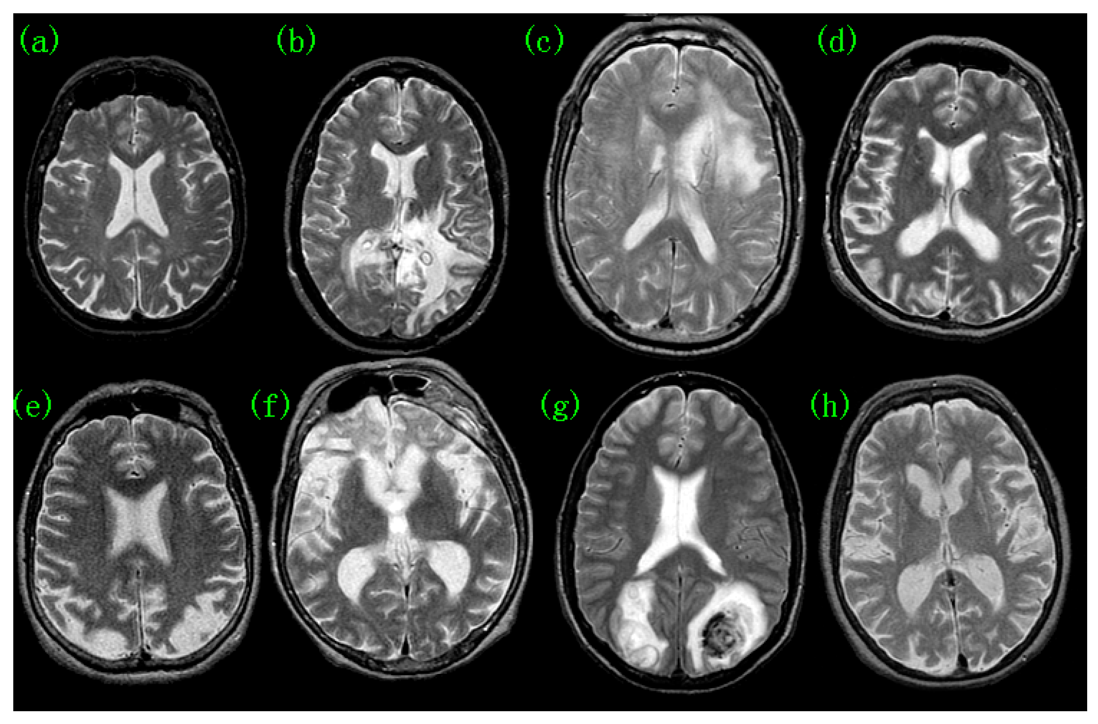
Figure 1. Pathological-brain samples: (a) normal; (b) glioma; (c) meningioma (d) Alzheimer’s disease; (e) Alzheimer’s disease with visual agnosia; (f) Pick’s disease; (g) sarcoma; (h) Huntington’s disease.
Preprints 93564 g001
Figure 2. Detection Techniques.
Preprints 93564 g002
Figure 3. Common computational methods for PBD.
Preprints 93564 g003
Figure 4. An example of VBM.
Preprints 93564 g004
Figure 5. Relationship between artificial intelligence, machine learning, and deep learning.
Preprints 93564 g005
Disclaimer/Publisher’s Note: The statements, opinions and data contained in all publications are solely those of the individual author(s) and contributor(s) and not of MDPI and/or the editor(s). MDPI and/or the editor(s) disclaim responsibility for any injury to people or property resulting from any ideas, methods, instructions or products referred to in the content.
Copyright: This open access article is published under a Creative Commons CC BY 4.0 license, which permit the free download, distribution, and reuse, provided that the author and preprint are cited in any reuse.
Prerpints.org logo

Preprints.org is a free preprint server supported by MDPI in Basel, Switzerland.

Subscribe

© 2024 MDPI (Basel, Switzerland) unless otherwise stated