Preprint
Article

Electrochemotherapy in Kaposi’s Sarcoma Patients. From the Gold Standard Strategy to Locally Advanced Cutaneous and Subcutaneous Lesions.

Altmetrics

Downloads

104

Views

31

Comments

0

A peer-reviewed article of this preprint also exists.

Submitted:

25 January 2024

Posted:

29 January 2024

You are already at the latest version

Alerts
Abstract
Electrochemotherapy (ECT) is one of the newest therapeutic strategies employed as medical procedure for skin neoplasms’ treatment, especially for Classic Kaposi’s Sarcoma (CKS). The aim of this study was to demonstrate ECT clinical response and local control of CKS disease. Primary endpoint was to value the worth and efficacy of this local therapy in CKS skin lesions’ treatment. Nineteen CKS patients were enrolled, 14 males and 5 females with median age at diagnosis of 72. Complete response (CR) has been gained in 12 patients after first ECT attempt, meanwhile 3 and 4 out of 19 patients obtained a Partial response (PR), so that they underwent to a second and a third ECT treatment, each other. Clinical response was evaluated during all the time of the follow-up, which ranged between 3 months and 4 years with a median of 18 months. CKS skin lesions control still represents a challenge for surgeons and oncologists. Nevertheless, according to this and other authors’ recent experiences, ECT could be considered the gold standard strategy for early-stage patients, but at the same time it could be considered as a valid option in controlling Kaposi’s Sarcoma locally advanced lesions.
Keywords: 
Subject: Public Health and Healthcare  -   Public Health and Health Services

1. Introduction

Kaposi’s sarcoma (KS) is an angio-proliferative malignancy, involving blood and lymphatic vessels, strongly related to infection with human herpesvirus 8 (HHV8), also known as Kaposi’s sarcoma-related herpesvirus (KSHV); HHV-8 infection is a necessary but not sufficient condition to cause KS. Thus, other factors such as immunosuppression have been demonstrated to play an important role in KS pathogenesis [1,2]. Although four different clinical-epidemiological variants were described, the classic form of KS is localized on the skin and subcutaneous tissues of the lower limbs (followed by upper limbs, head, and trunk) with purplish maculo-papular lesions, which can rapidly evolve into multicentric and ulcerated plaques, or nodules, often associated with venous stasis, lymphedema, and pain. Most of the time, the clinical course is silent and slowly progressive, but in rare cases it could be characterized by lymph-nodes and internal organs’ involvement, with prognosis and quality of life’s decay [2,3]. Numerous treatments are used in therapeutic strategies for non-advanced KS lesions: surgical excision (excisional biopsy), cryotherapy, radiotherapy, intralesional chemotherapy and the use of isotretinoin gel [4,5]. Systemic and locoregional therapies or radiotherapy represent the main options for the treatment of the disease, but however they are not always feasible due to the development of tumor resistance or the deterioration of the patient’s performance status (PS). Therefore, the alternatives we can rely on include photodynamic, intralesional and topical therapies, which have shown a high degree of effectiveness although they still lack standardized protocols to optimize their application [6,7]. Unfortunately, none of these could be considered effective in terms of curative response [8]. Over the last two decades there has been a notable breakthrough in the development of new treatment modalities, including one which combines transient tumor permeabilization achieved by appropriately tuned electrical impulses with cytotoxic agents (electrochemotherapy, ECT) [7,8,9,10]. Nowadays, Electrochemotherapy (ECT) has reached the greatest interest for the treatment of cutaneous and subcutaneous lesions in non-advanced KS patients (stage I and slowly progressive stage II) [11]. Electrochemotherapy (ECT) is a non-thermal tumor ablation technique, which uses high intensity pulsed electric fields to temporarily increase cell membrane permeability through the creation of pores, by which small molecules can diffuse inside cells before closing again [12,13,14,15,16]; it combines the phenomenon of electroporation with the administration of highly cytotoxic chemotherapy [8,12,13,14,15,16]. The electrical impulses induced through specific electrodes lead to the transient opening of pores on the cell membrane, and all this favors the massive entry of drug molecules into the cytosol. Therefore, the main advantage of ECT is precisely the intensity of the local dose, obtained through a high intra-tumoral concentration of the drug [7,14]. The drugs most used in ECT are Bleomycin and Cisplatin [11,12,13,14,15,16]; in fact, their cytotoxicity appears to be increased by 1,000 and 80 times respectively, in the presence of the electroporation phenomenon. In addition to the drug-induced cellular destruction, electroporation appears to be responsible for vascular changes within the tumor: a “vascular lock” is induced, with a temporary reduction of perfusion in the tumor tissue with an interstitial edema. The aim of our study is not only to validate and confirm that ECT represents the best therapeutical choice in terms of risk-benefit ratio for the treatment of cutaneous and subcutaneous lesions in non-advanced forms of KS, but also to demonstrate the valid use of ECT for the local control of locally advanced CKS.

2. Materials and Methods

This was a retrospective single-center study enrolling 20 patients with classic KS lesions (15 males and 5 females) with median age at diagnosis of 72 (Table 1). They were referred to the Division of Plastic Surgery at the I.R.C.C.S. - Centro di Riferimento Oncologico di Basilicata, Rionero in Vulture, Italy, from November 2018 to December 2022.
The final goal of this study was the assessment of the clinical effectiveness rate; the other purposes were the evaluation of side effects, local control of the neoplasm and the lifestyle impact. Patients’ enrollment was carried out according to Brambilla CKS staging system [17,18] and according to ESOPE criteria [19]; To be included into this study patients must satisfy some criteria, as follows: KS histological diagnosis with cutaneous and subcutaneous lesions which cannot be healed by local treatments like surgery, radiotherapy, or intralesional vincristine; no extracutaneous involvement demonstrated by diagnostic procedures; age >18 years, Karnofsky performance status >70; and a washout period of at least 4 weeks after previous treatments [8]. Selected patients were not eligible for all other standard therapeutic options or had already undergone them without obtaining any benefit for their clinical condition (surgery, radiotherapy, isolated limb infusion/perfusion, chemotherapy, immunotherapy). Among the exclusion criteria we have patients who have previously shown allergic and anaphylactoid reactions to bleomycin or to any component necessary for sedation, patients who have exceeded the maximum cumulative dose of 250 mg of bleomycin/m2 (400,000 IU bleomycin/m2), patients who had chronic renal dysfunction (serum creatinine > 150 mmol/L) or acute lung infection. Therefore, patients could be excluded if they presented abnormal respiratory parameters, cardiac pacemaker or arrhythmias, or history of seizures [7]. Only 1 male patient was excluded from the study because of comorbidities. 14 patients were classified as stage I while the other 5 patients were set into stage II (Table 1). A written informed consent was approved by every patient so that they could be part of the study; numerous patients’ epidemiologic distinguishing marks have been collected (ethnic group, age at arising lesions, gender), as well as clinical marks (location of the lesions, treatments carried out, clinical response, and time at disease relapse during follow-up). All of them underwent to diagnostic confirmation through pounch-biopsy. Because of multiple or disseminated cutaneous and subcutaneous lesions mainly located to the lower limbs, many patients should not undergo excisional biopsy, radiotherapy, or topic chemotherapy; so that, they underwent ECT treatment. Multiple lesions’ patients who were too difficult to treat in a single session or patients who obtained a partial response (PR) at a first ECT application were undergone to repeated treatments. Treatments were made at the IRCCS CROB with the CliniporatorTM device (IGEA, Modena, Italy). The anesthesiology technique was chosen through the ESOPE criteria, but single patient’s adaptations were allowed according to local protocols. Each patient underwent to general anesthesia or spinal anesthesia. Various electrodes have been used: Type I electrode, made up of two parallel stainless-steel plates with varying distances between 6 and 8 mm, was employed for small superficial lesions’ management; Type II electrode, made up of two parallel arrays of needles with a 4-mm gap, was employed for small nodules’ management; Type III electrode, made up of a hexagonal array of needles with a 8-mm gap, was employed for big nodules’ management. According to AIFA note, about drugs with consolidated use in the treatment of solid tumors in adults for indications even different from those provided by the marketing authorization measure [20], patients were previously treated with intravenous Bleomycin, promoting a homogeneous drug concentration. Intravenous Bleomycin was injected at a dose of 15000 UI/m2; Electric pulses were carried out 8 min after Bleomycin infusion obtaining the most favorable drug concentration in tissues. Treated lesions were studied at day 0, and followed-up at weeks 4, 8, 12, and every 3 to 6 months thereafter. Lesions’ sizes were established measuring the largest diameter of the lesion; Clinical response and the treatment’s efficacy were assessed according to the RECIST criteria [21], as follows: progressive disease (PD), if lesions increase in the larger diameter of >20%; partial response (PR), if lesions decrease of 30% after a 4 weeks follow-up; unchanged clinical situation, if lesions increase of <20% or a decrease of <50%; and complete response (CR), if all lesions have been completely disappeared. Treatments’ systemic toxicity and side effects were evaluated according to World Health Organization criteria. Life-style impact was assessed through the Patient Global Assessment (PGA) [22]. Statistical evaluation was performed by Minitab Analytics software for MacOS (© 2023 Minitab, LLC. All Rights Reserved.). Contingency tables and the chi- square test have been used as tests for the evaluation of the different response of the lesions after ECT treatments. The Kaplan-Meier analysis has been used as a method to assess the local control of the disease over time. It has been evaluated measuring the period from the successful ECT treatment response to either relapse’s appearance in CR results or >25% size augmentation in PR results, or last follow-up medical examination. Time to treatment failure was studied from the first day of treatment with ECT to either tumor relapse necessitating another available therapy, treatment interruption, death from any cause. Overall survival was assessed from the first day of treatment with ECT to either death from any cause or last date of follow-up, counting all deaths as events.

3. Results

3.1. ECT Treatments

In this study, a total of 19 patients with widespread and persistent KS cutaneous lesions were enrolled and undergone ECT treatments. According to RECIST criteria [21] clinical response, evaluated after 4 weeks, was gained by all patients; but a complete regression (CR) was obtained in 12 of 19 cases (63,2%) and a partial regression (PR) in the other 7 cases (36,8%) (Table 1). No differences have been demonstrated among the overall response rate according to the size of cutaneous lesions; but the CR rate of bigger lesions (>2cm) was lower than CR rate of smaller lesions (46.3% vs. 65.2%; 2 test, P= 0.012) (Figure 1).
After first ECT session, 7 patients (36.8 %) received a second ECT treatment; 3 of 7 patients gained CR, instead the other 4 cases presented PR (Table 1). So, these 4 cases underwent to a third ECT treatment and further achieved 4 PR with respect to the initial measurement. So, ECT treatment led to a complete regression in 15 of 19 patients (78.9%), in 12 patients after the first ECT attempt and in 3 patients after the second attempt (Table 1). The mean interval between two treatment was 90 days.

3.2. Follow-up

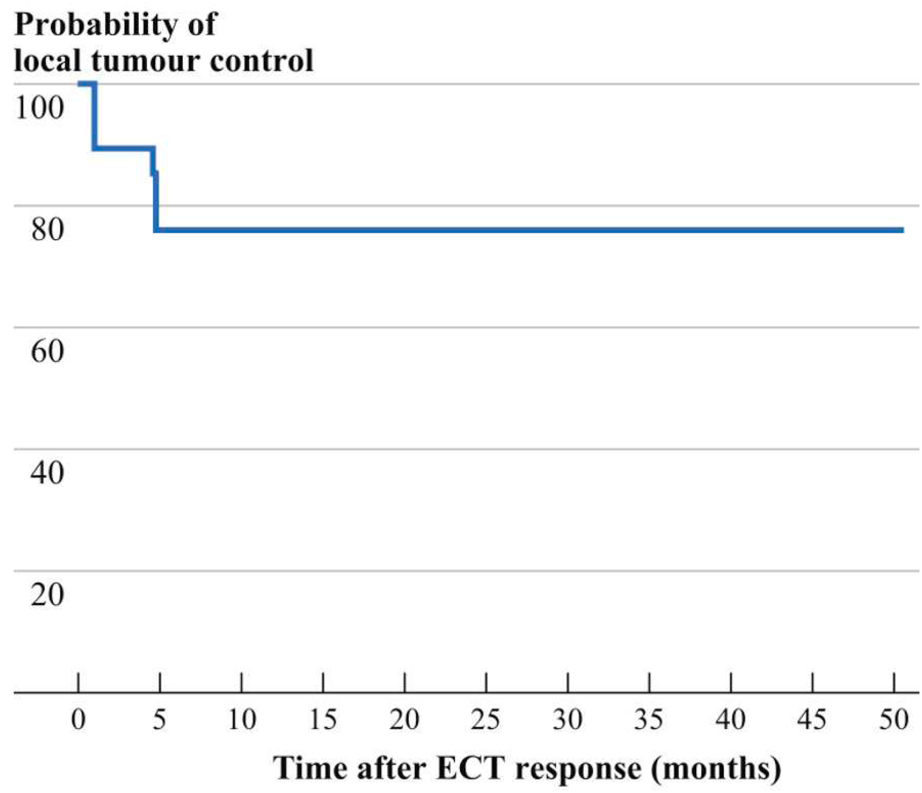
Clinical response was observed during the entire time of the follow-up, which ranged between 3 months and 4 years with a median of 18 months. CR was obtained in 10 cases with stage I, in 5 cases with stage II, and in 0 of stage III or IV cases (Table 1); After a median follow-up of 18 months, 15 patients maintained the response, but 3 cases only after repeated courses. Meanwhile the other 4 PR patients either were not retreated with ECT, or they underwent further therapies, because of the silent course of KS classic variant and the excellent impact of disease on lifestyle. (Figure 2).

3.3. Side Effects and Quality of Life

Among Side Effects and Quality of Life, the treatment was well tolerated with a very low complication rate. Pain and erythema were among the most reported side effects; cutaneous infections were noticed only in 3 patients, and they were healed with oral antibiotics, causing complete healing in less than few days. No other symptoms nor systemic toxicity occurred. According to PGA [22] in terms of quality of life, an improvement was obtained in all patients; this was not only related to an excellent result after treatment.

4. Discussion

Widespread presence of cutaneous and subcutaneous CKS lesions is an uncomfortable situation for many patients. The presence of multiple skin and subcutaneous lesions get worse patients’ quality of life, frequently burdened by pain, ulceration, and bleeding. Their management and treatment are an arduous challenge for operators, as they often depend on the number and size of the lesions, their anatomical sites, and the appearance of visceral lesions. When possible, definitive surgical removal is the most suitable therapeutic approach. However, when surgery cannot be performed due to the excessive number of lesions, their considerable extension with the compromise of a reasonable result from a functional point of view, other therapeutic options must be considered [1,7,14,23,24]. Unfortunately, there is no standard therapy policy for CKS [25]; According to Brambilla et al. staging system [18], in which each patient can be classified into 4 stages and further divided into two categories (A and B, based on the evolution rate of the disease) (Table 2), an accurate CKS patient’s staging allows the choice of the most appropriate therapeutic strategy [2,24]. After clinical staging, the patient should undergo specialist visits and instrumental investigations to exclude evidence of internal organs involvement (Table 3) [2,26].
Although complications are mainly frequent in advanced stages (stage III and IV), functional impairment and pain or lymphoedema, bleeding and ulceration can occur in all stages of the disease [2,18]. Visceral involvement, if present, usually occurs in the context of stage III or IV disease. Therefore, only after a correct staging of the patient, the diagnostic-therapeutic process can continue. Most CKS cutaneous and subcutaneous lesions treated are symptomatic lesions, unlike to asymptomatic or slowly evolving ones, for which a wait-and-see strategy can be chosen, because a spontaneous improvement could occur [4,5]. Therapeutic strategies are chosen after the assessment of the tumor staging, the localization and the evolution pattern of the lesions, clinical type, and immune status [6,7,8,9,10]. Local treatments should be performed alone in localized KS, or in combination with systemic treatments in patients with advanced or disseminated KS (Table 4) [2,4].
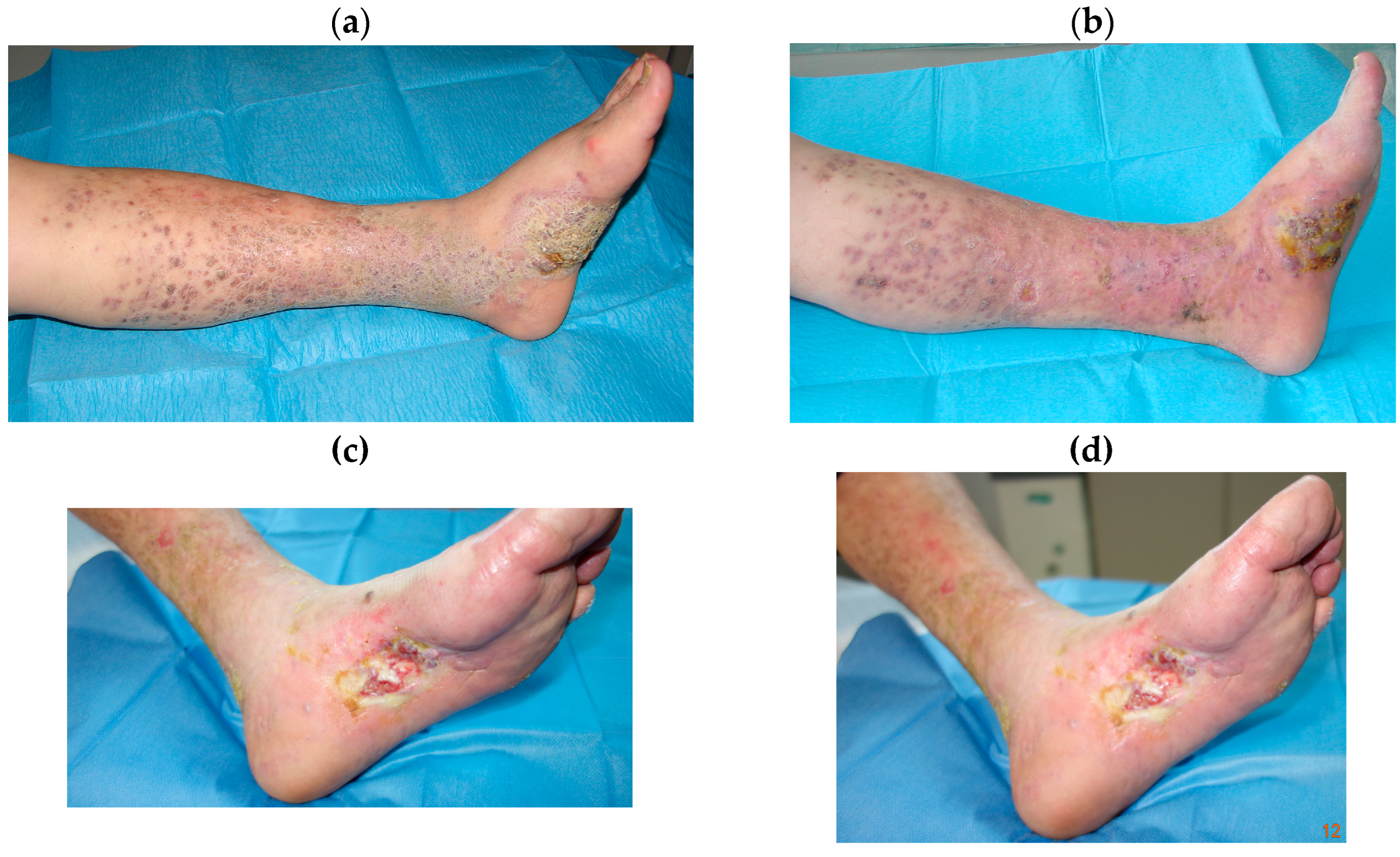
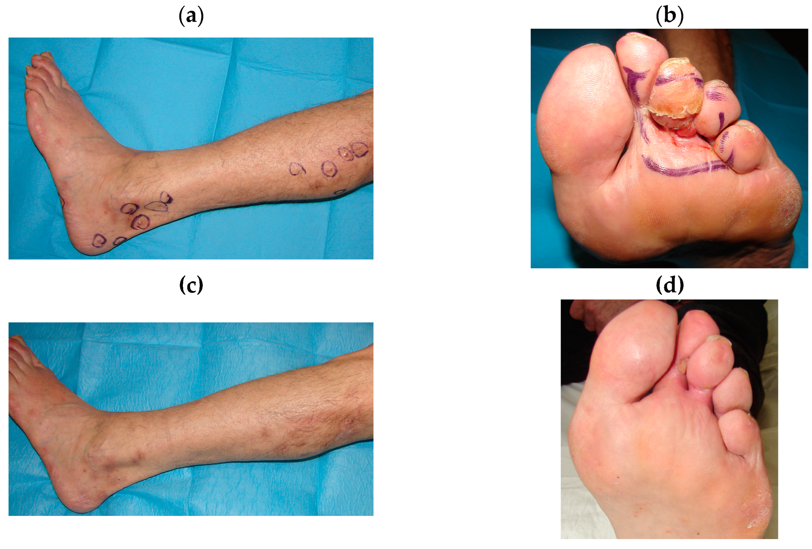
Surgical excision should be performed exclusively on a couple and well-defined lesions; also, radiotherapy or intralesional injections are included into local treatments. Despite the poor toxicity pattern induced by these therapies, their application on several lesions is not possible, because they can shatter the global status of the patients, who necessitate to preserve an immunocompetent status; so that many of these patients would have received systemic treatments, including single or combined antitumoral drugs (vinca alkaloids, etoposide, liposomal doxorubicin, bleomycin) [2]. This study shows the results of a retrospective trial aimed to evaluate clinical response and toxicities of ECT with intravenous bleomycin injection among the treatments of CKS cutaneous and subcutaneous lesions, demonstrating that ECT is non-inferior to other local and systemic treatments. In fact, clinical response was gained in all cases, apart from tumor dimensions, with 63% cases resulting a CR after the first ECT attempt; moreover, other additional CRs were gained after repeated sessions. Although the good overall response rates, greater CR rates were obtained by patients with nodules or plaques with < 2 cm diameter (65.2% vs. 46.3%). In this study ECT, after bleomycin previously administered, obtained positive results in the local control of the disease whereas other treatments, such as surgery or radiotherapy, would have been hazardous, because of the high risk of side effects like ulceration, bleeding, infection, and delayed healing. Furthermore, ECT has been successful also in the management of the disease for those patients affected by advanced and widespread CKS cutaneous lesions (Figure 3 and Figure 4).
Almost all patients presented pain and erythema after treatment and only 3 cases reported infection, then treated with antibiotics. No systemic toxicities occurred, compared to the higher toxicity due to classical treatments. Furthermore, an improvement of lifestyle is noticed, as established by the PGA [22], not only related to the stage of the disease.

5. Conclusions

This single center study, which is based on the largest prospective series published so far, strongly supports the efficacy of ECT in patients with superficial cutaneous and subcutaneous KS lesions. ECT generated a strong and long-lasting clinical response, with the development of very limited toxicity; in fact, it seems to preserve and even dramatically improve the health-related quality of life. Therefore, all this has translated into a very high acceptance rate among patients. Therefore, all ECT candidates can also undergo treatment with locoregional anesthesia, thus confirming the easy management of this treatment and above all that it is a treatment well tolerated by patients. Because of our and other experiences [7,9,14,27,28,29,30], we can affirm that ECT could be considered a valid therapeutical strategy for local control of widespread and advanced CKS cutaneous and subcutaneous lesions, and it is the gold standard therapy in non-advanced CKS patients. In conclusion, the clinical experience with ECT is growing and the results are promising.

Author Contributions

Conceptualization, T.F.; methodology, M.P.G. and F.C.; investigation, V.R., S.D.A., and A.D.R.; software, V.R., S.D.A and A.D.R.; validation, M.P.G. and F.C.; Formal analysis and data curation, V.R., S.D.A., A.D.R., M.P.G., and F.C..; visualization, V.R.; writing—original draft preparation, V.R..; writing—review and editing, M.P.G., F.C.; supervision, T.F.; project administration, T.F.; All authors have read and agreed to the published version of the manuscript.

Funding

This research received no external funding.

Institutional Review Board Statement

The study was conducted in accordance with the Declaration of Helsinki and approved by the Ethics Committee of I.R.C.C.S. - Centro di Riferimento Oncologico di Basilicata, Rionero in Vulture, Italy (protocol code 378; date of approval 30/01/2023).

Informed Consent Statement

Informed consent was obtained from all subjects involved in the study.

Data Availability Statement

The datasets used and/or analyzed during the current study are available from the corresponding author on reasonable request.

Conflicts of Interest

The authors declare no conflict of interest.

References

  1. Etemad SA, Dewan AK. Kaposi Sarcoma Updates. Dermatol Clin. 2019, 37, 505–517. [Google Scholar] [CrossRef]
  2. Brambilla L, Genovese G, Berti E, et al. Diagnosis and treatment of classic and iatrogenic Kaposi’s sarcoma: Italian recommendations. Ital J Dermatol Venerol. 2021, 156, 356–365. [Google Scholar] [CrossRef] [PubMed]
  3. COX FH, HELWIG EB. Kaposi’s sarcoma. Cancer. 1959, 12, 289–298. [Google Scholar] [CrossRef]
  4. Dupin, N. Update on oncogenesis and therapy for Kaposi sarcoma. Curr Opin Oncol. 2020, 32, 122–128. [Google Scholar] [CrossRef] [PubMed]
  5. Chak LY, Gill PS, Levine AM, et al. Radiation therapy for AIDS-related Kaposi’s sarcoma. J Clin Oncol 1998, 5, 863–867. [Google Scholar]
  6. Toschi E, Sgadari C, Monini P, et al. Treatment of Kaposi sar- coma-an update. Anticancer Drugs. 2002, 13, 977–87. [Google Scholar] [CrossRef]
  7. Campana LG, Testori A, Curatolo P, et al. Treatment efficacy with electrochemotherapy: A multi-institutional prospective observational study on 376 patients with superficial tumors. Eur J Surg Oncol. 2016, 42, 1914–1923. [Google Scholar] [CrossRef] [PubMed]
  8. Curatolo P, Quaglino P, Marenco F, et al. Electrochemotherapy in the treatment of Kaposi sarcoma cutaneous lesions: a two-center prospective phase II trial. Ann Surg Oncol. 2012, 19, 192–198. [Google Scholar] [CrossRef] [PubMed]
  9. Testori A, Tosti G, Martinoli C, et al. Electrochemotherapy for cutaneous and subcutaneous tumor lesions: a novel therapeutic approach. Dermatol Ther 2010, 23, 651–661. [Google Scholar] [CrossRef] [PubMed]
  10. Garbay JR, Billard V, Bernat C, Mir LM, Morsli N, Robert C. Successful repetitive treatments by electrochemotherapy of multiple unresectable Kaposi sarcoma nodules. Eur J Cancer 2006, 4 (Suppl. 4), 29–31. [Google Scholar] [CrossRef]
  11. Latini A, Bonadies A, Trento E, et al. Effective treatment of Kaposi’s sarcoma by electrochemotherapy and intravenous bleomycin administration. Dermatol Ther. 2012, 25, 214–218. [Google Scholar] [CrossRef] [PubMed]
  12. Giardino R, Fini M, Bonazzi V, Cadossi R, Nicolini A, Carpi A. Electrochemotherapy a novel approach to the treatment of metastatic nodules on the skin and subcutaneous tissues. Biomed Pharmacother. 2006, 60, 458–462. [Google Scholar] [CrossRef]
  13. Mir LM, Gehl J, Sersa G, et al. Standard operating procedures of the electrochemotherapy: instructions for the use of bleomycin or cisplatin administered either systemically or locally and electric pulses delivered by the CliniporatorTM by means of invasive or non-invasive electrodes. Eur J Cancer Suppl. 2006, 4, 14–25. [Google Scholar] [CrossRef]
  14. Solari N, Spagnolo F, Ponte E, et al. Electrochemotherapy for the management of cutaneous and subcutaneous metastasis: a series of 39 patients treated with palliative intent. J Surg Oncol. 2014, 109, 270–274. [Google Scholar] [CrossRef] [PubMed]
  15. Miklavcic D, Snoj M, Zupanic A, et al. Towards treatment planning and treatment of deep-seated solid tumors by electrochemotherapy. Biomed Eng Online. 2010, 9, 10. [Google Scholar] [CrossRef] [PubMed]
  16. Gehl, J. Electroporation: theory and methods, perspectives for drug delivery, gene therapy and research. Acta Physiol Scand. 2003, 177, 437–447. [Google Scholar] [CrossRef] [PubMed]
  17. Chachowa A, Kriegel R, Lafleur F,Ostreicher R, Speer M, Lauben- stein L, Wernz G, Rubenstein P, Zang E, Friedman-Kien A. Prognostic factors and staging classification of patients with epidemic Kaposi’s sarcoma. J Clin Oncol 1989, 7, 774–780. [Google Scholar] [CrossRef] [PubMed]
  18. Brambilla L, Boneschi V, Taglioni M, Ferrucci S. Staging of classic Kaposi’s sarcoma: a useful tool for therapeutic choices. Eur J Dermatol. 2003, 13, 83–86. [Google Scholar]
  19. Marty MSG, Garbay JR, Gehl J, et al. Electrochemotherapy an easy, highly effective and safe treatment of cutaneous and subcutaneous metastases: results of ESOPE (European Standard Operating Procedures of Electrochemotherapy) study. Eur J Cancer Suppl 2006, 4, 3–13. [Google Scholar] [CrossRef]
  20. DeVita, V.T. et al, Cancer: Principles and Practice of Oncology, 6th Ed. Bonadonna. G. et al, Medicina Oncologica, Settima Edizione.
  21. Therasse P, Arbuck SG, Eisenhauer EA, et al. New guidelines to evaluate the response to treatment in solid tumors. European Organization for Research and Treatment of Cancer, National Cancer Institute of the United States, National Cancer Institute of Canada. J Natl Cancer Inst. 2000, 92, 205–216. [Google Scholar] [CrossRef] [PubMed]
  22. Pincus T, Bergman M, Sokka T, Roth J, Swearingen C, Yazici Y. Visual analog scales in formats other than a 10 centimeter horizontal line to assess pain and other clinical data. J Rheumatol. 2008, 35, 1550–1558. [Google Scholar]
  23. Sullivan RJ, Pantanowitz L. New drug targets in Kaposi sarcoma. Expert Opin Ther Targets. 2010, 14, 1355–1366. [Google Scholar] [CrossRef] [PubMed]
  24. Brambilla L, Miedico A, Ferrucci S, et al. Combination of vinblastine and bleomycin as first line therapy in advanced classic Kaposi’s sarcoma. J Eur Acad Dermatol Venereol. 2006, 20, 1090–1094. [Google Scholar] [CrossRef] [PubMed]
  25. Radu O, Pantanowitz L. Kaposi sarcoma. Arch Pathol Lab Med. 2013, 137, 289–294. [Google Scholar] [CrossRef]
  26. Krigel RL, Laubenstein LJ, Muggia FM. Kaposi’s sarcoma: a new staging classification. Cancer Treat Rep. 1983, 67, 531–534. [Google Scholar]
  27. Fabrizio T, Cagiano L, De Terlizzi F, Grieco MP. Neoadjuvant treatment by ECT in cutaneous malignant neoplastic lesions. J Plast Reconstr Aesthet Surg. 2020, 73, 904–912. [Google Scholar] [CrossRef] [PubMed]
  28. Mali B, Jarm T, Snoj M, Sersa G, Miklavcic D. Antitumor effectiveness of electrochemotherapy: a systematic review and meta-analysis. Eur J Surg Oncol. 2013, 39, 4–16. [Google Scholar] [CrossRef] [PubMed]
  29. Di Monta G, Caracò C, Benedetto L, et al. Electrochemotherapy as “new standard of care” treatment for cutaneous Kaposi’s sarcoma. Eur J Surg Oncol. 2014, 40, 61–66. [Google Scholar] [CrossRef] [PubMed]
  30. Heller R, Jaroszeski MJ, Reintgen DS, et al. Treatment of cutaneous and subcutaneous tumors with electrochemotherapy using intralesional bleomycin. Cancer 1998, 83, 148–57. [Google Scholar] [CrossRef]
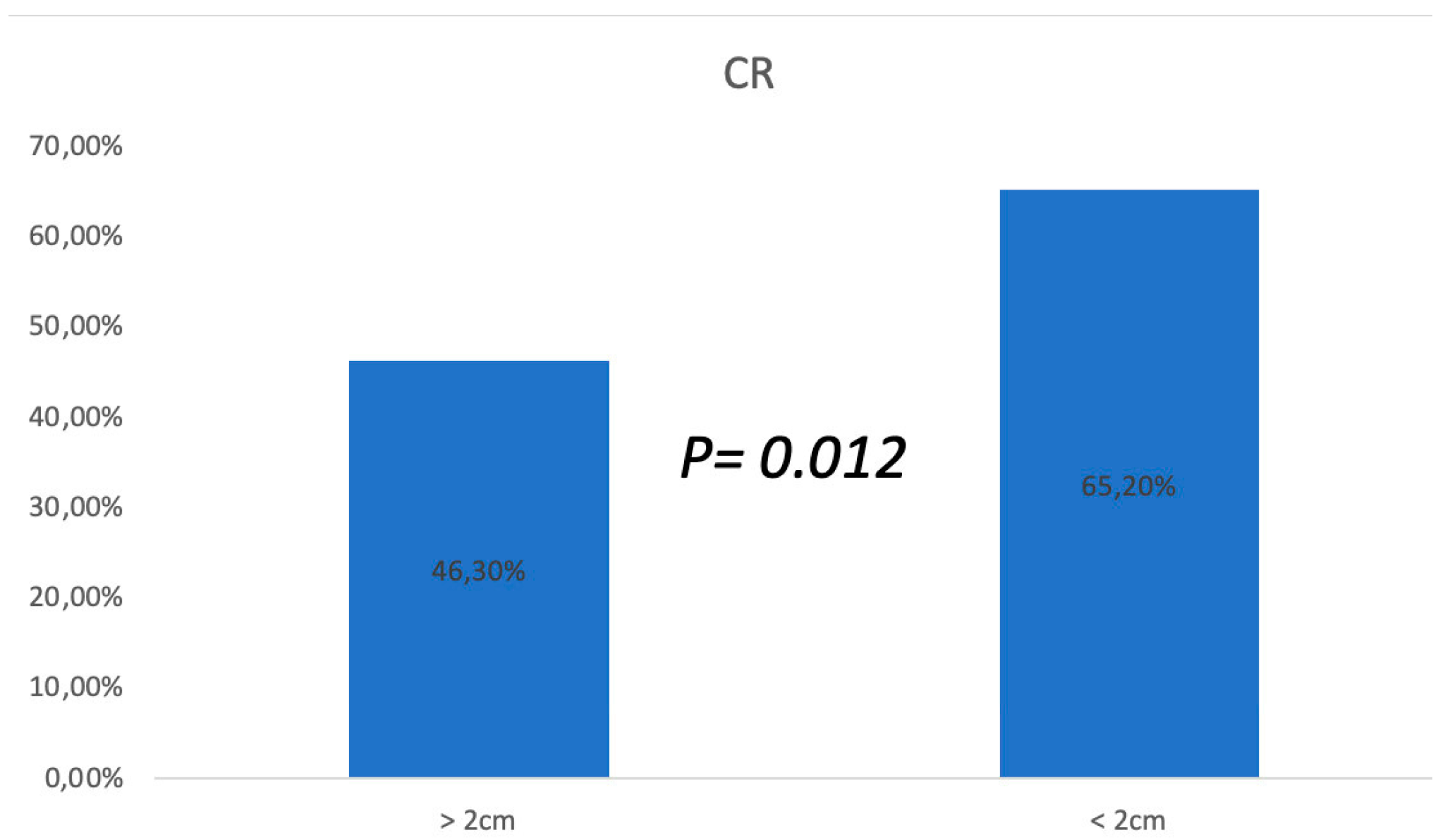
Figure 1. ECT success rate in KS cutaneous and subcutaneous different lesions.
Preprints 97331 g001
Figure 2. Local control of the disease in 19 CKS patients who undergone ECT.
Preprints 97331 g002
Figure 3. (a) Left leg widespread skin and subcutaneous Kaposi lesions in a 63-year-old patient. Before ECT treatment. (b) After three ECT treatments with systemic Bleomycin. (c), (d) Local control of the disease after 4 years follow-up.
Preprints 97331 g003
Figure 4. (a), (b) Widespread plaques and nodules on the leg and foot. (c) Complete remission of healed nodules and plaques with residual hyperpigmentation and hypotrophic scars. (d) Partial remission of treated lesion with residual nodule.
Preprints 97331 g004
Table 1. Patients’ characteristics and Results after 18 months. CR: Complete Response; PR: Partial Response.
Patients n° Sex, Age Localization Clinical Response Response Stage
1 F, 85 Right foot Present CR I
2 F, 74 Lower limb Present CR at second ECT I
3 F, 63 Lower limbs bilateral Foot Present PR after third ECT II
4 F, 55 Foot Present CR I
5 F,70 Lower limbs bilateral Foot Present CR I
6 M, 72 Foot Present CR at second ECT II
7 M,68 Foot Present CR I
8 M,60 Foot Present CR I
9 M,80 Lower limbs Present CR I
10 M, 75 Right foot Present CR I
11 M, 71 Bilateral foot Present PR after third ECT II
12 M, 78 Lower limbs Present CR I
13 M, 77 Lower limbs Present CR at second ECT I
14 M, 68 Foot Present CR I
15 M, 70 Lower limbs Present PR after third ECT II
16 M, 76 Left limb Present CR I
17 M,74 Right limb Present CR I
18 M, 84 Foot Present CR at second ECT I
19 M, 69 Bilateral foot Present PR after third ECT II
20 M, 71 Excluded for comorbidities.
Table 2. Staging of cutaneous Kaposi’s sarcoma.
Stage Prevalent cutaneous lesions Progression Visceral involvement
I macular-nodular macules and/or nodules predominantly on the lower limbs A, slow B, rapid ±V
II Infiltrative Plaques predominantly on the lower limbs A, slow B, rapid ±V
III florid exuberant angiomatous nodules predominantly on the lower limbs A, slow B, rapid ±V
IV Disseminate angiomatous lesions on the head, trunk, and mucosae A, slow B, rapid ±V
Table 3. Diagnostic tests at first presentation of Kaposi’s sarcoma.
Standard Personalized
Blood tests: full blood count, kidney and liver function, iron, protein electrophoresis, lymphocyte subpopulation analysis, HIV test, anti-HHV8 antibodies and HHV8 Dna
Fecal occult blood test on three samples, if positive ◊ Colonoscopy
Esophagogastroduodenoscopy (EGDS) Esophagogastroduodenoscopy (EGDS)
Abdomen and lymph nodes ultrasound Total body computerized tomography
Ears, nose, and throat (ent) visit
Table 4. Treatment of cutaneous Kaposi’s sarcoma.
Staging Standard Personalized
I Clinical observation Intralesional vincristine Curettage Elastocompression for edema or bullous Kaposi’s sarcoma Silver nitrate for soft, fungating and/or oozing lesions
II Elastocompression Radiotherapy for florid and localized lesions
III Systemic chemotherapy* Elastocompression for edema or bullous Kaposi’s sarcoma
IV Systemic chemotherapy* Elastocompression for edema or bullous Kaposi’s sarcoma
*Possible schemes: 1) first-line treatment (vinblastine: induction 4, 6, 8 mg i.v./week, then 10 mg i.v. every 3 weeks; vinblastine (as above) + bleomycin 15 mg i.m. every 3 weeks starting from the end of vinblastine induction; paclitaxel 75-100 mg/m2 i.v./week); 2) second-line treatment (pegylated liposomal doxorubicin 20 mg/ m2 every 3 weeks); 3) third-line treatment (gemcitabine 1200 mg/m2 i.v./week for 2 weeks, with an interval of three weeks; bleomycin 15 mg i.m. every 2 weeks; vinorelbine: induction: 20 mg/m2 every 2 weeks for 5 cycles; consolidation with 30 mg/m2 every 3 weeks (off label); etoposide oral 150 mg/m2 for 3 consecutive days every three weeks). every chemotherapy scheme should be maintained until the best therapeutic outcome, followed by three consolidation cycles with 3-, 6-, and 12-month follow up visits.
Disclaimer/Publisher’s Note: The statements, opinions and data contained in all publications are solely those of the individual author(s) and contributor(s) and not of MDPI and/or the editor(s). MDPI and/or the editor(s) disclaim responsibility for any injury to people or property resulting from any ideas, methods, instructions or products referred to in the content.
Copyright: This open access article is published under a Creative Commons CC BY 4.0 license, which permit the free download, distribution, and reuse, provided that the author and preprint are cited in any reuse.
Prerpints.org logo

Preprints.org is a free preprint server supported by MDPI in Basel, Switzerland.

Subscribe

© 2024 MDPI (Basel, Switzerland) unless otherwise stated