Preprint
Article

Improving Quality of Life in Chronic Fatigue Syndrome using Antioxidant Complex Twendee M®.

Altmetrics

Downloads

184

Views

228

Comments

0

Submitted:

29 January 2024

Posted:

06 February 2024

You are already at the latest version

Alerts
Abstract
Chronic fatigue syndrome (CFS) is a disease in which fatigue that interferes with daily life persists for six months or longer. The number of patients with CFS is increasing, as CFS-like symptoms have been reported to occur in the sequelae of both COVID-19 infection and the SARS-CoV-2 vaccine, both of which have become significant issues in recent years. While the pathogenesis mechanism is not yet fully understood, research suggests that oxidative stress (OS) may play a role in the development of CFS. In this paper, we discuss the antioxidant potential of the antioxidant formulation Twendee M® (TwM) and the results of a questionnaire that monitored changes in symptoms before and after TwM in a total of 23 men and women diagnosed with CFS. TwM is a supplement containing 15 different ingredients, and has a strong antioxidant capacity that cannot be achieved with a single antioxidant ingredient. The results of the questionnaire showed that TwM significantly improved all of the major symptoms of CFS, including fatigue, muscle pain, joint pain, sleep disturbance, decreased memory and concentration, and headache. TwM was shown to alleviate various symptoms of CFS and improve quality of life.
Keywords: 
Subject: Biology and Life Sciences  -   Life Sciences

1. Introduction

Chronic fatigue syndrome (CFS) is a disease characterized by persistent fatigue and malaise of unknown origin [1,2]. In addition to these symptoms, the patient can experience weakness, muscle pain, low-grade fever, headache, cognitive dysfunction, sleep disturbance, and neuropsychiatric symptoms that persist for a long time, making it difficult for the patient to lead a healthy social life [3]. Since CFS seriously affects patients' health and daily life, there is an urgent need to identify effective treatment strategies to alleviate these symptoms.
CFS is thought to often develop after immunocompromise due to strong stress or other factors, or after infection by viruses or other agents, and there is no established treatment for the condition. Recently, symptoms similar to those of CFS have been reported in the sequelae of COVID-19 infection and SARS-CoV-2 vaccine [4,5,6], and the number of patients is gradually increasing worldwide. There are multiple possible causes of CFS, including abnormal energy metabolism due to mitochondrial dysfunction [7] and inflammation in the brain.
In patients with CFS, inflammation is prevalent in various areas of the brain. Inflammation in the amygdala, thalamus, and midbrain is correlated with cognitive function, inflammation in the cingulate cortex and amygdala is correlated with headache and muscle pain, and inflammation in the hippocampus is correlated with depressive symptoms [8]. The presence of inflammation suggests that oxidative stress (OS) plays a significant role. Mitochondrial dysfunction and increased inflammatory substances are also observed in neurodegenerative diseases such as Alzheimer's disease related to OS [9], and it has been reported that the symptoms show a tendency to improve with the administration of antioxidants [10]. Even in COVID-19 infections that present with CFS-like symptoms, causative SARS-CoV-2 infections have been reported to cause OS in the body, resulting in the development of a variety of symptoms [11]. Antioxidant administration reduces various symptoms in long COVID and SARS-CoV-2 vaccine sequelae [4,5]. Recent studies about CFS have demonstrated that free radical production may play a role in its etiology [12,13,14], and OS clearly plays an important role in CFS.
Twendee M® (TwM) is an antioxidant formulation product based on Twendee X® (TwX) consisting of eight ingredients (vitamin C, L-glutamine, niacin, L-cystine, coenzyme Q10, vitamin B2, succinic acid, and fumaric acid) [15,16], with a small amount of seven additional ingredients (pantothenic acid, vitamin B1, vitamin B6, folic acid, biotin, vitamin B12, and lactoferrin). Although TwM and TwX are dietary supplements, both have undergone and passed all of the safety tests required for pharmaceutical products, including chromosomal aberration, toxicity, and mutation tests. The base TwX has been shown to protect cells and mitochondria, resulting in increased ATP production, a reduction in blood OS, and the maintenance of neurogenesis [17]. TwX has also been shown to prevent dementia in humans with mild cognitive impairment (MCI) in a multicenter, randomized, double-blind, placebo-controlled intervention clinical trial [18], and has been reported to have the potential to act on the intestinal microbiota [19]. In a mouse model of ischemic stroke, TwX has not only been found to reduce infarct size, but also to decrease the expression of OS, tumor necrosis factor-α (TNF-α), and inflammation markers [20]. In mice in which vitamin E deficiency causes increased OS and impaired cognitive function and coordination, TwX significantly improves vitamin-deficiency-induced cognitive function and coordination, and also shows significant increases in brain-derived neurotrophic factor and nerve growth factor levels [21]. Since TwM is an antioxidant formulation with more ingredients than TwX, which exhibits many of these benefits, it is expected to work in significantly alleviating the symptoms of CFS. This paper examines the antioxidant potential of TwM, and discusses the usefulness of TwM for CFS based on the results of a questionnaire survey of participants diagnosed with CFS.

2. Results and Discussion

2.1. OS Scavenging Ability of TwM

TwM is an antioxidant formulation containing 15 ingredients; the antioxidant capacity of the TwM solution (60 mg/ml) and the vitamin C (VC) solution, one of the ingredients in TwM, was measured using the OXY adsorption test.
The TwM solution was found to have 5.4 times higher antioxidant capacity than the solution with the same concentration of VC contained in TwM (20.5 mg/ml). Furthermore, the TwM solution had approximately twice as much antioxidant capacity than the VC solution at the same concentration (60 mg/ml) (Figure 1). These findings suggest that TwM possesses significantly greater antioxidant capacity than VC. Moreover, the results indicate that the combination of multiple beneficial components yields a higher capacity than a single-component antioxidant.
Reactive oxygen species (ROS) is a generic term for oxygen derivatives of free radicals (such as superoxide (O2-) and hydroxyl radical (OH)) and non-radicals (such as peroxides (hydrogen peroxide/H2O2) and oxygen ions/O2) [22]. All of them are highly reactive, and when they come into contact with any cellular biomolecule, they readily capture electrons from the molecule (oxidation), setting off a chain reaction that ultimately leads to damage to the cell structure. Of these, OH is the most reactive and is known to directly attack the DNA backbone, causing DNA damage [23,24]. The body's ROS concentration is closely related to the maintenance of homeostatic functions. While ROS are a necessary metabolic byproduct in physiological functions, high levels are toxic. Levels of these OS factors increase through lifestyle and aging, leading to mitochondrial dysfunction and damage to all parts of the body [25,26]. The elevation of OS is related to the risk of aging and disease development due to the accumulation of oxidation products in the body. Therefore, we consider it desirable for antioxidants to be able to scavenge both radicals and non-radicals, and additionally to increase the antioxidant capacity of the body. In addition to its ability to scavenge non-radicals, TwM has been shown to scavenge the radicals CH3, OH, and O2- at low concentrations [27]. Ascorbic-acid-containing formulations, including TwM, have been reported to produce ascorbyl radicals through high concentrations of VC [28,29], but the formation of ascorbyl radicals is suppressed in TwM [27], indicating that it is not adversely affected by VC. In addition to this, the base TwX has high ROS scavenging capacity and SOD-enhancing properties. TwM has shown to have higher OXY measurements than TwX [17]. This indicates that TwM has higher antioxidant capacity than TwX. In addition, TwM has been proven to be a safe antioxidant with significantly higher antioxidant capacity than the single component.

2.2. Effects of TwM on Various Symptoms in CFS

To investigate the impact of TwM on various symptoms of CFS, questionnaire participants were recruited (Eyez, Inc.). Participants were those who had been diagnosed with CFS by a physician at a hospital. The participants were informed of the purpose of the study and the dosage instructions on the website, and completed a pre-questionnaire to self-assess the impact of CFS on their daily lives and the extent of each symptom (fatigue, joint pain, muscle pain, sleep disturbance, headache, decrease in memory and concentration). The symptom severity was rated on a six-point scale ranging from none or low (0 points: 0P) to severe or high (5 points: 5P). Participants were considered to have agreed to participate by completing the pre-assessment questionnaire. TwM (13.51 mg/kg/day) was taken once daily for 2 months.
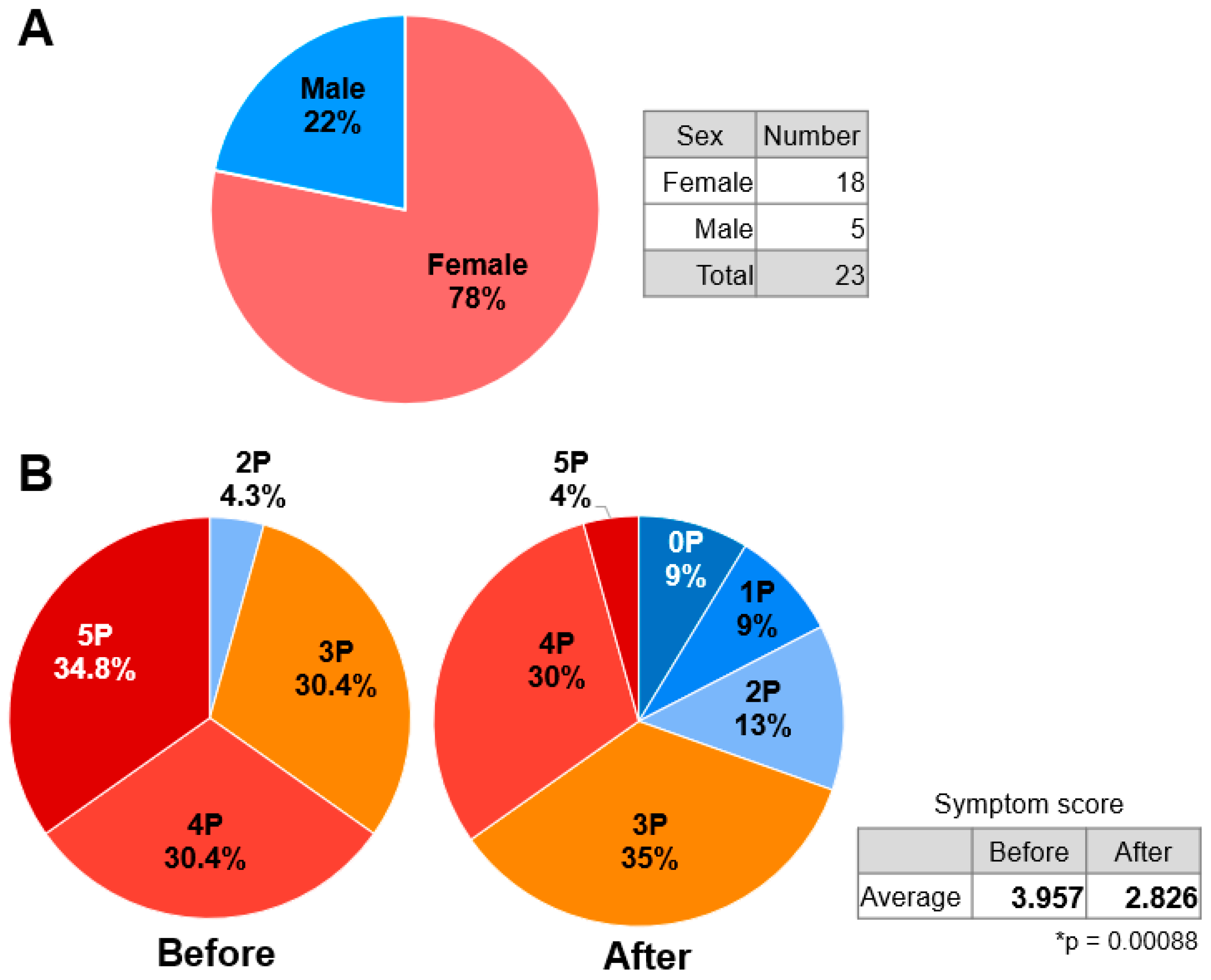
A total of 23 participants took part, including 5 males and 18 females diagnosed with CFS by a physician (Figure 2A). Before taking the medication, the responses regarding the impact of participants' CFS on their daily lives were 5P (severely impacted): 34.8% and 4P (very much impacted): 30.4%, which together accounted for more than half of the responses. No participants answered 0P (not affected) or 1P (slightly affected) (Figure 2B; Before). The results revealed that CSF has a severe impact on daily life, regardless of the degree of symptoms. In contrast, 2 months of TwM increased the number of participants who reported 0P-2P (0P: 9%, 1P: 9%, 2P: 13%), indicating being relatively unaffected by SSC, to 31%. Furthermore, the number of participants who reported 4P-5P (4P: 30%, 5P: 4%) decreased to a combined 34% (Figure 2B; After), significantly improving the extent to which TwM affects the daily life of individuals with CFS (p<0.001).
A diagnosis of CFS is only determined after fatigue from other etiologies has been ruled out, and only for medical conditions with a history of at least 6 months [30]. In many cases, CFS causes severe functional limitations in daily life and affects women more than men [31,32]. The reasons for this are unknown, but besides hormonal, viral, and immune causes [33], genetic and epigenetic origins have been suggested as possible explanations [34]. In the present study, female participants were also more common. Since TwM treatment showed a significant improvement trend in terms of the impact CFS on 23 patients’ daily lives, we considered the possibility that OS may play a major role in triggering the main symptoms of CFS.
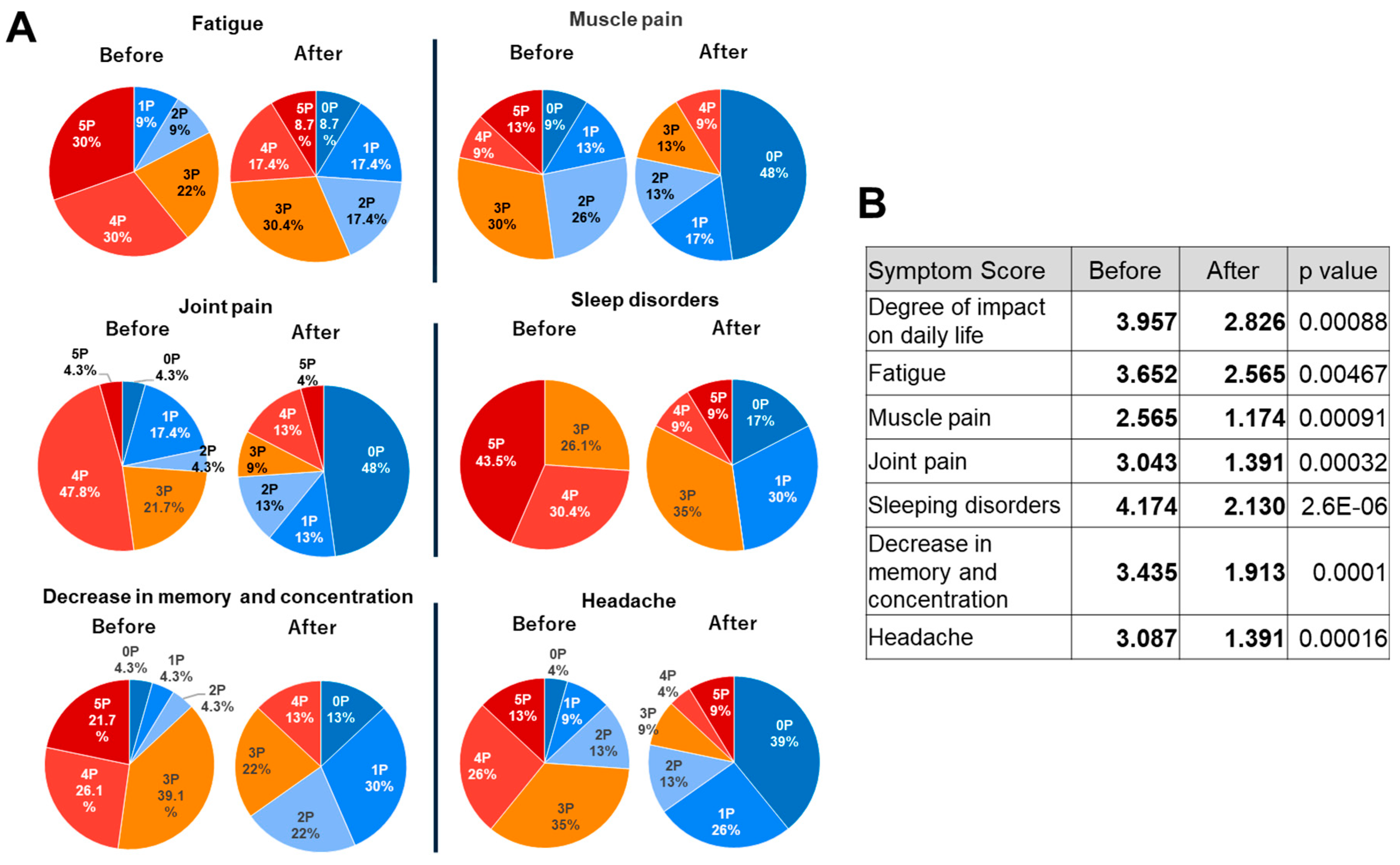
The six main symptoms of CFS defined in this questionnaire were fatigue, muscle pain, joint pain, sleep disturbance, decrease in memory and concentration, and headache. After taking TwM for 2 months, the participants completed a similar questionnaire about the extent of their CFS symptoms. The responses to the pretreatment questionnaire were not revealed to prevent bias. The responses were made on a six-point scale: no symptoms (0 points: 0P), a low degree of symptoms (1 point: 1P), a slightly low degree of symptoms (2 points: 2P), a moderate degree of symptoms (3 points: 3P), a slightly high degree of symptoms (4 points: 4P), and a high degree of symptoms (5 points: 5P). In terms of fatigue, 80% of the participants reported a relatively high degree of symptoms at 3P-5P (3P: 22%, 4P: 30%, 5P: 30%) before taking TwM, and no participants reported having no symptoms. However, after the TwM treatment, the number of respondents reporting 5P, which indicated the highest degree of symptoms, decreased substantially (8.7%), and the percentage of those reporting 3P-5P decreased to 56.5%. In addition to the participants who reported 0P (8.7%), nearly half reported a lower degree of symptoms (Figure 3; Fatigue). For muscle pain, 52% of the participants reported 3P-5P (3P: 30%, 4P: 9%, 5P: 13%) with a high degree of muscle pain before taking TwM; after the treatment, no participants reported 5P and almost half reported 0P (Figure 3; Muscle pain). For joint pain, more than half of the participants (73.8%) reported 3P-5P (3P: 21.7%, 4P: 47.8%, 5P: 4.3%) before taking TwM. After treatment, about half of the participants reported 0P, similar to the results for muscle pain, and the scores were reversed before and after taking TwM (Figure 3; Joint pain). In terms of the degree of sleep disturbance experienced before taking TwM, all participants selected 3P-5P (3P: 26.1%, 4P: 30.4%, 5P: 43.5%). This result suggests that the severity of this symptom was greater than for other symptoms and that sleep disturbance may have the highest impact on the daily life of individuals with CFS. In contrast, after taking TwM, 17% of the participants reported 0P, and nearly half reported 0P-1P (0P: 17%, 1P: 30%), indicating a lower level of symptoms. The proportion of participants who reported 5P, which accounted for nearly half of the participants, decreased to 9%, a significant improvement (Figure 3; Sleep disorders). Memory and concentration difficulties before taking TwM were one of the symptoms affecting daily life, with 86.9% of participants responding with 5P-3P (3P: 39.1%, 4P: 26.1%, 5P: 21.7%). After taking TwM, however, no participants reported 5P, and 65% of participants reported the lower 0P-2P (0P: 13%, 1P: 30%, 2P: 22%) (Figure 3; Decrease in memory and concentration). For headache, 74% of participants reported 3P-5P (3P: 35%, 4P: 26%, 5P: 13%) before taking TwM, while 78% of participants reported 0P-2P (0P: 39%, 1P: 26%, 2P: 13%) after taking TwM, indicating a reversal in the severity scores before and after TwM treatment (Figure 3; Headache). A comparison of the mean scores for each symptom before and after taking TwM showed a significant decrease in all items and a trend toward improvement after TwM treatment (p<0.005).
CFS is a multifaceted chronic neuroinflammatory disease, and various research data accumulated to date indicate that CFS is associated with redox imbalance, mitochondrial dysfunction, and inflammatory status. Increased OS and the chronic activation of the innate immune system have been reported in many CFS patients. Chronic activated immune-inflammatory responses and OS induce brain damage, including decreased cerebral perfusion/metabolism, neuroinflammation, DNA damage, mitochondrial dysfunction, secondary autoimmune responses to damaged protein and lipid membrane components, and the dysfunction of intracellular signaling pathways. In the frontal, cingulate, temporal, and occipital cortices, the basal ganglia, and the hippocampus of patients with CFS, hypoperfusion and the decreased biosynthesis of neurotransmitters such as glutamate, aspartate, and γ-aminobutyric acid via acetylcarnitine have been reported [35]. In addition, the serotonin transporter density in the rostral sector of the anterior cingulate cortex is decreased and negatively correlated with pain scores in patients with CFS [36]. Voxel-based morphometry studies have shown decreased volume in the bilateral prefrontal cortex in patients with CFS, and the level of this reduction is associated with the severity of fatigue [37]. Concentrations of inflammatory cytokines, which may be indicators of neuroinflammation, in peripheral blood and cerebrospinal fluid have been reported to be higher than in healthy controls, and neuroinflammation is likely to be related to the pathophysiology of CFS [38,39,40]. Furthermore, widespread brain inflammation (inflammation in the amygdala, thalamus, and midbrain: cognitive function and severe fatigue; inflammation in the cingulate cortex and amygdala: decreased pain suppression; inflammation in the hippocampus: depressive symptoms) is closely related to the severity of neuropsychological symptoms such as fatigue, cognitive impairment, pain, and depression [8]. The mechanism of neuroinflammation in CFS is unknown, but researchers have speculated that the many exertions required to compensate for the functional decline associated with CFS increase neuronal activation, and that this hyperactivity leads to elevated inflammatory cytokines [41].
Elevated inflammatory cytokines lead to elevated ROS. Mitochondria play a pivotal role in maintaining cell stability through energy production, the regulation of Ca2+ levels, the maintenance of ROS levels, and the regulation of apoptosis, whereby ROS rapidly lose their function by inducing damage to mitochondrial membrane lipids [42]. Therefore, elevated ROS levels lead to mitochondrial dysfunction. OS and energy metabolism have been elucidated as dysfunctional metabolic pathways in patients with CFS [43], and furthermore, the response of CFS patients to accumulative exercise is associated with elevated OS as well as noticeable changes in myofascial dysfunction that induce the post-exercise fatigue and muscle soreness reported by CFS patients [44,45].
OS has also been reported to be relevant in sleep disorders. Processes such as cognition, immunity, and metabolism are all dependent on sleep, and inadequate sleep is believed to cause serious health problems. Studies have reported changes in antioxidant responses in the brain during sleep deprivation [46,47,48]. When wild-type flies are treated with antioxidants to increase sleep, the overexpression of antioxidant genes reduces OS in fly neurons, resulting in decreased sleep and prolonged survival [47]. In addition, sleep deprivation in flies causes ROS to accumulate in the gut, causing OS in this organ. However, ceasing sleep deprivation gradually eliminates ROS and OS markers [48]. These results suggest that sleep plays an important role in protecting against OS [49], and it is asserted that there is a reciprocal relationship between ROS and neurons in regulating sleep. We hypothesize that the accumulation of ROS in the gut during sleep deprivation also affects the gut microbiota (GM). Short-term (<4 years) CFS patients show abnormalities in GM, particularly reduced butyrate production [50]. SCFAs have been studied as a byproduct of bacterial fermentation following soluble fiber intake, with acetate, propionate, and butyrate being the major bacterial products in the colon [51].
SCFAs have been reported to be released into the bloodstream, potentially reaching the brain. In particular, butyrate has anti-inflammatory effects [52], improves learning disabilities, and has been reported to improve dendritic spine density in hippocampal neurons in Tg2576 mice, a mouse model of AD [53]. It has been posited that if GM abnormalities are left untreated, they may lead to microglial activation, BBB destruction, and subsequent systemic inflammation that determines the crossing of pathogens and immune cells [54]. For all these reasons, the regulation of OS in CFS is important. TwM is an antioxidant combination drug based on TwX, which has been shown to reduce brain inflammation and blood OS, maintain neurogenic cells, prolong telomere, improve the balance of intestinal microflora, and increase butyrate-producing bacteria in addition to its mitochondrial protective effect based on previous basic experiments. TwM was suggested to show a tendency to improve various symptoms indicated in this questionnaire by reducing OS, maintaining ATP production through mitochondrial protection, and suppressing inflammation, including in the brain.
In addition to evaluating the degree of CFS symptoms, the survey included an open-ended question about subjective symptoms not covered by the items. Besides the muscle and joint pain included in the survey, the participants reported feeling less pain, including stiff shoulders and lower back pain, as well as feeling lighter and less mentally depressed and fatigued. They also reported feeling less susceptible to catching colds, which may be related to the immune system. Regarding sleep, the participants reported experiencing sound sleep and waking up refreshed (Table 1).
Many patients with CFS have been reported to have both anxiety and depressive disorders [55]. Depression is associated with altered brain function, neuronal plasticity, and decreased frontal cortical and hippocampal volume [56]. Increased ROS generation and the depletion of antioxidant defenses have been reported to be responsible for changes in brain structure in depression [57,58,59]. It is also associated with increased inflammatory cytokine levels and decreased nerve growth and subsequent neural progression. Mitochondrial dysfunction not only leads to cellular energy deficiency, but may also be involved in impaired neuronal communication and cellular resilience, which leads to mood and psychotic disorders [60,61]. Various types of stress have also been consistently observed to decrease hippocampal neurogenesis in adults, leading to depression. Increased mitochondrial genome and mitochondrial proteins are required for neuronal differentiation during neuronal development, and mitochondrial dysfunction plays an important role in impaired adult hippocampal neurogenesis in depression [62,63].
In addition, we speculate that the experience of fewer colds may have been due to a decrease in intestinal OS associated with improved sleep quality, which normalized GM and improved immune system function. Increased OS activates inflammatory signaling pathways, and increased inflammation also increases OS. Since OS and inflammation have a synergistic effect on each other, we speculate that a vicious cycle of OS and inflammation is established, which exacerbates and maintains the disease state. TwM could break the vicious cycle between OS and inflammation in CFS.

3. Materials and Methods

3.1. Materials

TwM consists of the following active ingredients: L-glutamine (33.9 wt%), ascorbic acid (33.5 wt%), L-cystine (17.8 wt%), coenzyme Q10 (3.6 wt%), succinic acid (3.6 wt%), fumaric acid (3.6 wt%), riboflavin (1.4 wt%), niacin amide (0.7 wt%), pantothenic acid (0.36 wt%), thiamin (0.07 wt%), pyridoxin (0.07 wt%), folic acid (0.01 wt%), cyanocobalamin (0.0002 wt%), biotin (0.004 wt%), and lactoferrin (1.4 wt%).
For the in vitro study, TwM was dissolved with Milli-Q water (Sigma-Aldrich, Tokyo, Japan) and stored at 4 °C until use. Participants who completed the questionnaire were provided with TwM by TIMA Tokyo INC. (Tokyo, Japan).

3.2. Antioxidant Measurement of Solutions

The antioxidant capacity of TwM and the vitamin C solutions at the concentration present in the TwM component (20.5 mg/mL) and at the same concentration as TwM (60 mg/mL) was determined using the OXY adsorption test (Diacron International Srl, Grosseto, Italy). The OXY adsorbent test can examine the total antioxidant capacity by evaluating the capacity of each sample to inactivate the oxidant solution (hypochlorous acid (HClO)) [64]. HClO is one of the most potent ROS produced by white blood cells.
The TwM solution (60 mg/mL) and vitamin C solutions (20.5 mg/mL, 60mg/mL) were prepared on the day of the assay, and the OXY adsorption test was performed according to the kit’s instructions. Briefly, each sample is mixed with the reagent, HClO, and the sample undergoes an antioxidant reaction against the HClO reagent. After 10 minutes, a coloring solution (N, N-Diethyl-p-phenylenediamine) is added to react with the remaining hypochlorous acid to cause a red coloring reaction (A-NH2 → [A-NH2・]+). Each sample was then subjected to the OXY adsorbent test based on the difference in coloration using REDOX LIBRA (Wismerll, Tokyo, Japan). The Student’s t-test was used for statistical analysis, and a p-value less than 0.05 was considered statistically significant.

3.3. Questionnaire Design

Questionnaires were conducted before and after TwM treatment by Eyez, Inc. A total of 23 volunteers who had been diagnosed as having ME/CFS were recruited online to participate in the survey.
On the website, the participants were given information regarding the purpose of the study and the dosing instructions for the TwM. The participants then completed a preliminary questionnaire. The questionnaire asked the participants to self-report the following: whether they had a diagnosis of ME/CFS, their sex, and the severity of each ME/CFS symptom (degree of impact on daily life, fatigue, muscle pain, joint pain, sleep disturbance, decrease in memory and concentration, and headache). The completion of the questionnaire was regarded as consent to participate in the study.
After completing the questionnaire, the participants were instructed to take TwM (13.51 mg/kg) orally at least 30 minutes before breakfast once a day for 2 months. During this period, the participants did not change their lifestyle except for taking TwM, and all participants completed the 2-month intake period.
After 2 months of taking TwM, the participants completed another questionnaire about the severity of their ME/CFS symptoms and any other changes after taking TwM. At the time of the post-treatment report, the participants' responses to the pretreatment questionnaire were not revealed to prevent any bias. The Student’s t-test was used for statistical analysis, and a p-value less than 0.05 was considered statistically significant.
The data collection was performed by Eyez, Inc. and is available on the company’s website (https://www.eyez.jp/media/%E3%82%B5%E3%83%97%E3%83%AA%E3%83%A1%E3%83%B3%E3%83%88%E3%80%8CTwendee%20Mtcontrol%E3%80%8D%E3%82%B5%E3%83%B3%E3%83%97%E3%83%AA%E3%83%B3%E3%82%B02022%E5%B9%B41%E6%9C%88%E5%AE%9F%E6%96%BD.pdf) and on the TIMA website (https://www.twendee.com/files/twendee/study-results/Chronic-fatigue-syndrome.pdf). This study made secondary use of these data with permission granted to the authors by both Eyez, Inc. and TIMA.

4. Conclusion

TwM was found to reduce the symptoms and improve the quality of life of individuals with CFS. Although the level of evidence may be low due to the results being based on a questionnaire, the fact that the participants diagnosed with CFS found that their symptoms tended to improve with TwM is very significant. To further confirm this conclusion, a randomized, double-blind, placebo-controlled intervention clinical trial should be conducted in the future. TwM is suggested to be a promising antioxidant formulation product that can be safely used on an ongoing basis as an antioxidant treatment for CFS.

Author Contributions

Conceptualization, H.I.; data curation, F.Y.; formal analysis, F.Y. and Y.H.; funding acquisition, H.I.; investigation, H.I. and F.Y.; project administration, H.I. and F.Y.; resources: H.I.; supervision, H.I. and T.Y.; validation, F.Y. and Y.H.; visualization, F.Y. and Y.H.; writing—original draft, F.Y. and H.I.; writing—review and editing, H.I., F.Y., and T.Y. All authors have read and agreed to the published version of the manuscript.

Funding

This research was funded by the TIMA Establishment (Liechtenstein), grant number 20170101. All patents and trademarks of Twendee X® and Twendee M® are the sole property of TIMA Establishment (Liechtenstein).

Institutional Review Board Statement

Not applicable.

Informed Consent Statement

Not applicable.

Data Availability Statement

All available data can be found at the URLs in the text.

Conflicts of Interest

F.Y., Y.H., and H.I. are employees of Gifu University. The Division of Antioxidant Research is a laboratory that has been established at the Life Science Research Center at Gifu University based on a research fund from TIMA Establishment (Liechtenstein). T.Y. is an advisor to TIMA Establishment (Liechtenstein). The sponsor had no control over the interpretation, writing, or publication of this work.

References

  1. Pinardi, G.; Scarlato, G. La sindrome della "fatica cronica". Approccio multifattoriale e possibilità di trattamento [The chronic fatigue syndrome. A multifactorial approach and the treatment possibilities]. Recenti Prog Med. 1990, 81, 773–777. [Google Scholar]
  2. Komaroff, A.L.; Buchwald, D. Symptoms and signs of chronic fatigue syndrome. Rev Infect Dis. 1991, 13 Suppl 1, S8–11. [Google Scholar] [CrossRef]
  3. Bjørklund, G.; Dadar, M.; Pen, J.J.; Chirumbolo, S.; Aaseth, J. Chronic fatigue syndrome (CFS): Suggestions for a nutritional treatment in the therapeutic approach. Biomed Pharmacother. 2019, 109, 1000–1007. [Google Scholar] [CrossRef]
  4. You, F.; Tanaka, S.; Yoshikawa, T.; von Greiffenclau, M.M.; Inufusa, H. Effects of Antioxidant composition Twendee X on side effects of SARS -COV-2 mRNA vaccine. Brain Supplement. 2022, 4, 1–6. [Google Scholar]
  5. You, F.; Tanaka, S.; Yoshikawa, T.; von Greiffenclau, M.M.; Inufusa, H. Antioxidant composition Twendee X may improve long COVID symptoms. Brain Supplement. 2022, 4, 7–12. [Google Scholar]
  6. Theoharides, T.C.; Cholevas, C.; Polyzoidis, K.; Politis, A. Long-COVID syndrome-associated brain fog and chemofog: Luteolin to the rescue. 2021, 47, 232-241. [CrossRef]
  7. Hirano, S.I.; Ichikawa, Y.; Sato, B.; Takefuji, Y.; Satoh, F. Molecular Hydrogen as a Medical Gas for the Trearment of Myalgic Ensephalomyelitis/Chronic Fatigue Syndrome: Possible Efficacy Based on Literture Review. Front Neurol. 2022, 13, 841310. [Google Scholar] [CrossRef]
  8. Nakatomi, Y.; Mizuno, K.; Ishii, A.; Wada, Y.; Tanaka, M.; Tazawa, S.; Onoe, K.; Fukuda, S.; Kawabe, J.; Takahashi, K.; et al. Neuroinflammation in Patients with Chronic Fatigue Syndrome/Myalgic Encephalomyelitis: An 11C-(R)-PK1195 PET Study. J Nucl Med. 2014, 55, 945–950. [Google Scholar] [CrossRef]
  9. Lin, M.T.; Beal, M.F. Mitochondrial dysfunction and oxidative stress in neurodegenerative diseases. Nature. 2006, 443, 787–795. [Google Scholar] [CrossRef] [PubMed]
  10. Liu, X.; Yamashita, T.; Shang, J.; Shi, X.; Morihara, R.; Huang, Y.; Sato, K.; Takemoto, M.; Hishikawa, N.; Ohta, Y.; et al. Clinical and Pathological Benefit of Twendee X in Alzheimer’s Disease Transgenic Mice with Chronic Cerebral Hypoperfusion. J. Stroke Cerebrovasc. Dis. 2019, 28, 1993–2002. [Google Scholar] [CrossRef] [PubMed]
  11. Cecchini, R.; Cecchini, A.L. SARS-CoV-2 infection pathogenesis is related to oxidative stress as a response to aggression. Med Hypotheses. 2020 Oct;143:110102. [CrossRef]
  12. Logan, A.C.; Wong, C. Chronic fatigue syndrome: oxidative stress and dietary modifications. Altern Med Rev. 2001, 6, 450–459. [Google Scholar]
  13. Morris, G.; Anderson, G.; Maes, M. Hypothalamic-pituitary-adrenal hypofunction in myalgic encephalomyelitis (ME)/chronic fatigue syndrome (CFS) as a consequence of activated immune-inflammatory and oxidative and nitrosative pathways. Mol Neurobiol. 2017, 54, 6806–6819. [Google Scholar] [CrossRef] [PubMed]
  14. Morris, G.; Stubbs, B.; Köhler, C.A.; Walder, K.; Slyepchenko, A.; Berk, M.; Carvalho, A.F. The putative role of oxidative stress and inflammation in the pathophysiology of sleep dysfunction across neuropsychiatric disorders: focus on chronic fatigue syndrome, bipolar disorder and multiple sclerosis. Sleep Med Rev. 2018, 41, 255–265. [Google Scholar] [CrossRef] [PubMed]
  15. Inufusa, H. Characterization of cell protection effects of Twendee X by oxidative stress. J. World Mitochondria Soc. 2016, 2, 42. [Google Scholar]
  16. Inufusa, H. Composition for protection against cytotoxic effects. TIMA Foundation. Patent No. 5777821, 2015-9-9. 9.
  17. You, F.; Harakawa, Y.; Yoshikawa, T.; Inufusa, H. Why Does the Antioxidant Complex Twendee X® Prevent Dementia? Int. J. Mol. Sci. 2023, 24, 13018. [Google Scholar] [CrossRef]
  18. Tadokoro, K.; Morihara, R.; Ohta, Y.; Hishikawa, N.; Kawano, S.; Sasaki, R.; Matsumoto, N.; Nomura, E.; Nakano, Y.; Takahashi, Y.; et al. Clinical Benefits of Antioxidative Supplement Twendee X for Mild Cognitive Impairment: A Multicenter, Randomized, Double-Blind, and Placebo-Controlled Prospective Interventional Study. J. Alzheimers Dis. 2019, 71, 1063–1069. [Google Scholar] [CrossRef]
  19. You, F.; Harakawa, Y.; Yoshikawa, T.; Inufusa, H. Controlling Gut Microbiota by Twendee X® May Contribute to Dementia Prevention. Int J Mol Sci. 2023, 24, 16642. [Google Scholar] [CrossRef]
  20. Kusaki, M.; Ohta, Y.; Inufusa, H.; Yamashita, T.; Morihara, R.; Nakano, Y.; Liu, X.; Shang, J.; Tian, F.; Fukui, Y.; et al. Neuroprotective Effects of a Novel Antioxidant Mixture Twendee X in Mouse Stroke Model. J. Stroke Cerebrovasc. Dis. 2017, 26, 1191–1196. [Google Scholar] [CrossRef] [PubMed]
  21. Fukui, K.; You, F.; Kato, Y.; Kimura, M.; Harakawa, Y.; Yoshikawa, T.; Inufusa, H. Twendee X, a mixed antioxidant supplement, improves cognitive function, coordination, and neurotrophic factor expression in long-term vitamin E-deficient mice. J Clin Biochem Nutr. 2023, 72, 93–100. [Google Scholar] [CrossRef]
  22. Halliwell, B. Oxidative stress and neurodegeneration: Where are we now? J. Neurochem. 2006, 97, 1634–1658. [Google Scholar] [CrossRef]
  23. Cooke, M.S.; Evans, M.D.; Dizdaroglu, M.; Lunec, J. Oxidative DNA damage: Mechanisms, mutation, and disease. FASEB J. 2003, 17, 1195–1214. [Google Scholar] [CrossRef]
  24. Cadet, J.; Delatour, T.; Douki, T.; Gasparutto, D.; Pouget, J.P.; Ravanat, J.L.; Sauvaigo, S. Hydroxyl radicals and DNA base damage. Mutat. Res. 1999, 424, 9–21. [Google Scholar] [CrossRef]
  25. Morrell, C.N. Reactive oxygen species: finding the right balance. Circ Res. 2008, 103, 571–572. [Google Scholar] [CrossRef] [PubMed]
  26. Ionescu-Tucker, A.; Cotman, C.W. Emerging roles of oxidative stress in brain aging and Alzheimer's disease. Neurobiol Aging. 2021, 107, 86–95. [Google Scholar] [CrossRef] [PubMed]
  27. Feng, T.; Yamashita, T.; Tsunoda, K.; Matsumoto, N.; Tadokoro, K.; Sasaki, R.; Abe, K. In Vitro Free Radical Scavenging Activities of Dietary Supplements by Electron Spin Resonance. Brain Suppl. 2020, 2, 1–12. [Google Scholar]
  28. Yamaguchi, F.; Yoshimura, Y.; Nakazawa, H.; Ariga, T. Free Radical Scavenging Activity of Grape Seed Extract and Antioxidants by Electron Spin Resonance Spectrometry in an H2O2/NaOH/DMSO System. J. Agric. Food Chem. 1999, 47, 2544–2548. [Google Scholar] [CrossRef]
  29. Yoshimura, Y.; Inomata, T.; Nakazawa, H.; Kubo, H.; Yamaguchi, F.; Ariga, T. Evaluation of Free Radical Scavenging Activities of Antioxidants with an H2O2/NaOH/DMSO System by Electron Spin Resonance. J Agric Food Chem. 1999, 47, 4653–4656. [Google Scholar] [CrossRef]
  30. Fukuda, K.; Straus, S.E.; Hickie, I.; Sharpe, M.C.; Dobbins, J.G.; Komaroff, A. The chronic fatigue syndrome: a comprehensive approach to its definition and study. Ann Intern Med. 1994, 121, 953–959. [Google Scholar] [CrossRef] [PubMed]
  31. Iacob, E.; Light, A.R.; Donaldson, G.W.; Okifuji, A.; Hughen, R.W.; White, A.T.; Light, K.C. Gene expression factor analysis to differentiate pathways linked to fibromyalgia, chronic fatigue syndrome, and depression in a diverse patient sample. Arthritis Care Res (Hoboken). 2016, 68, 132–140. [Google Scholar] [CrossRef]
  32. Faro, M.; Sàez-Francás, N.; Castro-Marrero, J.; Aliste, L.; Fernández, de Sevilla, T.; Alegre, J. Gender differences in chronic fatigue syndrome Reumatol Clin. 2016, 12, 72-77. [CrossRef]
  33. Blomberg, J.; Gottfries, C.G.; Elfaitouri, A.; Rizwan, M.; Rosén, A. Infection elicited autoimmunity and myalgic encephalomyelitis/chronic fatigue syndrome: an explanatory model. Front Immunol. 2018, 9, 229. [Google Scholar] [CrossRef]
  34. de Vega, W.C.; Vernon, S.D.; McGowan, P.O. DNA methylation modifications associated with chronic fatigue syndrome. PLoS One. 2014, 9, e104757. [Google Scholar] [CrossRef]
  35. Kuratsune, H.; Yamaguti, K.; Lindh, G.; Evengård, B.; Hagberg, G.; Matsumura, K.; Iwase, M.; Onoe, H.; Takahashi, M.; Machii, T.; et al. Brain regions involved in fatigue sensation: reduced acetylcarnitine uptake into the brain. Neuroimage. 2002, 17, 1256–1265. [Google Scholar] [CrossRef]
  36. Yamamoto, S.; Ouchi, Y.; Onoe, H.; Yoshikawa, E.; Tsukada, H.; Takahashi, H.; Iwase, M.; Yamaguti, K.; Kuratsune, H.; Watanabe, Y. Reduction of serotonin transporters of patients with chronic fatigue syndrome. Neuroreport. 2004, 15, 2571–2574. [Google Scholar] [CrossRef]
  37. Okada, T.; Tanaka, M.; Kuratsune, H.; Watanabe, Y.; Sadato, N. Mechanisms underlying fatigue: a voxel-based morphometric study of chronic fatigue syndrome. BMC Neurol. 2004, 4, 14. [Google Scholar] [CrossRef]
  38. Natelson, B.H.; Weaver, S.A.; Tseng, C.L.; Ottenweller, J.E. Spinal fluid abnormalities in patients with chronic fatigue syndrome. Clin Diagn Lab Immunol. 2005, 12, 52–55. [Google Scholar] [CrossRef]
  39. Natelson, B.H.; Haghighi, M.H.; Ponzio, N.M. Evidence for the presence of immune dysfunction in chronic fatigue syndrome. Clin Diagn Lab Immunol. 2002, 9, 747–752. [Google Scholar] [CrossRef]
  40. Morris, G.; Maes, M. A neuro-immune model of myalgic encephalomyelitis/chronic fatigue syndrome. Metab Brain Dis. 2013, 28, 523–540. [Google Scholar] [CrossRef] [PubMed]
  41. de Lange, F.P.; Kalkman, J.S.; Bleijenberg, G.; Hagoort, P.; van der Werf, S.P.; van der Meer, J.W.; Toni, I. Neural correlates of the chronic fatigue syndrome: an fMRI study. Brain. 2004, 127, 1948–1957. [Google Scholar] [CrossRef] [PubMed]
  42. Nicolson, G.L.; Gan, R.; Haier, J. Multiple co-infections (mycoplasma, chlamydia, human herpes virus-6) in blood of chronic fatigue syndrome patients: association with signs and symptoms. APMIS. 2003, 111, 557–566. [Google Scholar] [CrossRef] [PubMed]
  43. Wawrzyniak, N.R.; Joseph, A.M.; Levin, D.G.; Gundermann, D.M.; Leeuwenburgh, C.; Sandesara, B.; Manini, T.M.; Adhihetty, P.J. Idiopathic chronic fatigue in older adults is linked to impaired mitochondrial content and biogenesis signaling in skeletal muscle. Oncotarget. 2016, 7, 52695–52709. [Google Scholar] [CrossRef] [PubMed]
  44. Armstrong, C.W.; McGregor, N.R.; Lewis, D.P.; Butt, H.L.; Gooley, P.R. Metabolic profiling reveals anomalous energy metabolism and oxidative stress pathways in chronic fatigue syndrome patients. Metabolomics. 2015, 11, 1626–1639. [Google Scholar] [CrossRef]
  45. Jammes, Y.; Steinberg, J.G.; Mambrini, O.; Brégeon, F.; Delliaux, S. Chronic fatigue syndrome: assessment of increased oxidative stress and altered muscle excitability in response to incremental exercise. J Intern Med. 2005, 257, 299–310. [Google Scholar] [CrossRef]
  46. Alzoubi, K.H.; Khabour, O.F.; Rashid, B.A.; Damaj, I.M.; Salah, H.A. The neuroprotective effect of vitamin E on chronic sleep deprivation-induced memory impairment: the role of oxidative stress. Behav Brain Res. 2012, 226, 205–210. [Google Scholar] [CrossRef]
  47. Hill, V.M.; O’Connor, R.M.; Sissoko, G.B.; Irobunda, I.S.; Leong, S.; Canman, J.C.; Stavropoulos, N.; Shirasu-Hiza, M. A bidirectional relationship between sleep and oxidative stress in Drosophila. PLoS Biol. 2018, 16, e2005206. [Google Scholar] [CrossRef]
  48. Vaccaro, A.; Kaplan Dor, Y.; Nambara, K.; Pollina, E.A.; Lin, C.; Greenberg, M.E.; Rogulja, D. Sleep Loss Can Cause Death through Accumulation of Reactive Oxygen Species in the Gut. Cell 2020, 181, 1307–1328. [Google Scholar] [CrossRef]
  49. Reimund, E. The free radical flux theory of sleep. Med. Hypotheses. 1994, 43, 231–233. [Google Scholar] [CrossRef]
  50. Xiong, R.; Gunter, C.; Fleming, E.; Vernon, S.D.; Bateman, L.; Unutmaz, D.; Oh, J. Multi-'omics of gut microbiome-host interactions in short- and long-term myalgic encephalomyelitis/chronic fatigue syndrome patients. Cell Host Microbe. 2023, 31, 273–287. [Google Scholar] [CrossRef] [PubMed]
  51. Macfarlane, S.; Macfarlane, G.T. Regulation of short-chain fatty acid production. Proc. Nutr. Soc. 2003, 62, 67–72. [Google Scholar] [CrossRef] [PubMed]
  52. Cuervo-Zanatta, D.; Syeda, T.; Sánchez-Valle, V.; Irene-Fierro, M.; Torres-Aguilar, P.; Torres Ramos, M.A.; Shibayama-Salas, M.; Silva-Olivares, A.; Noriega, L.G.; Torres, N.; et al. Dietary Fiber Modulates the Release of Gut Bacterial Products Preventing Cognitive Decline in an Alzheimer's Mouse Model. Cell Mol Neurobiol. 2023, 43, 1595–1618. [Google Scholar] [CrossRef] [PubMed]
  53. Ricobaraza, A.; Cuadrado-Tejedor, M.; Marco, S.; Pérez-Otaño, I.; García-Osta, A. Phenylbutyrate rescues dendritic spine loss associated with memory deficits in a mouse model of Alzheimer disease. Hippocampus. 2012, 22, 1040–1050. [Google Scholar] [CrossRef] [PubMed]
  54. Sochocka, M.; Diniz, B.S.; Leszek, J. Inflammatory response in the CNS: Friend or foe? Mol. Neurobiol. 2017, 54, 8071–8089. [Google Scholar] [CrossRef] [PubMed]
  55. Janssens, K.A.; Zijlema, W.L.; Joustra, M.L.; Rosmalen, J.G. Mood and anxiety disorders in chronic fatigue syndrome, fibromyalgia, and irritable bowel syndrome: results from the LifeLines cohort study. Psychosom Med. 2015, 77, 449–457. [Google Scholar] [CrossRef]
  56. Belleau, E.L.; Treadway, M.T.; Pizzagalli, D.A. The impact of stress and major depressive disorder on hippocampal and medial prefrontal cortex morphology. Biol Psychiatry. 2019, 85, 443–453. [Google Scholar] [CrossRef]
  57. Michel, T.M.; Frangou, S.; Thiemeyer, D.; Camara, S.; Jecel, J.; Nara, K.; Brunklaus, A.; Zoechling, R.; Riederer, P. Evidence for oxidative stress in the frontal cortex in patients with recurrent depressive disorder - a post-mortem study. Psychiatry Res. 2007, 151, 145–150. [Google Scholar] [CrossRef]
  58. Michel, T.M.; Thome, J.; Martin, D.; Nara, K.; Zwerina, S.; Tatschner, T.; Weijers, H.G.; Koutsilieri, E. Cu, Zn- and Mn-superoxide dismutase levels in brains of patients with schizophrenic psychosis. J Neural Transm (Vienna). 2004, 111, 1191–1201. [Google Scholar] [CrossRef]
  59. Michel, T.M.; Camara, S.; Tatschner, T.; Frangou, S.; Sheldrick, A.J.; Riederer, P.; Grünblatt, E. Increased xanthine oxidase in the thalamus and putamen in depression. World J Biol Psychiatry. 2010, 11, 314–20. [Google Scholar] [CrossRef]
  60. Quiroz, J.A.; Gray, N.A.; Kato, T.; Manji, H.K. Mitochondrially mediated plasticity in the pathophysiology and treatment of bipolar disorder. Neuropsychopharmacology. 2008, 33, 2551–2565. [Google Scholar] [CrossRef] [PubMed]
  61. Rezin, G.T.; Gonçalves, C.L.; Daufenbach, J.F.; Fraga, D.B.; Santos, P.M.; Ferreira, G.K.; Hermani, F.V.; Comim, C.M.; Quevedo, J.; Streck, E.L. Acute administration of ketamine reverses the inhibition of mitochondrial respiratory chain induced by chronic mild stress. Brain Res. Bull. 2009, 79, 418–421. [Google Scholar] [CrossRef] [PubMed]
  62. Calingasan, N.Y.; Ho, D.J.; Wille, E.J.; Campagna, M.V.; Ruan, J.; Dumont, M.; Yang, L.; Shi, Q.; Gibson, G.E.; Beal, M.F. Influence of mitochondrial enzyme deficiency on adult neurogenesis in mouse models of neurodegenerative diseases. Neuroscience. 2008, 153, 986–996. [Google Scholar] [CrossRef] [PubMed]
  63. Kirby, D.M.; Rennie, K.J.; Smulders-Srinivasan, T.K.; Acin-Perez, R.; Whittington, M.; Enriquez, J.A.; Trevelyan, A.J.; Turnbull, D.M.; Lightowlers, R.N. Transmitochondrial embryonic stem cells containing pathogenic mtDNA mutations are compromised in neuronal differentiation. Cell Prolif. 2009, 42, 413–424. [Google Scholar] [CrossRef] [PubMed]
  64. Vassalle, C.; Masini, S.; Carpeggiani, C.; L'Abbate, A.; Boni, C.; CarloZucchelli, G. In vivo total antioxidant capacity: comparison of two different analytical methods. Clin Chem Lab Med. 2004, 42, 84–89. [Google Scholar] [CrossRef] [PubMed]
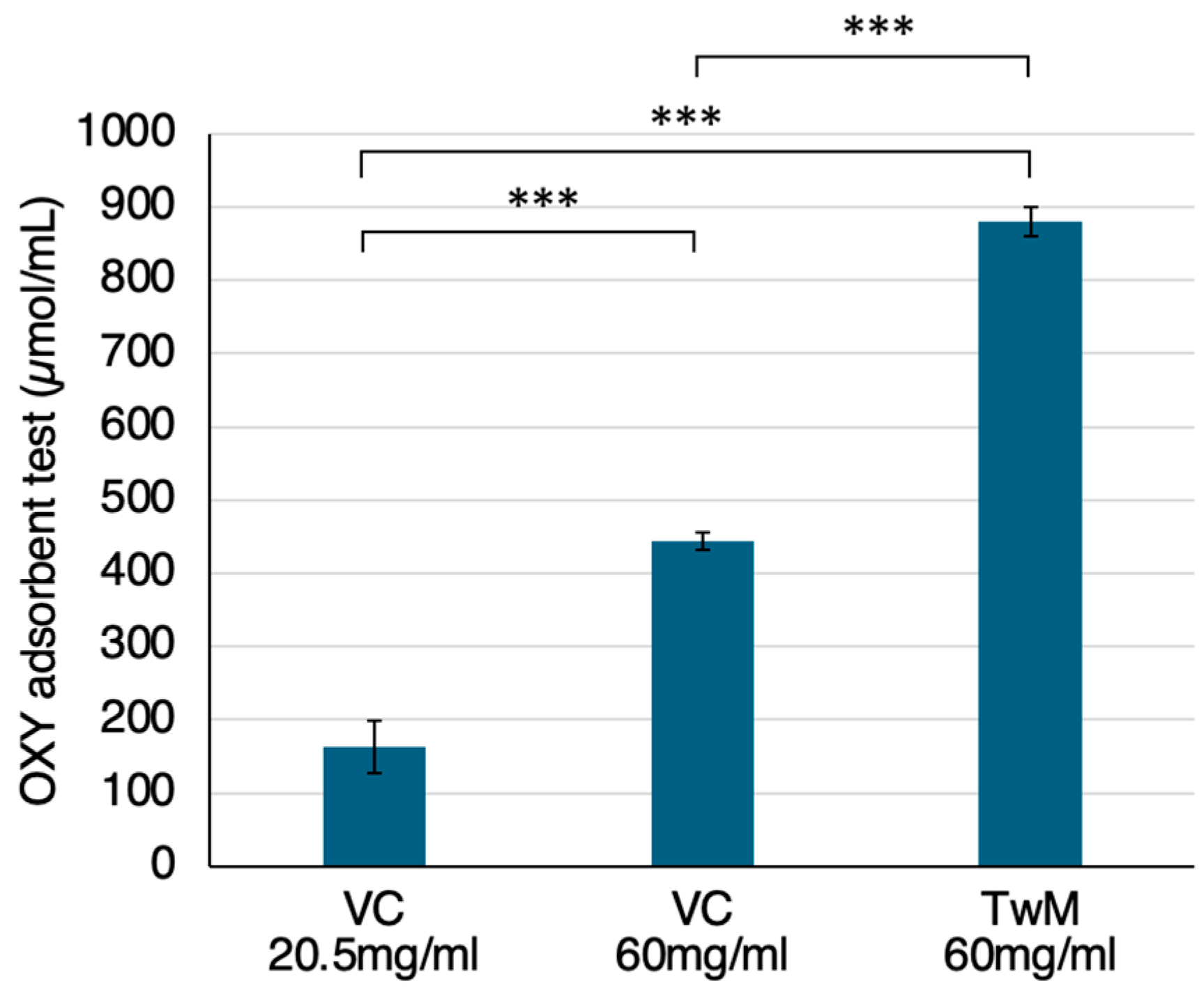
Figure 1. In vitro antioxidant capacity of Twendee M® (TwM) and vitamin C (VC) alone. VC at the concentration present in the TwM component (20.5 mg/mL, n = 6) and VC solution at the same concentration as TwM (60 mg/mL, n = 5) were compared to TwM (60 mg/mL, n = 5), respectively. Antioxidant capacity was determined via an OXY adsorbent test (Diacron International Srl, Grosseto, Italy). Values in the graphs represent mean ± SD. ***: p<0.001 (Student's t-test).
Preprints 97545 g001
Figure 2. Chronic fatigue syndrome (CFS) questionnaire overview. (A) Sex distribution of participants; (B) comparison of the degree of impact of chronic fatigue syndrome (CFS) on daily life before and after taking Twendee M® (TwM). Using a 6-point scale from 0 points (no impact) to 5 points (severe impact), the participants were asked to self-rate the extent to which CFS impacted their daily lives before and after taking TwM. The mean symptom scores of all participants were calculated and p-values were obtained using Student's t-test.
Preprints 97545 g002
Figure 3. Change in scores for each symptom of chronic fatigue syndrome (CFS) before and after taking Twendee M® (TwM). (A) Distribution of severity in each symptom. The participants were asked to rate their scores on typical symptoms of CFS (fatigue, muscle pain, joint pain, sleep disorders, decrease in memory and concentration, and headache before and after taking TwM) on a 6-point scale: no symptoms (0P), low degree of symptoms (1P), slightly low degree of symptoms (2P), moderate degree of symptoms (3P), slightly high degree of symptoms (4P), and high degree of symptoms (5P). (B) Mean of all participants' symptom scores for each symptom. p-values were obtained via Student's t-test.
Preprints 97545 g003
Table 1. Improved symptoms after taking TwM as indicated in the comments of the survey.
-
Poor quality of waking up, tiredness after sleeping, stiff shoulders were improved.
-
Sleep disturbances have improved and I am able to sleep better.
-
I catch less colds, and I feel less back pain, and I can sleep deeper.
-
Waking up and sleepiness is the same, but I don't feel sluggish when I wake up. I feel a little better about fatigue.
-
My body feels lighter and I don't feel tired when I wake up in the morning. (I am able to get up more easily.)
-
Dry skin. I feel like I recover from fatigue faster. (Especially during the daytime, I feel more energetic than before I started taking it.) Mentally, I feel less depressed.
Disclaimer/Publisher’s Note: The statements, opinions and data contained in all publications are solely those of the individual author(s) and contributor(s) and not of MDPI and/or the editor(s). MDPI and/or the editor(s) disclaim responsibility for any injury to people or property resulting from any ideas, methods, instructions or products referred to in the content.
Copyright: This open access article is published under a Creative Commons CC BY 4.0 license, which permit the free download, distribution, and reuse, provided that the author and preprint are cited in any reuse.
Prerpints.org logo

Preprints.org is a free preprint server supported by MDPI in Basel, Switzerland.

Subscribe

© 2024 MDPI (Basel, Switzerland) unless otherwise stated