1. Introduction
In this work, we aim to measure the response of the p-Therphenyl scintillator with a mono-energetic radiation source and determine the light output function. We were motivated by the fact that recently presented studies of light output functions [
8,
9] with the p-Therphenyl scintillator are measured only for neutron energies up to 8 MeV, which is limiting for practical applications in neutron fields with higher energies, e.g., in particle accelerator laboratories.
The high accuracy of the light output function determination is very important for Monte Carlo simulations of the response of scintillation detectors. The so-called light output function expresses the relation between proton and electron energies. It’s known that, protons and electrons of the same energy give light pulses of different amplitudes. The light output is defined as the equivalent electron of energy L which is the light output for an electron depositing the corresponding energy inside the scintillator, i.e., a proton of energy Ep (MeV) which gives the same light output as an electron of equivalent energy L (MeV).
The p-Therphenyl scintillator has been placed in a field of mono-energetic radiation over a wide energy range (2.5 to 19 MeV). In this energy range, a new more accurate light output function has been determined. Furthermore, we carried out a comparison of the PSD capability between the p-Terphenyl scintillator and the well-known NE-213 scintillator. We also calculate efficiency function for both scintillators.
2. Materials and Methods
2.1. Materials
The solid cylindrical scintillation detector p-Terphenyl of (45 x 45) mm with PSD properties has been studied in this work. The p-Terphenyl, also known as 1,4-Diphenylbenzene or p-Diphenylbenzene, is a white crystalline solid that is highly soluble in organic solvents such as ethyl acetate, benzene, and toluene.
The p-Terphenyl has a molecular formula of C6H5C6H4C6H5 (C18H14) and a molecular weight of 230.31 g/mol. The compound is derived from three phenyl groups connected by a single bond, giving it a linear structure. The p-Terphenyl product number: CAS 92-94-4 is meticulously manufactured to ensure a purity level of 99.5 %.
2.2. Digital Neutron-Gamma Spectrometer
The digital spectrometer is built as a modular system allowing the use of different types of scintillation detectors. The preamplifier splits the signal from the detector into two branches. Each branch is differently amplified and digitized by separate ADC. Different amplification increases the dynamic range of particles that the spectrometer is able to process.
The input analog signal from the p-Terphenyl detector is digitized with fast 12-bit analog to digital converter with a sampling frequency of 1 GHz. Digital signal processing is implemented into FPGA. FPGA is able to process all data flowing from ADC (12 Gbits per second). The spectrometer is connected with a computer via an optical ethernet of 10 Gbit.
2.3. Experimental Setup
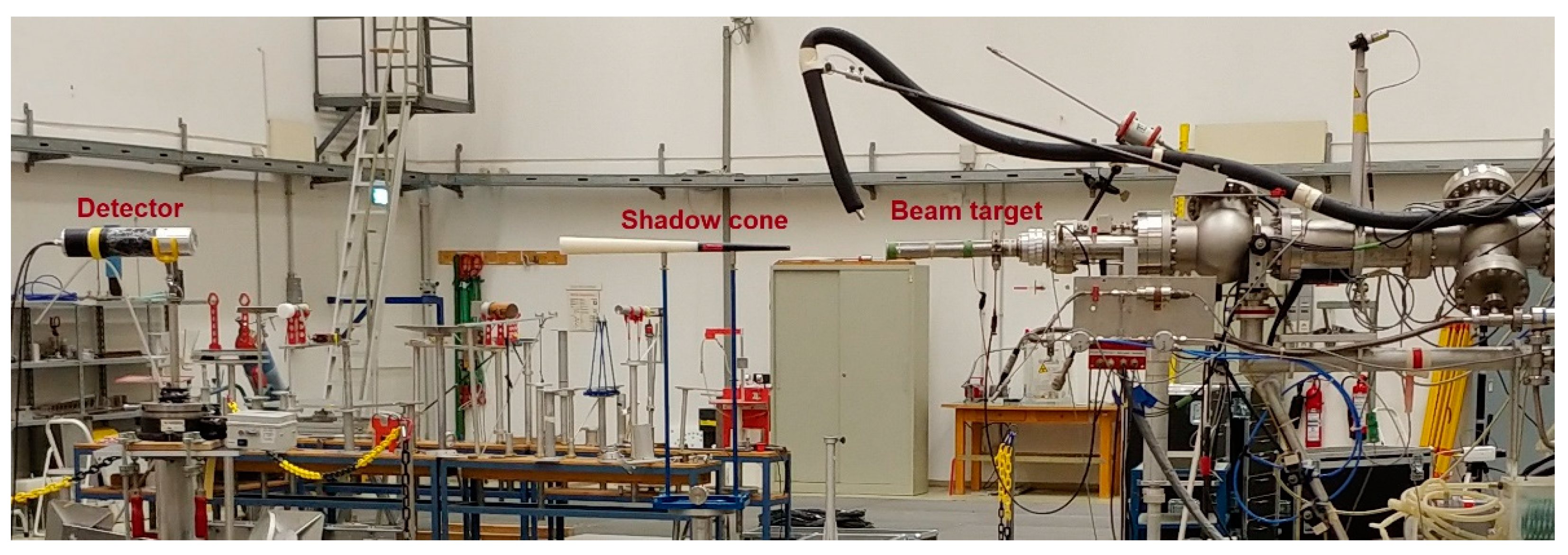
The first experimental measurements have been performed at the PTB Ion Accelerator Facility, where mono-energetic neutron fields are produced via selected reactions of proton and deuteron beams with light or medium-weight target nuclei, see
Figure 1. The measurements were carried out in open geometry in the low-scattering hall, where the contribution of scattered neutrons is minimized by having grid floors [
1,
2].
The two neutron energies considered in this campaign 2.5 and 19 MeV. Mean neutron energy was obtained at the neutron emission angle of zero degrees relative to the direction of the incident beam. Targets Ti(T) 1831 μg/cm
2 have been used for the
3H(p,n)
3He reaction and
3H(d,n)
4He reaction respectively. The experimental arrangement is shown in
Figure 2.
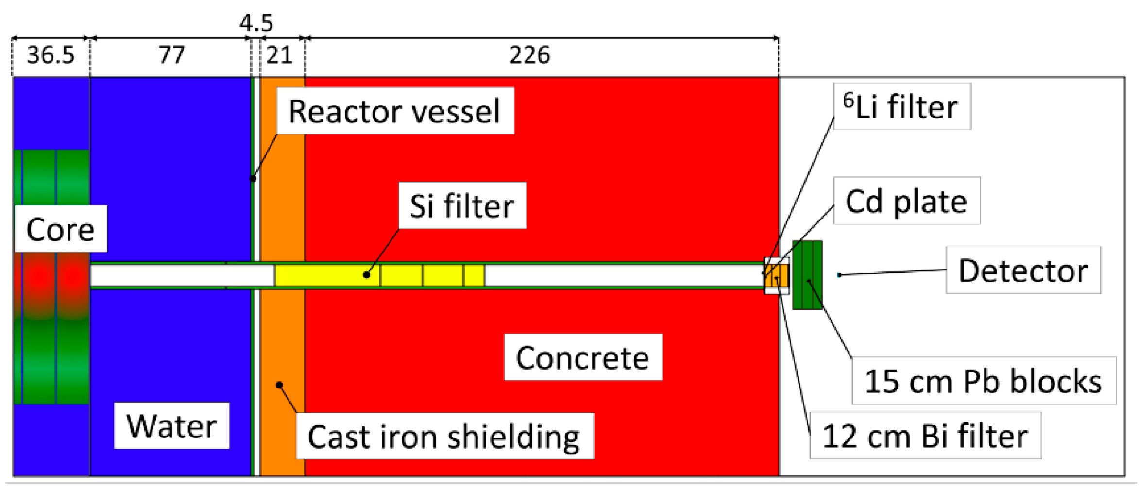
The second measurements have been carried out in the laboratory with experimental reactor LVR-15 at Research Center Rez (CVR), Prague. Well defined moderate neutron spectra with energies, see
Table 1, have been measured at the end of the horizontal beam port. Thermal neutrons and gammas was reduced via filter composed of
6Li, Cd, Bi, Pb. The experimental arrangement is shown in
Figure 3.
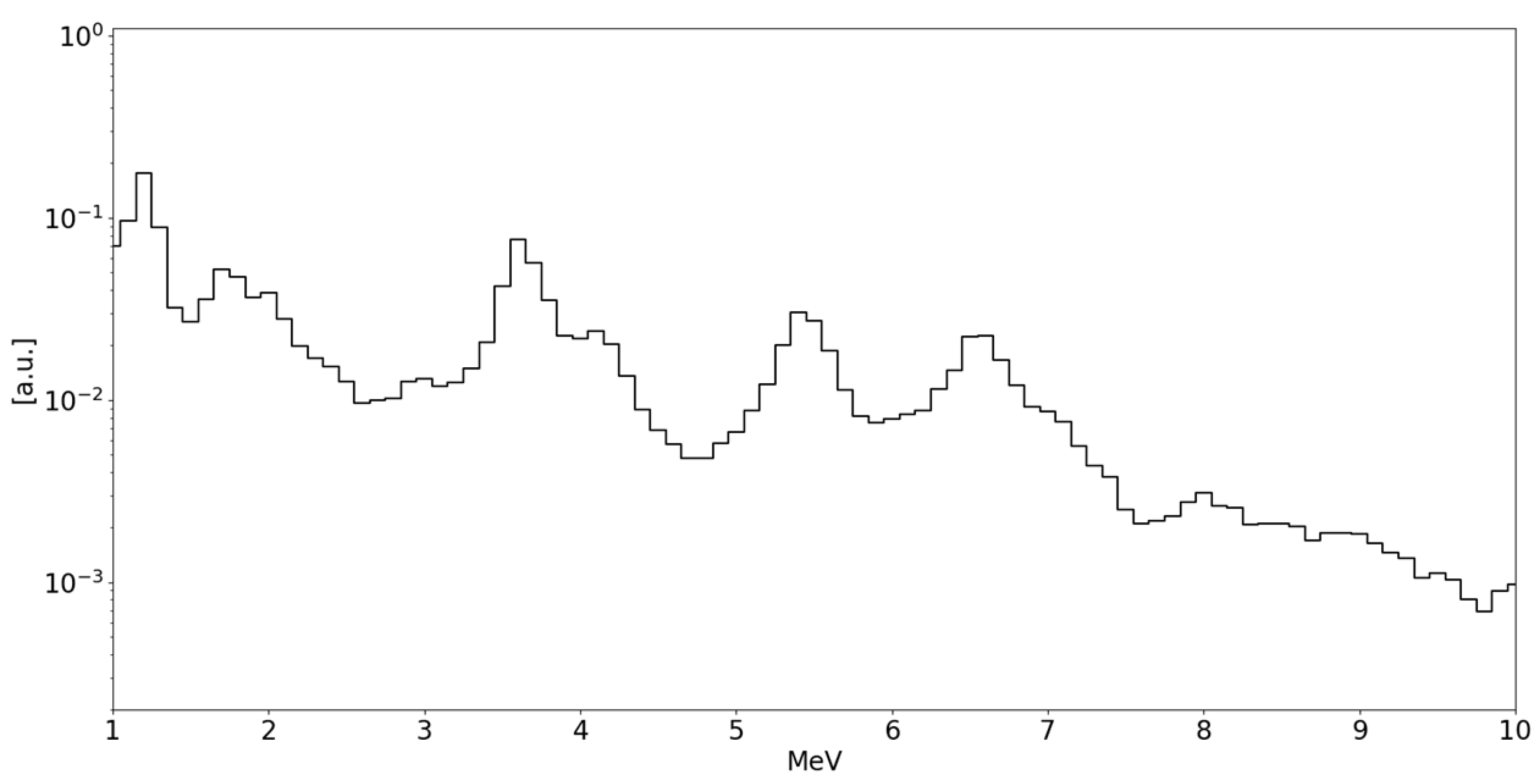
Neutron spectra have been moderated via 1 m wide silicon single crystal which provides spectrum with characteristic energy peaks, see
Figure 4.
2.4. Methods
The energy calibration has been performed using gamma-ray sources. Integrated digitized pulses have been linearly calibrated in keVee units, or keV electron equivalent. The linear transformation coefficients were derived from positions of the Compton edges [
4] in the spectra of two gamma-ray sources
137Cs and
60Co. Sources of activity 350 kBq have been placed on the center of the front face detector. Measurement time has been determined in accordance with count rates from the detectors. The surrounding background has been subtracted from the measured spectra.
Data were acquired over a time of 2 hours for each measurement. Subsequently these data have been used to calculate light-output parameters and PSD matrix of p-Terphenyl scintillator.
Digital spectrometer has incorporated the integration method [
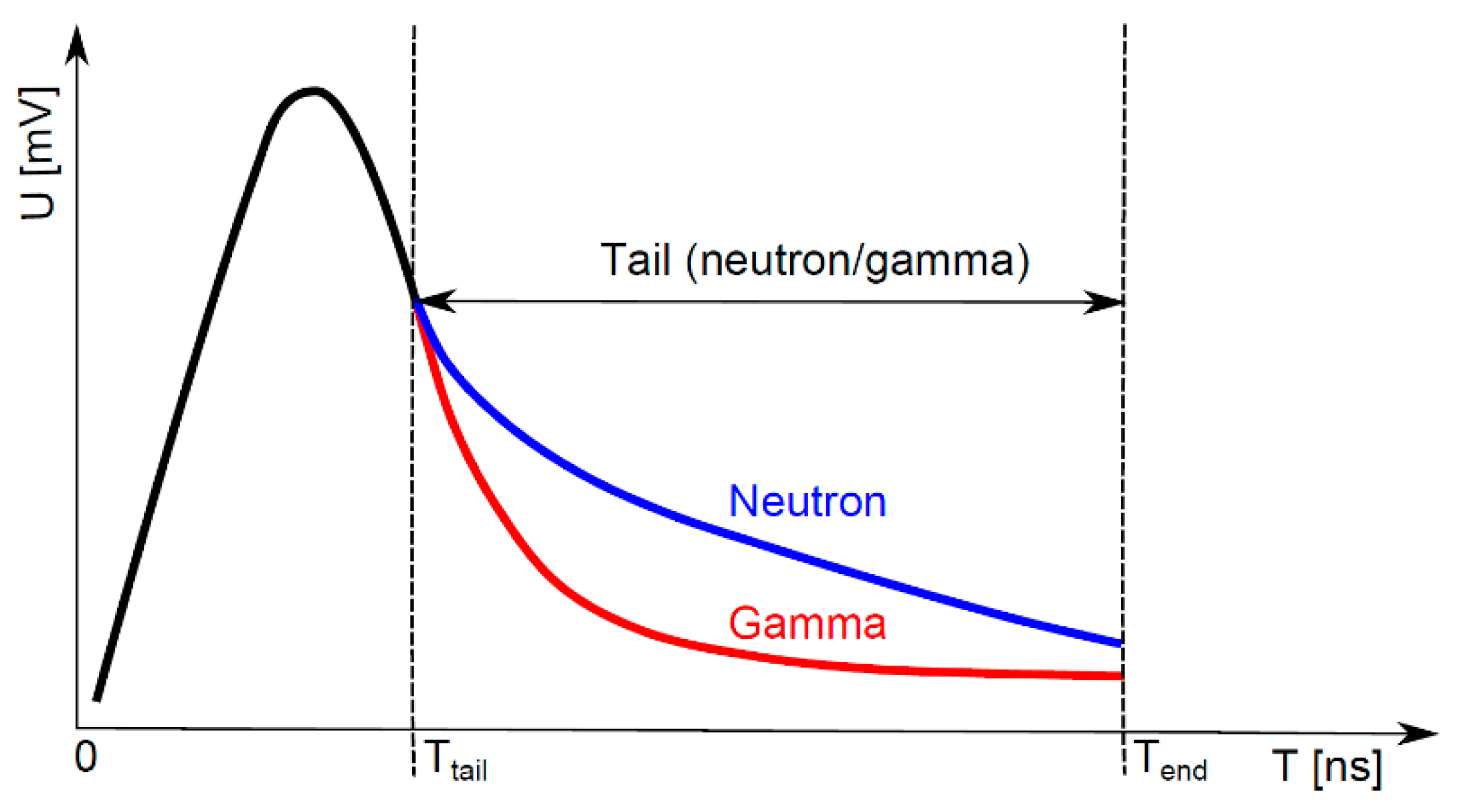
6] for recognition of neutron and photon pulses. Integration method is based on the principle of pulse charge comparison. The PSD parameter is calculated to recognize neutron and photon events:
where
Ttail is an optimized beginning of the tail part of the pulse and
Tend is an optimized end point of the pulse, see
Figure 5.
The experimental measurements of the scintillator response spectra were performed using the digital spectrometer in the laboratories CVR and PTB. The neutron response spectra have been identified employing the PSD method.
A summary of the incident neutron energies used in the experiments is given in
Table 1. The edge with the highest equivalent electron energy in the neutron response spectrum corresponds to the maximum energy deposited in the scintillator by the neutron-reflected proton.
3. Results
The pulse-shape discrimination capability of p-Terphenyl scintillator coupled to the fast digital spectrometer has been evaluated for selected energy of 14 MeV. We carried out the same measurement with the equivalent NE-213 detector. We studied neutron/gamma separation capabilities (see Eq. 1) for each measured detector. A two-dimensional graphs have been created, see
Figure 6.
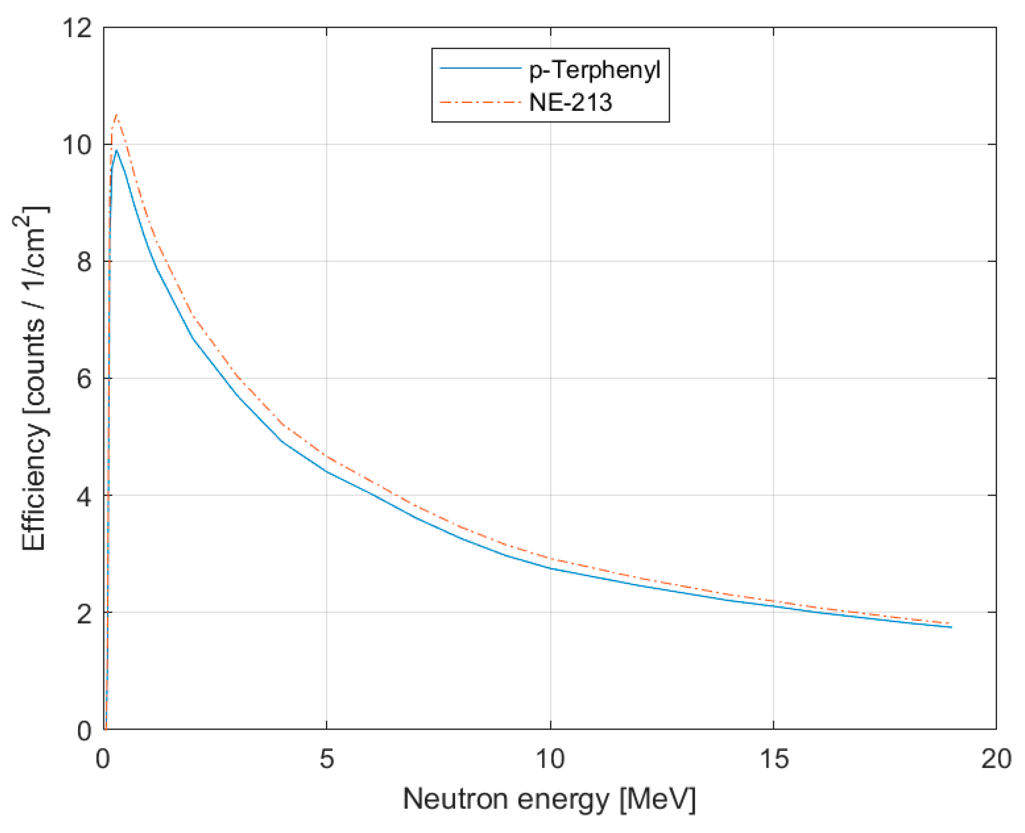
The efficiency calculations of the p-Terphenyl scintillator has been performed using Monte-Carlo MCNP 6.2 code. The efficiency curves for the p-Terphenyl and the NE-213 equivalent detector, with the same 100 keVee threshold, are shown in
Figure 7.
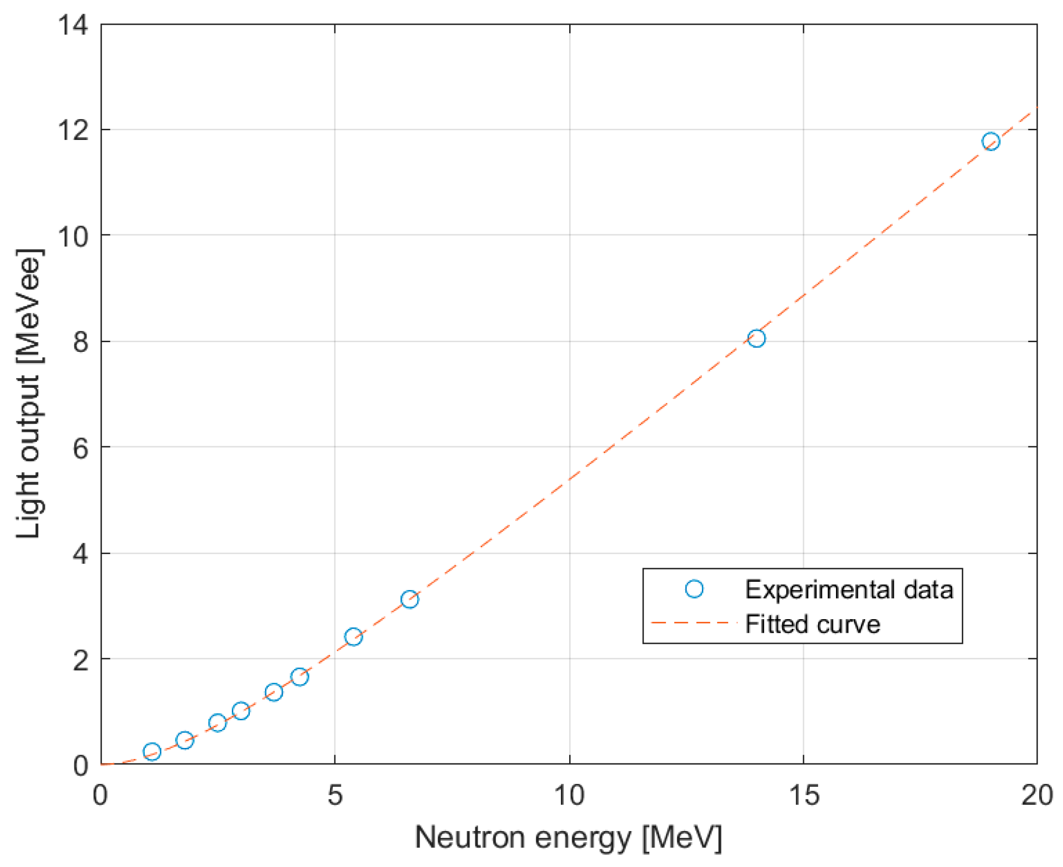
The light output function is important for unfolding the neutron spectra from the pulse height distribution. The light output function is described by following formula [
7]:
where
Ee is the electron energy in MeVee and
L0, L1 are fitting parameters. Calculated fit parameters are summarized in
Table 2. The light output function is shown in
Figure 8.
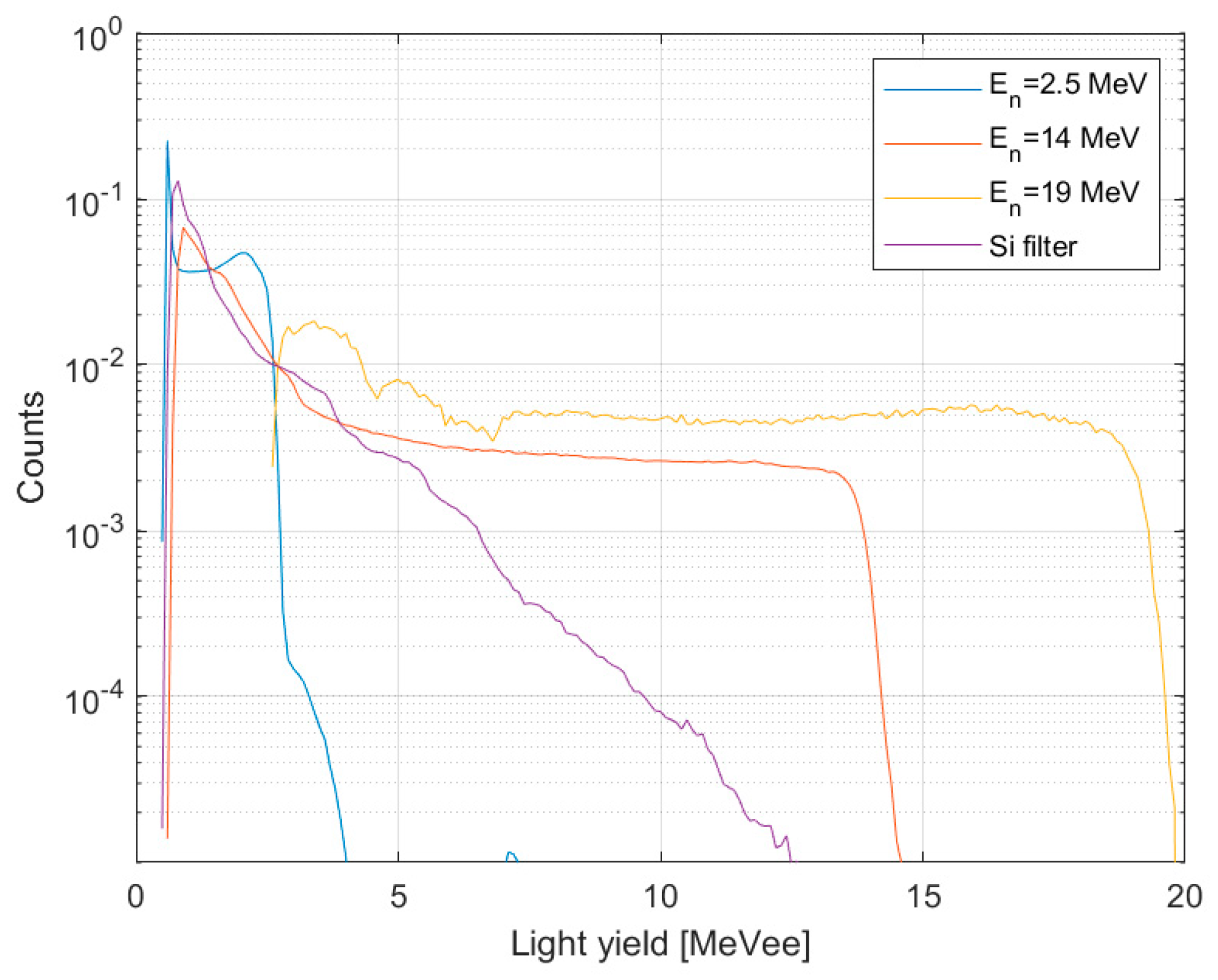
Normalized pulse-height spectra for p-Therphenyl scintillator in selected mono-energetic neutron beams are shown in
Figure 9.
4. Discussion
The light output function and pulse-shape discrimination capability of the p-Therphenyl scintillator have been measured and evaluated over a much wider energy range than previously reported.
Light output parameters, see
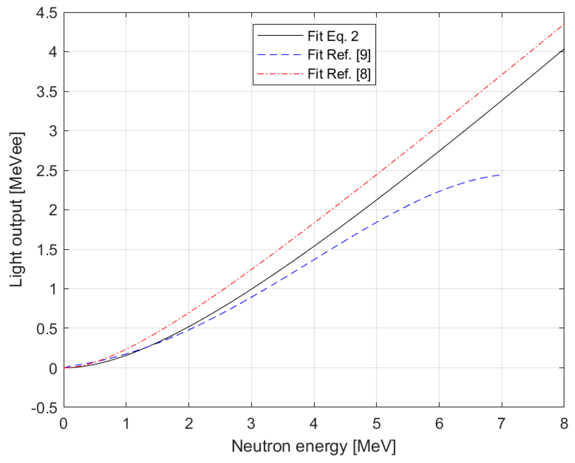
Table 2 of the p-Therphenyl scintillator, have been determined from the data fitted by the function described in equation (2). Previous studies concerned with the same issue have reported the results of light output functions in their publications, see ref. [
8,
9]. We compared our results with previously presented results of light output functions, see
Figure 10. Our light output function is in the band bounded by the functions from the cited papers. We assumed, based on our models, that the correct result would be among the presented functions. The measurement results confirmed our theory. This led to a refinement of the light output function for the p-Therphenyl scintillator.
The ability of the p-terphenyl scintillator to recognize the pulse shape was compared with the equivalent neutron detector NE-213. As can be seen in
Figure 7, the neutron-gamma separation is not nearly as sharp and sufficiently separated (separation parameter) as in the case of the NE-213 detector. The p-Therphenyl scintillator has worse properties in this respect than the NE-213 detector, which is to be expected.
In a study [
8], the efficiency of p-Therphenyl scintillator is discussed. The author C. Matei reported that the efficiency of this scintillator is greater than the NE-213 equivalent detector. Also, the author concluded his paper by stating that he could not explain this. We assume that the NE-213 detector has the best parameters, therefore the other detectors are compared with it. To support our hypothesis, we carried out Monte Carlo simulations of the efficiency of both detectors. The neutron efficiency comparison is shown in
Figure 8. It is clear that the NE-213 detector has a higher efficiency compared to the p-Therphenyl scintillator. The results also show that from 15 MeV energy upwards, the efficiencies of both detectors are very close.
The results will be used in the future especially in the design of new detectors and in simulations of radiation transport using Monte Carlo code. Using the new light output function parameters, see
Table 2, will ensure more accurate calculations and thus achieve more realistic results in practical applications.
Author Contributions
Conceptualization, A.J., Z.M. and M.K.; methodology, A.J. and Z.K.; software, Z.M.; validation, J.C.; data curation, A.J., Z.K and M.K.; formal analysis, A.J. and Z.K.; writing—original draft preparation, A.J.; supervision, Z.K. and Z.M.; project administration, A.J., Z.M. and M.K.; funding acquisition, A.J., Z.M. and M.K. All authors have read and agreed to the published version of the manuscript.
Funding
This research was funded by the Ministry of Education, Youth and Sports of the Czech Republic, project No. LM2018118 ”VR-1—Support for reactor operation for research activities”.
Data Availability Statement
Data will be made available upon request.
Acknowledgments
We are very grateful to the Physikalisch-Technische Bundesanstalt at Braunschweig for allowing the measurements in the laboratory with the Ion Accelerator.
Conflicts of Interest
The authors declare no conflicts of interest.
References
- Nolte, R.; Thomas, D. J. Monoenergetic fast neutron reference fields: I. neutron production. Metrologia 2011, 48, S263S273. [Google Scholar] [CrossRef]
- Nolte, R.; Thomas, D. J. Monoenergetic fast neutron reference fields: II. field characterization. Metrologia 2011, 48, S274S291. [Google Scholar] [CrossRef]
- Cvachovec, J.; Cvachovec, F. Maximum Likelihood Estimation of a Neutron Spectrum and Associated Uncertainties. Advances in Military Technology 2008, 3, 2nd ed., ISSN 1802-2308, 14.
- Dietze, G.; Klein, H. Gamma-calibration of NE 213 scintillation counters. Nuclear Instruments and Methods 1982, 193, 549–556. [Google Scholar] [CrossRef]
- Košťál, M.; Schulc, M.; Šoltés, J.; Losa, E.; Viererbl, L.; Matěj, Z.; Cvachovec, F.; Rypar, V. Measurements of neutron transport of well defined silicon filtered beam in lead. Applied Radiation and Isotopes 2018, 142, 160–166. [Google Scholar] [CrossRef] [PubMed]
- Brooks, F.D. A scintillation counter with neutron and gamma-ray discriminators. Nuclear Instruments and Methods 1959, 4, 151–163. [Google Scholar] [CrossRef]
- Kornilov, N.V.; Fabry, I.; Oberstedt, S.; Hambsch, F.-J. Total characterization of neutron detectors with a 252Cf source and a new light output determination. Nuclear Instruments and Methods 2009, 599, 226–233. [Google Scholar] [CrossRef]
- Matei, C.; Hambsch, F. - J.; Oberstedt, S. Proton light output function and neutron efficiency of a p-terphenyl detector using a 252Cf source. Nuclear Instruments and Methods 2012, 676, 135–139.
- Sardet, A.; Varignon, C.; Laurent, B.; Granier, T.; Oberstedt, A. p-Terphenyl: An alternative to liquid scintillators for neutron detection. Nuclear Instruments and Methods 2015, 792, 74–80. [Google Scholar] [CrossRef]
|
Disclaimer/Publisher’s Note: The statements, opinions and data contained in all publications are solely those of the individual author(s) and contributor(s) and not of MDPI and/or the editor(s). MDPI and/or the editor(s) disclaim responsibility for any injury to people or property resulting from any ideas, methods, instructions or products referred to in the content. |
© 2024 by the authors. Licensee MDPI, Basel, Switzerland. This article is an open access article distributed under the terms and conditions of the Creative Commons Attribution (CC BY) license (http://creativecommons.org/licenses/by/4.0/).

