Preprint
Article

Bioactive Compounds from Campsis radicans L.: Antioxidant and Antiproliferative Effects on Colon Cancer Cells

Altmetrics

Downloads

183

Views

123

Comments

0

This version is not peer-reviewed

Submitted:

16 February 2024

Posted:

19 February 2024

You are already at the latest version

Alerts
Abstract
This study focuses on the potential medicinal properties of Campsis radicans L. extracts and its aim is to investigate the antioxidant and antiproliferative effects of these extracts on the human colon adenocarcinoma cell line (HT-29). Cytotoxic activity in the HT-29 cell line was evaluated, and the 3-dimensional structures of phenolic compounds were obtained using the GC-MS method. Docking simulations were performed with AutoDock Vina, targeting proteins that are crucial in the apoptotic pathway (Bax, Bcl-2, and Caspase-3). Drug similarities and ADMET estimates of the phenolic compounds were obtained from databases. The highest antioxidant activity of Campsis radicans L. plant was observed in ethanol and water extracts, with notable inhibition capacities. While the total phenolic content was highest in ethanol, water, and ether extracts respectively the total flavonoid content was highest in ethanol, then ether, and water extracts. Certain elements were undetectable in mineral determination, and others exceeded the toxic doses for plants. The water extract exhibited dose-dependent cytotoxic activity in the HT-29 cell line, with an IC50 value of 4.47 mg/mL. Molecular docking results revealed the binding of phenolic molecules to the active sites of apoptotic pathway proteins. The findings suggest that Campsis radicans L. plant possesses cytotoxic properties, significant phenolic and flavonoid content, and a high antioxidant capacity. These properties make it a potential candidate for treatment use, emphasizing its potential therapeutic value in the HT-29 cell line. Further research and exploration on the identified phenolic compounds could contribute to the development of novel therapeutic agents.
Keywords: 
Subject: Biology and Life Sciences  -   Food Science and Technology

1. Introduction

Learning about the plants and seeds that cover a large portion of the earth and seeking remedies for many diseases by including plants in their lives has been a method used by humanity since the beginning of time., the active substances obtained with this method constitute the building blocks of drugs used for current diseases. In contrast to synthetic therapy (chemotherapy, radiotherapy, and other drug agents) beneficial herbs symbolize safety [1] and play a major role in treatment processes, including cancer treatment [2,3,4,5]. In additio experimental and clinical Chinese medicine treatment methods demonstrated effectiveness of many herbal formulations in the treatment of cancer at various stages [6]. Plants are source of biologically active substances, and in epidemiological studies, certain foods, such as vegetables, fruits, and dietary fiber, have been shown to act as chemo-preventive agents[7]. Additionally, in recent records, it is reported that 80% of people living in rural areas use medicinal plants to solve their basic health problems [8], and 700 plant species identified in the Middle East region have medicinal value[9]. The perennial pipe (Campsis radicans L.) plant is widely distributed in the USA, Canada, China, and South Asia [10] and is grown as an ornamental plant in parks and roadsides [11], and used in traditional medicine for the treatment of infections caused by Candida, Haemophilus, etc. [12].
DPPH scavenging activity, thrombolytic activity, analgesic activity, hypoglycemic activity, antidiarrheal activity, and CNS antidepressant activity of Campsis radicans L. plant were investigated by creating experimental models. According to the results of this study, it was revealed that C. radicans has significantly high antioxidant, thrombolytic, membrane stabilizing, analgesic hypoglycemic, antidiarrheal, and CNS antidepressant activities [13]. In our literature review, we could not find a significant study showing the antiapoptotic effects of C. radicans in the human colon adenocarcinoma cell line (HT-29). To compensate for this deficiency, cytotoxic activity experiments were also performed on the HT-29 cell line. is the decision whether the compounds obtained from plants can be used as medicine is made through many clinical studies and computer simulations. In this process, simulations can show the ADMET properties of ligands, drug affinities, and their interactions with the active site of the target protein with almost complete accuracy at a low cost. Knowing these properties is one of the first steps to decide whether the obtained ligand can be used in clinical trials. In our study, using computer simulations and some important databases, the properties of the phenolic compounds of the C. radicans plant, such as drug properties and whether they can bind to the active sites of proteins that play an important role in cancer, were also investigated.

2. Materials and Methods

2.1. Collection and Extraction of Campsis radicans L. Plant Samples

Plant material was collected as healthy plants from the coordinates 38.414438, 33.979856 (Aksaray/Turkey). Some of the collected plants were sent to Anadolu University Plant Medicine and Scientific Research Center (Eskişehir/Turkey) for identification of thier species. The plant was identified as Campsis radicans L. (Trumpet Vine) with the barcode number BMT 202200002. Three different solvents were chosen for obtaining the plant extracts: ethanol, diethyl ether, and hot water. In all three extractions, 300 mL of solvent was added to 50 g of dry herb powder. The extracts were kept in an ultrasonic sonicator (Bandelin Sonorex) for 30 minutes at 37oC. The shaker was then left on for 23 and a half hours. Afterwards, the solvent and plant particles were separated from each other by filtration using a paper filter. After ethanol and diethyl ether were evaporated, it was dissolved again in ethanol (50 mL). The obtained extracts were stored at 4oC to be used.

2.2. Determination of Campsis radicans L. Plant’s Mineral Content

Campsis radicans L. plant’s mineral content was determined using the ICP-MS method and the operating conditions of the device are shown in Table S1 In sample preparation: About 0.2 g of plant sample was placed in a microwave tube. 10 mL of nitric acid was added. The solution mixture was stirred. 10 minutes were given for pre-burning. Afterwards, the tubes were closed and incineration was performed in the microwave oven (Microwave Sample Combustion System, CEM MARS6). Burning program in the microwave oven: Initially, the microwave was heated to 190 °C for 20 minutes. It was then kept at 190 °C for 15 minutes. The pressure was set at 800 psi, and the power at 900-1800 watts. It was then cooled for 15 minutes from 190 °C to room temperature. After incineration, the samples taken from the tubes were made up to 50 mL with ultrapure water. ICP-MS Device Analysis (Bruker aurora M90): 27 standard and blank samples were read on the device. Calibrations were drawn. Then, the samples and the blanks of the samples were read and analyzed.

2.3. Determination of Total Phenolic Content

Total phenolic content analysis in Campsis radicans L. plant extracts was p by adding 900 µL of distilled water to 100 µl of extract and making minor changes in the 5 mL 0.2 M Folin-Ciocalteu method [14]. After adding 5 mL (7.5%) Na2CO3 and allowing it to incubate for 2 hours at room temperature, absorbance was measured at 765 nm in a spectrophotometer (Unico, S1205). Gallic acid was used to prepare a standard curve. Data are expressed as mg gallic acid equivalent (GAE) per gram of dry plant weight (DW).

2.4. Determination of Total Flavonoid Content

The total amount of flavonoid substances of Campsis radicans L. plant extracts [14] were determined based on the method used. Using AlCl3 and NaNO2 as reagents, 4 mL of distilled water and 0.3 mL of NaNO2 (5%) were added to the 0.4ml extract and incubated for 5 minutes. Then 0.5 mL of 10% AlCl3 was added to the mixture and waited for another 6 minutes. Then, 2 mL of 1 M NaOH and 3 mL of distilled water were added and the absorbance of the mixture was read at 510 nm. Catechin concentrations of 0.01–0.25 mg/mL were used to generate a calibration curve and data are expressed as mg catechin equivalents (CE) per g. plant weight.

2.5. Determination of antioxidant capacity of Campsis radicans L. plant extracts

To determine the antioxidant capacity of Campsis radicans L. plant extracts: DPPH free radical scavenging activity and ABTS radical cation scavenging activity tests were performed.

2.5.1. DPPH method

The antioxidant capacity of the Campsis radicans L. plant was determined by the DPPH method. The free radical scavenging activity of Campsis radicans L. extracts was measured with minor modifications to the DPPH method previously described by[15]. A 25 mg/L DPPH solution was prepared with methanol. 0.1 mL extract was added to 3.9 mL DPPH solution and kept in a shaker for 30 minutes in a dark environment at room temperature. Absorbance measurements were made using a spectrophotometer (Concepcion Sanchez-Moreno) at a wavelength of 517 nm. % calculations of antioxidant activity were performed using formula 1.
% D P P H   i n h i b i t i o n   c a p a c i t y = [ ( A c A e ) A c ] x 100
Ac is the absorbance of the DPPH solution and Ae is the absorbance of the sample.

2.5.2. ABTS method

The antioxidant capacity of the Campsis radicans L. plant was determined by the ABTS method. The antioxidant capacity of Campsis radicans L. extracts was determined by making some changes in the spectrophotometric measurement method developed by [16]. Briefly, 2,2′-Azinobis-(3-Ethylbenzthiazoline-6-Sulfonic Acid (ABTS+) cation radical solution was produced by reacting ABTS (2 mM) in H2O and potassium persulfate (2.45 mM) for 12 hours at room temperature. The ABTS+ cation radical solution is dark blue-green with a characteristic absorbance at 734 nm. Before using the ABTS+ radical solution, the absorbance of the control solution at 734 nm was adjusted to a value between 0.750 nm and 0.800 nm. Phosphate buffer 0.1 M and pH 7.4. 80 µL by adding 1ml ABTS solution at different concentrations to the extract samples, the total volume was made up to 4 mL with phosphate buffer. These samples were vortexed and incubated for 30 minutes, followed by their absorbance. Measured at 734 nm. ABTS radical scavenging activity % inhibition values were calculated by formula 2.
% A B T S   i n h i b i t i o n   c a p a c i t y = [ ( A c A e ) A c ] x 100
Ac is the absorbance of the ABTS solution and Ae is the absorbance of the sample.

2.6. Cell culture

HT-29 human colon adenocarcinoma cells, Roswell Park Memorial Institute (RPMI) 1640) (Life Technologies), in medium supplemented with 10% inactivated fetal bovine serum (Life Technologies) and 1% penicillin-streptomycin (100 U/mL) (Life Technologies). It was grown in 75 cm2 flasks in a culture environment of 37 °C and 5% CO2. Switching to new medium was done every 2 days [17].

2.7. Cell Proliferation and Viability

Cells were collected by centrifugation (300 x g for 5 min); the culture medium was removed and the pellet was suspended in PBS (phosphate buffered saline). 10 µL of trypan blue was added to 10 µL of cell sample, and after 3 minutes, cells were counted using a hemocytometer (Neubauer)[18].

2.8. MTT test

Cancer cell line viability was evaluated by the 3-(4,5-dimethylthiazol-2-yl)-2, 5-diphenyltetrazolium bromide (MTT) method, first described by Mosmann [19]. Briefly, 2x10⁴ HT-29 cells were seeded in 96-well plates and various concentrations of ethanol, ether, and water extracts (50-5000 µg/mL) were added to the cells after 24 hours of incubation. Cells were incubated in 5% CO2 at 37oC for 24 and 48 hours. MTT (5 mg/mL phosphate buffered saline, pH 7.2) was then added to all wells. After 3 hours of incubation with MTT, the medium was removed and DMSO was added to dissolve the water-insoluble formazan crystals. Cell viability was determined as absorbance at 492 nm via a microplate reader (ChroMate®ELISA reader) after MTT assay. MTT experiments were repeated three times individually with separate cells from the same passage on different days. Cell viability is then defined by equation 3:
Formule   1 ( T e s t   O D C o n t r o l   O D ) x 100
Polynomial regression analysis was performed using Microsoft Excel to calculate the half-maximum inhibitory concentration (IC50) values of the investigated compounds.

2.9. ADMET and drug similarity estimation for ligands

Computer-based ADMET and drug similarity estimations represent an advantage for drug discovery[20]. For ADMET (Absorption, Distribution, Metabolism, Excretion, Toxicity) estimations (http://biosig.unimelb.edu.au/pkcsm/) database and BMDRC (https://preadmet.qsarhub.com/) database were used [21,22]. SwissAdme (http://www.swissadme.ch/) database was used for drug similarity estimates. During the drug similarity process, Lipinski, Ghose, and Veber rules, additionally gastrointestinal system absorptions, blood brain barrier absorptions, bioavailability scores, hepatotoxicity, skin sensitivity, oral acute toxic doses and maximum tolerable doses in rats were also investigated.

2.10. Finding and Preparing the Target Protein

Proteins involved in the apoptotic process Bax (PDBID: 1F16)[23], Bcl-2 (PDBID: 4LVT)[24], Caspase-3 (PDBID: 3GJQ)[25] were obtained for free using data from the Protein Data Bank (https://www.rcsb.org/) database. During the protein preparation process, water molecules were removed, polar hydrogens were added, and everything was made ready for molecular docking using the Discovery Studio 2021 program.

2.11. Molecular Docking

Docking simulations of phenolic compounds obtained by GC-MS scanning (Table S2) to proteins that play an important role in apoptosis were performed with AutoDock Vina (version 1.1.2) program using 3-way multiplex Lamarc Genetic Algorithm[26] and by entering the coordinates of the protein’s active site.

2.12. Statistical analysis

All statistical analyzes were performed using the Graphpad Prism 9 (https://www.graphpad.com) statistical software suite. Statistical significance was determined using a one-way variance analysis (ANOVA) followed by a post hoc test (Tukey’s multiple comparison tests). The results were evaluated at 95% confidence interval and at p<0.05 significance level.

3. Results

The heavy mineral content of the Campsis radicans plant L. is shown in Table 1 and elements such as Ag, As, Ba, Cd, Co, Sn, and Tl could not be detected. Evaluations were performed according to the normal and toxic concentrations found in medicinal plants, which were previously studied [27]. As a result, while Mn, Pb and Zn concentrations in Campsis radicans L. plant were observed to be within safe limits, while Cr, Cu, Fe, Mo, Ni, and V elements were found to be above the safe limits.
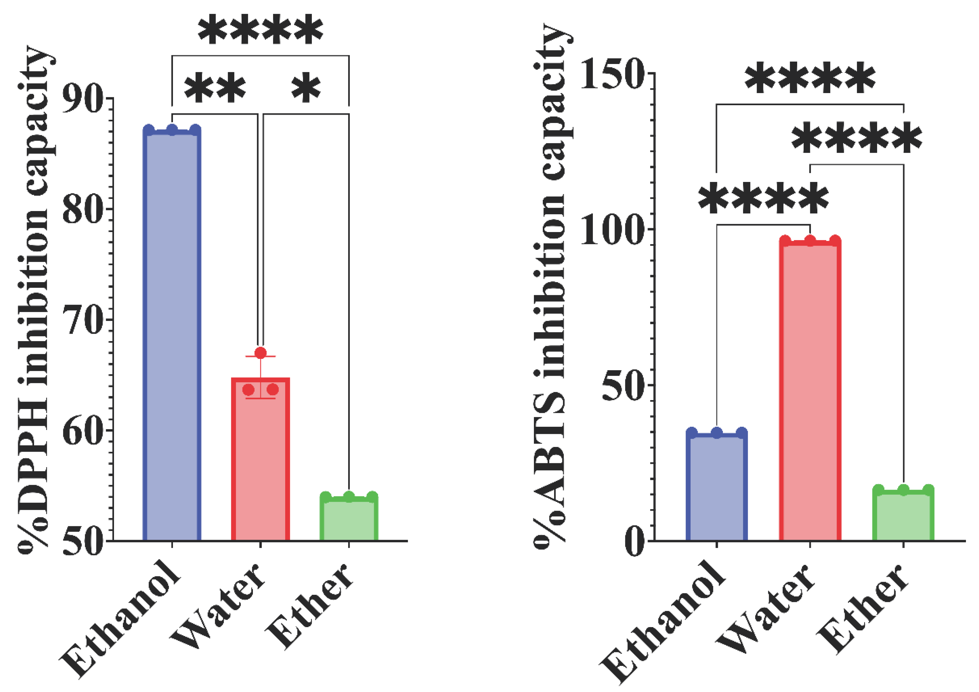
Total phenolic and flavonoid contents in the extracts (Tables S3 and S4) of Campsis radicans L. plant prepared with three different solvents are shown in Figure 1 Significant differences were found between total phenolic and flavonoid amounts in the extracts prepared with water, ethanol, and ether (p<0.0001). % DPPH and % ABTS inhibition capacities in extracts prepared with three different solvents are shown in Figure 2 Significant differences were found between the three methods and the %DDPH and %ABTS inhibition capacities (p<0.0001).
Common names, carbon numbers, and PubChem CID numbers of fatty acids obtained from GC-MS fatty acid and phenolic content analysis of Campsis radicans L. plant are shown in Table 2.
Campsis radicans L. plant extracts (except Ether) were found to have a cytotoxic effect on the HT-29 cell line. The cytotoxic activity increased depending on the dose and the IC50 values of ethanol, water, and ether extracts were found to be, 9.8 mg/mL, 8.47 mg/mL, and 5.8 mg/mL, respectively (Figure S1).
While Campsis radicans L. is mentioned as an anticarcinogenic plant in Chinese medicine [28], there is no study in the literature to support this idea. In order to close this gap, we performed cytotoxic activity experiments on the HT-29 cell line and found that the aqueous extract of the plant showed increased cytotoxic activity in a dose-dependent manner. This completes the information on the cytotoxic activity of Campsis radicans L. and acts as the first step to closing a major gap in the literature. Plants can demonstrate their anticarcinogenic and antimicrobial activities with their phenolic and flavonoid contents. Phenolic and flavonoid content plays an important role in determining the antioxidant capacity of a plant. There is little so little data in the literature about the antioxidant capacity, as well as phenolic and flavonoid content of the Campsis radicans L. plant. In the extraction study performed with different solvents, the phenolic capacity of the Campsis radicans L. plant was found to be between 6.38 and 60.13 mg/GAE [13]. In our study, the total phenolic content was found to be 0.42 – 2.07 mg/L extract, and the total flavonoid content was found to be 83.4-300.6 mg/L of extract. In addition, the Campsis radicans L. plant can be a source of antioxidant supplement according to its %DPPH and %ABTS scavenging activities. Heavy metal content analysis of Campsis radicans L. plant was a missing data in the literature soin order to close this gap with our study, heavy metal content analysis of the plant was performed by ICP-MS. When the region where Campsis radicans L. is collected is evaluated in terms of air and soil quality, we think that this plant is a plant that can accumulate some minerals. While As, Cd, Co, and Ti metals were not found in the plant, Fe, Cu, Ni, Cr, Mo, and V metals were found above normal concentrations and even at toxic levels for plants. Since the heavy metal content at this high concentration is above medicinal plant standards, we think that the Campsis radicans L. plant should be grown specifically for medicinal purposes.
Molecular interactions of phenolic compounds obtained by GC-MS method with target proteins; the binding energy, the number of hydrogen bonds, and bonded amino acids are shown in Table 3.
The interactions developed by ligands in the protein active site are conventional hydrogen bonds, carbon-hydrogen bonds, halogen bonds, van der Waals interactions, pi-sigma, pi-alkyl, and alkyl interactions. According to the results obtained in the bonding study, all ligands interacted with the active site of the proteins by making bonds in the appropriate conformation. According to these results, 4-Vinylbenzoic acid, which is one of the phenolics obtained from the aqueous extracts of the plant, has higher binding energy (-5.2 kcal/mol) and 1 conventional hydrogen bond compared to the other aqueous extract ligands, demonstrate distinct interactions with 6 amino acids while binding to the Bax protein active site. Propanoic acid, 2-hydroxy-, 1-methylethyl ester, (2S)-, which bonded with the lowest binding energy (-3.3 kcal/mol) did not form conventional hydrogen bonds but developed different favorable interactions with 4 different amino acids. Trimethyl citrate, which makes 5 conventional hydrogen bonds, is bound to the protein active site with a binding energy of -3.8 kcal/mol. 4-Methoxyphenethyl alcohol, which binds to the active site of the Bcl-2 protein with the highest binding energy, did not make any conventional hydrogen bonds but bonded to the protein’s active site by developing different interactions with 5 different amino acids. 4-Methyl-3,6,9-trioxadecan-1-ol was bound to the protein active site with the lowest binding energy (-3.2 kcal/mol) and made 1 conventional hydrogen bond. Trimethyl citrate, which binds to the protein active site with the most conventional hydrogen bond, made 5 conventional hydrogen bonds. The binding energy to the protein active site was -4.2 kcal/mol. Levoglucosan, which binds to the Caspase-3 protein active site with the highest binding energy (-4.9 kcal/mol), was bound to the protein active site by conventional hydrogen bonds and van der Waals interactions. Propanoic acid, 2-hydroxy-, 1-methylethyl ester, (2S)- bonded with the lowest binding energy (-3.7 kcal/mol) to the protein active site by developing appropriate interactions with 4 different amino acids that have made 2 conventional hydrogen bonds. Trimethyl citrate, which made ten conventional hydrogen bonds, was bound to the protein active site with a binding energy of -4.3 kcal/mol. 6-nitro-3-phenyl-1H-indol-2-amine, which is one of the phenolics obtained from the ethanolic extracts of the plant, develops a conventional hydrogen bond with the highest binding energy (-5.8 kcal/mol) to the Bax protein active site and performed different interactions with 4 different amino acids. While vitamin E, which binds with the lowest binding energy (-4.4 kcal/mol), does not develop any conventional hydrogen bonds, it binds to the active site of Bax protein by developing different interactions with 4 different amino acids. 2-methyl-2,3-dihydro-1H-inden-1-ol, which interacted with most amino acids, did not develop conventional hydrogen bonds and was bound to the active site of the Bax protein with a binding energy of -5.3 kcal/mol. The 6-nitro-3-phenyl-1H-indole-2-amine Bcl-2 protein binds to its active site with the highest binding energy (-6.9 kcal/mol) and also has different interactions with 1 amino acid and made 1 conventional hydrogen bond. Vitamin E, which binds with the lowest binding energy, binds to the active site of the Bcl-2 protein by performing different interactions with 5 different amino acids and made 1 conventional hydrogen bond.
The caspase-3 protein binds to the active site of the protein by performing 2 conventional hydrogen bonds and different interactions with 2 different amino acids to 6-nitro-3-phenyl-1H-indole-2-amine, which binds to the active site with the highest binding energy (-6.8 kcal/mol). 2-methyl-2,3-dihydro-1H-inden-1-ol, which binds to Caspase-3 protein with the lowest binding energy (-4.8 kcal/mol), did not develop any conventional hydrogen bonds while bonded to the protein active site by developing different interactions with 3 different amino acids.
The relationships of the phenolic compounds obtained from Campsis radicans L plant extracts by GC-MS method with the active sites of three proteins (Bax, Bcl-2, and Caspase-3) that play an important role in the apoptotic pathway was observed by using the molecular coupling method; additionally, the drug properties of these phenolic compounds were also analyzed by using databases. In the literature, there is information about the production of Campsis radicans L. plant phenolics, but the lack of in-silico studies on these phenolic compounds creates a big gap in the literature. To close this gap, our study tested the ADMET properties of phenolic compounds and their compliance with the Lipinski, Veber, and Ghose rules, which are the rules for drug specifications. According to the Lipinski rules, in order to be considered as a drug, a ligand must have some specifications: molecular weight <500g/mol, Log P ≤ 5, hydrogen bond donor ≤5, hydrogen bond acceptor ≤10, and molar break value between 4-130 [29]. According to Veber’s rules, required specifications for a ligand to be considered as a drug; hydrogen bonding ≤12, rotatable bonds ≤10, polar surface area ≤140, and oral bioavailability >20% [30]. According to Ghose’s rules, the properties that the drug candidate ligand should have are LogP -0.4 - 5.6, molar refraction 40 - 150, molecular weight 160 - 480 g/mol, the atomic number between 20 and 70, and polar surface area <140 [31].
The findings of our study show that the Campsis radicans L. plant can be used as an antioxidant source due to its phenolic and flavonoid capacity, and this antioxidant feature also contributes to anticarcinogenic activity. Compliance of phenolic compounds with drug rules and ADMET properties (Tables S5 and S6) was also investigated. It was shown that the obtained phenolic compounds were bonded to the active sites of proteins by in-silico studies. However, bonding and other in-silico studies are the results of predictions made within algorithms. For this reason, more clinical studies on these phenolic compounds are required.

4. Discussion

Molecular interactions of phenolic compounds obtained from the GC-MS method with target proteins were investigated, and the results are summarized in Table 3 The findings provide insight about the binding energy, number of hydrogen bonds, and amino acids involved in the interactions. In-silico studies indicated that the phenolic compounds effectively interacted with the active sites of the proteins, showcasing potential therapeutic relevance.
To close the gap in the literature regarding in-silico information on these phenolic compounds, we explored their ADMET properties and compliance with drug-specific rules. The study confirmed that the phenolic compounds adhere to drug rules, showing potential as drug candidates. However, these in-silico predictions warrant further validation through clinical studies to establish their actual therapeutic benefits.

Supplementary Materials

The following supporting information can be downloaded at the website of this paper posted on Preprints.org.

Author Contributions

The study conception and design were contributed by TK. Formal analysis and investigation were conducted by TK and CS. Manuscript’s first draft was written by TK and CS. The authors read and approved the manuscript.

Funding

Not applicable.

Data Availability Statement

Raw data of the this study are available from the corresponding author on reasonable request.

Conflicts of Interest

The authors declare no conflicts of interest.

References

  1. Singh, M.; Kumar, D.; Sharma, D.; Singh, G. TYPHONIUM FLAGELLIFORME: A MULTIPURPOSE PLANT. Int. Res. J. Pharm. 2013, 4, 45–48. [Google Scholar] [CrossRef]
  2. Nobili, S.; Lippi, D.; Witort, E.; Donnini, M.; Bausi, L.; Mini, E.; Capaccioli, S. Natural Compounds for Cancer Treatment and Prevention. Pharmacol. Res. 2009, 59, 365–378. [Google Scholar] [CrossRef]
  3. Jing, L.; Mohamed, M.; … A.R.-J. of M.; 2010, undefined Phytochemicals, Antioxidant Properties and Anticancer Investigations of the Different Parts of Several Gingers Species (Boesenbergia Rotunda, Boesenbergia. academia.eduLJ Jing, M Mohamed, A Rahmat, MFA BakarJournal of Medicinal Plants Research, 2010•academia.edu.
  4. Research, H.M.-J. of M.P.; 2010, undefined Studies of the Anticancer Effect of Sesquiterpene Lactone from Carpesium Rosulatum. academicjournals.org.
  5. Ghavami, G.; Sardari, S.; Res, M.S.-J.M.P.; 2010, undefined Anticancerous Potentials of Achillea Species against Selected Cell Lines. academicjournals.orgG Ghavami, S Sardari, MA ShokrgozarJ Med Plants Res, 2010•academicjournals.org 2010, 4, 2411–2417.
  6. Cho, W. Supportive Cancer Care with Chinese Medicine; 2010. [Google Scholar]
  7. Borrelli, F.; Capasso, R.; … A.R.-A. pharmacology; 2004, undefined Green Tea and Gastrointestinal Cancer Risk. Wiley Online LibraryF Borrelli, R Capasso, A Russo, E ErnstAlimentary pharmacology & therapeutics, 2004•Wiley Online Library 2004, 19, 497–510. [CrossRef]
  8. Sakarkar, D.; Deshmukh, V. Ethnopharmacological Review of Traditional Medicinal Plants for Anticancer Activity. 2011.
  9. Azaizeh, H.; Saad, B.; Khalil, K.; … O.S.--B.C. and; 2006, undefined The State of the Art of Traditional Arab Herbal Medicine in the Eastern Region of the Mediterranean: A Review. hindawi.comH Azaizeh, B Saad, K Khalil, O SaidEvidence-Based Complementary and Alternative Medicine, 2006•hindawi.com 2006, 3, 229–235. [CrossRef]
  10. Wen, J.; Jansen, R.K. Morphological and Molecular Comparisons of Campsis Grandiflora and C. Radicans (Bignoniaceae), an Eastern Asian and Eastern North American Vicariad Species Pair. Plant Syst. Evol. 1995, 196, 173–183. [Google Scholar] [CrossRef]
  11. Uddin, M.; of, M.H.-J. of the A.S.; 2016, undefined Plant Diversity of Dhaka University Campus, Bangladesh. researchgate.netMZ Uddin, MA HassanJournal of the Asiatic Society of Bangladesh, Science, 2016•researchgate.net 2016, 42, 49–68. [CrossRef]
  12. Moore, M. Medicinal Plants of the Desert and Canyon West; UNM Press, 1989; ISBN 0890135916. [Google Scholar]
  13. Islam, M.; Jannat, T.; Kuddus, Md.R.; Rashid, M.A.; Haque, M.R. In Vitro and in Vivo Evaluation of Pharmacological Potentials of Campsis Radicans L. Clin. Phytoscience 2019, 5. [Google Scholar] [CrossRef]
  14. Capanoglu, E.; De Vos, R.C.H.; Hall, R.D.; Boyacioglu, D.; Beekwilder, J. Changes in Polyphenol Content during Production of Grape Juice Concentrate. Food Chem. 2013, 139, 521–526. [Google Scholar] [CrossRef]
  15. Brand-Williams, W.; Cuvelier, M.E.; Berset, C. Use of a Free Radical Method to Evaluate Antioxidant Activity. LWT - Food Sci. Technol. 1995, 28, 25–30. [Google Scholar] [CrossRef]
  16. Re, R.; Pellegrini, N.; Proteggente, A.; Pannala, A.; Yang, M.; Rice-Evans, C. Antioxidant Activity Applying an Improved ABTS Radical Cation Decolorization Assay. Free Radic Biol. Med. 1999, 26, 1231–1237. [Google Scholar] [CrossRef] [PubMed]
  17. Karacaer, N.T.; Kerimoğlu, B.; Baran, T.; Tarhan, M.; Menteş, A.; Öztürk, K. Antitumor and Apoptotic Effects of New-Generation Platinum Compounds on Human Leukemia Cell Lines HL-60 and K562. Biol. (Bratisl) 2022, 77, 249–260. [Google Scholar] [CrossRef]
  18. Bestwick, C.S.; Milne, L. Influence of Galangin on HL-60 Cell Proliferation and Survival. Cancer Lett. 2006, 243, 80–89. [Google Scholar] [CrossRef]
  19. Mosmann, T. Rapid Colorimetric Assay for Cellular Growth and Survival: Application to Proliferation and Cytotoxicity Assays. J. Immunol. Methods 1983, 65, 55–63. [Google Scholar] [CrossRef] [PubMed]
  20. Ntie-Kang, F.; Lifongo, L.L.; Mbah, J.A.; Owono Owono, L.C.; Megnassan, E.; Mbaze, L.M.; Judson, P.N.; Sippl, W.; Efange, S.M.N. In Silico Drug Metabolism and Pharmacokinetic Profiles of Natural Products from Medicinal Plants in the Congo Basin. Silico Pharmacol. 2013, 1. [Google Scholar] [CrossRef] [PubMed]
  21. Zoete, V.; Daina, A.; Bovigny, C.; Michielin, O. SwissSimilarity: A Web Tool for Low to Ultra High Throughput Ligand-Based Virtual Screening. J. Chem. Inf. Model. 2016, 56, 1399–1404. [Google Scholar] [CrossRef] [PubMed]
  22. Daina, A.; Michielin, O.; Zoete, V. SwissADME: A Free Web Tool to Evaluate Pharmacokinetics, Drug-Likeness and Medicinal Chemistry Friendliness of Small Molecules. Sci. Rep. 2017, 7. [Google Scholar] [CrossRef] [PubMed]
  23. Suzuki, M.; Youle, R.J.; Tjandra, N. Structure of Bax: Coregulation of Dimer Formation and Intracellular Localization. Cell 2000, 103, 645–654. [Google Scholar] [CrossRef]
  24. Souers, A.J.; Leverson, J.D.; Boghaert, E.R.; Ackler, S.L.; Catron, N.D.; Chen, J.; Dayton, B.D.; Ding, H.; Enschede, S.H.; Fairbrother, W.J.; et al. ABT-199, a Potent and Selective BCL-2 Inhibitor, Achieves Antitumor Activity While Sparing Platelets. Nat. Med. 2013, 19, 202–208. [Google Scholar] [CrossRef]
  25. Fang, B.; Fu, G.; Agniswamy, J.; Harrison, R.W.; Weber, I.T. Caspase-3 Binds Diverse P4 Residues in Peptides as Revealed by Crystallography and Structural Modeling. Apoptosis 2009, 14, 741–752. [Google Scholar] [CrossRef]
  26. Trott, O.; chemistry, A.O.-J. of computational; 2010, undefined AutoDock Vina: Improving the Speed and Accuracy of Docking with a New Scoring Function, Efficient Optimization, and Multithreading. Wiley Online LibraryO Trott, AJ OlsonJournal of computational chemistry, 2010•Wiley Online Library 2010, 31, 455–461. [CrossRef]
  27. Bil Der, T.; StAnojkovIC-SebIC, A.; PIvIC, R.; joSIC, D.; DInIC, Z.; StAnojkovIC, A.; Author, C.; Formülasyonlarda Yaygın Olarak Kullanılan Seçilmiş Tıbbi Bitkilerin Ağır Metal İçeriği ESER BİLGİSİ Araştırma Makalesi Sorumlu Yazar, B. Heavy Metals Content in Selected Medicinal Plants Commonly Used As. dergipark.org.trA Stanojkovic-Sebic, R Pivic, D Josic, Z Dinic, A StanojkovicJournal of Agricultural Sciences, 2015•dergipark.org.tr 2015, 21, 317–325. [CrossRef]
  28. Cai, Y.; Luo, Q.; Sun, M.; Corke, H. Antioxidant Activity and Phenolic Compounds of 112 Traditional Chinese Medicinal Plants Associated with Anticancer. Life Sci. 2004, 74, 2157–2184. [Google Scholar] [CrossRef]
  29. Lipinski, C.A.; Lombardo, F.; Dominy, B.W.; Feeney, P.J. Experimental and Computational Approaches to Estimate Solubility and Permeability in Drug Discovery and Development Settings. Adv. Drug Deliv. Rev. 2001, 46, 3–26. [Google Scholar] [CrossRef]
  30. Veber, D.F.; Johnson, S.R.; Cheng, H.-Y.; Smith, B.R.; Ward, K.W.; Kopple, K.D. Molecular Properties That Influence the Oral Bioavailability of Drug Candidates. ACS Publ. 2002, 45, 2615–2623. [Google Scholar] [CrossRef] [PubMed]
  31. Ghose, A.; … V.V.-J. of combinatorial; 1999, undefined A Knowledge-Based Approach in Designing Combinatorial or Medicinal Chemistry Libraries for Drug Discovery. 1. A Qualitative and Quantitative Characterization Of. ACS Publ. 1999, 1, 55–68. [CrossRef]
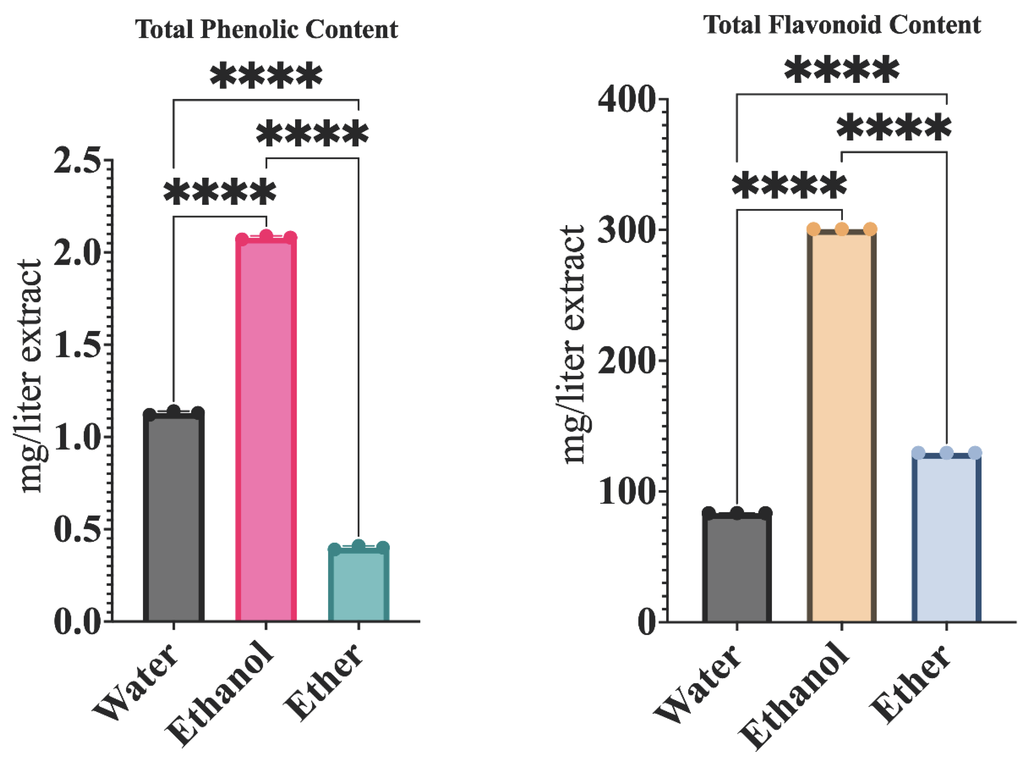
Figure 1. Total phenolic and flavonoid contents of Campsis radicans L. extra plant dissolved with three different solvents.
Preprints 99089 g001
Figure 2. DPPH and % ABTS inhibition capacities in extracts prepared with three different solvents.
Preprints 99089 g002
Table 1. Reference values for the total heavy metal content (mg kg-1) of Campsis radicans L. and the normal and toxic concentrations of trace elements in plants.
Element Campsis radicans L. Heavy Metal Concentrations (mg/kg) Normal Concentrations in Plants (mg/kg) Toxic Concentrations in Plants (mg/kg)
As Not Detected 10-60 <2
Cd Not Detected <0.1-1 10
Co Not Detected 0.05-0.5 30-40
Cr 119.66 ± 0,06 0.1-1 2
Cu 1372.9 ± 4.59 3-15 20
Fe 5742.15 ± 3.24 50-200 >500
Mn 31.92 ± 0.16 15-100 400
Mo 1.92 ± 0.17 0.1-0.2 >0.2
Ni 649.3 ± 1.8 0.1-5 30
Pb 3.51 ± 0.18 1.0-5.0 20
Ti Not Detected 5-50 >50
V 11.66 ± 0.03 0.5-1 >1
Zn 25.62 ± 0.61 15-100 200
Table 2. Fatty acid content of Campsis radicans L.
Systematic name Carbon number PubChem CID
Pentadecanoic Acid C 15:0 13849
Palmitic Acid C 16:0 985
Stearic acid C 18:0 5281
Linoleic acid C 18:2 ω6 5280450
Table 3. Molecular Docking Results of Water Extracts Phenolics.
Protein Ligands Binding Energy (kj/mol) Number of Hydrogen Bonds Bonded Amino Acids
Bax Levoglucosan -3.7 3 ASP 71 - LYS 119
Propanoic acid, 2-hydroxy-, 1-methylethyl ester, (2S)- -3.3 0 PRO 88 - LEU 120 - ALA 124 - LEU 132
4-Methyl-3,6,9-trioxadecan-1-ol -3.4 3 ARG 34 - LYS 119
4-Vinylbenzoic acid -5.2 1 THR 85 - PRO 88 - LEU 120 - LYS 123 - ALA 124 - LEU 132 - ILE 136
Dimethyl malate -3.3 3 ARG 34 - LYS 119
Levoglucosenone -3.8 2 LYS 123 - THR 127
L-Proline, 1-(1-methylethyl)-5-oxo-, methyl ester -3.7 4 LYS 119 - LUE 122 - LYS 123
4-Methoxyphenethyl alcohol -4.3 0 PRO 88 - LYS 123 - ALA 124
Trimethyl citrate -3.8 5 ILE 80 - LYS 119 - LYS 123
Bcl-2 Levoglucosan -4.3 2 ALA 97 - GLY 142
Propanoic acid, 2-hydroxy-, 1-methylethyl ester, (2S)- -3.6 2 ALA 97 - GLY 142
4-Methyl-3,6,9-trioxadecan-1-ol -3.2 1 GLY 142
4-Vinylbenzoic acid -4.5 1 ALA 97 - TRP 141 - VAL 145 - LEU 198
Dimethyl malate -3.9 2 ALA 97 - ARG 104
Levoglucosenone -4.4 1 GLY 142
L-Proline, 1-(1-methylethyl)-5-oxo-, methyl ester -4.3 0 PHE 101 - ARG 104
4-Methoxyphenethyl alcohol -4.7 0 ALA 97 - ASP 100 - PHE 101 - ARG 104 - VAL 145
Trimethyl citrate -4.2 5 PHE 101 - ARG 104 - GLY 142
Caspase-3 Levoglucosan -4.9 4 ARG 207 - PHE 250
Propanoic acid, 2-hydroxy-, 1-methylethyl ester, (2S)- -3.7 2 HIS 121 - TYR 204 - SER 205 - TRP 206 - ARG 207 - PHE 256
4-Methyl-3,6,9-trioxadecan-1-ol -3.8 2 TYR 204 - SER 205 - TRP 206 - ARG 207 - PHE 256
4-Vinylbenzoic acid -4.5 2 CYS 163 - SER 205 - TRP 206 - PHE 256
Dimethyl malate -3.9 2 ARG 207
Levoglucosenone -3.9 2 TRP 214 - PHE 250
L-Proline, 1-(1-methylethyl)-5-oxo-, methyl ester -4.3 2 SER 205 - TRP 206 - ARG 207
4-Methoxyphenethyl alcohol -4.0 1 TRP206 - PHE 250 - PHE 256
Trimethyl citrate -4.3 10 THR 62 - SER 63 - SER 65 - SER. 205 - ARG 207 - SER 209
Disclaimer/Publisher’s Note: The statements, opinions and data contained in all publications are solely those of the individual author(s) and contributor(s) and not of MDPI and/or the editor(s). MDPI and/or the editor(s) disclaim responsibility for any injury to people or property resulting from any ideas, methods, instructions or products referred to in the content.
Copyright: This open access article is published under a Creative Commons CC BY 4.0 license, which permit the free download, distribution, and reuse, provided that the author and preprint are cited in any reuse.
Prerpints.org logo

Preprints.org is a free preprint server supported by MDPI in Basel, Switzerland.

Subscribe

© 2024 MDPI (Basel, Switzerland) unless otherwise stated