Preprint
Article

Phytochemical Composition of 50 Medicinal Plants, Their Antioxidant and Acetylcholinesterase Inhibitory Activities

Altmetrics

Downloads

189

Views

108

Comments

0

Submitted:

21 February 2024

Posted:

21 February 2024

You are already at the latest version

Alerts
Abstract
Pakistan flora is enriched with potent bioactive compounds that must be explored while Alzheimer's disease (AD) poses a global challenge driven by neurochemical imbalances. Acetylcholinesterase (AChE) inhibitors traditionally alleviate symptoms, but addressing the root cause remains elusive. This gap has led to the exploration of herbal sources for interventions. In this context, we analyzed fifty 50 medicinal plants for their phytochemical composition and antioxidant and acetylcholinesterase inhibition activities. AChE activity was measured using Ellman’s method. Santalum album exhibited significant AChE inhibition (86.2%), surpassing galantamine hydrobromide (81.8%). Promising inhibition was also seen in Bombax ceiba (77.8%), Acacia karoo (52.9%), and Phaseolus lunatus (50.1%). Detailed analysis revealed Santalum album's abundance in terpenoids, flavonoids, tannins, and phenolics, potentially correlating with its antioxidant and AChE inhibitory effects. Acacia karoo demonstrated elevated levels of total alkaloids, antioxidants, terpenoids, and tannins. Both Acacia karoo and Santalum album show promise for AD intervention due to their AChE inhibitory activities and unique phytochemical attributes, warranting further exploration, particularly for Acacia karoo's novel potential in AChE inhibition, to advance targeted AD therapeutics utilizing natural resources. This study contributes to the search for effective AD strategies from nature's pharmacological resources.
Keywords: 
Subject: Biology and Life Sciences  -   Food Science and Technology

1. Introduction

Neurochemical abnormalities are leading causes of Alzheimer’s disease (AD), which is the most common form of dementia and is a continuous growing global threat [1,2]. It is an irreversible illness and every fifth person (aged > 80 years) in the world is affected with this. As per World Health Organization (WHO), the prevalence of AD will increase to double, up to 2030 [3], however still the ultimate treatment of this disease is yet needs to be discovered [4]. The reduced cholinergic transmission at the synapse is considered to be the prominent cause related to the incidence and development of AD [3], and so currently, its treatment is dependent on acetylcholinesterase inhibitors (AChE) for short-term efficacy to mitigate the symptoms rather than reversing or curing it [5]. In this connection, several AChE inhibitors are approved by the drug regulatory authorities as a treatment for AD [6,7]. The leading natural inhibitors of acetylcholine (AChE), include galanthamine and rivastigmine, which are being commercially used in AD treatment [3]. Herbs, as a source of bioactive phytochemicals, have long been used for the treatment of various diseases, leading to their massive consumption among a wide population as a cheaper and readily available source [8,9,10]. However, sometimes, these plant-based drugs possess side effects due to the misidentification of plants, malpractices of collection and processing, etc [10]. Similarly, traditional herbal compounds’ cognitive enhancement and neuroprotective effects as Alzheimer’s protective agents are also well-reported [11] as complementary and alternative interventions. Still, there is a considerable gap and limited literature on the plants already being used in the industry for treating various diseases and can be a valuable source for developing drug candidates for AD [12]. This generated the desire to explore further herbal plants to identify potential leads to cure AD. Nature is the best laboratory, and the present drug discovery is based on identifying the active compounds for known drug targets with a super reductionist approach [13]. There is significant variability of AChE among various plants and within different parts of the same plant [14], so selecting a specific plant or any specific part of this plant is a tactful question [15]. In such cases, traditional knowledge is a paved path to identify the potential candidates for drug discovery [12]. In this study, we identified plants from specific families known for their applications in herbal medicine and evaluated them for acetylcholinesterase inhibitory activity, as well as their phytochemical profiles, focusing on plants with significant inhibitory potential.

2. Materials and Methods

2.1. Collection of plants

Fifty (50) plants (Table 1) from 19 families were selected based on their usage in various treatments and potential applications for memory enhancement in traditional practices. MAPICC (Medicinal and Aromatic Plants Introduction and Conservation Center) Azad Jammu and Kashmir, Pakistan identified, collected, and provided the plant materials. All the samples were collected and frozen in liquid nitrogen, transported in closed containers, and dried by using a freeze drier (Alpha 1-2 LD plus, Crist, Germany). Later, the freeze-dried samples were ground to a fine powder with a high-speed (24000rpm) grinder (Biobase, China) and stored in airtight plastic jars at room temperature.

2.2. Sample preparation

Powdered plant material (10gm) was extracted in 100ml of 80% methanol by using ultrasonication (Biobase, China) for 30 minutes at room temperature three times every 8 hours and filtered by Whatman filter No. 1. All the extracts were mixed together and concentrated through the rotary evaporator at 40 °C. The residual water-based part of the extract was completely dried further by using a spin vacuum drier (VC36R) at 30°Cand stored at –20°Cuntil used for any analysis.

2.3. Acetylcholinesterase inhibitory activity

All plant extracts were analyzed with a micro-plate assay based on Ellman’s colorimetric method [16,17] with modifications. In the 96-well plate 60µL of assay buffer KH2PO4 (100mM, pH 7.7), 10 µL of AChE (0.2U/mL in assay buffer) was added with 10µL of plant extract (1mg/ml in MeOH) or standard compound, Galantamine (0.1 mM in MeOH). After 10 minutes of incubation (37 °C), initial absorbance was taken at 405nm in a UV-Vis spectrophotometer. ATCI (5mM in assay buffer) and DTNB (5mM in assay buffer) 10 µL each, were added and the final absorbance was taken again at the same wavelength after 20 minutes of incubation. All samples were performed in triplicates. Percent inhibition was calculated against normal control (10µl methanol in assay buffer in place of the sample).
P e r c e n t   i n h i b i t i o n = 100 F i n a l   a b s I n i t i a l   A b s F i n a l   a b s   o f   n o r m a l   c o n t r o l 100

2.4. Free radical scavenging activity

The spectrophotometric method was used to analyze free radical scavenging activity, followed by [18] with modifications. As a standard, ascorbic acid (1-100µg/ml) in methanol was used. 100µl of DPPH (0.02% w/v) solution in methanol was mixed separately with 100µl of sample solution and standard solution. Solutions were kept in the dark for half an hour, and absorbance was taken at 517nm using a spectrophotometer. As a blank, 100µl of DPPH solution in methanol (0.02% w/v) with 100µl of methanol was used. Percent inhibition of DPPH was calculated by following the formula.
A n t i o x i d a n t   a c t i v i t y ( % ) = A b s   o f   B l a n k A b s   o f   s a m p l e   o r   s t a n d a r d A b s   o f   B l a n k 100

2.5. Total phenolics

Total phenolics were determined using the Folin-Ciocalteu method [23]. In this method, 10µl of plant extract (1 mg/ml) was mixed with 10µl of FC reagent and diluted with 150µl of distilled water. Then, 20µl of 35% sodium carbonate was added, followed by 10µl of distilled water. After shaking and incubating at room temperature for 30 minutes, absorbance was measured at 725nm. Distilled water served as blank and gallic acid was used as the standard in place of the sample. Results were expressed as mean±SD of three replicates in mg/g of extract as gallic acid equivalents (GAE).

2.6. Total flavonoids

Flavonoid content was determined using a modified aluminum chloride method [20]. In a 96-well plate, quercetin (25-200µg/ml) or methanolic extract (0.5mg/ml) was mixed with sodium nitrate, aluminum chloride, and sodium hydroxide. After 15 minutes of incubation at 37°C, absorbance at 415nm was measured against a methanol blank. Flavonoid content was expressed as mg quercetin equivalent (QE) per 100g dry weight.

2.7. Total terpenoids

Terpenoid content was determined with modifications to the method by [21]. Methanolic extract (0.5mg/ml) was mixed with chloroform and sulfuric acid, resulting in a reddish-brown precipitate. After adding methanol and measuring absorbance at 538nm, terpenoid content was calculated using a linalool standard curve (20-120mg/ml). Terpenoid content was expressed as mg linalool equivalents (LE) per gram of sample weight.

2.8. Total alkaloids

Total alkaloids were estimated spectrophotometrically using bromocresol green (BCG), following the modified method of [22]. A BCG solution was prepared by dissolving 34.9mg of BCG in 1.5 ml of NaOH (2N) and 2.5 ml of distilled water, diluted to a final volume of 500 ml. Precise aliquots (0.2, 0.3, 0.4, and 0.5ml) of the boldine solution or plant sample (1mg/ml) in HCl (pH 2.5) were individually transferred to a separating funnel, followed by the addition of 5ml each of Na2HPO4 and BCG solution. After vigorous shaking, the formation of a yellow-colored complex was measured at 470nm against a blank. The results were articulated as mg of Boldine equivalents (BE) per gram of sample weight.

2.9. Total tannins

Total tannins were determined using the method of Ali et al. [23] with adaptations for the 96-well plate technique. 25 µL of sample solution was combined with 150 µL of 4% vanillin solution in a 96-well plate. 25 µL of 32% sulfuric acid was added to the mixture and incubated at 25 °C for 15 minutes. The absorbance was then measured at 500 nm. Measurements were taken three times for each sample, and quantification was performed by creating a standard curve using a catechin solution ranging from 0 to 1000 µg/mL in methanol. The results were quantified as milligrammes of catechin equivalents (CE) per gramme of the samples’ dry weight.

2.10. Statistical analysis

PCA and OPLS-DA were performed with the SIMCA-P software (v. 11.0, Umetrics, Umea, Sweden) based on a unit-variance scaling method [24]. All the values are articulated as mean±Standard deviation of three replicates. Data was analyzed using analysis of variance (ANOVA) at a 5% significance level, followed by a factorial design that was applied to compare the groups using computer software Statistix 8.1 [25].

3. Results and Discussion

3.1. Acetylcholinesterase inhibitory activity of selected medicinal plants

In this study, the focus was on investigating the potential acetylcholinesterase (AChE) inhibitory activity of various plant extracts in comparison to galantamine hydrobromide, a known reference compound with AChE inhibition of 81.79%, as that in the present work (Table 1). The assessment revealed that all 50 selected plant extracts exhibited some degree of AChE inhibitory activity, underscoring their potential significance in the field of neurology and pharmacology. The inhibitory activity of the plant extracts varied considerably, with inhibitions ranging from moderate to high. Among the range of inhibitions observed, the lowest inhibition was recorded for Melilotus indica at 5.7%, while the most notable AChE inhibition was displayed by Santalum album at 86.2%. This remarkable activity level in Santalum album is particularly noteworthy when contrasted with galantamine hydrobromide’s inhibition percentage. Bombax ceiba also revealed substantial AChE inhibitory activity of 77.8%. On the contrary, Phaseolus lunatus and Acacia karoo also exhibited moderate inhibition percentages (50.1% and 52.9%, respectively), surpassing the 50% threshold of minimum target screening level, indicating their potential as sources of AChE inhibitors. Acacia karroo and Phaseolus lunatus are not commonly reported for acetylcholine cholinesterase inhibitory activity in previous studies.
Santalum album, Bombax ceiba, Acacia karroo and Phaseolus lunatus exhibiting above than 50% AChE inhibitory activity were further analyzed for antioxidant and phytochemical analysis.

3.2. Antioxidant potential of selected medicinal plants

A closer examination of the antioxidant activity of the selected plants revealed their potential bioactive constitution. Table 2 provides data on the DPPH (2,2-diphenyl-1-picrylhydrazyl) scavenging activity of selected plant materials. It can be observed from Table 2 that Phaseolus lunatus exhibited the highest DPPH scavenging activity at 90.9%, followed closely by ascorbic acid 91.7%. Acacia karroo and Santalum album also demonstrated significant DPPH scavenging activity at 88.6% and 87.1%, respectively. Bombax ceiba, while lower than the other treatments, still exhibited moderate DPPH scavenging activity at 61.4%. This data suggests that Phaseolus lunatus emerged as on among others, as top rated to possess antioxidant activity by demonstrating a remarkable free radical scavenging rate of 90.9% (Table 2). This anti-oxidative property could be indicative of its potential in combating oxidative stress-related conditions.

3.3. Phytochemical composition of selected medicinal plants

The phenolic compounds possess significant free radical scavenging ability, crucial for combating various chronic diseases caused by free radicals [26]. Santalum album exhibited the highest phenolic content at 71.9mg GAE/g, highlighting its strong antioxidant potential. Conversely, Bombax ceiba showed the lowest phenolic content at 3.9mg GAE/g. Phaseolus lunatus and Acacia karroo displayed moderated phenolic content at 18.7mg GAE/g and 15.9mg GAE/g, respectively (Table 2).
Flavonoids, which are polyphenolic compounds found widely in nature, exhibit antioxidative characteristics due to their aromatic rings with multiple hydroxyl groups [27]. Santalum album demonstrated the highest flavonoid content at 31.1mg QE/g, highlighting its antioxidant potential. However, all three samples, except Santalum album, displayed relatively low flavonoid content, supporting the antioxidant potential of Santalum ablum [27,28]. Bombax ceiba exhibited the lowest flavonoid content at 7.2mg QE/g. The other two plants showed moderate flavonoid content, with values of 16.1mg QE/g and 18.8mg QE/g, respectively (see Table 2).
Terpenoids, a prominent class of secondary metabolites in higher plants, are valuable sources of chemical compounds with therapeutic significance and various industrial applications such as solvents, perfumes, and biofuels [29,30]. The terpenoid content of the selected medicinal plants is presented in Table 2, with Santalum album exhibiting the highest terpenoid content of 20mg LE/g, followed by Acacia karroo at 16.3mg LE/g and Phaseolus lunatus at 11.2mg LE/g. In contrast, Bombax ceiba displayed the lowest terpenoid content at 6mg LE/g. These findings suggest that the selected plants have relatively lower terpenoid contents compared to other phytochemicals.
Alkaloids, a diverse group of nitrogen-containing phytochemical compounds, are found in various organisms including plants, bacteria, fungi, and animals. They exhibit compelling biological activities and are utilized in pharmaceuticals, stimulants, narcotics, and poisons [31]. In this study, alkaloid content ranged from 1.8 to 4.6mg BE/g, with Acacia karroo exhibiting the highest alkaloid content at 4.6mg BE/g and Phaseolus lunatus showing the lowest at 1.8mg BE/g. Bombax ceiba and Santalum album displayed moderate alkaloid content at 2.7mg BE/g and 1.9mg BE/g, respectively, as outlined in Table 2.
Tannins, naturally occurring water-soluble polyphenols with molecular weights between 500 to 3000 g/mol, are known for their antioxidant properties and are commonly found in plant materials. They have diverse industrial applications, including stabilizing colors in the wine industry, balancing complexity in wines, and inhibiting enzymes in infected fruits. Tannins also can precipitate proteins and alkaloids [27]. The total tannin content of selected medicinal plants is presented in Table 2, indicating that Santalum album and Phaseolus lunatus possess higher quantities of total tannins at 34.1 and 24.6mg GAE/g, respectively. Acacia karroo and Bombax ceiba displayed moderate tannin content at 25.1 and 17.9mg GAE/g, respectively.
The data underscores these plant materials’ diverse antioxidant and phytochemical profiles, potentially suggesting their varied therapeutic potentials. The historical and ethnopharmacological context of the selected plants further supports their potential therapeutic applications. Santalum album has been traditionally used for various medicinal purposes, including memory enhancement, antioxidant activity, and anti-inflammatory effects [32,33,34,35]. Phaseolus lunatus, Acacia karoo, and Bombax ceiba have also been reported to possess a spectrum of pharmacological activities ranging from anti-fungal and anti-microbial to anti-diabetic and anti-inflammatory effects [36,37,38,39]. These attributes suggest that Santalum album could hold promise as a multifaceted therapeutic agent, potentially addressing a spectrum of health concerns.

3.4. Chemometric analysis

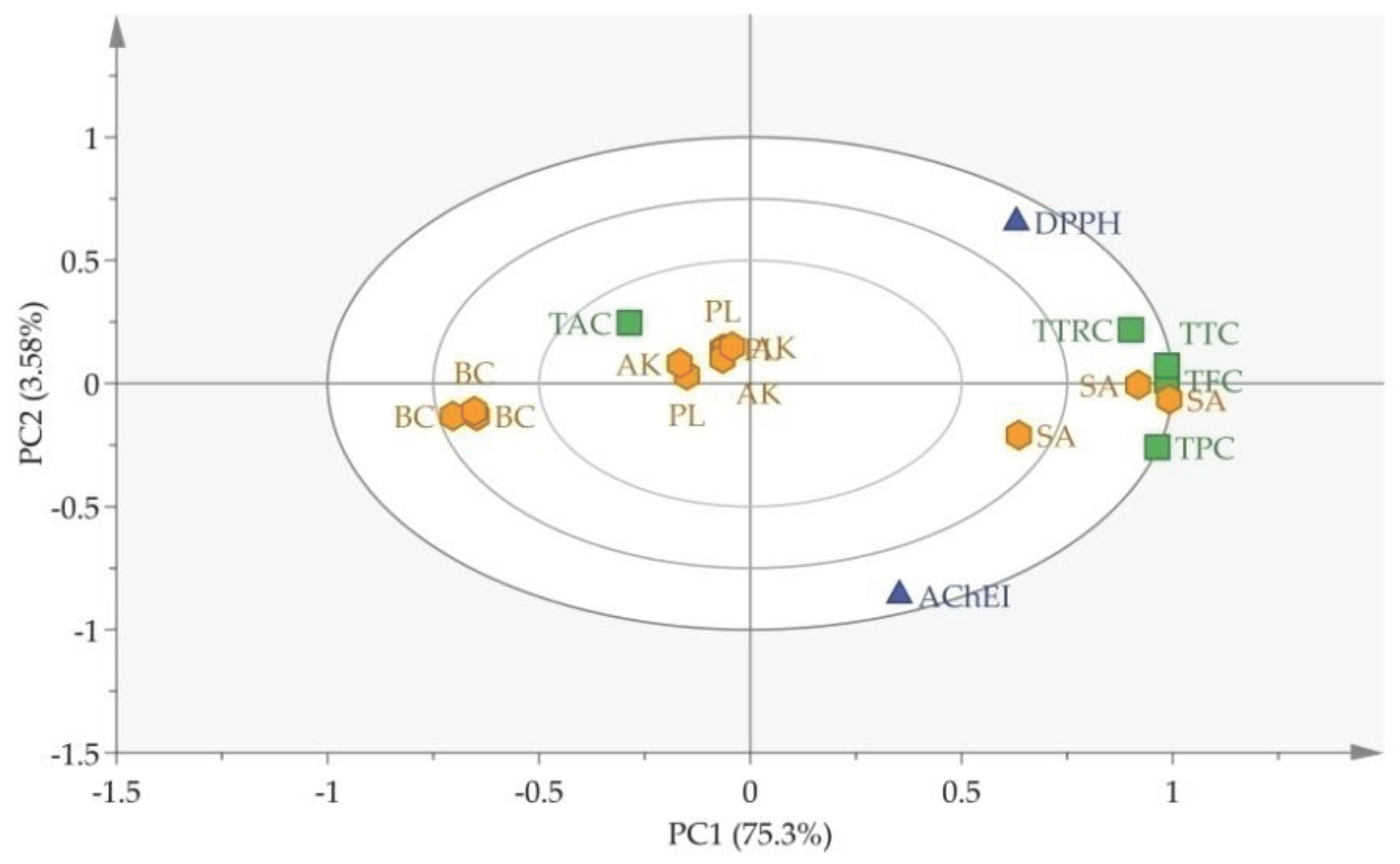
Principal component analysis (PCA) was employed as a statistical tool to elucidate the interplay between plant species and their correlations [40]. The PCA (Figure 1) illustrates that most plants exhibited AChE inhibitory activity below 50%, except for Galantamine hydrobromide (STD), Santalum album (SA), Bombax ceiba (BC), Acacia karoo (AK), and Phaseolus lunatus (PL), which demonstrated inhibitory activity above 50%. Two-dimensional OPLS-biplot (Figure 2) where PC1 explained a variance of 75.3% and PC2 explained a variance of 3.58%. The graphical representation of the PCA revealed distinct clusters of plant species based on their phytochemical constituents, antioxidant potential, and AChE inhibition. Notably, the discrimination of Santalum album in PC1 (Figure 2) from others is due to higher antioxidant potential, total flavonoids, total phenolics, and total terpenoids. Further, AChE also played its role in discriminating Santalum album in PC1 and PC2 (Figure 2). In contrast, Bombax ceiba displayed a positive association with AChE inhibitory activity. Nevertheless, its phytochemical analysis indicated an opposing connection, showcasing a negative relationship with phenolics, flavonoids, tannins, and terpenoids, but revealing a positive link with alkaloids. On a different note, Acacia karoo and Phaseolus lunatus exhibited an adverse correlation with AChE inhibition yet exhibited favorable associations with total alkaloids. The diverse range of inhibitory activities observed, coupled with the distinct phytochemical profiles, highlights the importance of further research to identify and isolate active compounds responsible for the observed effects. The integration of traditional knowledge and modern pharmacological approaches offers a promising avenue for the development of new therapeutic interventions.

4. Conclusions

This study highlights the promising potential of diverse plant extracts as sources of acetylcholinesterase (AChE) inhibitors and underscores the need for innovative approaches to address Alzheimer’s disease (AD) using natural compounds. Evaluating fifty medicinal plants for AChE inhibitory activity revealed promising candidates, with Santalum album standing out for its potent inhibition, surpassing galantamine hydrobromide. Additionally, Bombax cieba, Phaseolus lunatus, and Acacia karoo showed significant AChE inhibition with positive associations with total alkaloids. Furthermore, Santalum album’s rich phytochemical profile, especially its terpenoids, flavonoids, and phenolics, correlates with its antioxidant and AChE inhibitory effects. These findings suggest the potential of Santalum album and Acacia karoo as candidates for AD therapy, urging further research and development in the pursuit of novel treatments.

Funding

This work was funded by the National Research Program for Universities, Higher Education Commission, Pakistan [Project# 3109, 2015). This research was also supported by the Researchers Supporting Project Number (RSP2024R154), King Saud University, Riyadh, Saudi Arabia.

Data Availability Statement

All the data generated and analyzed as part of this study are thoroughly presented and integrated within the contents of the article.

Acknowledgments

Authors are grateful to Prof. Dr. Jamshed Iqbal, Department of Pharmacy, Center of Advanced Drug Research Lab, COMSATS University, Abbottabad campus for protocol standardization. We are also grateful to the Medicinal and Aromatic Plants Introduction and Conservation Center (MAPICC), Azad Jammu and Kashmir, Pakistan for providing and identifying plant materials. The authors also sincerely thank the Researchers Supporting Project Number (RSP2024R154), King Saud University, Riyadh, Saudi Arabia, for providing support.

Conflicts of Interest

The authors declare no conflicts of interest.

References

  1. Bortolami, M.; Rocco, D.; Messore, A.; Di-Santo, R.; Costi, R.; Madia, V.N.; Scipione, L.; Pandolfi, F. Acetylcholinesterase inhibitors for the treatment of Alzheimer’s disease-a patent review (2016-present). Expert Opinion on Therapeutic Patents 2021. [Google Scholar] [CrossRef]
  2. Ali, A.; Cottrell, J.J.; Dunshea, F.R. Identification and characterization of anthocyanins and non-anthocyanin phenolics from Australian native fruits and their antioxidant, antidiabetic, and anti-Alzheimer potential. Food Res. Int. 2022, 162, 111951. [Google Scholar] [CrossRef]
  3. Arya, A.; Chahal, R.; Rao, R.; Rahman, M.H.; Kaushik, D.; Akhtar, M.F.; Saleem, A.; Khalifa, S.M.; El-Seedi, H.R.; Kamel, M.; Albadrani, G.M. Acetylcholinesterase Inhibitory Potential of Various Sesquiterpene Analogues for Alzheimer’s Disease Therapy. Biomolecules. 2021, 11, 350. [Google Scholar] [CrossRef]
  4. Mukherjee, P.K.; Kumar, V.; Mal, M.; Houghton, P.J. Acetylcholinesterase inhibitors from plants. Phytomedicine. 2007, 14, 289–300. [Google Scholar] [CrossRef] [PubMed]
  5. Alzobaidi, N. Quasimi, H.; Emad, N.A.; Alhalmi, A.; Naqvi, M. Bioactive Compounds and Traditional Herbal Medicine: Promising Approaches for the Treatment of Dementia. Degener.Neurol. Neuromuscul Dis. 2021, 11, 1–14. [Google Scholar] [PubMed]
  6. Mukherjee, P.K.; Kumar, V.; Houghton, P.J. Screening of Indian medicinal plants for acetylcholinesterase inhibitory activity. Phytotherapy Research: An International Journal Devoted to Pharmacological and Toxicological Evaluation of Natural Product Derivatives 2007, 21, 1142–1145. [Google Scholar] [CrossRef]
  7. Zhou, Y.; Fu, Y.; Yin, W.; Li, J.; Wang, W.; Bai, F.; Xu, S.; Gong, Q.; Peng, T.; Hong, Y.; Zhang, D. Kinetics-Driven Drug Design Strategy for Next-Generation Acetylcholinesterase Inhibitors to Clinical Candidate. J. Med. Chem. 2021, 64, 1844–1855. [Google Scholar] [CrossRef]
  8. Kiani, H.S.; Ali, B.; Al-Sadoon, M.K.; Al-Otaibi, H.S.; Ali, A. Lc-ms/ms and gc-ms identification of metabolites from the selected herbs and spices, their antioxidant, anti-diabetic potential, and chemometric analysis. Processes 2023, 11, 2721. [Google Scholar] [CrossRef]
  9. Ali, A.; Cottrell, J.J.; Dunshea, F.R. Characterization, antioxidant potential, and pharmacokinetics properties of phenolic compounds from native Australian herbs and fruits. Plants 2023, 12, 993. [Google Scholar] [CrossRef] [PubMed]
  10. Ali, A.; Cottrell, J.J.; Dunshea, F.R. Antioxidant, alpha-glucosidase inhibition activities, in silico molecular docking and pharmacokinetics study of phenolic compounds from native Australian fruits and spices. Antioxidants 2023, 12, 254. [Google Scholar] [CrossRef]
  11. Yizibula, M.; Wusiman, Z.; Abudouhalike, N.; Maimaitiming, B. Cognitive enhancement and neuroprotective effects of a traditional Chinese herbal compound medicine on Aβ1–42 induced Alzheimer’s disease in rats. Folia Neuropathol. 2020, 58, 365–376. [Google Scholar] [CrossRef]
  12. Gregory, J.; Vengalasetti, Y.V.; Bredesen, D.E.; Rao, R.V. Neuroprotective Herbs for the Management of Alzheimer’s Disease. Biomolecules. 2021, 11, 543. [Google Scholar] [CrossRef]
  13. Derakhshan, A.R. Natural Treatments for Fissure in Ano Used by Traditional Persian Scholars, Razi (Rhazes) and Ibn Sina (Avicenna). Evid. Based Complement. Alternat. Med. 2017, 22, 324–333. [Google Scholar] [CrossRef]
  14. Adsersen, A.; Gauguin, B.; Gudiksen, L.; Jäger, A.K. Screening of plants used in Danish folk medicine to treat memory dysfunction for acetylcholinesterase inhibitory activity. J. Ethnopharmacol. 2006, 104, 418–422. [Google Scholar] [CrossRef]
  15. Guzman-Martinez, L.; Calfío, C.; Farias, G.A.; Vilches, C.; Prieto, R.; Maccioni, R.B. New Frontiers in the Prevention, Diagnosis, and Treatment of Alzheimer’s Disease. J. Alzheimer’s Dis.2021, Preprint, 1-13.
  16. Ellman, G.L.; Courtney, K.D.; Andres, V.; Featherstone, R.M. A new and rapid colorimetric determination of acetylcholinesterase activity. Biochem. Pharmacol. 1961, 7, 88–95. [Google Scholar] [CrossRef]
  17. Shah, M.S.; Najam-ul-Haq, M.; Shah, H.S.; Rizvi, S.U.F.; Iqbal, J. Quinoline containing chalcone derivatives as cholinesterase inhibitors and their in silico modeling studies. Comput.Biol.Chem. 2018, 76, 310–317. [Google Scholar] [CrossRef]
  18. Afsar, V.; Reddy, Y.M.; Saritha, K. In vitro antioxidant activity and anti-inflammatory activity of methanolic leaf extract of Boswellia serrata. Int. J. Life Sci. Biotechnol. Pharma. Res. 2012, 1, 15–23. [Google Scholar]
  19. Siddiqui, N.; Rauf, A.; Latif, A.; Mahmood, Z. Spectrophotometric determination of the total phenolic content, spectral and fluorescence study of the herbal Unani drug Gul-e-Zoofa (Nepeta bracteata Benth). J. Taibah Univ. Medical Sci. 2017, 12, 360–363. [Google Scholar] [CrossRef] [PubMed]
  20. Nguyen, N.; Minh, L.V.; Trieu, L.H.; Bui, L.M.; Lam, T.D.; Hieu, V.Q.; Khang, T.V.; Trung, L.N.Y. Evaluation of total polyphenol content, total flavonoid content, and antioxidant activity of Plectranthus amboinicus leaves. in IOP Conference Series: Materials Science and Engineering. 2020. IOP Publishing.
  21. Ghorai, N.; Chakraborty, S.; Gucchait, S.; Saha, S.K.; Biswas, S. Estimation of total Terpenoids concentration in plant tissues using a monoterpene, Linalool as standard reagent. Protoc. Exch. 2012, 5, 1038. [Google Scholar] [CrossRef]
  22. Shamsa, F.; Monsef, H.; Ghamooshi, R.; Verdian-rizi, M. Spectrophotometric determination of total alkaloids in some Iranian medicinal plants. Thai. J. Pharm. Sci. 2008, 32, 17–20. [Google Scholar]
  23. Ali, A.; Wu, H.; Ponnampalam, E.N.; Cottrell, J.J.; Dunshea, F.R.; Suleria, H.A.R. Comprehensive profiling of most widely used spices for their phenolic compounds through LC-ESI-QTOF-MS2 and their antioxidant potential. Antioxidants 2021, 10, 721. [Google Scholar] [CrossRef]
  24. Jahangir, M.; Abdel-Farid, I.B.; Choi, Y.H.; Verpoorte, R. Metal ion-inducing metabolite accumulation in Brassica rapa. J. Plant Physiol. 2008, 165, 1429–1437. [Google Scholar] [CrossRef]
  25. Steel, R.G.D.; Torrie, J.H. Principles and procedures of statistics, a biometrical approach. 1980: McGraw-Hill Kogakusha, Ltd.
  26. Zahid, H.F.; Ali, A.; Ranadheera, C.S.; Fang, Z.; Ajlouni, S. Identification of phenolics profile in freeze-dried apple peel and their bioactivities during in vitro digestion and colonic fermentation. Int. J. Mol. Sci. 2023, 24, 1514. [Google Scholar] [CrossRef]
  27. Mohammed, S.; Manan, F.A. Aalysis of total phenolics, tannins and flavonoids from Moringa oleifera seed extract. J. Chem. Pharm. Res. 2015, 7, 132–135. [Google Scholar]
  28. Asem, N.; Abdul-Gapar, N.A.; Abd-Hapit, N.H.; Omar, E.A. Correlation between total phenolic and flavonoid contents with antioxidant activity of Malaysian stingless bee propolis extract. J. Apic. Res. 2020, 59, 437–442. [Google Scholar] [CrossRef]
  29. Malik, S.K. Qualtitative and quantitative estimation of terpenoid contents in some important plants of Punjab, Pakistan. Pak. J. Sci. 2017, 69. [Google Scholar]
  30. Harman-Ware, A.E.; Sykes, R.; Peter, G.F.; Davis, M. Determination of terpenoid content in pine by organic solvent extraction and fast-GC analysis. Front. Energy Res. 2016, 4, 2. [Google Scholar] [CrossRef]
  31. John, B.I.J.U; Sulaiman, C.T.; George, S.; Reddy, V.R.K. Spectrophotometric estimation of total alkaloids in selected Justicia species. Int. J. Pharm. Sci. 2014, 6, 647–648. [Google Scholar]
  32. Mohammad, A.; Syed, B.; Malay, B.; Kumar, B. Effect of Santalum album Linn on memory enhancing activity on mice. J. Chem. Pharm. Sci. 2010, 3, 172–177. [Google Scholar]
  33. Sindhu, R.K.; Upma, K.A.; Arora, S. Santalum album linn: a review on morphology, phytochemistry and pharmacological aspects. Intl. J. Pharm. Tech. Res. 2010, 2, 914–919. [Google Scholar]
  34. Kumar, D.N.; Alex, S.A.; Kumar, R.S.S.; Chandrasekaran, N.; Mukherjee, A. Acetylcholinesterase inhibition-based ultrasensitive fluorometric detection of malathion using unmodified silver nanoparticles. Colloids Surf. A: Physicochem. Eng. Asp. 2015, 485, 111–117. [Google Scholar] [CrossRef]
  35. Mehvish, S.; Barkat, M.Q. Phytochemical And Antioxidant Screening of Amomum Subulatum, Elettaria Cardamomum, Emblica Officinalis, Rosa Damascene, Santalum album and Valeriana officinalis And Their Effect On Stomach, Liver And Heart. Matrix Sci. Medica. 2018, 2, 28–33. [Google Scholar] [CrossRef]
  36. Saleem, Z.M.; Ahmed, S.; Hasan, M.M. Phaseolus lunatus linn: Botany, medicinal uses, phytochemistry and pharmacology. World J. Pharm. Pharm. Sci. 2016, 5, 87–93. [Google Scholar]
  37. Granito, M.; Brito, Y.; Torres, A. Chemical composition, antioxidant capacity and functionality of raw and processed Phaseolus lunatus. J. Sci. Food Agric. 2007, 87, 2801–2809. [Google Scholar] [CrossRef]
  38. Maroyi, A. Acacia karroo Hayne: Ethnomedicinal uses, phytochemistry and pharmacology of an important medicinal plant in southern Africa. Asian Pac. J. Trop. Med. 2017, 10, 351–360. [Google Scholar] [CrossRef] [PubMed]
  39. Sinha, S.; Kumar, B.; Singh, D.K.; Luqman, S.; Singh, M.; Singh, A. Antioxidant and Choline Esterase Inhibitory Activity of Phenolic Rich Extracts from Bombax ceiba L. Flowers. Free Radic. Antioxid. 2018, 8. [Google Scholar] [CrossRef]
  40. Granato, D.; Santos, J.S.; Escher, G.B.; Ferreira, B.L.; Maggio, R.M. Use of principal component analysis (PCA) and hierarchical cluster analysis (HCA) for multivariate association between bioactive compounds and functional properties in foods: A critical perspective. Trends Food Sci. Technol. 2018, 72, 83–90. [Google Scholar] [CrossRef]
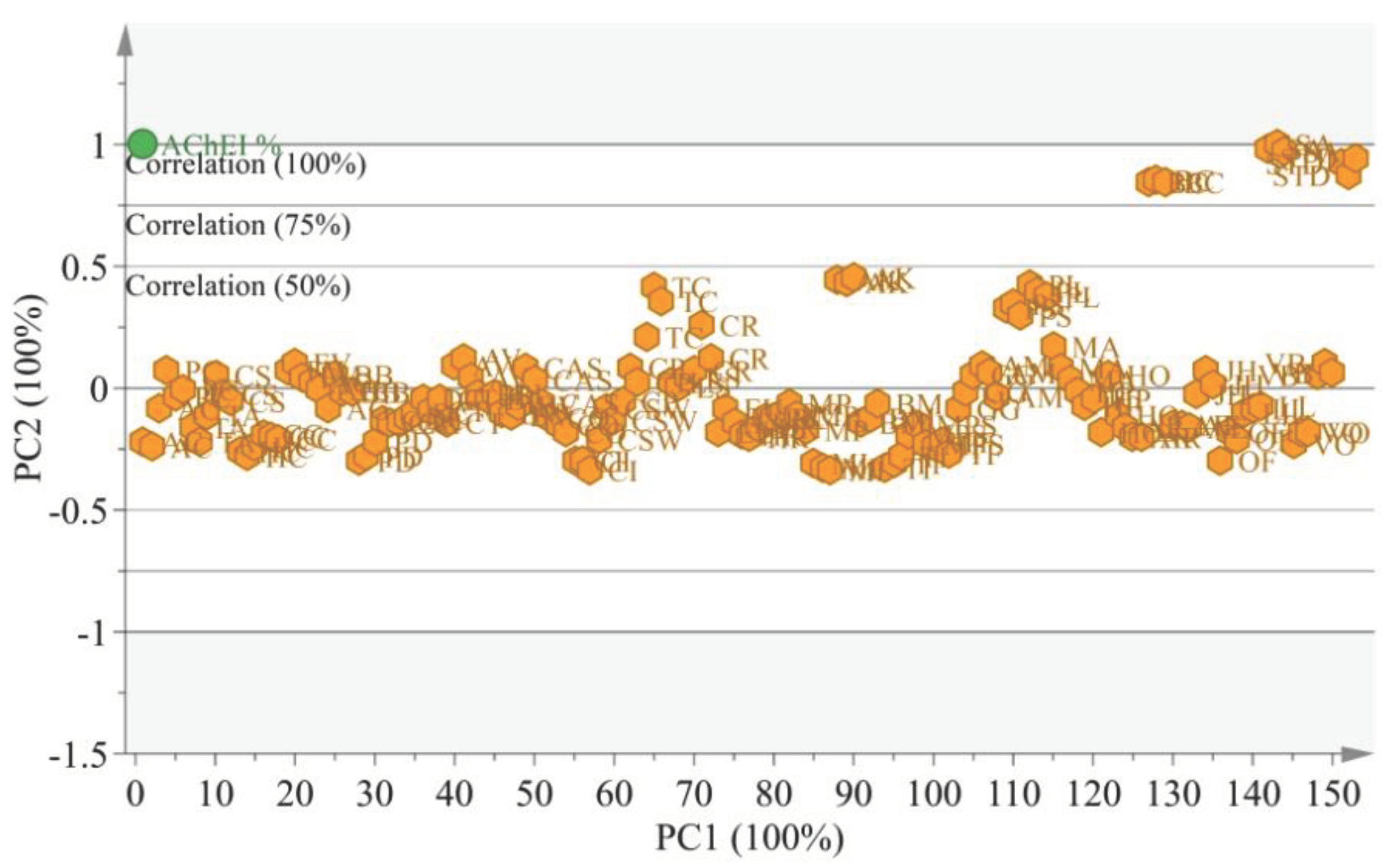
Figure 1. Principal component analysis (PCA) loading plot constructed from AChE inhibitory activity of fifty Pakistani medicinal plants.
Preprints 99510 g001
Figure 2. OPLS biplot for discriminating AChE inhibitory activity, antioxidant, and phytochemical analysis.
Preprints 99510 g002
Table 1. Acetylcholinesterase inhibitory activity (%) of selected plant’s parts against galantamine hydrobromide (0.1mM), as standard.
S. No Code Family Name English Name Scientific Name Part Used AChEI %
1 S87 Acoraceae Sweet Flag, Calamus Acorus calamus RT 14.6±5.3
2 S92 Apiaceae Dill, True dill Anethum graveolens SD 24.4±8.3
3 S3 Apiaceae Black cumin Bunium bulbocastanum SD 26.0±2.5
4 S94 Apiaceae Indian pennywort Centella asiatica WP 17.1±0.4
5 S158 Apiaceae Coriander, Chinese parsley Coriandrum sativum SD 25.1±5.4
6 S20 Apiaceae Cumin Seed Cuminum cyminum SD 13.5±0.8
7 S188 Apiaceae Wild Carrot, Bishop’s lace Daucus carota SD 20.2±7.3
8 S106 Apiaceae Fennel Seed Foeniculum vulgare SD 30.1±1.6
9 S44 Apiaceae Hogweed, Tookar Heracleum candicans TB 9.6±3.1
10 S145 Apiaceae Parsley Petroselinum crispum SD 26.4±8.5
11 S73 Apiaceae Sweet Cumin, Pimpenella Pimpinella diversifolia SD 9.3±5.2
12 S16 Apiaceae Ajowan Trachyspermum ammi SD 15.6±8.3
13 S177 Asclepiadaceae Bitter Cress Caralluma tuberculata WP 20.0±3.1
14 S136 Asphodelaceae Aloe vera Aloe vera LV 31.1±6.3
15 S40 Berberidaceae Tree Turmeric, Indian Lycium Berberis lyceum FR 23.7±0.7
16 S154 Berberidaceae Barberry Berberis vulgaris FR 19.8±1.3
17 S162 Caesalpiniaceae Jasmeejaz Cassia absus SD 26.8±8.3
18 S45 Caesalpiniaceae Cloves Cassia occidentalis WP 16.1±1.8
19 S43 Cannabinaceae Canna lilly, Marijuana, Arrowroot Cannabis sativa WP 16.9±4.1
20 S169 Cannaceae Canna lilly, Arrowroot Canna indica WP 6.7±1.3
21 S6 Caricaceae Papaya Carica papaya PU 26.7±4.5
22 S79 Combretaceae French lavender Lavandula stoechas FW 27.2±1.1
23 S26 Combretaceae Black myrobalan, Chebulic myrobalan Terminalia chebula SD 45.7±6.6
24 S197 Cyperaceae Nut-grass Cyperus rotundus RZ 34.9±6.1
25 S52 Elaeagnaceae Autumn olive, Spreading oleaster Elaeagnus umbellate FR 17.2±7.9
26 S23 Elaeagnaceae Sea buckthorns Hippophae rhamnoides SD 14.3±1.3
27 S172 Euphorbiaceae Kamala tree Mallotus philippensis FR 18.5±13.3
28 S103 Euphorbiaceae Castor oil plant Ricinus cummunis FR 18.4±0.7
29 S216 Fabaceae Acacia Tree Bark Acacia karoo ST 52.9±0.6
30 S39 Fabaceae Bear’s breeches Atylosia mollis SD 28.0±4.2
31 S196 Fabaceae Sacred Tree Butea monosperma FW 19.0±2.7
32 S48 Fabaceae Liquorice Glycyrrhiza glabra RT 24.9±4.2
33 S173 Fabaceae White sweet clover Melilotus albus WP 31.4±4.4
34 S59 Fabaceae Blue sweet clover Melilotus indica WP 5.7±0.7
35 S64 Fabaceae Velvet Beans, Cowhage, Cow-itch Mucuna pruriens SD 13.9±2.2
36 S156 Fabaceae Lima Seed, Sieva bean Phaseolus lunatus SD 50.1±1.5
37 S85 Fabaceae Red Sandalwood Pterocarpus santalinus BR 45.7±1.5
38 S127 Fabaceae Tamarind Tamarindus indica FR 7.1±4.8
39 S69 Fabaceae Fenugreek Trigonella foenum-graecum SD 10.7±1.3
40 S160 Hyperiaceae St. John’s wort Hypericum oblongifolium LV 20.7±7.5
41 S54 Hyperiaceae Perforate St John’s-wort Hypericum perforatum WP 23.1±1.8
42 S191 Malvaceae Lady’s finger, Okra Abelmoschus esculentus SD 16.3±0.3
43 S34 Malvaceae Hollyhocks Alcea rosea FW 14.1±1.5
44 S142 Malvaceae Silk-cotton tree Bombax ceiba RT 77.8±0.3
45 S185 Oleaceae Yellow jasmine Jasminum humile LV 26.9±2.9
46 S27 Oleaceae Broad leaf privet, Glossy privet Ligustrum lucidum LV 20.1±0.8
47 S60 Oleaceae Indian olive, Wild olive Olea ferruginea SD 11.5±3.5
48 S77 Santalaceae White sandalwood, Sandal peel Santalum album BR 86.2±0.8
49 S82 Violaceae Mountain violet, Showy violet Viola betonicifolia WP 30.2±1.4
50 S83 Violaceae Sweet violet, Viola Viola odorata WP 13.7±1.8
Galatamine hydrobromide (0.1mM) 81.8±2.09
Values are articulated as mean ± Standard deviation of three replications. LV, RT, WP, SD, BU, BR, FR, PU and RZ indicate Leaf, Root, Whole plant, Seed, Bud, Bark, Fruit, Pulp and Rhizome, respectively.
Table 2. Antioxidant and phytochemical potential of selected medicinal plants.
Sample DPPH (%) TFC (mg QE/g) TPC (mg GAE/g) TTRC (mg LE/g) TAC (mg BE/g) TTC (mg GAE/g)
PL 90.9±0.2a 16.1±1.6b 18.7±1.6b 11.2±1.2c 1.8±0.1b 24.6±0.6b
AK 88.6±1.1bc 16.84±2.8b 15.9±3.2b 16.3±1.0b 4.7±2.0a 25.1±2.5b
BC 61.4±2.1d 7.18±0.2c 3.9±0.2c 6.0±0.5d 2.7±0.3b 17.9±0.6c
SA 87.1±0.2c 31.12±2.1a 71.94±2.4a 19.9±2.5a 1.9±0.5b 34.1±2.0a
AA 91.7±0.7a
Values are articulated as mean ± Standard deviation of three replicates. Whereas a, b, c, and d show significant (p ≤ 0.05) variations among groups. Phaseolus lunatus (PL), Acacia karroo (AK), Bombax ceiba (BC), Santalum album (SA) and AA Ascorbic acid. Total terpenoid content (TTC), Total tannin-related content (TTRT).
Disclaimer/Publisher’s Note: The statements, opinions and data contained in all publications are solely those of the individual author(s) and contributor(s) and not of MDPI and/or the editor(s). MDPI and/or the editor(s) disclaim responsibility for any injury to people or property resulting from any ideas, methods, instructions or products referred to in the content.
Copyright: This open access article is published under a Creative Commons CC BY 4.0 license, which permit the free download, distribution, and reuse, provided that the author and preprint are cited in any reuse.
Prerpints.org logo

Preprints.org is a free preprint server supported by MDPI in Basel, Switzerland.

Subscribe

© 2024 MDPI (Basel, Switzerland) unless otherwise stated