Submitted:
01 March 2024
Posted:
04 March 2024
You are already at the latest version
Abstract
Keywords:
1. Introduction
2. Materials and Methods
2.1. Study Design and Patients
2.2. Inclusion and Exclusion Criteria
2.3. Surgical Procedures
2.4. Statistics
3. Results
3.1. Clinical Data
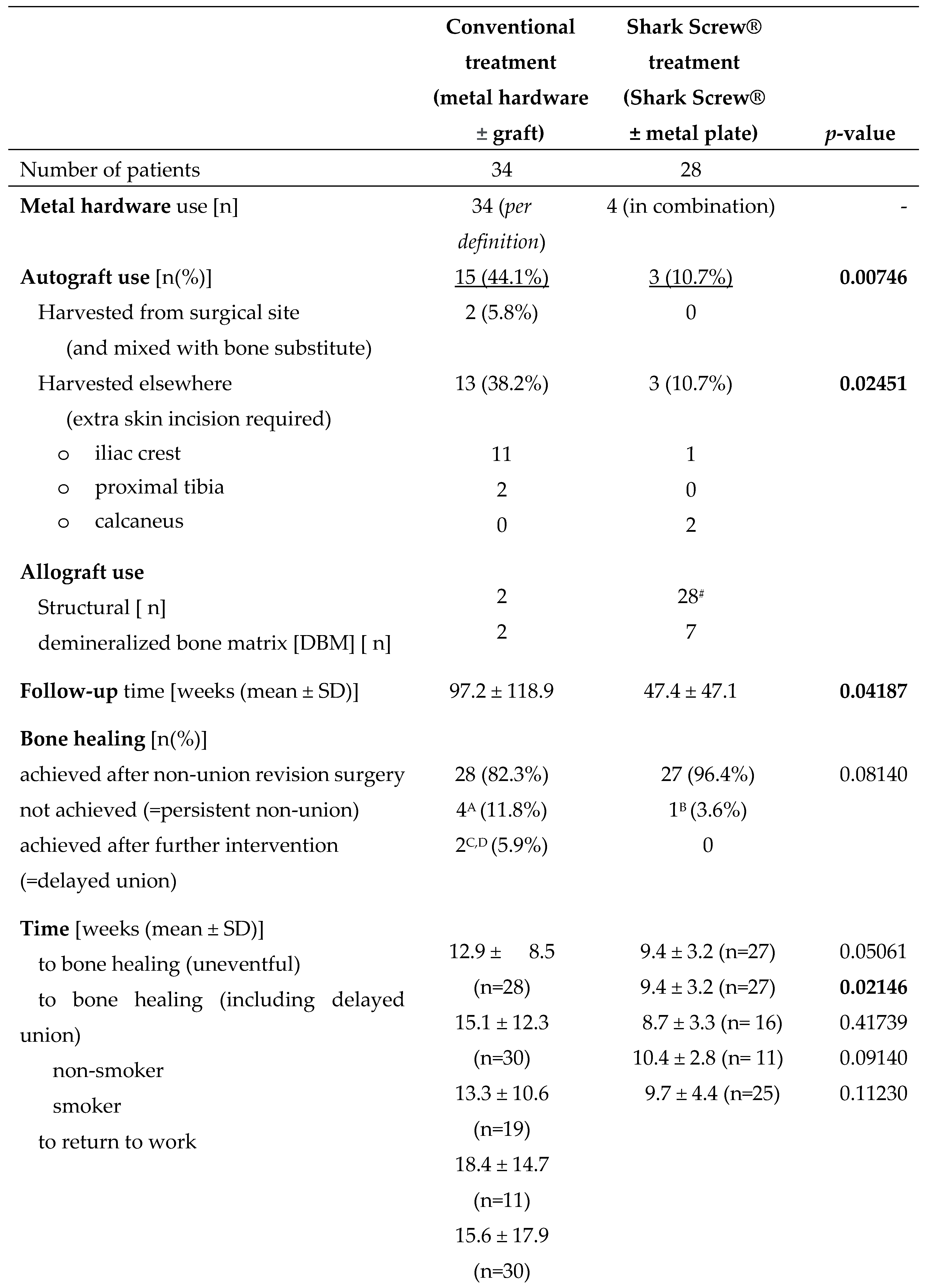
3.2. Bone Healing Rate (Union Rate)
3.3. Time to Bone Healing (Time to Union) and Return to Work
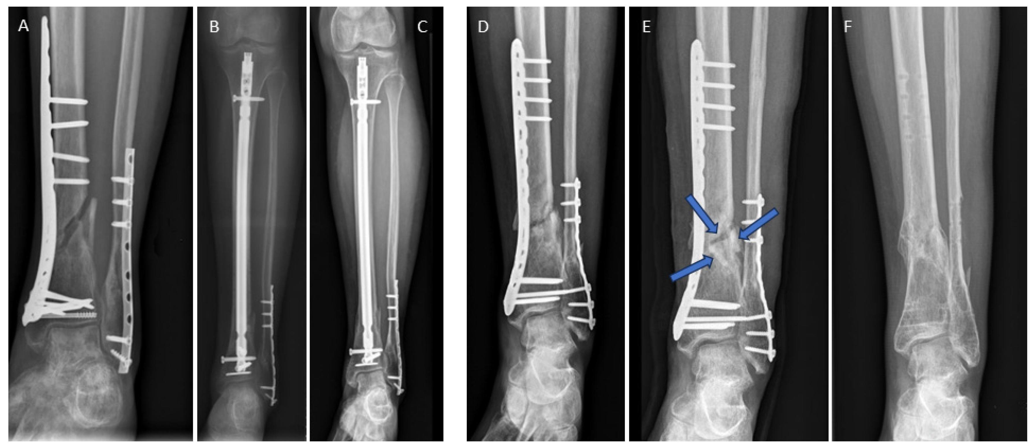
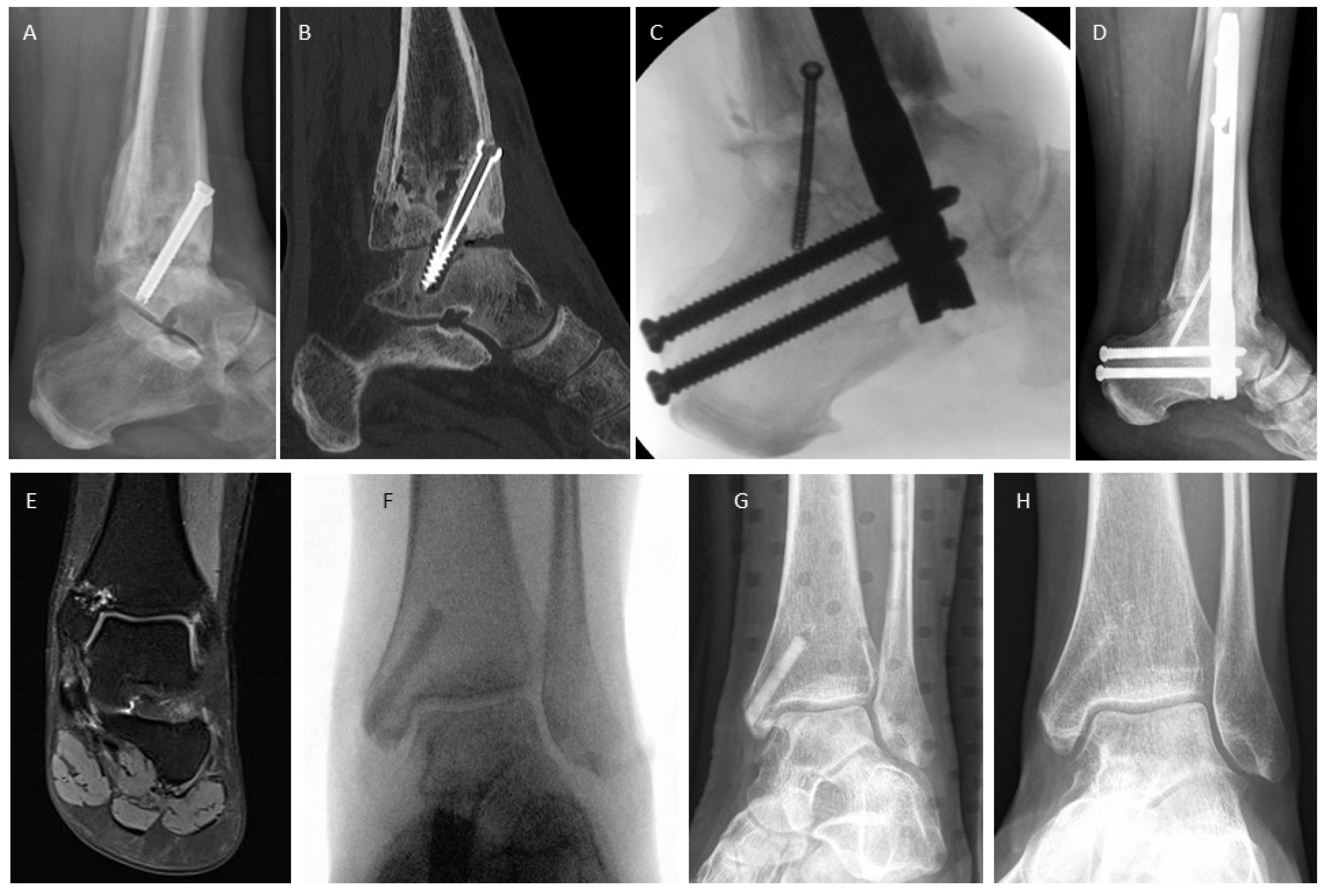
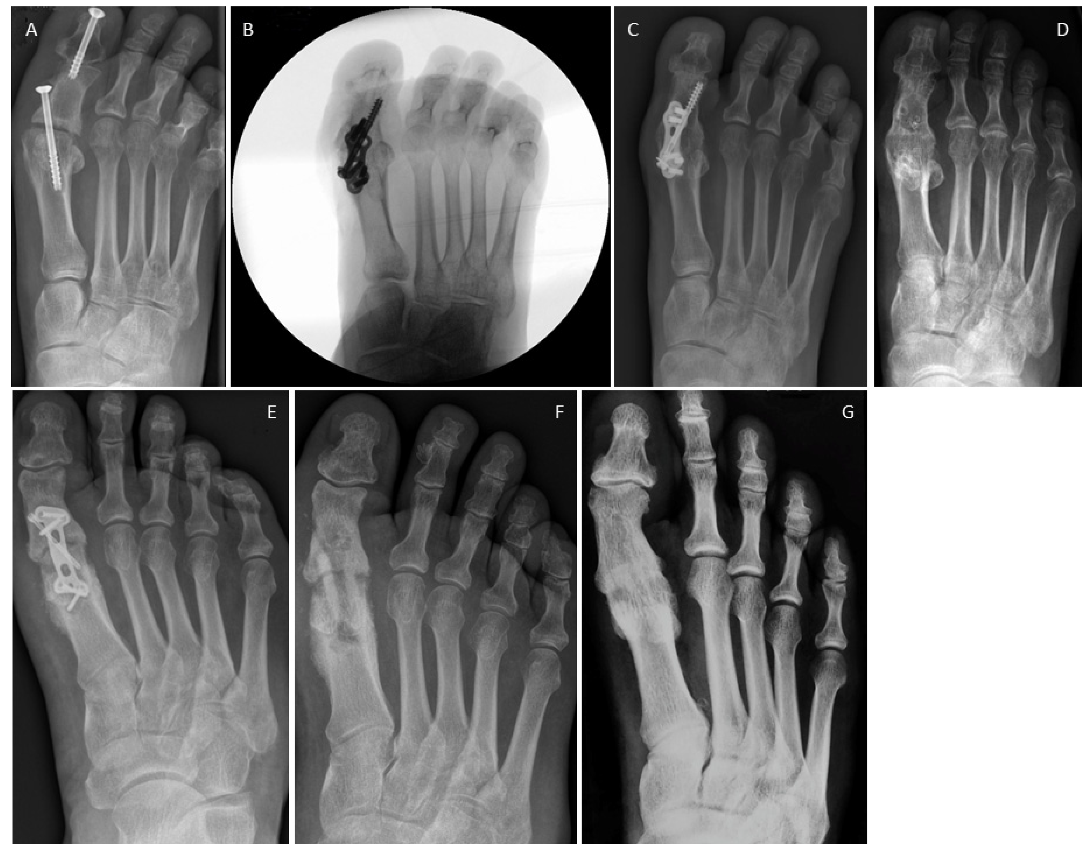
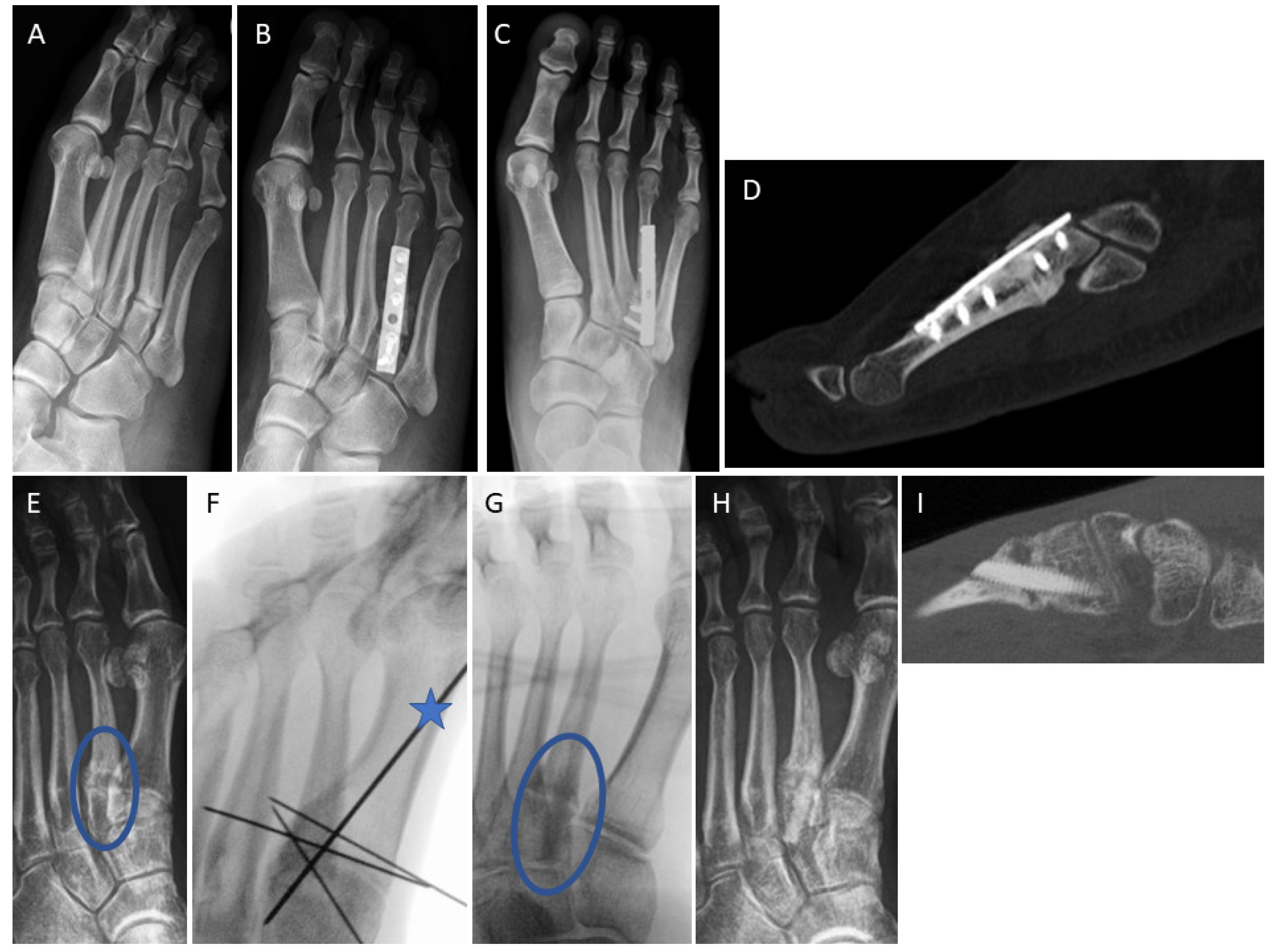
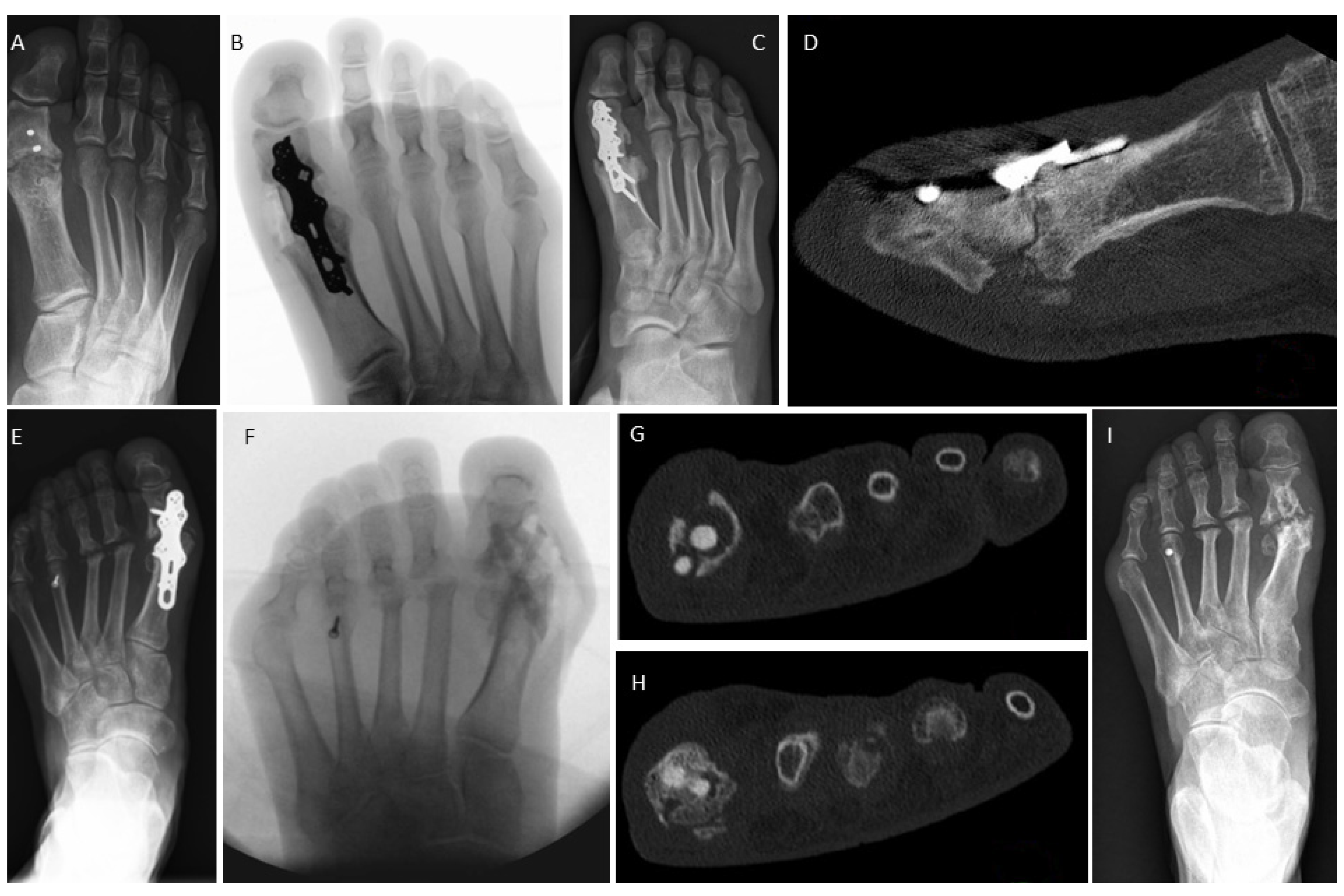
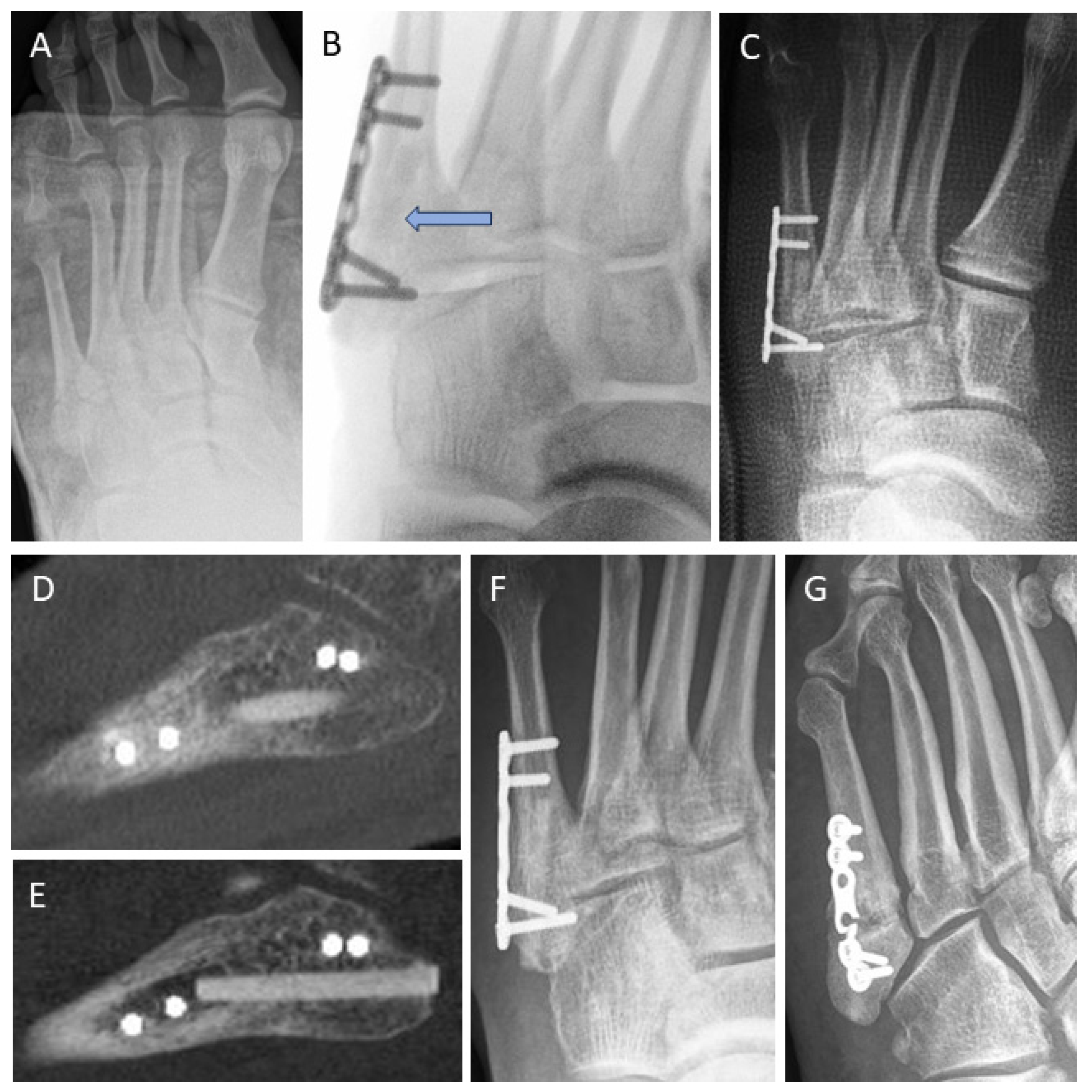
3.4. Examples for Bone Healing
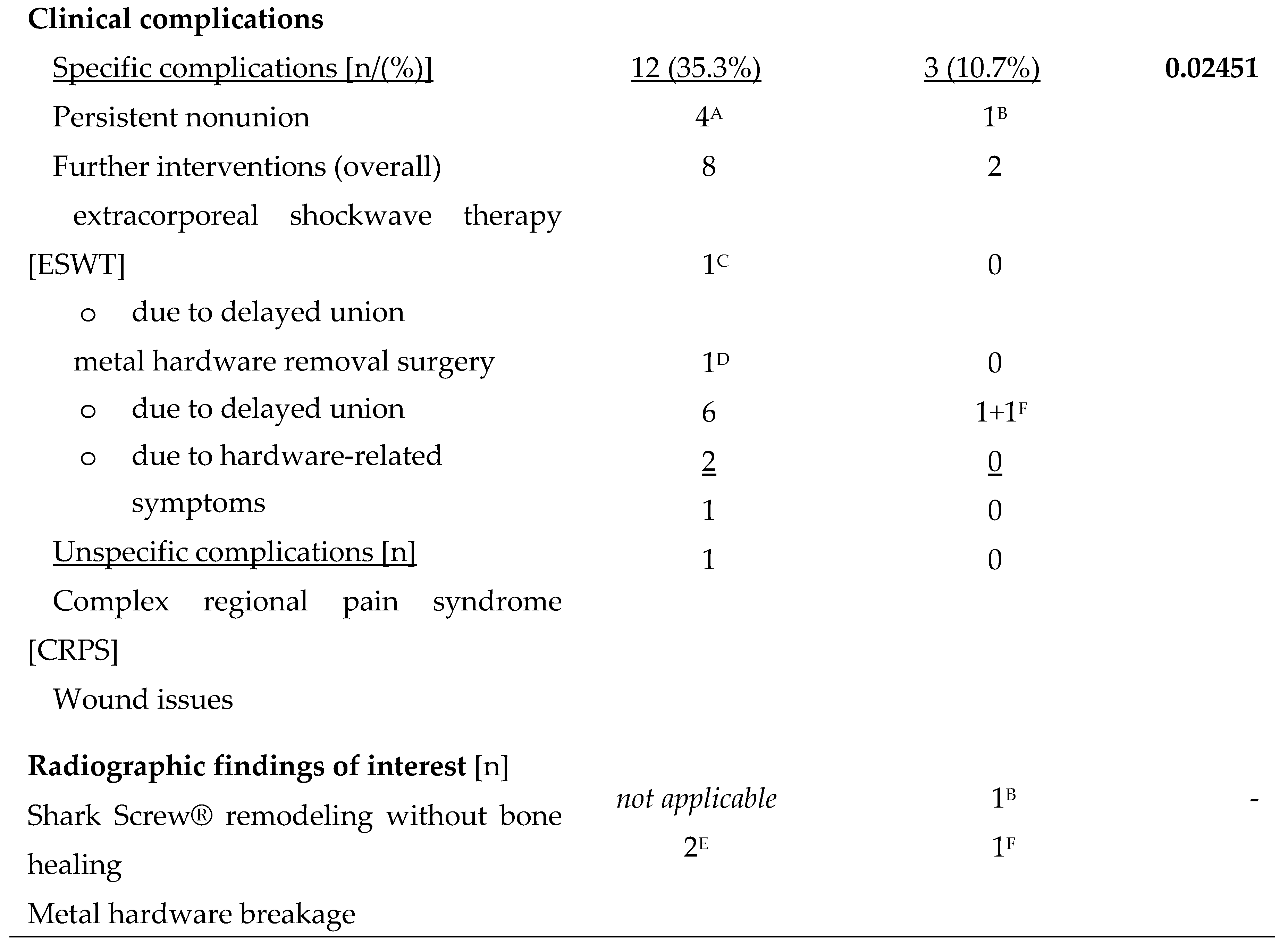
3.5. Clinical Complications
3.6. Radiological Findings of Interest
4. Discussion
5. Conclusion
6. Patents
Supplementary Materials
Author Contributions
Funding
Institutional Review Board Statement
Informed Consent Statement
Data Availability Statement
Conflicts of Interest
References
- Elliott, D.S.; Newman, K.J.; Forward, D.P.; Hahn, D.M.; Ollivere, B.; Kojima, K.; Handley, R.; Rossiter, N.D.; Wixted, J.J.; Smith, R.M.; Moran, C.G. A unified theory of bone healing and nonunion: BHN theory. Bone Joint J 2016, 98-b, 884–891. [Google Scholar] [CrossRef]
- Perren, S.M. Evolution of the internal fixation of long bone fractures. The scientific basis of biological internal fixation: choosing a new balance between stability and biology. The Journal of bone and joint surgery. British volume 2002, 84, 1093–1110. [Google Scholar] [CrossRef]
- Krause, F.; Younger, A.S.; Baumhauer, J.F.; Daniels, T.R.; Glazebrook, M.; Evangelista, P.T.; Pinzur, M.S.; Thevendran, G.; Donahue, R.M.; DiGiovanni, C.W. Clinical Outcomes of Nonunions of Hindfoot and Ankle Fusions. J Bone Joint Surg Am 2016, 98, 2006–2016. [Google Scholar] [CrossRef]
- Schmal, H.; Brix, M.; Bue, M.; Ekman, A.; Ferreira, N.; Gottlieb, H.; Kold, S.; Taylor, A.; Toft Tengberg, P.; Ban, I. Nonunion - consensus from the 4th annual meeting of the Danish Orthopaedic Trauma Society. EFORT Open Rev 2020, 5, 46–57. [Google Scholar] [CrossRef]
- Smolle, M.A.; Leitner, L.; Böhler, N.; Seibert, F.J.; Glehr, M.; Leithner, A. Fracture, nonunion and postoperative infection risk in the smoking orthopaedic patient: a systematic review and meta-analysis. EFORT Open Rev 2021, 6, 1006–1019. [Google Scholar] [CrossRef] [PubMed]
- Hamilton, G.A.; Mullins, S.; Schuberth, J.M.; Rush, S.M.; Ford, L. Revision lapidus arthrodesis: rate of union in 17 cases. J Foot Ankle Surg 2007, 46, 447–450. [Google Scholar] [CrossRef] [PubMed]
- O'Connor, K.M.; Johnson, J.E.; McCormick, J.J.; Klein, S.E. Clinical and Operative Factors Related to Successful Revision Arthrodesis in the Foot and Ankle. Foot Ankle Int 2016, 37, 809–815. [Google Scholar] [CrossRef] [PubMed]
- Krannitz, K.W.; Fong, H.W.; Fallat, L.M.; Kish, J. The effect of cigarette smoking on radiographic bone healing after elective foot surgery. J Foot Ankle Surg 2009, 48, 525–527. [Google Scholar] [CrossRef] [PubMed]
- Zura, R.; Xiong, Z.; Einhorn, T.; Watson, J.T.; Ostrum, R.F.; Prayson, M.J.; Della Rocca, G.J.; Mehta, S.; McKinley, T.; Wang, Z.; Steen, R.G. Epidemiology of Fracture Nonunion in 18 Human Bones. JAMA Surg 2016, 151, e162775. [Google Scholar] [CrossRef] [PubMed]
- Walsh, W.R.; Cotton, N.J.; Stephens, P.; Brunelle, J.E.; Langdown, A.; Auld, J.; Vizesi, F.; Bruce, W. Comparison of poly-L-lactide and polylactide carbonate interference screws in an ovine anterior cruciate ligament reconstruction model. Arthroscopy 2007, 23, 757–765. [Google Scholar] [CrossRef] [PubMed]
- Kreder, H.J. Tibial nonunion is worse than having a myocardial infarction: Commentary on an article by Mark R. Brinker, MD, et al.: "The devastating effects of tibial nonunion on health-related quality of life". J Bone Joint Surg Am 2013, 95, e1991. [Google Scholar] [CrossRef]
- Khurana, S.; Karia, R.; Egol, K.A. Operative treatment of nonunion following distal fibula and medial malleolar ankle fractures. Foot Ankle Int 2013, 34, 365–371. [Google Scholar] [CrossRef]
- Giannoudis, P.V.; Einhorn, T.A.; Marsh, D. Fracture healing: the diamond concept. Injury 2007, 38 Suppl 4, S3–6. [Google Scholar] [CrossRef]
- Giza, E.; Sarcon, A.K.; Kreulen, C. Tibiotalar nonunion corrected by hindfoot arthrodesis. Foot Ankle Spec 2010, 3, 76–79. [Google Scholar] [CrossRef] [PubMed]
- Kloen, P.; Wiggers, J.K.; Buijze, G.A. Treatment of diaphyseal non-unions of the ulna and radius. Arch Orthop Trauma Surg 2010, 130, 1439–1445. [Google Scholar] [CrossRef]
- Tonk, G.; Yadav, P.K.; Agarwal, S.; Jamoh, K. Donor site morbidity in autologous bone grafting – A comparison between different techniques of anterior iliac crest bone harvesting: A prospective study. Journal of Orthopaedics, Trauma and Rehabilitation 2022, 29, 22104917221092163. [Google Scholar] [CrossRef]
- Zermatten, P.; Wettstein, M. Iliac wing fracture following graft harvesting from the anterior iliac crest: literature review based on a case report. Orthopaedics & traumatology, surgery & research : OTSR 2012, 98, 114–117. [Google Scholar] [CrossRef]
- Baumhauer, J.F.; Glazebrook, M.; Younger, A.; Quiton, J.D.; Fitch, D.A.; Daniels, T.R.; DiGiovanni, C.W. Long-term Autograft Harvest Site Pain After Ankle and Hindfoot Arthrodesis. Foot Ankle Int 2020, 41, 911–915. [Google Scholar] [CrossRef]
- Ahlmann, E.; Patzakis, M.; Roidis, N.; Shepherd, L.; Holtom, P. Comparison of anterior and posterior iliac crest bone grafts in terms of harvest-site morbidity and functional outcomes. J Bone Joint Surg Am 2002, 84, 716–720. [Google Scholar] [CrossRef]
- Müller, S.A.; Barg, A.; Vavken, P.; Valderrabano, V.; Müller, A.M. Autograft versus sterilized allograft for lateral calcaneal lengthening osteotomies: Comparison of 50 patients. Medicine 2016, 95, e4343. [Google Scholar] [CrossRef]
- Slevin, O.; Ayeni, O.R.; Hinterwimmer, S.; Tischer, T.; Feucht, M.J.; Hirschmann, M.T. The role of bone void fillers in medial opening wedge high tibial osteotomy: a systematic review. Knee Surg Sports Traumatol Arthrosc 2016, 24, 3584–3598. [Google Scholar] [CrossRef]
- Suchomel, P.; Barsa, P.; Buchvald, P.; Svobodnik, A.; Vanickova, E. Autologous versus allogenic bone grafts in instrumented anterior cervical discectomy and fusion: a prospective study with respect to bone union pattern. Eur Spine J 2004, 13, 510–515. [Google Scholar] [CrossRef] [PubMed]
- Faldini, C.; Traina, F.; Perna, F.; Borghi, R.; Nanni, M.; Chehrassan, M. Surgical treatment of aseptic forearm nonunion with plate and opposite bone graft strut. Autograft or allograft? International orthopaedics 2015, 39, 1343–1349. [Google Scholar] [CrossRef] [PubMed]
- Lareau, C.R.; Deren, M.E.; Fantry, A.; Donahue, R.M.; DiGiovanni, C.W. Does autogenous bone graft work? A logistic regression analysis of data from 159 papers in the foot and ankle literature. Foot Ankle Surg 2015, 21, 150–159. [Google Scholar] [CrossRef]
- Reeh, F.M.; Sachse, S.; Wedekind, L.; Hofmann, G.O.; Lenz, M. Nonunions and Their Operative Treatment—a DRG-Based Epidemiological Analysis for the Years 2007-2019 in Germany. Dtsch Arztebl Int 2022, 119, 869–875. [Google Scholar] [CrossRef]
- Kothari, A.; Monk, P.; Handley, R. Percutaneous Strain Reduction Screws-A Safe and Simple Surgical Option for Problems With Bony Union. A Technical Trick. J Orthop Trauma 2019, 33, e151–e157. [Google Scholar] [CrossRef]
- Grambart, S.T.; Reese, E.R. Trephine Procedure for Revision Tarsometatarsal Arthrodesis Nonunion. Clin Podiatr Med Surg 2020, 37, 447–461. [Google Scholar] [CrossRef] [PubMed]
- Takács, I.M.; Swierstra, B.A. Pseudoarthrosis repair after failed metatarsophalangeal 1 arthrodesis. Acta Orthop 2011, 82, 114–115. [Google Scholar] [CrossRef] [PubMed]
- Habbu, R.A.; Marsh, R.S.; Anderson, J.G.; Bohay, D.R. Closed intramedullary screw fixation for nonunion of fifth metatarsal Jones fracture. Foot Ankle Int 2011, 32, 603–608. [Google Scholar] [CrossRef]
- Lifka, S.; Baumgartner, W. A Novel Screw Drive for Allogenic Headless Position Screws for Use in Osteosynthesis-A Finite-Element Analysis. Bioengineering (Basel) 2021, 8. [Google Scholar] [CrossRef]
- Huber, T.; Hofstätter, S.G.; Fiala, R.; Hartenbach, F.; Breuer, R.; Rath, B. The Application of an Allogenic Bone Screw for Stabilization of a Modified Chevron Osteotomy: A Prospective Analysis. Journal of Clinical Medicine 2022, 11, 1384. [Google Scholar] [CrossRef]
- Pastl, K.; Schimetta, W. The application of an allogeneic bone screw for osteosynthesis in hand and foot surgery: a case series. Arch Orthop Trauma Surg 2021. [Google Scholar] [CrossRef] [PubMed]
- Sailer, S.; Lechner, S.; Floßmann, A.; Wanzel, M.; Habeler, K.; Krasny, C.; Borchert, G.H. Treatment of scaphoid fractures and pseudarthroses with the human allogeneic cortical bone screw. A multicentric retrospective study. J Orthop Traumatol 2023, 24, 6. [Google Scholar] [CrossRef] [PubMed]
- Amann, P.; Bock, P. Clinical and Radiological Results after Use of A Human Bone Graft (Shark Screw®) in TMT II/+II Arthrodesis. Foot Ankle Orthop 2022, 7, 2473011421s2473000077. [Google Scholar] [CrossRef]
- Hanslik-Schnabel, B.; Flöry, D.; Borchert, G.H.; Schanda, J.E. Clinical and Radiologic Outcome of First Metatarsophalangeal Joint Arthrodesis Using a Human Allogeneic Cortical Bone Screw. Foot & Ankle Orthopaedics 2022, 7, 24730114221112944. [Google Scholar] [CrossRef]
- Pastl, K.; Pastl, E.; Flöry, D.; Borchert, G.H.; Chraim, M. Arthrodesis and Defect Bridging of the Upper Ankle Joint with Allograft Bone Chips and Allograft Cortical Bone Screws (Shark Screw(®)) after Removal of the Salto-Prosthesis in a Multimorbidity Patient: A Case Report. Life (Basel) 2022, 12. [Google Scholar] [CrossRef]
- Brcic, I.; Pastl, K.; Plank, H.; Igrec, J.; Schanda, J.E.; Pastl, E.; Werner, M. Incorporation of an Allogenic Cortical Bone Graft Following Arthrodesis of the First Metatarsophalangeal Joint in a Patient with Hallux Rigidus. Life (Basel) 2021, 11. [Google Scholar] [CrossRef] [PubMed]
- Butscheidt, S.; Moritz, M.; Gehrke, T.; Püschel, K.; Amling, M.; Hahn, M.; Rolvien, T. Incorporation and Remodeling of Structural Allografts in Acetabular Reconstruction: Multiscale, Micro-Morphological Analysis of 13 Pelvic Explants. J Bone Joint Surg Am 2018, 100, 1406–1415. [Google Scholar] [CrossRef]
- Hope, M.; Savva, N.; Whitehouse, S.; Elliot, R.; Saxby, T.S. Is it necessary to re-fuse a non-union of a Hallux metatarsophalangeal joint arthrodesis? Foot Ankle Int 2010, 31, 662–669. [Google Scholar] [CrossRef]
- Walter, N.; Hierl, K.; Brochhausen, C.; Alt, V.; Rupp, M. The epidemiology and direct healthcare costs of aseptic nonunions in Germany - a descriptive report. Bone Joint Res 2022, 11, 541–547. [Google Scholar] [CrossRef]
- Kanakaris, N.K.; Giannoudis, P.V. The health economics of the treatment of long-bone non-unions. Injury 2007, 38 Suppl 2, S77–84. [Google Scholar] [CrossRef]
- Antonova, E.; Le, T.K.; Burge, R.; Mershon, J. Tibia shaft fractures: costly burden of nonunions. BMC Musculoskelet Disord 2013, 14, 42. [Google Scholar] [CrossRef]
- Hak, D.J.; Fitzpatrick, D.; Bishop, J.A.; Marsh, J.L.; Tilp, S.; Schnettler, R.; Simpson, H.; Alt, V. Delayed union and nonunions: epidemiology, clinical issues, and financial aspects. Injury 2014, 45 Suppl 2, S3–7. [Google Scholar] [CrossRef]
- Vaughn, J.; Gotha, H.; Cohen, E.; Fantry, A.J.; Feller, R.J.; Van Meter, J.; Hayda, R.; Born, C.T. Nail Dynamization for Delayed Union and Nonunion in Femur and Tibia Fractures. Orthopedics 2016, 39, e1117–e1123. [Google Scholar] [CrossRef]
- Webb, L.X.; Winquist, R.A.; Hansen, S.T. Intramedullary nailing and reaming for delayed union or nonunion of the femoral shaft. A report of 105 consecutive cases. Clin Orthop Relat Res 1986, 133–141. [Google Scholar]
- Sandberg, O.H.; Aspenberg, P. Inter-trabecular bone formation: a specific mechanism for healing of cancellous bone. Acta Orthop 2016, 87, 459–465. [Google Scholar] [CrossRef]
- Walter, E.; Schalle, K.; Voit, M. Cost-Effectiveness of a bone transplant fixation „SHARK SCREW“ compared to metal devices in osteosynthesis in Austria. Value in Health 2016, 19. [Google Scholar] [CrossRef]
- Jentzsch, T.; Renner, N.; Niehaus, R.; Farei-Campagna, J.; Deggeller, M.; Scheurer, F.; Palmer, K.; Wirth, S.H. Radiological and Clinical Outcome After Reversed L-Shaped Osteotomy: A Large Retrospective Swiss Cohort Study. J Foot Ankle Surg 2019, 58, 86–92. [Google Scholar] [CrossRef] [PubMed]
- Bence, M.; Kothari, A.; Riddick, A.; Eardley, W.; Handley, R.; Trompeter, A. Percutaneous Strain Reduction Screws Are a Reproducible Minimally Invasive Method to Treat Long Bone Nonunion. J Orthop Trauma 2022, 36, e343–e348. [Google Scholar] [CrossRef] [PubMed]
- Levine, S.E.; Myerson, M.S.; Lucas, P.; Schon, L.C. Salvage of pseudoarthrosis after tibiotalar arthrodesis. Foot Ankle Int 1997, 18, 580–585. [Google Scholar] [CrossRef] [PubMed]
- Easley, M.E.; Montijo, H.E.; Wilson, J.B.; Fitch, R.D.; Nunley, J.A. , 2nd. Revision tibiotalar arthrodesis. J Bone Joint Surg Am 2008, 90, 1212–1223. [Google Scholar] [CrossRef]
- Anderson, J.G.; Coetzee, J.C.; Hansen, S.T. Revision ankle fusion using internal compression arthrodesis with screw fixation. Foot Ankle Int 1997, 18, 300–309. [Google Scholar] [CrossRef]
- Dekker, T.J.; White, P.; Adams, S.B. Efficacy of a Cellular Allogeneic Bone Graft in Foot and Ankle Arthrodesis Procedures. Foot Ankle Clin 2016, 21, 855–861. [Google Scholar] [CrossRef]
- Egol, K.A.; Bechtel, C.; Spitzer, A.B.; Rybak, L.; Walsh, M.; Davidovitch, R. Treatment of long bone nonunions: factors affecting healing. Bull NYU Hosp Jt Dis 2012, 70, 224–231. [Google Scholar]
- Chuckpaiwong, B.; Reingrittha, P.; Harnroongroj, T.; Mawhinney, C.; Tharmviboonsri, T. Sport and Exercise Activity After Isolated Ankle Arthrodesis for Advanced-Stage Ankle Osteoarthritis: A Single-Center Retrospective Analysis. Foot Ankle Orthop 2023, 8, 24730114231177310. [Google Scholar] [CrossRef]
- Alcelik, I.; Fenton, C.; Hannant, G.; Abdelrahim, M.; Jowett, C.; Budgen, A.; Stanley, J. A systematic review and meta-analysis of the treatment of acute lisfranc injuries: Open reduction and internal fixation versus primary arthrodesis. Foot Ankle Surg 2020, 26, 299–307. [Google Scholar] [CrossRef]
- Tay, W.H.; de Steiger, R.; Richardson, M.; Gruen, R.; Balogh, Z.J. Health outcomes of delayed union and nonunion of femoral and tibial shaft fractures. Injury 2014, 45, 1653–1658. [Google Scholar] [CrossRef]
- Masterson, S.; Laubscher, M.; Maqungo, S.; Ferreira, N.; Held, M.; Harrison, W.J.; Graham, S.M. Return to Work Following Intramedullary Nailing of Lower-Limb Long-Bone Fractures in South Africa. JBJS 2023, 105, 518–526. [Google Scholar] [CrossRef] [PubMed]
- Prat, D.; Haghverdian, B.A.; Pridgen, E.; Lee, W.; Wapner, K.L.; Chao, W.; Farber, D.C. Revision First Metatarsophalangeal Joint Fusion for Non-Union, Implant Failures, and Failed Hallux Valgus Correction: Does the Indication Matter? Foot & Ankle Orthopaedics 2022, 7, 2473011421S2473000403. [Google Scholar] [CrossRef]
- van den Heuvel, S.B.M.; Doorgakant, A.; Birnie, M.F.N.; Blundell, C.M.; Schepers, T. Open Ankle Arthrodesis: a Systematic Review of Approaches and Fixation Methods. Foot Ankle Surg 2021, 27, 339–347. [Google Scholar] [CrossRef] [PubMed]
- Sangeorzan, B.J.; Ledoux, W.R.; Shofer, J.B.; Davitt, J.; Anderson, J.G.; Bohay, D.; Coetzee, J.C.; Maskill, J.; Brage, M.; Norvell, D.C. Comparing 4-Year Changes in Patient-Reported Outcomes Following Ankle Arthroplasty and Arthrodesis. J Bone Joint Surg Am 2021, 103, 869–878. [Google Scholar] [CrossRef] [PubMed]
- Norvell, D.C.; Ledoux, W.R.; Shofer, J.B.; Hansen, S.T.; Davitt, J.; Anderson, J.G.; Bohay, D.; Coetzee, J.C.; Maskill, J.; Brage, M.; et al. Effectiveness and Safety of Ankle Arthrodesis Versus Arthroplasty: A Prospective Multicenter Study. J Bone Joint Surg Am 2019, 101, 1485–1494. [Google Scholar] [CrossRef] [PubMed]
- Daniels, T.R.; Younger, A.S.; Penner, M.; Wing, K.; Dryden, P.J.; Wong, H.; Glazebrook, M. Intermediate-term results of total ankle replacement and ankle arthrodesis: a COFAS multicenter study. J Bone Joint Surg Am 2014, 96, 135–142. [Google Scholar] [CrossRef] [PubMed]
- SooHoo, N.F.; Zingmond, D.S.; Ko, C.Y. Comparison of reoperation rates following ankle arthrodesis and total ankle arthroplasty. J Bone Joint Surg Am 2007, 89, 2143–2149. [Google Scholar] [CrossRef] [PubMed]
- Wennergren, D.; Bergdahl, C.; Selse, A.; Ekelund, J.; Sundfeldt, M.; Möller, M. Treatment and re-operation rates in one thousand and three hundred tibial fractures from the Swedish Fracture Register. Eur J Orthop Surg Traumatol 2021, 31, 143–154. [Google Scholar] [CrossRef] [PubMed]
- Prat, D.; Haghverdian, B.A.; Pridgen, E.M.; Lee, W.; Wapner, K.L.; Chao, W.; Farber, D.C. High complication rates following revision first metatarsophalangeal joint arthrodesis: a retrospective analysis of 79 cases. Arch Orthop Trauma Surg 2022. [Google Scholar] [CrossRef]
- Eurostat. 18.4% of EU population smoked daily in 2019. Available online: https://ec.europa.eu/eurostat/web/products-eurostat-news/-/edn-20211112-1 (accessed on 20/11/2023).
- Dailey, H.L.; Wu, K.A.; Wu, P.S.; McQueen, M.M.; Court-Brown, C.M. Tibial Fracture Nonunion and Time to Healing After Reamed Intramedullary Nailing: Risk Factors Based on a Single-Center Review of 1003 Patients. J Orthop Trauma 2018, 32, e263–e269. [Google Scholar] [CrossRef]
- Mills, L.A.; Simpson, A.H. The relative incidence of fracture non-union in the Scottish population (5.17 million): a 5-year epidemiological study. BMJ Open 2013, 3. [Google Scholar] [CrossRef]
- Best, M.J.; Buller, L.T.; Miranda, A. National Trends in Foot and Ankle Arthrodesis: 17-Year Analysis of the National Survey of Ambulatory Surgery and National Hospital Discharge Survey. J Foot Ankle Surg 2015, 54, 1037–1041. [Google Scholar] [CrossRef]
- Milstrey, A.; Domnick, C.; Garcia, P.; Raschke, M.J.; Evers, J.; Ochman, S. Trends in arthrodeses and total joint replacements in Foot and Ankle surgery in Germany during the past decade-Back to the fusion? Foot Ankle Surg 2021, 27, 301–304. [Google Scholar] [CrossRef]
- Eurostat. New indicator on annual average salaries in the EU. Available online: https://ec.europa.eu/eurostat/web/products-eurostat-news/w/ddn-20221219-3 (accessed on 16/11/2023).
- Gagne, O.J.; Veljkovic, A.N.; Glazebrook, M.; Penner, M.; Wing, K.; Younger, A.S.E. Agonizing and Expensive: A Review of Institutional Costs of Ankle Fusion Nonunions. Orthopedics 2020, 43, e219–e224. [Google Scholar] [CrossRef]
- Vanderkarr, M.F.; Ruppenkamp, J.W.; Vanderkarr, M.; Holy, C.E.; Blauth, M. Risk factors and healthcare costs associated with long bone fracture non-union: a retrospective US claims database analysis. J Orthop Surg Res 2023, 18, 745. [Google Scholar] [CrossRef] [PubMed]
- Partio, N.; Huttunen, T.T.; Mäenpää, H.M.; Mattila, V.M. Reduced incidence and economic cost of hardware removal after ankle fracture surgery: a 20-year nationwide registry study. Acta Orthop 2020, 91, 331–335. [Google Scholar] [CrossRef] [PubMed]
- Fenelon, C.; Murphy, E.P.; Galbraith, J.G.; Kearns, S.R. The burden of hardware removal in ankle fractures: How common is it, why do we do it and what is the cost? A ten-year review. Foot Ankle Surg 2019, 25, 546–549. [Google Scholar] [CrossRef] [PubMed]
- Sugi, M.T.; Ortega, B.; Shepherd, L.; Zalavras, C. Syndesmotic Screw Removal in a Clinic Setting Is Safe and Cost-effective. Foot Ankle Spec 2020, 13, 144–151. [Google Scholar] [CrossRef]
- Lalli, T.A.; Matthews, L.J.; Hanselman, A.E.; Hubbard, D.F.; Bramer, M.A.; Santrock, R.D. Economic impact of syndesmosis hardware removal. Foot (Edinb) 2015, 25, 131–133. [Google Scholar] [CrossRef]

| Conventional treatment (metal hardware ± graft) |
Shark Screw® treatment (Shark Screw® ± metal plate) | p-value | |
|---|---|---|---|
| Number of patients | 34 | 28 | |
| Age [years] | 50.2 ± 15.4 | 49.8 ± 19.2 | 0.99993 |
| BMI [kg/m²] | 29.1 ± 6.2 | 25.4 ± 4.0 | 0.01780 |
| Gender [male/female] | 18/16 | 11/17 | 0.28353 |
| Smoker [yes/no] | 12/22 | 11/17 | 0.74610 |
| Comorbidities [yes/no] | 19/15 | 7/21 | 0.02017 |
| Non-union revision following: Elective surgery (arthrodesis, osteotomy) Surgical fracture treatment Conservative fracture treatment |
17 8 9 |
13 3 12 |
0.26197 |
| Anatomic location of non-union | specific | Conventional treatment (metal hardware ± graft) | Shark Screw® treatment (Shark Screw® ± metal plate) | |
|---|---|---|---|---|
| Number of patients | 34 | 28 | ||
| Forefoot | 1st metatarsophalangeal joint | 6 | 8 | |
| 1st metatarsal bone | 1 | 1 | ||
| 2nd to 5th metatarsal bone | subcapital | 3 | 1 | |
| 2nd to 5th metatarsal bone | base | 6 | 8 | |
| Midfoot | 1st tarsometatarsal joint | 2 | 1 | |
| Tarsometatarsal joints | Lisfranc | 3 | 1 | |
| Naviculocuneiform joint | 1 | 1 | ||
| Navicular bone | - | 1 | ||
| Talonavicular joint | 4 | - | ||
| Calcaneocuboid joint | 1 | - | ||
| Ankle | Medial malleolus | - | 1 | |
| Lateral malleolus | 3 | - | ||
| Lateral and medial malleolus | 1 | 1 | ||
| Talocrural joint | 1 | - | ||
| Lower leg | Tibia | distal | 1 | - |
| Tibia | shaft | 1 | 3 | |
| Tibia | proximal | - | 1 | |
Disclaimer/Publisher’s Note: The statements, opinions and data contained in all publications are solely those of the individual author(s) and contributor(s) and not of MDPI and/or the editor(s). MDPI and/or the editor(s) disclaim responsibility for any injury to people or property resulting from any ideas, methods, instructions or products referred to in the content. |
© 2024 by the authors. Licensee MDPI, Basel, Switzerland. This article is an open access article distributed under the terms and conditions of the Creative Commons Attribution (CC BY) license (http://creativecommons.org/licenses/by/4.0/).


