Preprint
Article

Novel Tetracyclic Azaphenothiazines With The Quinoline Ring As New Anticancer And Antibacterial Derivatives Of Chlorpromazine

Altmetrics

Downloads

88

Views

33

Comments

0

A peer-reviewed article of this preprint also exists.

Submitted:

10 March 2024

Posted:

12 March 2024

You are already at the latest version

Alerts
Abstract
Phenothiazine derivatives are widely studied in various fields such as biology, chemistry, and medicine research because of their pharmaceutical effects. The first reagent that was used suc-cessfully in the treatment of psychosis was a phenthiazine derivative, chlorpromazine. Apart from their activity in neurons, chlorpromazine have also been reported to display anticancer and antibacterial properties. In these studies, we present the synthesis and research on the activ-ity against A549, MDA, MiaPaCa, PC3 and HCT116 cancer cell lines and against S. aureus, S. epi-dermidis, E. coli and P. aeruginosa bacterial strains a series of new tetracyclic chlorpromazine ana-logues containing a quinoline scafford in their structure instead of the benzene ring and various substituents at the thiazine nitrogen. The structure of these novel molecules has been deter-mined by 1H NMR, 13C NMR and HRMS spectral techniques. The seven most active of the twen-ty-four new chlorpromazine analogues tested were selected for study of the mechanism of cyto-toxic action. Their ability to induce apoptosis or necrosis in cancer cells was assessed by flow cytometry analysis. The results obtained confirmed the proapoptotic activity of selected com-pounds, especially in terms of inducing late apoptosis or necrosis in cancer cell lines A549, Mi-aPaCa-2 and HCT-116. Furthermore, studies on the induction of cell cycle arrest suggest that the new chlorpromazine analogues exert antiproliferative effects by inducing cell cycle arrest in the S phase and, consequently, apoptosis.
Keywords: 
Subject: Chemistry and Materials Science  -   Medicinal Chemistry

1. Introduction

Heterocyclic compounds containing sulfur and nitrogen atoms occupy a special place in medicinal chemistry due to their wide range of biological activities. This group includes aromatic tricyclic compounds in which two benzene rings are linked by sulfur and nitrogen atoms – phenothiazines. These compounds were initially used in the dye and pigment industry and as insecticides, and quickly became the parent molecule of a multitude of drugs that have varied use throughout medical and veterinary practice. The 10H-dibenzo-1,4-thiazine itself shows to possess insecticidal, antifungal, antibacterial and anthelmintic properties [1].
One of the first important uses of phenothiazines, in addition to their use in the dye industry, was their toxic effect on mosquito larvae and as anthelmintics (effective against swine roundworm) and antimalarials, however, they were not widely used for these purposes [2].
The history of these compounds goes back to the second half of the 19th century and chlorpromazine, initially used as an anesthetic, which belongs to this group, revolutionized the treatment of psychiatric disorders. It was originally synthesized by scientists at Rhone–Poulenc with the hopes that it would be an effective antimalarial. Chlorpromazine has been one of the most widely used antipsychotic medications for the treatment of schizophrenia and other psychiatric disorders. Although chlorpromazine is a first-generation antipsychotic medication, it is still widely used in psychiatry. Chlorpromazine demonstrates a high affinity for dopamine (D1–D4) receptors and acts as a receptor antagonist by inhibiting adenylatecyclase activity. Chlorpromazine also inhibits other receptors, including receptors for 5-HT, H1 histamine, α1 and α2 adrenaline, and M1 and M2 muscarinic acetylcholine receptors. N-Methyl-D-aspartate (NMDA) receptor inhibitory effects have also been described at high concentrations of chlorpromazine [3,4,5,6,7,8].
Several studies have reported on the potential anticancer activity of dopamine receptor antagonists. Among the drugs belonging to the neuroleptic phenothiazine, not only chlorpromazine, but also perphenazine, prochlorperazine, fluphenazine, and thioridazine have been tested for anticancer activity [9,10,11].
From the very beginning of the use of chlorpromazine in psychiatry, its impact on the course of cancer diseases has been observed. Among other things, studies conducted in Denmark in the second half of the twentieth century suggested a reduced risk of cancer in chlorpromazine-treated psychiatric patients. Significant inhibition of tumor growth has also been reported in a patient with laryngeal squamous cell carcinoma after direct injection of chlorpromazine into the tumor. Chlorpromazine was also shown to inhibit the growth of sarcoma tumors in mice. In line with the drug repurposing strategy, chlorpromazine was tested for its potential antitumorigenic effects among others, against colorectal, breast, brain, lung, skin, pancreatic, and oral cancers and also against leukemia, lymphoma, sarcoma, mastocytoma and glioblastoma [12,13,14,15,16,17,18,19,20,21,22,23,24,25,26,27,28].
The good results obtained in studies on cancer cell lines coupled with the animal studies drew attention to chlorpromazine as a potential antitumor medication, leading to follow-up studies focused on chlorpromazine’s anticancer mechanisms. Published research results have found that chlorpromazine inhibits cancer growth through multiple independent pathways, through various targets, ranging from histone deacetylases to ion channels. The overlap of molecular pathways between schizophrenia and cancer has been suspected for many years [29,30,31,32,33].
Therefore, in parallel with multidirectional research on neuroleptic phenothiazines, syntheses and research on the activity of new phenothiazine derivatives are carried out. New syntheses are realized in many ways, new substituents are introduced to the thiazine nitrogen atom and/or to the carbons of benzene rings. It is also increasingly common to replace one or both benzene rings with monocyclic or bicyclic azine systems (pyridine, pyrimidine, pyridazine, pyrazine, or quinoline) [34,35,36,37,38]. Among the many activities determined for new phenothiazine analogues, such as antibacterial, antifungal, anti-tubercular, antiviral, anti-inflammatory, anti-filarial, antimalarial, anti-parasitic, and multidrug resistance reversal, the anticancer properties are of particular interest [34,35,36,37,38,39].
Phenothiazines containing one or two quinoline moieties instead of benzene rings are quinobenzothiazines and diquinothiazines. Selected from substituted quinobenzothiazines I and II and diquinothiazines III-VIII (Figure 1) show significant anticancer activity against dozens of cancer cells derived from leukemia, melanoma, non-small cell lung, colon, CNS, ovary, prostate, breast and skin cancers [34,37,40,41]. These compounds also show promising antioxidant effects on rat liver microsomal membranes to protect non-enzymatic lipid peroxidation, inhibitory effects on mitogen-induced proliferation of human peripheral blood mononuclear cells, production of tumor necrosis factor alpha (TNFα) in human whole blood cultures, against butyrylcholinesterase, and free radical scavenging, antiglycation and alpha-glucosidase and alpha-amylase inhibition [34,37,42]. They exerted a suppressive effect in in vivo models: delayed-type hypersensitivity to ovalbumin and cutaneous reaction to carrageenan, contact sensitivity to oxazolone, and experimental psoriasis in mice and showed inhibitory effects on the expression of IFNβ and IFNβ-dependent further genes and proteins involved in the pathogenesis of autoimmune diseases [34,37]. Some of the phenothiazines modified with the quinoline ring have also shown therapeutic effects in mouse experimental colitis [43], and prolongation of skin graft survival in mice [44].
In these studies, we developed an efficient synthesis of 6H-8-chloroquinobenzothiazine as a substrate to obtain a series of new chlorpromazine analogues, which have a quinoline scafford in their structure instead of the benzene ring and various substituents at the thiazine nitrogen atom. The starting point for planning such a modification were the significant and promising in vitro and in vivo activities of previously obtained 9-chloroquinobenzothiazine derivatives and the great importance of chlorpromazine as a leading structure in medicinal chemistry.
Figure 2. Chlorpromazine structure modification scheme.
Preprints 101039 g002

2. Results and discussion

2.1. Synthesis

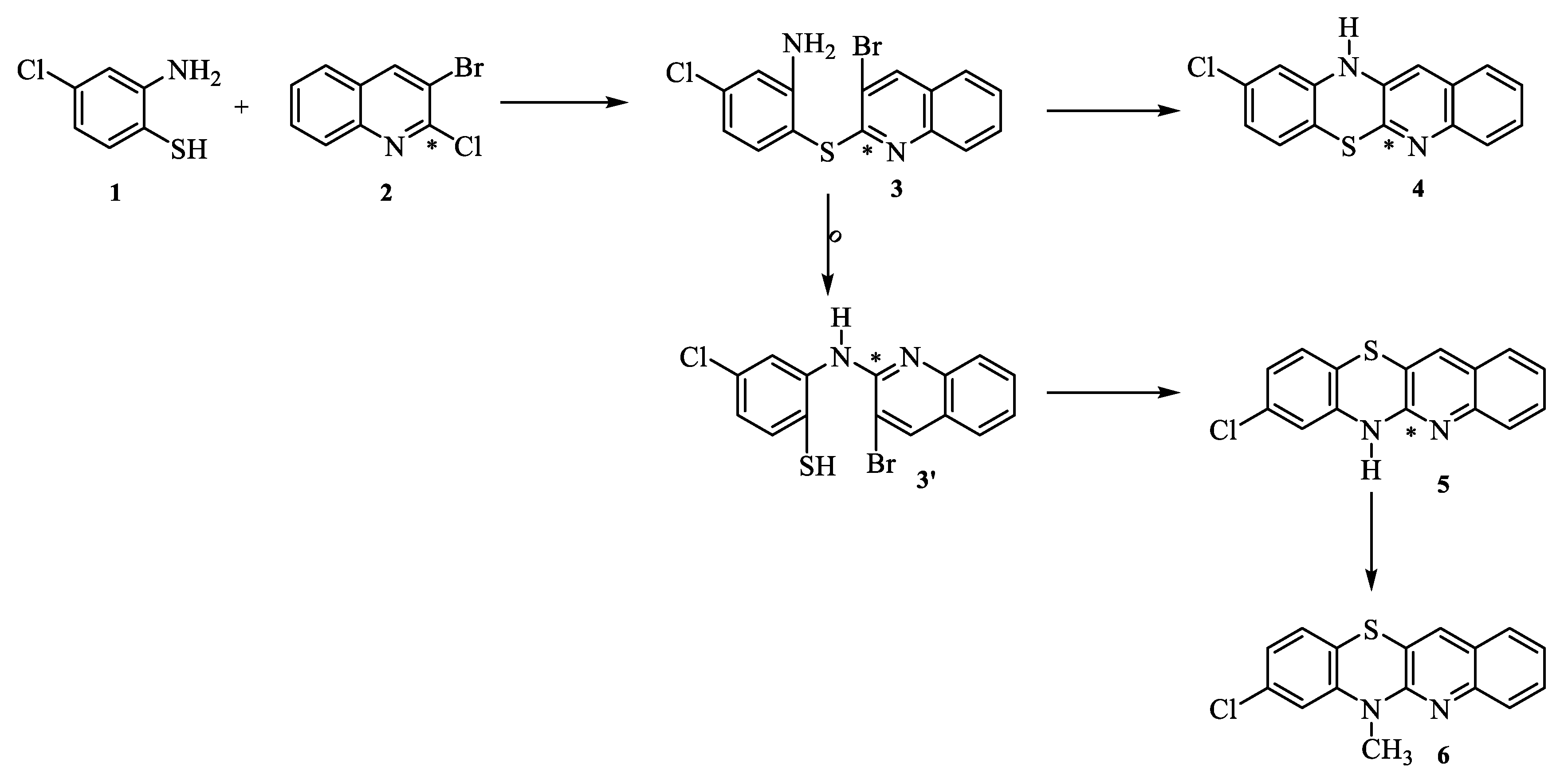
For the synthesis of 6H-8-chloroquinobenzothiazine 5, 2-amino-4-chlrobenzenethiol 1 and 3-bromo-2-chloroquinoline 2 (Scheme 1) were used as starting material. The reaction was carried out in boiling DMF for 1 hour. Phenyl quinoline sulfide 3 is formed as an intermediate product in this type of phenothiazine ring synthesis reactions. Sulfide 3 can then undergo transformations in two directions. There is the possibility of direct cyclization (the Ullmann cyclization) toward quinobenzothiazine 4. The second variant leads through the Smiles rearrangement reaction (the S→N type, quinolinyl part migrates from sulfur atom to the nitrogen atom, not isolated) to an amine 3’, which then undergoes cyclization to quinobenzothiazine 5. Literature data show that the course of this type of reaction for the synthesis of phenothiazine systems most often depends on the conditions used. Sometimes, it is impossible to state whether a reaction goes with or without the rearrangement because Ullmann’s and Smiles’s products are the same. The rearrangement proceeds under basic (most often) conditions, but also under acidic and neutral conditions. Using substituted o-aminobenzenethiol as a substrate, as in the reaction described, the possibility of creating two phenothiazines 4 and 5 should be considered.
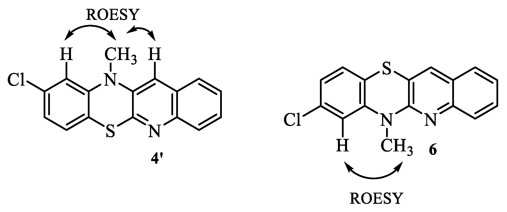
6H-8-Chloroquinobenzothiazine 5 was obtained in our previous studies by reacting 2,2'-dichloro-3,3'-diquinolinyl disulfide with 2,5-dichloroaniline [45]. Comparison of the substances obtained in these reactions allowed the preliminary assumption that the reaction of 2-amino-4-chlrobenzenethiol 1 and 3-bromo-2-chloroquinoline 2 involves a Smiles rearrangement. In order to identify unequivocally the structure of quinobenzothiazine 5, we transformed it into 6-methyl derivative 6 and we carried out 1H NMR and two-dimensional NOESY and COSY spectra (Table 1).
The 1H NMR spectra were very useful for identification of the product as 8-substituted quinobenzothiazine 6. The proton signals of the quinoline part were found at low field (over 7.3 ppm) as a singlet (proton H12), doublet-shaped multiplet signal with one ortho-coupling (proton H4), triplet-shaped multiplet signal with two ortho-couplings (proton H2), and multiplet containing a doublet and a triplet derived from the remaining two protons of the quinoline ring (H1 and H3). The proton signals of the benzene ring were found at high field (below 7.1 ppm) and are observed as 3 doublets differing in shape and multiplicity depending on the proton to which they are assigned. The H10 and H9 (as double doublet signals) proton signals appear in the form of wide doublets with a coupling constant of 7, while the H7 proton signal appears in the form of a very narrow doublet with a coupling constant of 1.5.
In order to fully document the structure of derivative 6, a 13C NMR spectrum and two-dimensional HSQC and HMBC spectra were also performed, which allowed for the assignment of appropriate C atoms to individual signals. Therefore, the products were identified as 8-chloroquino[3,2-b]benzo[1,4]thiazines (8-chlorobenzo[b]-1-azaphenothiazine).
The next step in the modification of the phenothiazine system was the introduction of N,N-dialkylaminoalkyl, N-acylaminoalkyl, N-sulfonylaminoalkyl, and groups, and 1,2,3-triazole substituents to the thiazine nitrogen atom in position 6. The N,N-dialkylaminoalkyl substituents were introduced in the N-alkylation reactions with hydrochlorides of selected acyclic and cyclic dialkylaminoalkyl chlorides in boiling dioxane in the presence of sodium hydroxide. As a result of such syntheses, the following was obtained five different 6-dialkylaminoalkyl derivatives 7-11 in 64-86% yield (Scheme 3).
Preparation of 6-substituted 8-chloroquinobenzothiazines with N-acylaminoalkyl and N-sulfonylaminoalkyl groups required a three-step synthesis. In the first stage 6H-8-chloroquinobenzothiazine 1 was alkylated with phthalimidopropyl and phthalimidobutyl bromides in dry toluene in the presence of sodium hydride into the phthalimidoalkyl derivatives 12 and 13. Next, these compounds underwent reactions with hydrazine hydrate in aqueous ethanol to give aminoapropyl derivative 14 and aminobutyl derivative 15 with yields of 85 and 84%, respectively (Scheme 4).
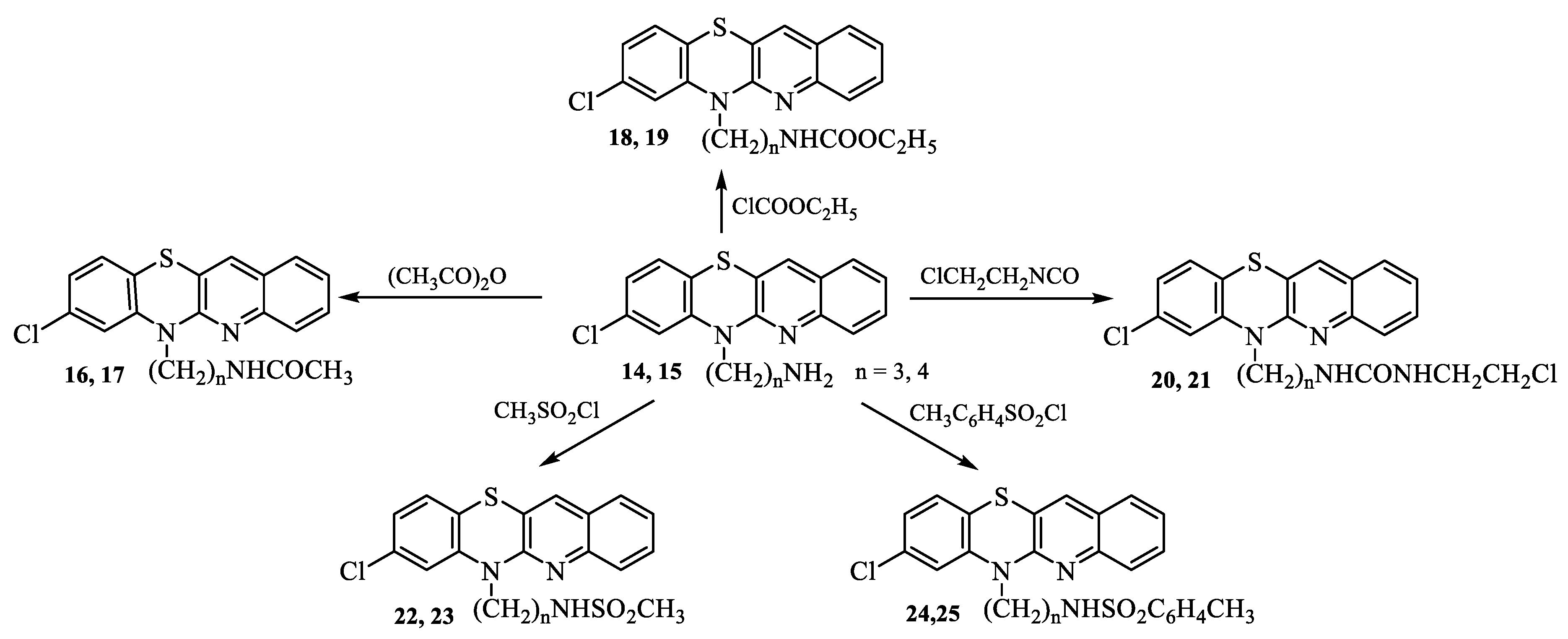
Aminoalkylquinobenzothiazines 14 and 15 were transformed into the N-acyl derivatives. The reactions with acetic anhydride, ethyl chloroformate and 2-chloroethyl isocyanate gave two 8-chloro-6-acetylaminoalkylquinobenzothiazines 16, 17, two 8-chloro-6-ethoxycarbonylaminoalkylquinobenzothiazines 18 and 19, and two 8-chloro-6-chloroethylureidoalkylquinobenzothiazines 20 and 21 (possessing a half-mustard unit) in 65-86% yield. Aminoalkylquinobenzothiazines 14 and 15 were also transformed into the N-sulfonyl derivatives. The reactions with methanesulfonyl and p-toluenesulfonyl chlorides led to the sulfonamide derivatives: two 8-chloro-6-methanesulfonylaminoalkyl- and two 8-chloro-6-p-toluenesulfonylaminoalkylquinobenzothiazines 22-25 in 74-77% yield (Scheme 5).
Due to the high pharmaceutical potential of substances containing the 1,2,3-triazole ring in their structure, we designed and synthesised a series of triazole derivatives of 8-chloroquinobenzothiazine [46,47,48,49,50,51]. These derivatives were obtained using 1,3-dipolar cycloaddition reaction between 2-propynyl derivative of 8-chloroquinobenzothiazine 1 and some selected organic azides. Starting quinobenzothiazine 1 was transformed with 2-propynyl bromide into propynyl derivatives 26 according to the synthesis described synthesis [34], and further using ‘click chemistry’ 1,3-dipolar cycloadditon (with selected azides, in toluene, in the presence of CuI as a catalyst) into substituted triazole derivatives of 8-chloroquinobenzothiazine 27-33. Taking into account the fact of significant biological activity of triazole systems with various phenyl and benzyl substituents [52,53,54], selected azides (phenyl azide, p-trifluoromethylbenzyl azide, benzyl azide, p-fluorobenzyl azide, p-chlorobenzyl azide, p-cyanobenzyl azide and phenylthiomethyl azide) were selected for 1,3-dipolar cycloaddition.

2.2. Biological evaluation

2.2.1. Cytotoxic activity

Our biological study aimed to assess the cytotoxicity of newly synthesized derivatives of chlorpromazine (Schemes 1 to 6) and their potential utility in cancer treatment. Initially, all derivatives were tested against two human carcinoma cell lines (A549 - lung cancer, MDA-MB-231 - breast cancer) and a normal cell line HaCaT (immortalized human keratinocytes) to determine their IC50 values using the MTT method [55], with comparisons made to the IC50 values of the common chemotherapeutic agent, doxorubicin, are presented in Table 3.
Most derivatives exhibited moderate antiproliferative potency, with lower IC50 values observed in A549 cells compared to MDA-MB-231 cells. Among them, eight compounds (5, 8, 9, 11, 20, 21, 23, 25, and 27) showed promising activity against A549 cells without affecting HaCaT cells. Compounds 8, 20, 21, and 25 demonstrated the highest cytotoxicity against A549 cells. The selectivity index (SI) for these compounds ranged from 7.6 to 10.7, which was higher than that of doxorubicin (0.14–0.15). Based on these findings, eight compounds were further evaluated on three additional cancer cell lines (MiaPaca-2, PC3, and HCT-116) using the MTT method. Apart from the A549 cell line, HCT116 cells exhibited the highest sensitivity to the tested substances, while PC3 cells showed lower sensitivity.
It's important to note that while doxorubicin demonstrated high cytotoxicity against all cancer cell lines, it also affected normal cells. The compounds 8 and 23 displayed the highest selectivity index (143), with IC50 values of 1.6 ± 0.24 µM and 0.7 ± 0.01 µM, respectively, against HCT-116 cells, while showing no cytotoxic effects on HaCaT cells.
Derivatives 5 and 11 showed moderate effectiveness against most tested cell lines in terms of antitumor activity (11.1 to 14.9 µM and 16.1 to 16.7 µM, respectively), but compound 11 exhibited notably higher cytotoxicity specifically against HCT-116 cells (7.7 µM) (Table 3 and Table 4).
Additionally, compound 21 was effective toward four cancerous cell lines at concentrations of 6.4–10.4 µM, respectively. However, despite relatively strong inhibitory effects on cancer cells, also showed low selectivity (Table 3 and Table 4).

2.2.2. In vitro antibacterial activity

The antibacterial efficacy of new synthesized derivatives 533 was assessed by initially screening them for their minimal inhibitory concentrations (MICs) [56].
The compounds were tested against standard Gram-positive bacteria, including various strains of S. aureus (NCTC 4163, ATCC 25923, ATCC 6538, and ATCC 29213) and Staphylococcus epidermidis (ATCC 12228 and ATCC 35984), as well as Gram-negative rods such as Escherichia coli (ATCC 25922) and P. aeruginosa (ATCC 15442).
The results showed that investigated compounds (5-9, 20, 21) exhibited the potential to moderate antibacterial potency, mainly against standard staphylococcal strains (Table 5).
In general, the most prominent activity against standard strains was observed for compound 21, with MIC values ranging from 2 to 8 µg/ml.
On the other hand, moderate activity (MIC range 8–16 µg/ml) against staphylococci was observed for compounds 5-9 and 20. Additionally, compound 11 also exhibited moderate activity against one strain of S. aureus, NCTC 4163 (MIC 8 µg/ml), while its activity against the other three strains of S. aureus was very low (MIC >128 µg/ml). All tested compounds were inactive against the Gram- Negative rods P. aeruginosa strain (ATCC 15442) and E. coli, except for compound 21, which showed strong activity against E. coli ( MIC 4 µg/ml) (Table 5).
The above results indicate that derivatives 8, 11, 20 and 21 have both antibacterial and cytotoxic properties, which makes them promising compounds.
The most potent derivatives (5, 8, 11, 20, 21, 23 and 25) were selected for further investigations of their mechanisms of cytotoxic action.

2.2.3. Mechanism of cytotoxicity of newly derivatives

  • Apoptotic activity:
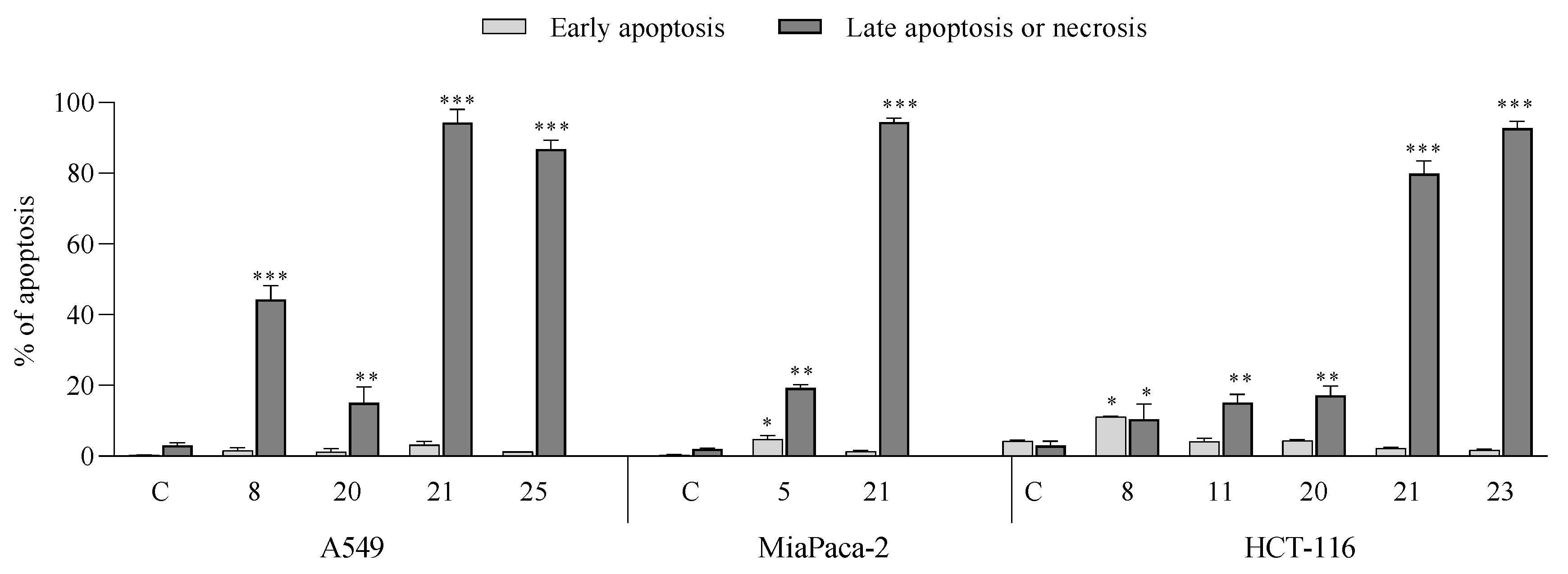
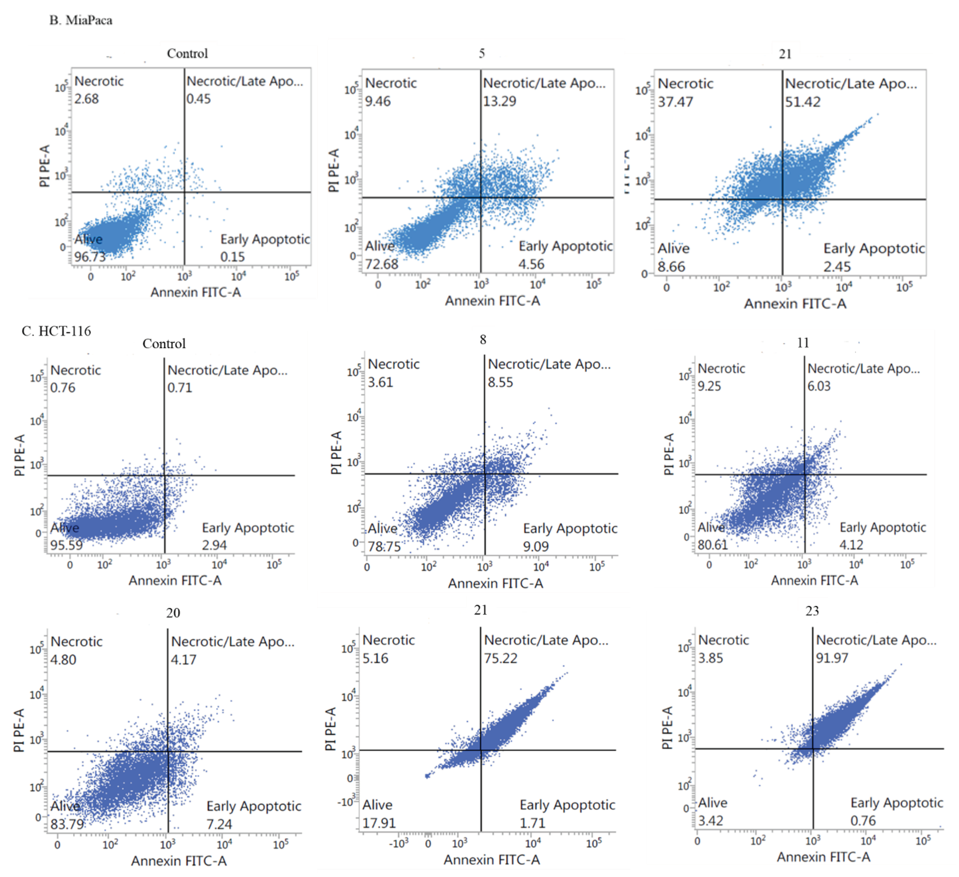
Given the significant cytotoxic potential observed in the newly synthesized chlorpromazine-derived compounds, namely 5, 8, 11, 20, 21, 23 and 25, against cancer cell lines such as A549 (lung cancer), MiaPaCa-2 (breast cancer), and HCT-116 (colon cancer), with concentrations not exceeding 11 μM, these compounds were selected for further investigation to elucidate their mechanisms of biological action. Their ability to induce apoptosis or necrosis in cancer cells was assessed through flow cytometry analysis. As depicted in Figure 1; Figure 2, the tested derivatives, when administered at their IC50 concentrations, exhibited proapoptotic properties in the selected cell lines compared to untreated controls.
The obtained results indicated that the selected derivative 21 exhibited potent proapoptotic activity across all tested cancer cell lines (Figure 1, 2). For derivative 8, the strong late apoptosis and necrosis inducing effect was found in A549 (42% ± 0.76), whereases in HCT-116 the same compound induced mainly early and late apoptosis/necrosis (9.09 ± 0.5 and 8.55 ± 0.33) (Figure 1). Furthermore, incubation with derivative 20 led to a significantly higher percentage of A549 and HCT-116 cells in late apoptosis or necrosis (ranging from 15.2% to 17%) compared to the control. Additionally, a similar noticeable pro-apoptotic effect as compound 20 was observed with derivative 11 in HCT-116 cells (15.64% in late apoptosis or necrosis). The compound 5 acted similarly, as it activated not only early, but also late apoptosis/necrosis in MiaPaCa-2 cells, which accounted for 4.56% and 23%, respectively (Figure 1, 2 B). The strongest of late apoptosis and necrotic activity (82% ± 2.89) was found for 25 towards A549 cancer cells and for compound 23 high percentage of late apoptosis/necrosis (93% ± 3.29) in HCT-116 cells (Figure 1, 2 C).
Our study confirmed the proapoptotic activity of selected compounds, especially in terms of inducing late apoptosis or necrosis in the A549, MiaPaCa-2 and HCT-116 cancer cell lines (Figure 1).
Figure 1. The effects of newly synthesized compounds 5, 8, 11, 20, 21, 23, and 25 on early and late apoptosis or necrosis were assessed in A549, MiaPaca-2, and HaCaT cell lines. Cells were treated with the compounds at their IC50 concentrations for 72 hours, followed by staining with annexin V-FITC and PI, and analysis using flow cytometry. The results are presented as the percentage of cells in the early stage of apoptosis and the percentage of cells in the late stage of apoptosis or necrosis. ***p ≤ 0.0001, **p ≤ 0.001, *p ≤ 0.01, as compared to the control (C).
Preprints 101039 g003
Figure 2. Representative results (%) as dot plots from apoptosis analysis of (A) A549, (B) MiaPaCa-2 and (C) HCT-116 cancer cells treated with compounds 5, 8, 11, 20, 21, 23 and 25 determined by flow cytometry, using the Annexin V-FITC/PI staining bioassay.
Preprints 101039 g004
  • Induction of cell cycle arrest
The dysregulation of the cell cycle represents a hallmark of cancer cells, manifesting disruptions in various cellular pathways, particularly those governing the cell cycle and apoptosis. This dysregulation frequently enables cancer cells to evade crucial processes such as apoptosis or senescence, consequently leading to unchecked tumor proliferation and growth. Consequently, targeting this dysregulation has emerged as a promising therapeutic strategy in cancer treatment [57,58,59].
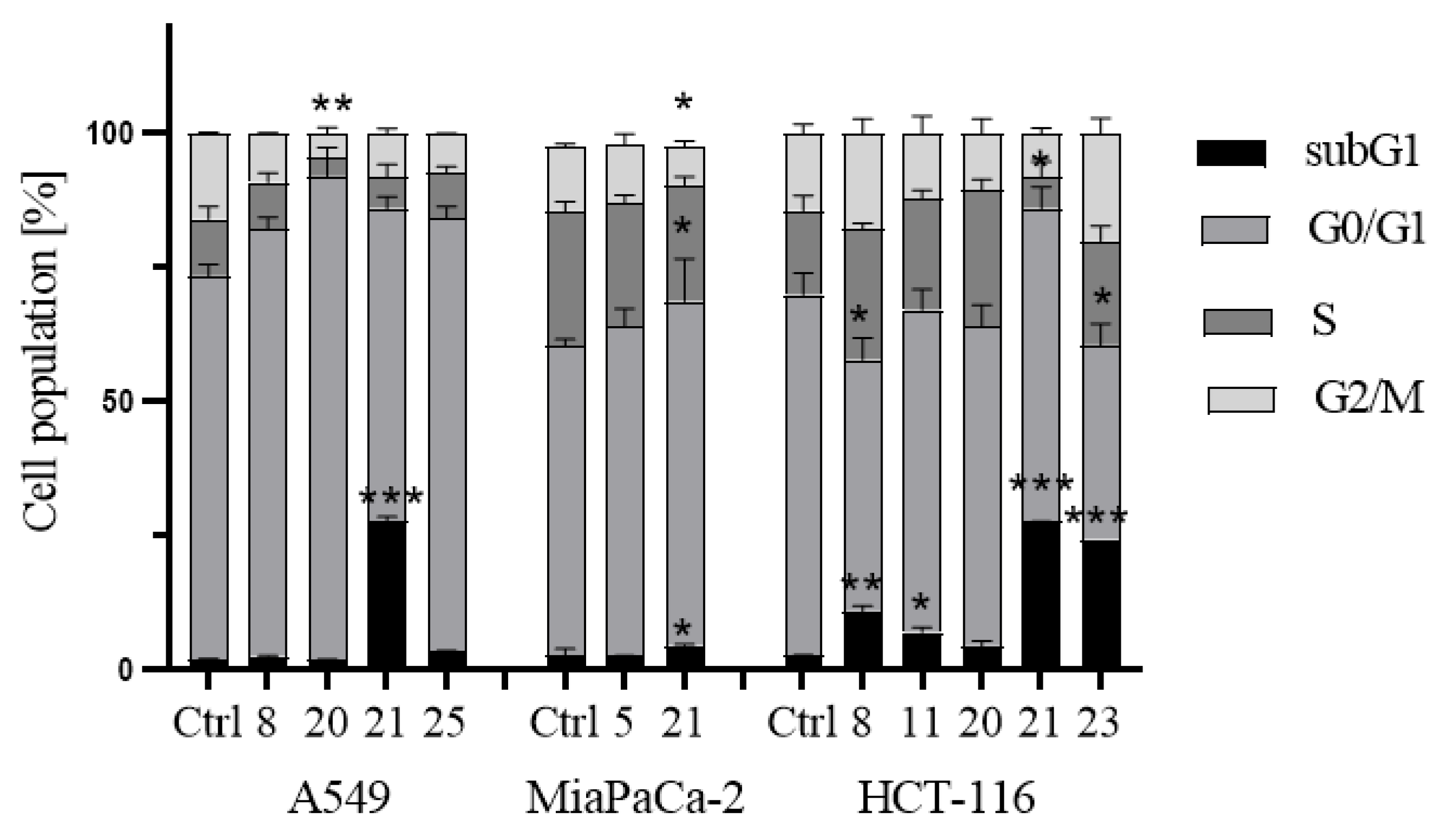
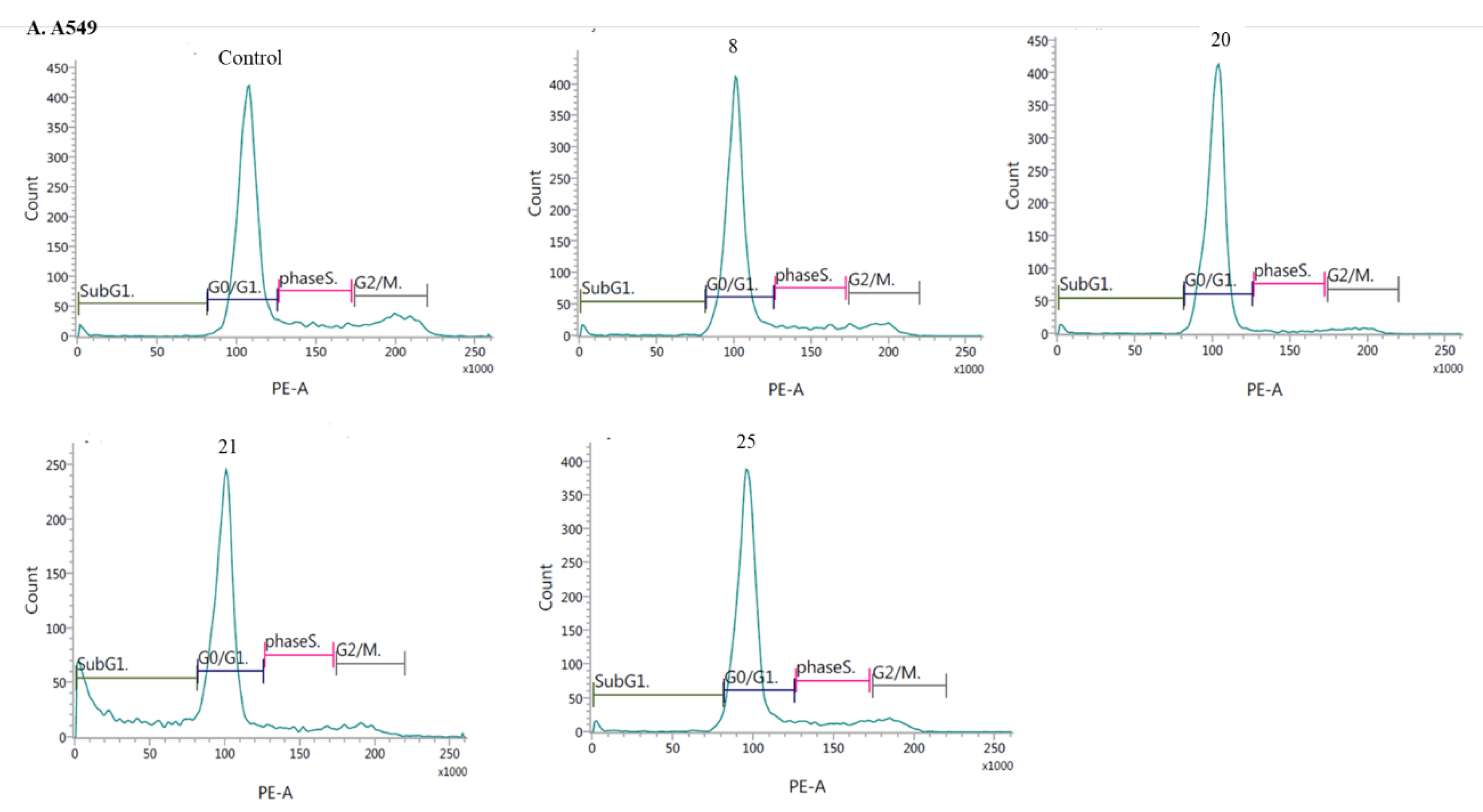
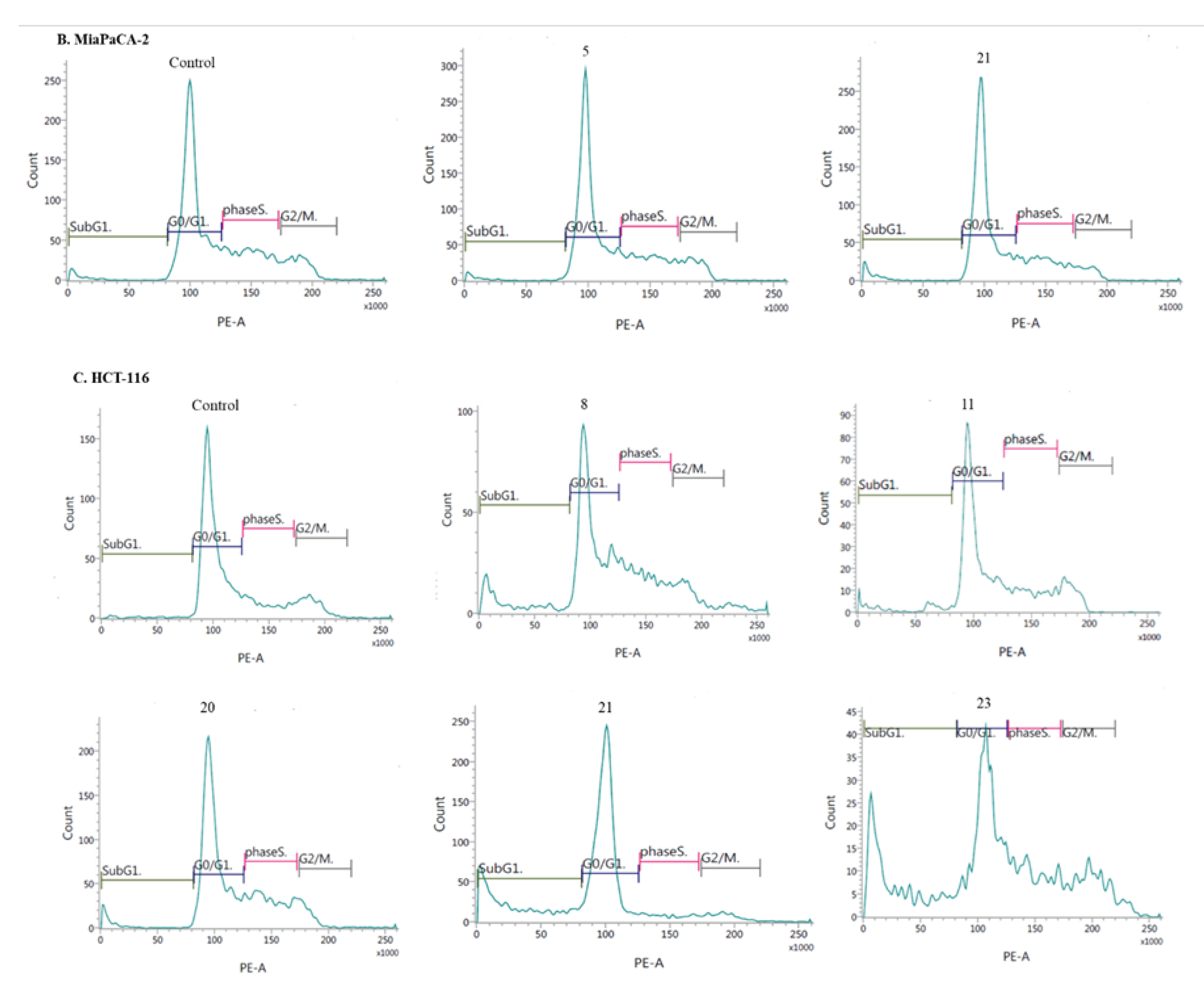
Therefore, the effects of new derivatives on the cell cycle were investigated to elucidate the inhibition mechanisms. The cells were exposed to IC50 concentrations of compounds 5, 8, 20, 21, 23, 25 for 24 hours. It was found that, compounds 5, 8, 20, 21, 25, they induced G0/G1 phase arrest in both cell lines (the A549 and MiaPaca-2, respectively) (Figure 3 and Figure 4A, B). A concomitant reduction in the number of cells in the S and G2/M phases was also observed (Figure 3).
This increase in the G0/G1-phase cell population was mostly at the expense of G2/M cells. These results clearly suggest that studied derivatives exerts its antiproliferative effect by inducing cell cycle arrest at S phase and, consequently, apoptosis. The addition of derivative 21 significantly increased the percentage Sub G1 phase A549 cells when tested at IC50 (27.6%, p = 0.0001) and reduced the cell population in G0/G1, and G2/M phase, compared to the cell cycle distribution monitored in untreated A459 cells (Figure 3 and 4A). Upon, treatment of HCT-116 cells with compounds 8, 21, 23, a significant increase of the cell amount at the sub-G1 phase (5, 12 and 11-folds, respectively). Moreover, these three derivatives led to a significant increase in the number of cells accumulated in the S phase (approximately 2-fold) and in the G2/M phase (approximately 1.3-fold) as compared to control. In addition, on exposure to compounds 11, 20, there was a little increase in the sub-G1 population (rage 2 and 3-folds, respectively) and statistically significant growth at the S-phase (range 20.4 to 25.81%). The compounds 11, 20 decreased HCT-116 cells at their G0/G1 phase, and arrested the cell population at S-phase (Figure 3 and 4 C). Furthermore, cell cycle distribution in HCT116 cells indicates proapoptotic action (Figure 3).

3. Methods and materials

Melting points were determined in open capillary tubes on a Boetius melting point apparatus and were uncorrected. The standard NMR spectra were recorded on Bruker Avance spectrometers (1H at 600 MHz, 13C at 150 MHz) in CDCl3 or DMSOd6. Two-dimensional COSY, NOESY, HSQC and HMBC spectra of selected compounds were recorded on a Bruker Avance spectrometer at 600 MHz, using COSYGPSW, NOESYGPPHSW, HSQCGPPH and HMBCGP experiments. The HRMS spectra (EI - electroimpact ionization) were run on a Brucker Impact II. Thin-layer chromatography was performed on aluminum oxide 60 F254 neutral (type E) (Merck 1.05581) with CH2Cl2 as eluents.

3.1. Synthesis of compounds

3.1.1. General procedure for the synthesis of 8-chloroquinobenzothiazine derivatives:

  • Synthesis of 6H-8-chloroquinobenzothiazine 5.
To a solution of 0.24 g (1 mmol) 3-bromo-2-chloroquinoline 1 in 5 mL dry DMF, 0.16 g (1 mmol) 2-amino-4-chlorothiophenol 2 was added. The reaction mixture was heated at reflux for 1 hour. After cooling, the reaction mixture was poured into water (25 mL). The precipitate was filtered off, washed with water and, after drying, purified by crystallization from ethanol to give 6H-8-chloroquinobenzothiazine 5. Yield: 79%. M.p.: 230-231°C [45]. 1H NMR (DMSO-d6) δ: 6.86 (d, 1H, H-9), 6.92 (d, 1H, H-7), 7.04 (d, 1H, H-10), 7.25 (m, 1H, H-2), 7.45 (m, 1H, H-1,H-3), 7.56 (d, 1H, H-4), 7.84 (s, 1H, H-12), 9.99 (s, 1H, NH). 13C NMR (75 MHz, DMSO-d6) δ: 114.66, 115.22, 115.25, 122.23, 124.48, 126.20, 126.64, 127.15, 127.55, 129.98, 132.04, 132.23, 140.87, 146.08, 150.35. HR MS (ESI) calcd for C15H10N2S [M+H]+: 285.0253, found: 285.0256.
  • Synthesis of 8-chloro-6-methylquinobenzothiazine 6.
To a solution of 6H-8-chloroquinobenzothiazines 0.14 (0.5 mmol) 1 in dry DMF (5 mL) NaH (0.12 g, 5 mmol, 60% NaH in mineral oil was washed out with hexane) was added. The reaction mixture was stirred at room temperature for 0.5 h, methyl iodide (0.05 mL, 0.75 mmol) was added and the stirring was continued for 24 h. The reaction mixture was poured into water (25 mL). The resulting solid was filtered off, washed with water and purified by column chromatography (aluminum oxide, CHCl3) to give 6-methyl-8-chloroquinobenzothiazine 2.
Yield: 86%. M.p.: 120-121°C. 1H NMR (600 MHz, CDCl3) δ: 3.61 (s, 3H, CH3), 6.94 (d, 1H, H7), 6.96 (d, 1H, H9), 7.05 (d, 1H, H10), 7.32 (m, 1H, H2), 7.54-7.57 (m, 2H, H1, H3), 7.70 (s, 1H, H12), 7.79 (d, 1H, H4). 13C NMR (75 MHz, CDCl3) δ: 29.73, 115.70, 118.19, 118.95, 122.48, 124.53, 125.99, 126.30, 127.23, 127.47, 129.33, 132.21, 133.60, 144.17, 145.74, 152.77. HR MS (ESI) calcd for C16H12ClN2S [M+H]+: 299,0410, found: 299,0410.
  • Synthesis of 8-chloro-6-dialkylaminoalkylquinobenzothiazines 7-11.
A mixture of 8-chloro-6H-quinobenzothiazine 1 (0.14 g, 0.5 mmol), sodium hydroxide (0.30 g, 7.5 mmol) and hydrochloride of dialkylaminoalkyl chloride (1.5 mmol, 2-diethylaminoethyl - 0.26 g, 3-dimethylaminopropyl - 0.24 g, 2-(1-pyrrolidyl)ethyl - 0.26 g, 2-(1-piperidyl)ethyl - 0.28 g, 2-(1-methyl-2-piperydinyl)ethyl - 0.30 g) in dry dioxane (5 mL) was refluxed for 3 h. After cooling the reaction mixture was poured into water (25 mL) and extracted with chloroform (3 × 10 mL). The combined extracts were washed with water to pH = 7 and dried over Na2SO4. Chloroform was evaporated in vacuo and the residue was purified by column chromatography (Al2O3, CHCl3) to give compounds 7-11:
6-
(2-Diethylaminoethyl)-8-chloroquinobenzothiazine (7):
Yield: 78%. Yellow oil. 1H NMR (CDCl3) δ: 1.18 (m, 6H, 2CH3), 2.70 (m, 4H, 2CH2), 2.80 (m, 2H, CH2), 4.24 (m, 2H, NCH2), 6.80 (d, 1H, H-7), 6.88 (d, 1H, H-9), 7.06 (d, 1H, H-10), 7.20 (t, 1H, H-2), 7.42 (m, 2H, H-1, H-3), 7.51 (s, 1H, H-12), 7.61 (d, 1H, H-4). 13C NMR (75 MHz, CDCl3) δ: 12.07, 47.86, 48.11, 116.03, 117.73, 118.21, 122.25, 124.72, 125.94, 127.18, 127.39, 129.20, 129.51, 131.62, 133.52, 142.67, 145.66, 151.23. HR MS (ESI) calcd for C21H23ClN3S [M+H]+: 384.1301, found: 384.1302.
6-
(3-Dimethylaminopropyl)-8-chloroquinobenzothiazine (8):
Yield: 86%. M.p.: 73-75 °C. 1H NMR (CDCl3) δ: 2.07 (m, 2H, CH2), 2.35 (s, 6H, 2CH3), 2.52 (t, 2H, CH2), 4.23 (m, 2H, NCH2), 6.88 (d, 1H, H-7), 6.96 (d, 1H, H-9), 7.00 (d, 1H, H-10), 7.27 (t, 1H, H-2), 7.49 (d, 1H, H-1), 7.50 (t, 1H, H-3), 7.57 (s, 1H, H-12), 7.71 (d, 1H, H-4). 13C NMR (75 MHz, CDCl3) δ: 24.25, 44.09, 45.63, 57.36, 115.92, 117.95, 118.58, 122.23, 124.39, 126.04, 126.14, 127.26, 127.45, 129.16, 131.72, 133.45, 142.73, 145.68, 151.60. HR MS (ESI) calcd for C20H21ClN3S [M+H]+: 370.1145, found: 370.1151.
6-
(1-Pyrrolidylethyl)-8-chloroquinobenzothiazine (9):
Yield: 79%. Yellow oil. 1HNMR (CDCl3) δ: 1.89 (m, 4H, 2CH2), 2.90 (m, 4H, 2CH2), 3.09 (m, 2H, CH2), 4.40 (m, 2H, NCH2), 6.80 (d, 1H, H-7), 6.88 (d, 1H, H-9), 7.03 (d, 1H, H-10), 7.20 (t, 1H, H-2), 7.43 (m, 2H, H-1, H-3), 7.50 (s, 1H, H-12), 7.62 (d, 1H, H-4). 13C NMR (75 MHz, CDCl3) δ: 23.53, 43.84, 51.54, 54.22, 115.95, 117.90, 118.35, 122.68, 124.68, 126.12, 126.22, 127.35, 127.35, 129.32, 131.89, 133.80, 142.36, 145.46, 151.15. HR MS (ESI) calcd for C21H21ClN3S [M+H]+: 382.1145, found: 382.1145.
6-
(1-Piperidylethyl)-8-chloroquinobenzothiazine (10):
Yield: 76%. M.p.: 99-100°C. 1HNMR (CDCl3) δ: 1,51 (m, 2H, CH2), 1.70 (m, 4H, 2CH2), 2.62 (m, 4H, 2CH2), 2.84 (m, 2H, CH2), 4.35 (m, 2H, NCH2), 6.88 (d, 1H, H-7), 6.96 (d, 1H, H-9), 7.20 (d, 1H, H-10), 7.51 (m, 2H, H-1, H-3), 7.57 (s, 1H, H-12), 7.71 (d, 1H, H-4). 13C NMR (75 MHz, CDCl3) δ: 24.86, 26.07, 44.18, 55.10, 55.32, 116.26, 117.85, 118.41, 122.26, 124.42, 126.07, 126.19, 127.17, 127.47, 129.16, 131.63, 133.52, 142.79, 145.65, 151.39. HR MS (ESI) calcd for C22H22ClN3S [M+H]+: 396.1310, found: 396.1310.
6-
(1-Methyl-2-piperidylethyl)-8-chloroquinobenzothiazine (11):
Yield: 64%. M.p.: 143-144ºC. 1H NMR (CDCl3) d: 1.67 (m, 4H, 2CH2), 1.85 (m, 2H, CH2), 2.11 (m, 4H, 2CH2), 2.48 (s, 3H, CH3), 2.92 (m, 1H, CH), 4.21 (m, 2H, CH2), 6.87 (d, 1H, H-7), 6.91 (d, 1H, H-9), 6.96 (d, 1H, H-10), 7.27 (t, 1H, H-2), 7.50 (m, 2H, H-1, H-3), 7.57 (s, 1H, H-12), 7.69 (d, 1H, H-4). 13C NMR (75 MHz, CDCl3) δ: 24.36, 25.86, 28.63, 31.03, 43.01, 43.44, 57.13, 62.59, 115.59, 117.76, 118.38, 122.11, 124.38, 126.03, 126.12, 127.24, 127.37, 129.15, 131.58, 133.38, 142.67, 145.72, 151.24. HR MS (ESI) calcd for C23H25ClN3S [M+H]+: 410.1458, found: 410.1468.
  • Synthesis of 8-chloro-6-phthalimidoalkylquinobenzothiazines 12 and 13.
To a stirred solution of 8-chloro-6H-quinobenzothiazine 1 (0.14 g, 0.5 mmol) in dry toluene (5 mL) NaH (0.12 g, 10 mmol, washed out with hexane) was added. The mixture was refluxed for 30 min and a solution of N-(bromoalkyl)phthalimide [1.1 mmol, N-(3-bromopropyl)phthalimide 0.30 g, N-(4-bromobutyl)phthalimide 0.31 g] in dry toluene (5 mL) was added. The mixture was refluxed for 24 h. Next toluene was evaporated in vacuo and the residue was extracted with CHCl3 (2.5 mL). The extract was concentrated and purified by column chromatography (silica gel, CHCl3) to give compounds 12 or 13:
  • 8-Chloro-6-phthalimidopropylquinobenzothiazine (12):
Yield: 85%. M.p.: 168-169 °C. 1H NMR (CDCl3) δ: 2.35 (m, 2H, CH2), 3.94 (t, 2H, NCH2), 4.32 (t, 2H, NCH2), 6.84 (s, 1H, H-7), 6.87 (d, 1H, H-9), 6.95 (d, 1H, H-10), 7.26 (t, 1H, H-2), 7.44 (t, 1H, H-3), 7.47 (d, 1H, H-1), 7.56 (m, 2H, H-4, H-12), 7.68 (m, 2H, 2H phthal.), 7.80 (m, 2H, 2H phthal.). 13C NMR (75 MHz, CDCl3) δ: 25.35, 29.83, 35.92, 115.81, 119.09, 122.56, 123.19, 123.36, 125.99, 126.10, 127.29. 127.46, 129.28, 130.92, 132.00, 132.07, 133.50, 133.88, 142.44, 151.59, 168.29, 168.39. HR MS (ESI) calcd for C26H19ClN3O2S [M+H]+: 472,0887, found: 472,0871.
  • 8-Chloro-6-phthalimidobutylquinobenzothiazine (13):
Yield: 84%. M.p.: 148-149 °C. 1H NMR (CDCl3) δ: 1.93 (m, 4H, 2CH2), 3.80 (m, 2H, NCH2), 4.33 (m, 2H, NCH2), 6.89 (d, 1H, H-9), 6.91 (s, 1H, H-7), 6.98 (d, 1H, H-10), 7.29 (d, 1H, H-2), 7.51 (m, 2H, H-1, H-3), 7.64 (s, 1H, H-12), 7.73 (m, 2H, H phthal.), 7.75 (d, 1H, H-4), 7,84 (m, 2H, H phthal.). 13C NMR (75 MHz, CDCl3) δ: 23.66. 26.00, 37.61, 45.45, 116.42, 119.31, 122.80, 123.24, 123.30, 124.74, 125.88, 126.18, 126.86, 127.51, 129.60, 132.05, 132.57, 133.60, 133.91, 134.03, 142.35, 151.91, 168.45. HR MS (ESI) calcd for C27H21ClN3O2S [M+H]+: 486,1043, found: 486.1040.
  • Synthesis of 8-chloro-6-aminoalkylquinobenzothiazines 14 and 15.
To a boiling solution of 6-phthalimidoalkylquinobenzothiazines 12 or 13 (0.5 mmol) in EtOH (25 mL) 80% aqueous solution of hydrazine (0.1 mL, 2.5 mmol) was added. The mixture was refluxed for 2 h. After cooling the reaction mixture was acidified to pH ≈ 2 with conc. hydrochloric acid and evaporated. Water (10 mL) was added to the residue, the resulting solid was filtered off and washed with 10% hydrochloric acid. Combined filtrates were alkalized to pH ≈ 10 and the resulted solid was filtered off, washed with water, dried and purified by column chromatography (SiO2, CHCl3-EtOH 10:1) to give compounds 14 or 15:
  • 6-Aminopropyl-8-chloroquinobenzothiazine (14):
Yield: 76%. M.p.: 89-90 °C. 1H NMR (CDCl3) δ: 2.11 (m, 2H, CH2), 3.01 (t, 2H, NCH2), 4.32 (t, 2H, NCH2), 6.90 (d, 1H, H-9), 6.95 (s, 1H, H-7), 6.99 (d, 1H, H-10), 7.26 (t, 1H, H-2), 7.49 ( m, 2H, H-1, H-3), 7.61 (s, 1H, H-12), 7.73 (d, 1H, H-4). HR MS (ESI) calcd for C18H17ClN3S [M+H]+: 342.0832, found: 342,0844.
  • 6-Aminobutyl-8-chloroquinobenzothiazine (15):
Yield: 70%. M.p.: 127-128 °C. 1H NMR (DMSO-d6) δ: 1.73 (m, 2H, CH2), 1.80 (m, 2H, CH2), 2.87 (m, 2H, NCH2), 4.23 (m, 2H, NCH2), 7.05 (d,1H, H-9), 7.16 (d, 1H, H-7), 7.22 (d, 1H, H-10), 7.34 (t, 1H, H-2), 7.57 (t, 1H, H-3), 7.69 (d, 1H, H-1), 7.79 (s, 1H, H-12), 8.02 (d,1H, H-4). 13C NMR (75 MHz, CDCl3) δ: 23.65, 25.17, 39.07, 44.39, 116.48, 117.63, 118.96, 123.07, 125.14, 126.23, 127.05, 127.46, 128.44, 130.06, 132.97, 133.17, 142.58, 145.38, 151.72. HR MS (ESI) calcd for C19H19ClN3S [M+H]+: 356,0988, found: 356.0987.
  • Synthesis of 8-chloro-6-acetylaminoalkylquinobenzothiazines 16-25.
To a suspension of aminoalkylquinothiazines 14 or 15 (0.5 mmol) in pyridine (5 mL) acetic anhydride (3 mL, 32 mmol) was added and the mixture was stirred at rt for 24 h. The reaction mixture was poured into water (10 mL) and the resulting solid was filtered off, washed with water, air-dried and purified by column chromatography (Al2O3, CHCl3) to give compounds 16 or 17:
  • 6-Acetylaminopropyl-8-chloroquinobenzothiazine (16):
Yield: 84%. M.p.: 179-180 °C. 1H NMR (CDCl3) δ: 2.02 (s, 3H, CH3), 2.11 (m, 2H, CH2), 3.46 (m, 2H, NCH2), 4.34 (m, 2H, NCH2), 6.17 (s, 1H, NH), 6.92 (s, 1H, H-7), 6.99 (d, 1H, H-9), 7.00 (d, 1H, H-10), 7.31 (t, 1H, H-2), 7.54 (m, 2H, H-1, H-3), 7.65 (s, 1H, H-12), 7.71 (d, 1H, H-4). 13C NMR (75 MHz, CDCl3) δ: 23.43, 26.38, 37.43, 42.58, 115.94, 117.98, 118.84, 122.73, 124.75, 126.14, 126.35, 126.93, 127.51, 129.54, 132.24, 133.60, 142.28, 145.34, 151.84, 170.19. HR MS (ESI) calcd for C20H19ClN3OS [M+H]+: 384,0937, found: 384,0931.
  • 6-Acetylaminobutyl-8-chloroquinobenzothiazine (17):
Yield: 86%. M.p.: 149-150 °C. 1H NMR (CDCl3) δ: 1.73 (m, 2H, CH2), 1.93 (m, 2H, CH2), 1.98 (s, 3H, CH3), 3.39 (m, 2H, NCH2), 4.36 (m, 2H, NCH2), 6.95 (m, 2H, H-7, H-8), 7.04 (d, 1H, H-10), 7.35 (t, 1H, H-2), 7.57 (m, 2H, H-1, H-3), 7.72 (s, 1H, , H-12), 7.93 (m, 1H, H-4). 13C NMR (75 MHz, CDCl3) δ: 23.28, 23.91, 26.40, 29.72, 38.82, 117.79, 119.87, 122.47, 124.66, 125.31, 126.20, 126.56, 127.86, 128.40, 131.32, 132.83, 134.32, 141.50, 141.85, 151.37, 170.57. HR MS (ESI) calcd for C21H21ClN3OS [M+H]+: 398,1094, found: 398.1092.
  • Synthesis of 8-chloro-6-etoxycarbonylaminoalkylquinobenzothiazines 18 and 19.
To a stirred solution of aminoalkylquinobenzothiazines 14 or 15 (0.5 mmol) in a mixture of CH2Cl2 (5 mL) and 10% Na2CO3 solution (5 mL), a solution of ethyl chloroformate (0.65 mL, 0.65 mmol) in CH2Cl2 (3 mL) was added. The mixture was stirred at rt for 24 h. The organic phase was separated and aqueous phase was extracted with CH2Cl2 (2×5 mL). The combined extracts were washed with water (2×10 mL) and dried over Na2SO4. The drying agent was filtered off and filtrate was evaporated. The resulting residue was purified by column chromatography (Al2O3, CHCl3) to give compounds 18 or 19:
  • 6-Ethoxycarbonylaminopropyl-8-chloroquinobenzothiazine (18):
Yield: 78%). M.p.: 130-131 °C. 1H NMR (CDCl3) δ: 1.30 (t, 3H, CH3), 2.10 (m, 2H, CH2), 3.40 (m, 2H, NCH2), 4.17 (m, 2H, CH2), 4.29 (m, 2H, NCH2), 5.75 (s, 1H, NH), 6.91 (m, 2H, H-7, H-9), 6.98 (d, 1H, H-10), 7.30 (t, 1H, H-2), 7.52 (m, 2H, H-1, H-3), 7.62 (s, 1H, H-12), 7.85 (d, 1H, H-4). 13C NMR (75 MHz, CDCl3) δ: 26.76, 27.34, 38.79, 42.69, 60.74, 115.70, 117.79, 118.56, 122.56, 124.66, 126.12, 126.20, 127.12, 127.37, 129.39, 132.05, 133.48, 142.34, 145.43, 151.54, 156.84. HR MS (ESI) calcd for C21H21ClN3O2S [M+H]+: 414,1043, found: 414,1055.
  • 8-Chloro-6-ethoxycarbonylaminobutylquinobenzothiazine (19):
Yield: 81%. M.p.: 138-139 °C. 1H NMR (CDCl3) δ: 1.26 (d, 3H, CH3), 1.75 (m, 2H, CH2), 1.93 (m, 2H, CH2), 3.32 (m, 2H, NCH2), 4.12 (m, 2H, CH2), 4.32 (m, 2H, NCH2), 4.92 (m, 1H, NH), 6.94 (m, 2H, H-7, H-9), 7.03 (d, 1H, H-10), 7.33 (d, 1H, H-2), 7.55 (m, 2H, H-1, H-3), 7.69 (s, 1H, H-12), 7,91 (d, 1H, H-4). 13C NMR (75 MHz, CDCl3) δ: 14.66, 23.72, 27.07, 40.23, 40.36, 60.69, 116.01, 117.58, 119.82, 122.73, 123.16, 125.40, 125.86, 126.41, 127.61, 127.80, 130.07, 130.92, 134.16, 141.57, 151.58, 156.75. HR MS (ESI) calcd for C22H23ClN3O2S+: 428.1200, found: 428.1198.
  • Synthesis of 8-chloro-6-chloroethylureidoalkylquinobenzothiazines 20 and 21.
To a stirred solution of 6-aminoalkyldiquinothiazines 14 or 15 (0.5 mmol) in ethanol (10 mL) at 0 °C 2-chloroethyl isocyanate (0.08 mL, 1 mmol) was added. The mixture was stirred at 0 °C for 1 h and at rt for 24 h. After evaporation of EtOH in vaccuo the residue was purified by column chromatography (Al2O3, CHCl3) to give compounds 20 or 21:
  • 6-Chloroethylureidopropyl-8-chloroquinobenzothiazine (20):
Yield: 73%. M.p.: 165-166 °C. 1H NMR (CDCl3) δ: 2.11 (m, 2H, CH2), 3.42 (m, 2H, CH2), 3.55 (m, 2H, NCH2), 3.62 (m, 2H, NCH2), 4.35 (m, 2H, NCH2), 6.94 (m, 2H, H-7, H-9), 7.03 (d, 1H, H-10), 7.32 (t, 1H, H-2), 7.56 (m, 2H, H-1, H-3), 7.68 (s, 1H, H-12), 7,80 (d, 1H, H-4). 13C NMR (75 MHz, CDCl3) δ: 26.68, 26.96, 37.62, 43.43, 48.27, 119.38, 120.89, 123.09, 124.51, 125.80, 126.95, 127.11, 128.03, 128.39, 128.45, 133.30, 133.44, 135.35, 138.46, 139.73, 159.21. HR MS (ESI) calcd for C21H21Cl2N4OS [M+H]+: 447.0831, found: 447,0827.
  • 6-Chloroethylureidobutyl-8-chloroquinobenzothiazine (21):
Yield: 65%. M.p.: 169-170 °C. 1H NMR (DMSO-d6) δ: 1.89 (t, 2H, CH2), 3.19 (m, 2H, CH2), 3.31 (m, 4H, 2CH2), 3.56 (m, 2H, CH2), 4.22 (m, 2H, CH2), 6.20 (m, 1H, 1NH2), 6.24 (m, 1H, 1NH2), 7.04 (d, 1H, H-9), 7.11 (s, 1H, H-7), 7.21 (d, 1H, H-10), 7.34 (t, 1H, H-2), 7.55 (t, 1H, H-3), 7.67 (m, 2H, H-1, H-4), 7.99 (s, 1H, H-12). 13C NMR (75 MHz, DMSO-d6) δ: 27.34, 37.69, 41.94, 43.12, 45.01, 55.39, 116.30, 117.60, 118.91, 122.94, 125.08, 126.21, 127.00, 127.49, 128.38, 130.00, 132.84, 133.09, 142.64, 145.43, 151.64, 158.31. HR MS (ESI) calcd for C22H23Cl2N4OS [M+H]+: 461.0970, found: 461.0942.
  • Synthesis of 8-chloro 6-methanesulfonylaminoalkylquinobenzothiazines 22 and 23.
To a stirred solution of aminoalkyldiquinothiazines 14 or 15 (0.5 mmol) in a mixture of CH2Cl2 (5 mL) and 10% Na2CO3 solution (7 mL), a solution of methanesulfonyl chloride (0.06 mL, 0.75 mmol) was added. The mixture was stirred at rt for 24 h. The organic phase was separated and aqueous phase was extracted with CH2Cl2 (2×5 mL). The combined extracts were washed with water (2×10 mL) and dried over Na2SO4. The drying agent was filtered off and filtrate was evaporated. The resulting residue was purified by column chromatography (Al2O3, CHCl3) to give compounds 22 or 23:
  • 8-Chloro-6-methanesulfonylaminopropylquinobenzothiazine (22):
Yield: 71%. M.p.: 138-139 °C. 1H NMR (CDCl3) δ: 2,18 (m, 2H, CH2), 2,89 (s, 3H, CH3), 3,33 (m, 2H, NCH2), 4,41 (m, 2H, NCH2), 5,91 (s, 1H, NH), 6,94 (m, 2H, H-7, H-9), 7,01 (d, 1H, H-10), 7,34 (t, 1H, H-7), 7,54 (d, 1H, H-1), 7,58 (t, 1H, H-3), 7,66 (s, 1H, H-12), 7,91 (d, 1H, H-4). 13C NMR (75 MHz, CDCl3) δ: 28.99, 30.57, 38.89, 67.77, 120.14, 121.06, 121.21, 122.11, 124.09, 124.54, 126.80, 126.99, 127.88, 128.38, 129.50, 133.41, 153.23, 138.51, 151.51. HR MS (ESI) calcd for C19H19ClN3O2S2 [M+H]+: 420.0607, found 420.0607.
  • 8-Chloro-6-methanesulfonylaminobutylquinobenzothiazine (23):
Yield: 76%. M.p.: 169-170 ⁰C. 1H NMR (CDCl3) δ: 1,83 (m, 2H, CH2), 1,98 (m, 2H, CH2), 2,99 (s, 3H, CH3), 3,29 (m, 2H, NCH2), 4,34 (m, 2H, NCH2), 6,93 (s, 1H, H-7), 6,96 (d, 1H, H-9), 7,04 (d, 1H, H-10), 7,34 (d, 1H, H-7), 7,57 (m, 2H, H-1, H-3), 7,70 (s, 1H, H-12), 7,90 (m, 1H, H-4). 13C NMR (75 MHz, CDCl3) δ: 23.38, 27.43, 40.33, 42.67, 42.78, 116.43, 116.84, 118.57, 119.28, 123.13, 124.67, 125.07, 125.84, 126.31, 127.61, 127.96, 129.96, 133.72, 142.17, 151.65. HR MS (ESI) calcd for C20H21ClN3O2S2 [M+H]+: 434.0764, found 434.0763.
  • Synthesis of 8-chloro-6-p-toluenesulfonylaminolkylquinobenzothiazines 24 and 25.
To a stirred solution of aminoalkyldiquinothiazines 14 or 15 (0.5 mmol) in a mixture of CH2Cl2 (5 mL) and 10% Na2CO3 solution (7 mL), a solution of p-toluenesulfonyl chloride (0.14 g, 0.75 mmol) in CH2Cl2 (6 mL) was added. The mixturewas stirred at rt for 24 h. The organic phase was separated and aqueous phase was extracted with CH2Cl2 (2x5 mL). The combined extracts were washed with water (2x10 mL) and dried over Na2SO4. The drying agent was filtered off and filtrate was evaporated. The resulting residue was purified by column chromatography (Al2O3, CHCl3) to give: compounds 24 or 25:
  • 8-Chloro-6-p-toluenesulfonylaminopropylquinobenzothiazine (24):
Yield: 68% M.p.: 121-122 ⁰C. 1H NMR (CDCl3) δ: 2,03 (m, 2H, CH2), 2,39 (s, 3H, CH3), 3,15 (m, 2H, NCH2), 4,24 (m, 2H, NCH2), 6,09 (s, 1H, NH), 6,86 (s, 1H, H-7), 6,93 (d, 1H, H-9), 6,99 (d, 1H, H-10), 7,18 (d, 2H, 2Hph), 7,37 (t, 1H, H-7), 7,56 (d, 1H, H-1), 7,61 (t, 1H, H-3), 7,64 (m, 4H, 2Hph, H-4, H-12). 13C NMR (75 MHz, CDCl3) δ: 21.52, 26.44, 40.78, 42.98, 116.23, 118.74, 123.28, 125.30, 125.87, 126.15, 126.27, 127.01, 127.47, 129.63, 130.32, 133.00, 133.67, 136.80, 141.50, 143.16, 151.29. HR MS (ESI) calcd for C25H23ClN3O2S2 [M+H]+: 496.0920, found 496.0924
  • 8-Chloro-6-p-toluenesulfonylaminobutylquinobenzothiazine (25):
Yield: 73%. M.p.: 148-149 °C. 1H NMR (CDCl3) δ: 1,68 (m, 2H, CH2), 1,85 (m, 2H, CH2), 2,42 (s, 3H, CH3), 3,09 (m, 2H, NCH2), 4,28 (m, 2H, NCH2), 6,88 (s, 1H, H-7), 6,95 (d, 1H, H-9), 7,03 (d, 1H, H-10), 7,28 (d, 2H, Hph), 7,35 (d, 1H, H-2), 7,57 (m, 2H, H-1, H-3). 13C NMR (75 MHz, CDCl3) δ: 21.54, 23.35, 26.85, 42.62, 45.81, 116.67, 119.41, 123.39, 125.26, 125.73, 126.32, 127.10, 127.64, 129.62, 129.69, 130.23, 133.28, 133.81, 137.00, 142.04, 143.30, 151.59. HR MS (ESI) calcd for C26H25ClN3O2S2 [M+H]+: 510.1077, found 510.1076
  • Synthesis of 8-chloroquinobenzothiazines with triazole substituents 27-33.
To a solution of 8-chloro-6-propynyquinobenzothiazine 26 (0.16 g, 0.5 mmol) and copper iodide (I) (0.06 g) in dry toluene (5 mL), a corresponding organic azide (0.510 mmol) was added. The reaction mixture was stirred and heated at 70 °C for 48 h. Then the solvent mixture was distilled under reduced pressure. The dry residue was dissolved in CH2Cl2 and purified by column chromatography (aluminum oxide 90 active neutral, Merck 1.01077.2000, CH2Cl2 as eluent) to give pure triazole derivatives 27-33.
8-Chloro-6-
[(1-phenyl-1H-1,2,3-triazol-4-yl)methyl]-quinobenzo[1,4]thiazine (27):
Yield: 73% M.p.: 180-181 ⁰C. 1H NMR (CDCl3) δ: 5.47 (s, 2H, CH2), 6.80 (d, 1H, H-7), 6.88 (d, 1H, H-9), 7.25 (d, 1H, H-2), 7.32 (d, 1H, H-Ph), 7.40 (m, 3H, 2H-Ph, CH), 7.47 (m, 2H, H-1, H-3), 7.60 (m, 3H, H-10, 2H-Ph), 7.67 (d, 1H, H-4), 8.11 (s, 1H, H-12). 13C NMR (75 MHz, CDCl3) δ: 42.85, 116.74, 117.99, 118.07, 120.57, 122.44, 122.89, 124.78, 126.25, 126.33, 127.02, 127.14, 128.68, 129.56, 129.68, 131.99, 133.82, 137.08, 142.35, 145.35, 152.25. HR MS (ESI) calcd for C24H17ClN5S [M+H]+: 442.0893, found 442.0892.
8-Chloro-6-
[(1-(4-(trifluoromethyl)phenyl)-1H-1,2,3-triazol-4-yl)methyl]-quinobenzo[1,4]thiazine (28):
Yield: 77% M.p.: 188-189 ⁰C. 1H NMR (CDCl3) δ: 5.57 (s, 2H, CH2), 6.90 (d, 1H, H-2), 6.98 (d, 1H, H-9), 7.35 (t, 1H, H-2), 7.45 (s, 1H, H-10), 7.57 (m, 2H, H-1, H-3), 7.59 (s, 1H, CH), 7.64 (m, 3H, H-4, 2H-Ph), 7.86 (d, 2H, 2H-Ph) 8.32 (s, 1H, H-12). 13C NMR (75 MHz, CDCl3) δ: 42.69, 53.46, 116.65, 118.03, 118.15, 120.48, 123.00, 124.44, 124.91, 126.26, 126.44, 127.00, 127.03, 127.06, 127.08, 127.10, 129.67, 132.12, 133.83, 142.24, 145.22, 151.18. HR MS (ESI) calcd for C25H16ClF3N5S [M+H]+: 510.0767, found 510.0772.
8-Chloro-6-
[(1-Benzyl-1H-1,2,3-triazol-4-yl)-methyl]-quinobenzo[1,4]thiazine (29):
Yield: 82% M.p.: 183-184 ⁰C. 1H NMR (CDCl3) δ: 5.32 (s, 2H, CH2), 5.50 (s, 2H, CH2), 6.91 (d, 1H, H-7), 6.98 (d, 1H, H-9), 7.20 (m, 2H, H-2, 1H-Ph), 7.33(m, 4H,4H-Ph), 7.40 (d, 1H, H-10), 7.52 (H, 1H, H-3), 7,57 (d, 1H, H-1), 7.66 (d,1H, H-4), 7.67 (s, 1H, H-12), 7.72 (s, 1H, CH). 13C NMR (75 MHz, CDCl3) δ: 43.40, 54.10, 117.13, 118.52, 123.22, 123.22, 124.48, 125.00, 125.99, 126.34, 126.42, 127.10, 127.81, 127.88, 128.10, 128.59, 128.83, 129.04, 129.11, 129.14, 129.75, 132.56, 133.88, 134.72, 142.30, 151.22. HR MS (ESI) calcd for: C25H19ClN5S [M+H]+: 456.1050, found 456.1052.
8-Chloro-6-
[(1-(4-fluorobenzyl)-1H-1,2,3-triazol-4-yl)methyl]-quinobenzo[1,4]thiazine (30):
Yield: 83% M.p.: 164-165 ⁰C. 1H NMR (CDCl3) δ: 5.46 (s, 2H, 2CH2), 6.05 (s, 2H, CH2), 6.92 (m, 2H, H-7, H-9), 7.13 (m, 4H, H-1, H-10, 2H-Ph), 7.31 (d, 1H, H-3), 7.50 (t, 1H, H-2), 7.65 (m, 2H, 2H-Ph), 7.94 (s, 1H, H-12), 8.40 (s, 1H, CH) 8.57 (s, 1H, H-4). 13C NMR (75 MHz, CDCl3) δ: 46.48, 53.47, 115.97, 116.11, 119.31, 120.57, 121.64, 122.25, 124.80, 125.51, 126.05, 126.72, 127.29, 127.90, 129.57, 129.63, 130.37, 130.39, 132.35, 134.76, 136.72, 141.28, 150.28. HR MS (ESI) calcd for: C25H18ClFN5S [M+H]+: 474.0955, found 474.0944.
8-Chloro-6-
[(1-(4-chlorobenzyl)-1H-1,2,3-triazol-4-yl)methyl]-quinobenzo[1,4]thiazine (31):
Yield: 83% M.p.: 184-185 ⁰C. 1H NMR (CDCl3) δ: 5.47 (s, 2H, CH2), 5.50 (s, 2H, CH2), 6.91 (d, 1H, H-7), 6.98 (d, 1H, H-9), 7.13 (d, 2H, 2H-Ph), 7.30 (m, 2H, 2H-Ph), 7.34 (d, 1H, H-2), 7.36 (d, 1H, H-10), 7.54 (m, 2H, H-1, H-3), 7.63 (d, 1H, H-4), 7.66 (s, 1H, H-12), 7.70 (s, 1H, CH). 13C NMR (75 MHz, CDCl3) δ: 43.25, ,53.34, 116.98, 118.40, 118.46, 123.14, 124.41, 124.99, 126.03, 126.37, 126.49, 127.10, 129.18, 129.25, 129.71, 132.44, 133.22, 133.83, 134.64, 142.30, 144.47, 144.79, 151.20. HR MS (ESI) calcd for: C25H18Cl2N5S [M+H]+: 490,0668, found 490,0668.
8-Chloro-6-
[(1-(4-cyanobenzyl)-1H-1,2,3-triazol-4-yl)methyl]-quinobenzo[1,4]thiazine (32)
Yield: 75% M.p.: 192-193 ⁰C. 1H NMR (CDCl3) δ: 5.57 (s, 2H, CH2), 5.76 (s, 2H, CH2), 7.02 (d, 1H, H-7), 7.06 (d, 1H, H-9), 7.25 (d, 2H, 2H-Ph), 7.28 (d, 1H, H-2), 7.33 (d, 1H, H-10), 7.43 (d, 1H, H-3), 7.56 (d, 2H, 2H-Ph), 7.62 (m, 2H, H-4, H-12), 7.80 (s, 1H, CH). 13C NMR (75 MHz, CDCl3) δ: 46.92, 53.41, 112.65, 118.10, 119.64, 120.87, 124.62, 126.15, 126.57, 126.88, 127.79, 128.03, 128.06, 128.13, 128.32, 132.75, 132.78, 132.81, 134.96, 137.44, 139.71, 141.14, 150.25. HR MS (ESI) calcd for: C26H18ClN6S [M+H]+: 481.1002, found 481.1011.
8-Chloro-6-
[(1-phenylthiomethyl-1H-1,2,3-triazolo-4-ylo)methyl]-quinobenzo[1,4]thiazine (33)
Yield: 80% M.p.: 141-142 ⁰C. 1H NMR (CDCl3) δ: 5.44 (s, 2H, CH2), 5.55 (s, 2H, CH2), 6.90 (s, 1H, H-7), 6.98 (s, 1H, H-9), 7.16 (t, 2H, 2H-Ph), 7.18 (d, 1H, H-2), 7.21 (d, 2H, 2H-Ph), 7.34 (m, 2H, H-10, H-Ph), 7.54 (t, 1H, H-3), 7.57 (d, 1H, H-1), 7.62 (d, 1H, H-4), 7.63 (s, 1H, H-12), 7.67 (s,1H, CH). 13C NMR (75 MHz, CDCl3) δ: 42.83, 54.16, 116.61, 117.87, 118.12, 122.12, 123.76, 124.75, 126.20, 126.29, 127.01, 127.26, 128.81, 129.34, 129.43, 129.48, 131.50, 131.94, 132.93, 133.75, 142.32, 145.10, 145.24, 151.13. HR MS (ESI) calcd for: C25H19ClN5S2 [M+H]+: 488.0770, found 488.0770.

3.2. Biological assays

3.2.1. Cell line and culture

The human cell lines, such as A549 (lung cancer), MDA-MB-231 (breast cancer), MiaPaca-2 (pancreatic cancer), PC3 (metastatic prostate cancer), HCT-116 (colon carcinoma), and HaCaT (immortalized keratinocytes), were sourced from the American Type Culture Collection (ATCC) in Rockville, USA. Culturing conditions varied: HCT116 cells were cultured in MEM (ThermoSci, USA), A549, MDA-MB-231, MiaPaca, HaCaT cells in DMEM High Glucose and PC3 cells were cultured in RPMI (Biowest SAS, France). The growth medium consisted of 10% fetal bovine serum (FBS, Sigma-Aldrich, St. Louis, MO, USA), 20 mM HEPES (Biowest, Nuaillé, France), and antibiotics (100 U/mL of penicillin and 100 μg/mL of streptomycin) from Gibco, Grand Island, NY, USA. All cells were maintained in a humidified incubator at 37 °C with a 5% CO2 atmosphere until they reached 80–90% confluence.

3.2.2. MTT assay

To evaluate the cytotoxic effects of the newly synthesized compounds, a preliminary MTT (3-(4,5-dimethylthiadiazol-2-yl)-2,5-diphenyl-2H-tetrazolium bromide) test was performed on two different cancer cell lines, A549 and MDA-MB231, as well as on one healthy HaCaT cell line. After obtaining the results, additional MTT tests were performed on three additional cancer cell lines (MiaPaca-2, PC3 and HCT-116), only on a selected group of compounds showing cytotoxic activity (5, 8, 21, 23, 25). The study derivatives were subjected to testing at various concentrations (ranging from 5 to 140 µM), alongside the reference drug doxorubicin. These compounds were added to 96-well plates containing study cells (1 × 10⁴ cells per well) and incubated for 72 hours. MTT analysis, as previously described [60,61], was employed.
Cell absorbance results were incorporated into the formula for calculating the relative MTT level (%), enabling the assessment of cell viability following exposure to the test compounds. The cell viability percentage represents the MTT reduction in cells treated with the test compounds compared to the control sample, where only the medium was added to the cells. The IC50 values, representing the concentration at which 50% of cell viability is inhibited, were calculated using Prism 8.0.1, GraphPad software.

3.2.3. Apoptosis and cell cycle analysis by flow cytometry (FCM)

To analyze the number of cells in early apoptosis, late apoptosis, or necrosis, A549, MiaPaca-2, and HCT-116 cell lines were cultured in 6-well plates with a seeding density of 1 × 10⁵ cells per well. These cells were then treated with selected compounds (4, 5, and 8) at their respective IC50 concentrations and incubated for 72 hours. Subsequently, a commercially available kit, the FITC: Annexin V Apoptosis Detection Kit I from BD Biosciences Pharmingen in San Jose, CA, USA, was utilized to assess apoptosis. After 72 h the cells were harvested, washed, and labeled with Annexin V-FITC and propidium iodide (PI) following the manufacturer's protocol (Becton Dickinson), as previously described [60]. The stained cells were analyzed by flow cytometry. The cells were identified as early apoptotic (Annexin V+/PI−) or late apoptotic/necrotic (Annexin V+/PI+).
  • Cell cycle analysis:
In brief, cells were seeded at a density of 1 × 10⁵ cells/well on a six-well plate and allowed to adhere for 24 hours. Subsequently, they were treated with selected compounds at their IC50 concentrations for an additional 24 hours. Both populations cells (the attached and detached) were collected and centrifuged at 400g for 5 minutes at 4 °C. After centrifugation, the cells were washed twice with 0.9% NaCl and subsequently fixed in 500 μL of 70 % cold ethanol overnight.
Before analysis, the fixed cells underwent another centrifugation step at 850g for 5 minutes at 4 °C and were washed with PBS. Following the washes, the cells were incubated with 50 μL of RNase (100 μg/mL) and 200 μL of propidium iodide (PI) (50 μg/mL) at 37°C for 30 minutes in the dark. Finally, 100 μL of PBS was added to each sample. Flow cytometry (Becton Dickinson FACS Verse) was then used to analyze the cell cycle distribution, identifying cells in various stages including sub-G1, G0/G1, S, and G2/M phases. Each assay was performed in fourplicates.

3.2.4. In vitro antibacterial studies

To evaluate the antibacterial effectiveness of chloroquinobenzothiazine derivatives, various bacterial strains from international microbe collections such as the American Type Culture Collection (ATCC) and the National Collection of Type Culture (NCTC) were examined, alongside a panel of clinical rods. This included two Gram-negative organisms, E. coli ATCC 25922 and P. aeruginosa ATCC 15442, and a series of six Gram-positive strains: S. aureus NCTC 4163; S. aureus ATCC 29213, 25923, and 6538; and S. epidermidis ATCC 12228 and 35984.
The antimicrobial activity was examined using the Minimal Inhibitory Concentration (MIC) method, following standard CLSI procedures with slight adjustments. MIC was determined using the two-fold serial broth microdilution method in 96-well microtitration plates containing Mueller–Hinton II broth medium (Becton Dickinson, Franklin Lakes, NJ, USA). The final inoculum for all tested bacteria was adjusted to 10^6 CFU/mL (colony forming units per milliliter). The stock solution of the tested compounds was prepared in dimethyl sulfoxide (DMSO) and diluted to a maximum of 1% solvent content with a sterile medium. The MIC value recorded represents the lowest concentration of the tested antimicrobial agents (expressed in µg/mL) that inhibits visible growth of the microorganism after 19 hours of incubation at 35 °C.
Additional details regarding the conducted biological studies, including cell culture, appropriate conditions, and methodology, were provided in our previous publication [62].

3.2.5. Statistical analysis

The statistical analysis was performed using GraphPad Prism 9 software (GraphPad Software). The results were reported as mean ± SD from a minimum of three independent experiments. Statistical significance of differences between values was assessed using analysis of variance with Dunnett's multiple comparison post hoc test, and significance was defined as p < 0.05.
Początek formularza

4. Conclusions

We report here efficient synthesis of new derivatives of 6H-8-chloroquinobenzothiazines in the reactions of 2-amino-4-chlorobenzenothiol and 3-bromo-2-chloroquinoline. On the basis of spectroscopic studies, it was found that the thiazine ring formation reaction involves the Smiles rearrangement. The tetracyclic quinobenzothiazine ring system was identified using advanced two-dimensional 1H and 13C NMR spectra (COSY, ROESY, HSQC and HMBC) of N-methyl derivatives. The efficient reaction to obtain 6H-8-chloroquinobenzothiazine allowed it to be used for the synthesis of a series of new N-substituted quinobenzothiases modified with the quinolyl ring, which are new analogues of chlorpromazine. 6H-8-chloroquinobenzothiazine was further transformed into N-dialkylaminoalkyl, N-acylaminoalkyl, N-sulfonylaminoalkyl and N-methyltriazolyl derivatives.
All derivatives were tested against two human carcinoma cell lines (A549, MDA-MB-231) and a normal cell line HaCaT to determine their IC50 values using the MTT method. Of the twenty-four compounds tested, eight compounds (5, 8, 9, 11, 20, 21, 23, 25 and 27) showed promising activity against A549 cells without affecting HaCaT cells. Compounds 8, 20, 21 and 25 showed the most promising cytotoxicity towards A549 cells and a higher selectivity index (SI = 7.6-10.7) than the reference compound - doxorubicin (SI = 0.14-0.15). Based on these results, these most active new chlorpromazine analogues were further evaluated in three additional cancer cell lines (MiaPaca-2, PC3, and HCT-116) using the MTT method. Apart from the A549 cell line, HCT 116 cells showed the highest sensitivity to the tested substances, while PC3 cells showed lower sensitivity. Compounds 8 – with dimethylaminopropyl substituent and 23 – with methanesulfonylaminobutyl substituent displayed the highest selectivity index (143), with IC50 values of 1.6 µM and 0.7 µM, respectively, against HCT-116 cells, while showing no cytotoxic effects on HaCaT cells. Compound 11 – with 1-methyl-2-piperidylethyl substituent exhibited notably higher cytotoxicity specifically against HCT-116 cells (7.7 µM).
Studies on the mechanisms of cytotoxic action have also been carried out. These studies confirmed the pro-apoptotic activity of the tested chlorpromazine analogues, especially in terms of inducing late apoptosis or necrosis in the A549, MiaPaCa-2 and HCT-116 cancer cell lines.
The effects of the new derivatives on the cell cycle were also investigated to elucidate the inhibition mechanisms. Compounds 5, 8, 20, 21, 25 were found to induce G0/G1 phase arrest in cell lines (A549 and MiaPaca-2, respectively). A simultaneous reduction in the number of cells in the S and G2/M phases was also observed. The results clearly suggest that the tested derivatives exert antiproliferative effects by inducing cell cycle arrest in the S phase and, consequently, apoptosis.
New chlorpromazine analogues were also tested against selected standard Gram-positive bacteria (various strains of S. aureus and S. epidermidis) as well as Gram-negative bacteria (E. coli and P. aeruginosa). Seven of the tested 8-chloroquinobenzothiazines (5-9, 20, 21) showed moderate antibacterial activity, mainly against standard strains of staphylococci. The most prominent activity against standard strains was observed for compound 21 (MIC = 2-8 µg/ml.
The obtained test results indicate five new chlorpromazine analogues: 8 (6-(3-dimethylaminopropyl)-8-chloroquinobenzothiazine), 11 (6-(1-methyl-2-piperidylethyl)-8-chloroquinobenzothiazine), 20 (6-chloroethylureidopropyl-8-chloroquinobenzothiazine), 21 (6-chloroethylureidobutyl-8-chloroquinobenzothiazine) and 23 (8-chloro-6-methanesulfonyl-aminobutylquinobenzothiazine) as very promising compounds. Compounds 8, 11, 20 and 21 have both antibacterial and cytotoxic properties, while derivative 23 shows high activity against the HCT116 cell line with no toxicity on HaCaT cells.

Supplementary Materials

The following supporting information can be downloaded at the website of this paper posted on Preprints.org.

Author Contributions

Conceptualization, M.J.; methodology, M.S., D.O.Ś.; software, B.M.M.; validation, M.J.; M.S.; formal analysis, M.S., D.O.Ś.; investigation, M.J., D.O.Ś.; resources, M.S., D.O.Ś., M.J.; data curation, M.S., D.O.Ś.; writing—original draft preparation, M.J., M.S., D.O.Ś.; writing—review and editing, M.J., M.S., D.O.Ś.; visualization, B.M.M.; supervision, M.J.; project administration, M.J., M.S.; funding acquisition, M.J., M.S., B.M.M. All authors have read and agreed to the published version of the manuscript.

Funding

This research was funded by Medical University of Silesia in Katowice, grant number. BNW-1-013/N/3/F.

Institutional Review Board Statement

Not applicable.

Informed Consent Statement

Not applicable.

Data Availability Statement

Not applicable

Conflicts of Interest

The authors declare no conflicts of interest.

References

  1. Mitchell, S.C. Phenothiazine: the parent molecule. Curr. Drug. Targets. 2006, 7, 1181–1189. [Google Scholar] [CrossRef] [PubMed]
  2. Kristiansen, J.E.; Dastidar, S.G.; Palchoudhuri, S.; Roy, D.S.; Das, S.; Hendricks, O.; Christensen, J.B. Phenothiazines as a solution for multidrug resistant tuberculosis: from the origin to the present. Int. Microbiol. 2015, 18, 1–12. [Google Scholar] [CrossRef]
  3. Remington, G.; Hahn, M.K.; Agarwal, S.M.; Chintoh, A.; Agid, O. Schizophrenia: antipsychotics and drug development. Behav. Brain Res. 2021, 414, 113507. [Google Scholar] [CrossRef]
  4. Adams, C.E.; Awad, G.A.; Rathbone, J.; Thornley, B.; Soares-Weiser, K. Chlorpromazine versus placebo for schizophrenia. Cochrane Database Syst. Rev. 2014, 1, CD00028. [Google Scholar] [CrossRef] [PubMed]
  5. Boyd-Kimball, D.; Gonczy, K.; Lewis, B.; Mason, T.; Siliko, N.; Wolfe, J. Classics in chemical neuroscience: chlorpromazine. ACS Chem. Neurosci. 2019, 10, 79–88. [Google Scholar] [CrossRef]
  6. Rai, S.; Tanaka, H.; Suzuki, M.; Espinoza, J.L.; Kumode, T.; Tanimura, A.; Yokota, T.; Oritani, K.; Watanabe, T.; Kanakura, Y.; Matsumura, I. Chlorpromazine eliminates acute myeloid leukemia cells by altering the subcellular localisation of FLT3-ITD and KIT-D816V. Nat. Commun. 2020, 11, 4147. [Google Scholar] [CrossRef] [PubMed]
  7. Lidsky, T.; Yablonsky-Alter, E.; Zuck, L.; Banerjee, S. Antipsychotic drug effects on glutamatergic activity. Brain Res. 1997, 764, 46–52. [Google Scholar] [CrossRef]
  8. López-Muñoz, F.; Alamo, C.; Cuenca, E.; Shen, W.; Clervoy, P.; Rubio, G. History of the discovery and clinical introduction of chlorpromazine. Ann. Clin. Psychiatry. 2005, 17, 113–135. [Google Scholar] [CrossRef]
  9. Otręba, M.; Kośmider, L. In vitro anticancer activity of fluphenazine, perphenazine and prochlorperazine. A review. J. Appl. Toxicol. 2021, 41, 82–94. [Google Scholar] [CrossRef]
  10. Kamgar-Dayhoff, P.; Brelidze, T.I. Multifaceted effect of chlorpromazine in cancer: implications for cancer treatment. Oncotarget. 2021, 12, 1406–1426. [Google Scholar] [CrossRef]
  11. Hendouei, N.; Saghafi, F.; Shadfar, F.; Hosseinimehr, S.J. Molecular mechanisms of antipsychotic drugs for improvement of cancer treatment. Eur. J. Pharmacol. 2019, 856, 172402. [Google Scholar] [CrossRef] [PubMed]
  12. Csatary, L.K. Chlorpromazines and cancer. The Lancet. 1972, 300, 338–339. [Google Scholar] [CrossRef] [PubMed]
  13. Mortensen, P.B. Neuroleptic treatment and other factors modifying cancer risk in schizophrenic patients. Acta Psychiatr. Scand. 1987, 75, 585–590. [Google Scholar] [CrossRef] [PubMed]
  14. Yde, C.W.; Clausen, M.P.; Bennetzen, M.V.; Lykkesfeldt, A.E.; Mouritsen, O.G.; Guerra, B. The antipsychotic drug chlorpromazine enhances the cytotoxic effect of tamoxifen in tamoxifen-sensitive and tamoxifen-resistant human breast cancer cells. Anti-Cancer Drugs. 2009, 20, 723–735. [Google Scholar] [CrossRef]
  15. Yang, C.E.; Lee, W.Y.; Cheng, H.W.; Chung, C.H.; Mi, F.L.; Lin, C.W. The antipsychotic chlorpromazine suppresses YAP signaling, stemness properties, and drug resistance in breast cancer cells. Chem. Biol. Interact. 2019, 302, 28–35. [Google Scholar] [CrossRef]
  16. Lee, W.Y.; Lee, W.T.; Cheng, C.H.; Chen, K.C.; Chou, C.M.; Chung, C.H.; Sun, M.S.; Cheng, H.W.; Ho, M.N.; Lin, C.W. Repositioning antipsychotic chlorpromazine for treating colorectal cancer by inhibiting sirtuin 1. Oncotarget. 2015, 6, 27580–27595. [Google Scholar] [CrossRef]
  17. Lee, M.S.; Johansen, L.; Zhang, Y.; Wilson, A.; Keegan, M.; Avery, W.; Elliott, P.; Borisy, A.A.; Keith, C.T. The novel combination of chlorpromazine and pentamidine exerts synergistic antiproliferative effects through dual mitotic action. Cancer Res. 2007, 67, 11359–11367. [Google Scholar] [CrossRef]
  18. Kurita, J.; Hirao, Y.; Nakano, H.; Fukunishi, Y.; Nishimura, Y. Sertraline, chlorprothixene and chlorpromazine characteristically interact with the REST-binding site of the corepressor mSin3, showing inhibitory activities of medulloblastoma cell growth. Sci. Rep. 2018, 8, 13763. [Google Scholar] [CrossRef]
  19. Oliva, C.R.; Zhang, W.; Langford, C.; Suto, M.J.; Griguer, C.E. Repositioning chlorpromazine for treating chemoresistant glioma through the inhibition of cytochrome c oxidase bearing the COX4-1 regulatory subunit. Oncotarget. 2017, 8, 37568–37583. [Google Scholar] [CrossRef] [PubMed]
  20. Shin, S.; Kim, C.; Kim, S.; Kim, S. H.; Kim, Y. S.; Lim, Y.; Lee, Y.H. Chlorpromazine activates p21Waf1/Cip1 gene transcription via early growth response-1 (Egr-1) in C6 glioma cells. Exp. Mol. Med. 2010, 42, 395–405. [Google Scholar] [CrossRef] [PubMed]
  21. Shin, S.Y.; Lee, K.S.; Choi, Y.K.; Lim, H.J.; Lee, H.G.; Lim, Y.; Lee, Y.H. The antipsychotic agent chlorpromazine induces autophagic cell death by inhibiting the Akt/mTOR pathway in human U-87MG glioma cells. Carcinogenesis. 2013, 34, 2080–2089. [Google Scholar] [CrossRef]
  22. Gil-Ad, I.; Shtaif, B.; Levkovitz, Y.; Nordenberg, J.; Taler, M.; Korov, I.; Weizman, A. Phenothiazines induce apoptosis in a B16 mouse melanoma cell line and attenuate in vivo melanoma tumor growth. Oncol. Rep. 2006, 15, 107–112. [Google Scholar] [CrossRef]
  23. Zhelev, Z.; Ohba, H.; Bakalova, R.; Hadjimitova, V.; Ishikawa, M.; Shinohara, Y.; Baba, Y. Phenothiazines suppress proliferation and induce apoptosis in cultured leukemic cells without any influence on the viability of normal lymphocytes. Cancer Chemother. Pharmacol. 2004, 53, 267–275. [Google Scholar] [CrossRef]
  24. Zong, D.; Zielinska-Chomej, K.; Juntti, T.; Mörk, B.; Lewensohn, R.; Hååg, P.; Viktorsson, K. Harnessing the lysosome-dependent antitumor activity of phenothiazines in human small cell lung cancer. Cell Death Dis. 2014, 5, 1111. [Google Scholar] [CrossRef]
  25. Eisenberg, S.; Giehl, K.; Henis, Y.I.; Ehrlich, M. Differential interference of chlorpromazine with the membrane interactions of oncogenic K-Ras and its effects on cell growth. J. Biol. Chem. 2008, 283, 27279–27288. [Google Scholar] [CrossRef]
  26. Jhou, A.J.; Chang, H.C.; Hung, C.C.; Lin, H.C.; Lee, Y.C.; Liu, W.; Lee, C.H. Chlorpromazine, an antipsychotic agent, induces G2/M phase arrest and apoptosis via regulation of the PI3K/AKT/mTOR-mediated autophagy pathways in human oral cancer. Biochem. Pharmacol. 2021, 184, 114403. [Google Scholar] [CrossRef]
  27. Abbruzzese, C.; Matteoni, S.; Persico, M.; Villani, V.; Paggi, M.G. Repurposing chlorpromazine in the treatment of glioblastoma multiforme: analysis of literature and forthcoming steps. J. Exp. Clin. Cancer Res. 2020, 39, 1–3. [Google Scholar] [CrossRef]
  28. Xu, F.; Xi, H.; Liao, M.; Zhang, Y.; Ma, H.; Wu, M.; Xue, Q.; Sun, H.; Zhang, Y.; Xia, Y. Repurposed antipsychotic chlorpromazine inhibits colorectal cancer and pulmonary metastasis by inducing G2/M cell cycle arrest, apoptosis, and autophagy. Cancer Chemother. Pharmacol. 2022, 89, 331–346. [Google Scholar] [CrossRef] [PubMed]
  29. Kamgar-Dayhoff, P.; Brelidze, T.I. Multifaceted effect of chlorpromazine in cancer: implications for cancer treatment. Oncotarget. 2021, 12, 1406–1426. [Google Scholar] [CrossRef] [PubMed]
  30. Baker, N.C.; Ekins, S.; Williams, A.J.; Tropsha, A. A bibliometric review of drug repurposing. Drug Discovery Today. 2018, 23, 661–672. [Google Scholar] [CrossRef] [PubMed]
  31. Varga, B.; Csonka, Á.; Csonka, A.; Molnár, J.; Amaral, L.; Spengler, G. Possible biological and clinical applications of phenothiazines. Anticancer Res. 2017, 37, 5983–5993. [Google Scholar] [CrossRef]
  32. Yap, K.Y.; Tay, W.L.; Chui, W.K.; Chan, A. Clinically relevant drug interactions between anticancer drugs and psychotropic agents. Eur. J. Cancer Care. 2011, 20, 6–32. [Google Scholar] [CrossRef]
  33. Hendouei, N.; Saghafi, F.; Shadfar, F.; Hosseinimehr, S.J. Molecular mechanisms of anti-psychotic drugs for improvement of cancer treatment. Eur. J. Pharmacol. 2019, 5, 856:172402. [Google Scholar] [CrossRef] [PubMed]
  34. Pluta, K.; Jeleń, M.; Morak-Młodawska, B.; Zimecki, M.; Artym, J.; Kocięba, M.; Zaczyńska, E. Azaphenothiazines - promising phenothiazine derivatives. An insight into nomenclature, synthesis, structure elucidation and biological properties. Eur. J. Med. Chem. 2017, 138, 774–806. [Google Scholar] [CrossRef]
  35. Pluta, K.; Morak-Młodawska, B.; Jeleń, M. Synthesis and properties of diaza-, triaza- and tetraazaphenothiazines. J. Heterocycl. Chem. 2009, 46, 355–391. [Google Scholar] [CrossRef]
  36. Morak-Młodawska, B.; Jeleń, M.; Pluta, K. Phenothiazines modified with the pyridine ring as promising anticancer agents. Life (Basel). 2021, 11, 206. [Google Scholar] [CrossRef] [PubMed]
  37. Jeleń, M.; Morak-Młodawska, B.; Korlacki, R. Anticancer activities of tetra-, penta-, and hexacyclic phenothiazines modified with quinoline moiety. J. Mol. Struct. 2023, 1287, 1–12. [Google Scholar] [CrossRef]
  38. Kumar, A.; Vigato, C.; Boschi, D.; Lolli, M.L.; Kumar, D. Phenothiazines as anti-cancer agents: SAR overview and synthetic strategies. Eur. J. Med. Chem. 2023, 254, 115337. [Google Scholar] [CrossRef]
  39. Posso, M.C.; Domingues, F.C.; Ferreira, S.; Silvestre, S. Development of phenothiazine hybrids with potential medicinal interest: A review. Molecules. 2022, 27, 276. [Google Scholar] [CrossRef]
  40. Jeleń, M.; Pluta, K.; Latocha, M.; Morak-Młodawska, B.; Suwińska, K.; Kuśmierz, D. Evaluation of angularly condensed diquinothiazines as potential anticancer agents. Bioorg. Chem. 2019, 87, 810–820. [Google Scholar] [CrossRef]
  41. Jeleń, M.; Pluta, K.; Szmielew, M.; Morak-Młodawska, B.; Herman, K.; Giercuszkiewicz, K.; Kasprzycka, A.; Skonieczna, M. 14-Substituted diquinothiazines as a new group of anticancer agents. Molecules 2023, 28, 3248. [Google Scholar] [CrossRef]
  42. Jeleń, M.; Ying, P.; Ting, C.; Hao, Y. J.; Balachandran, A.; Anamalay, K.; Morak-Młodawska, B.; Gaurav, A.; Lavilla, C.A.; Uy, M.M.; Billacura, M.P.; Okechukwu Nwabueze, P. In vitro study of antioxidant, antigylycation, sugar hydrolysis enzyme inhibitory effect and molecular in silico docking study of angularly condensed diquinothiazines. J. Mol. Struct. 2024, 1296, 1–12. [Google Scholar] [CrossRef]
  43. Artym, J.; Kocięba, M.; Zaczyńska, E.; Zimecki, M.; Kałas, W.; Strządała, L.; Pawlak, A.; Jeleń, M.; Morak-Młodawska, B.; Pluta, K.; Kaleta-Kuratewicz, K.; Madej, J.P.; Kuropka, P.; Kuryszko, J. Therapeutic effects of an azaphenothiazine derivative in mouse experimental colitis. Histol. Histopathol. 2020, 35, 691–699. [Google Scholar] [CrossRef]
  44. Artym, J.; Kocięba, M.; Zaczyńska, E.; Kochanowska, I.; Zimecki, M.; Kałas, W.; Strządała, L.; Zioło, E.; Jeleń, M.; Morak-Młodawska, B.; Pluta, K. Prolongation of skin graft survival in mice by an azaphenothiazine derivative. Immunol. Lett. 2019, 208, 1–7. [Google Scholar] [CrossRef]
  45. Jeleń, M.; Pluta, K. Synthesis of quinobenzo-1,4-thiazines from diquino-1,4-dithiin and 2,2'-dichloro-3,3'-diquinolinyl disulfide. Heterocycles. 2009, 78, 2325–2336. [Google Scholar] [CrossRef]
  46. Matin, M.M.; Matin, P.; Rahman, M.R.; Hadda, T. B.; Almalki, F.A.; Mahmud, S.; Ghoneim, M.M.; Alruwaily, M.; Alshehri, S. Triazoles and their derivatives: chemistry, synthesis, and therapeutic applications. Front. Mol. Biosci. 2022, 9, 864286. [Google Scholar] [CrossRef]
  47. Tian, G.; Song, Q.; Liu, Z.; Guo, J.; Cao, S.; Long, S. Recent advances in 1,2,3- and 1,2,4-triazole hybrids as antimicrobials and their SAR: A critical review. Eur. J. Med. Chem. 2023, 259, 115603. [Google Scholar] [CrossRef] [PubMed]
  48. Zhao, S.; Liu, J.; Lv, Z.; Zhang, G.; Xu, Z. Recent updates on 1,2,3-triazole-containing hybrids with in vivo therapeutic potential against cancers: A mini-review. Eur. J. Med. Chem. 2023, 251, 115254. [Google Scholar] [CrossRef] [PubMed]
  49. Zhang, B. Comprehensive review on the anti-bacterial activity of 1,2,3-triazole hybrids. Eur. J. Med. Chem. 2019, 168, 357–372. [Google Scholar] [CrossRef]
  50. Bozorov, K.; Zhao, J.; Aisa, H.A. 1,2,3-Triazole-containing hybrids as leads in medicinal chemistry: A recent overview. Bioorg. Med. Chem. 2019, 27, 3511–3531. [Google Scholar] [CrossRef] [PubMed]
  51. Alam, M.M. 1,2,3-Triazole hybrids as anticancer agents: A review. Arch. Pharm. 2022, 355, 2100158. [Google Scholar] [CrossRef] [PubMed]
  52. Reddyrajula, R.; Dalimba, U.; Kumar, S. Molecular hybridization approach for phenothiazine incorporated 1,2,3-triazole hybrids as promising antimicrobial agents: Design, synthesis, molecular docking and in silico ADME studies. Eur. J. Med. Chem. 2019, 168, 263–282. [Google Scholar] [CrossRef] [PubMed]
  53. Zhang, J.X.; Guo, J.M.; Zhang, T.T.; Lin, H.J.; Qi, N.S.; Li, Z.G.; Zhou, J.C.; Zhang, Z.Z. Antiproliferative phenothiazine hybrids as novel apoptosis inducers against MCF-7 breast cancer. Molecules. 2018, 23, 1–10. [Google Scholar] [CrossRef] [PubMed]
  54. Liu, N.; Jin, Z.; Zhang, J.; Jin, J. Antitumor evaluation of novel phenothiazine derivatives that inhibit migration and tubulin polymerization against gastric cancer MGC-803 cells. Invest. New Drugs. 2019, 37, 188–198. [Google Scholar] [CrossRef]
  55. Sylvester, P.W. Optimization of the tetrazolium dye (MTT) colorimetric assay for cellular growth and viability. Methods Mol. Biol. 2011, 716, 157–168. [Google Scholar] [CrossRef] [PubMed]
  56. Clinical and Laboratory Standards Institute Methods for Dilution Antimicrobial Susceptibility Tests for Bacteria That Grow Aerobically. CLSI; Wayne, PA, USA: 2006.
  57. Bonelli, M.; La Monica, S.; Fumarola, C.; Alfieri, R. Multiple effects of CDK4/6 inhibition in cancer: from cell cycle arrest to immunomodulation. Biochem. Pharmacol. 2019, 170, 113676. [Google Scholar] [CrossRef]
  58. Zhang, J.; Wang, L.; Zhang, Y. Downregulation of NIMA-related kinase-7 inhibits cell proliferation by inducing cell cycle arrest in human retinoblastoma cells. Exp. Ther. Med. 2018, 15, 1360–1366. [Google Scholar] [CrossRef]
  59. Plati, J.; Bucur, O.; Khosravi-Far, R. Dysregulation of apoptotic signaling in cancer: Molecular mechanisms and therapeutic opportunities. J. Cell. Biochem. 2008, 104, 1124–1149. [Google Scholar] [CrossRef]
  60. Otto-Ślusarczyk, D.; Mielczarek-Puta, M.; Graboń, W. The real cytotoxic effect of artemisinins on colon cancer cells in a physiological cell culture setting. How composition of the culture medium biases experimental findings. Pharmaceuticals (Basel). 2021, 14, 976. [Google Scholar] [CrossRef]
  61. Antoszczak, M.; Otto-Ślusarczyk, D.; Kordylas, M.; Struga, M.; Huczyński, A. Synthesis of lasalocid-based bioconjugates and evaluation of their anticancer activity. ACS Omega. 2022, 7, 1943–1955. [Google Scholar] [CrossRef]
  62. Struga, M.; Roszkowski, P.; Bielenica, A.; Otto-Ślusarczyk, D.; Stępień, K.; Stefańska, J.; Zabost, A.; Augustynowicz-Kopeć, E.; Koliński, M.; Kmiecik, S.; Myslovska, A.; Wrzosek, M. N-Acylated ciprofloxacin derivatives: synthesis and in vitro biological evaluation as antibacterial and anticancer agents. ACS Omega. 2023, 8, 18663–18684. [Google Scholar] [CrossRef] [PubMed]
Figure 1. Linearly and angularly condensed quinobenzothiazines and diquinothiazines.
Preprints 101039 g001
Scheme 1. Possible directions of the reaction of 2-amino-4-chlrobenzenethiol 1 and 3-bromo-2-chloroquinoline 2.
Preprints 101039 sch001
Scheme 2. 1H-1H ROESY connection in compounds 4’ and 6.
Preprints 101039 sch002
Scheme 3. Synthesis of 6-dialkylaminoalkylquinobenzothiazines 7-11.
Preprints 101039 sch003
Scheme 4. Synthesis of 6-aminoalkylquinobenzothiazines 14 and 15.
Preprints 101039 sch004
Scheme 5. Scheme of the synthesis of N-acyl- and N-sulfonyl-8-chloroquinobenzothiazines.
Preprints 101039 sch005
Figure 3. Flow cytometer analysis of cell cycle distribution in A549, MiaPaCa-2 and HCT-116 cells after 24 h of incubation tested compounds (5, 8, 11, 20, 21, 23 and 25) with doses of IC50. The SubG1 fraction represents apoptotic and dead cells. Results are presented as the mean from four experiments with standard deviation (+/- SD). ***p < 0.001, **p < 0.01, *p < 0.05, as compared to the control (Ctl).
Preprints 101039 g005
Figure 4. Flow cytometry analysis of cell cycle distribution after incubation of (A) A549, (B) MiaPaCa-2 and (C) HCT-116 cancer cells with tested compounds (5, 8, 11, 20, 21, 23 and 25) at their IC50 concentration for 24 h. The SubG1 fraction represents apoptotic and dead cells. Results are presented as the mean from four experiments with standard deviation (+/- SD). ***p < 0.001, **p < 0.01, *p < 0.05, as compared to the control (Ctl).
Preprints 101039 g006aPreprints 101039 g006b
Table 1. The proton-proton correlation of compound 6.
1H NMR (ppm) ROESY COSY
3.61 CH3 6.94
6.94 H7 3.61
6.96 H9 7.05
7.05 H10 6.96
7.32 H2 7.54-7.57
7.54-7.57 H1 and H3 7.32
7.70 H12
7.79 H4 7.54-7.57
Table 2. The proton-carbon correlation of compound 6.
13C NMR HSQC HMBC
29.73 3.61
115.70 3.61 and 6.94 C7 6.96
118.19 6.96 and 6.94 C8
118.95 6.94 C4a
122.48 6.96 C9
124.53 7.32 C2
125.99 7.32 C11a
126.30 7.54-7.57 C1
127.23 7.79 C4
127.47 7.05 C10
129.33 7.54-7.57 C3 7.54-7.57
132.21 7.70 C12
133.60 6.94 and 7.05 C10a
144.17 7.05 and 3.61 C6a
145.74 7.70 and 7.54-7.57 C12a
152.77 7.70 and 3.61 C5a
Table 3. Cytotoxic activity (IC50, µM) of studied compounds estimated by the MTT assaya.
Compound Cancer cells Normal cells
A549d MDAe HaCaTf
IC50b SIc IC50 SI IC50
5 14.9 ± 2.7 3.8 76.5 ± 0.8 0.7 56.5 ± 4.5
7 27.2 ± 8.4 3.7 < 100 1.0 < 100
8 8.2 ± 3.6 7.6 52.1 ± 2.7 1.2 62.3 ± 2.5
9 17.4 ± 4.1 5.7 77.1 ± 1.6 1.3 < 100
10 63.8 ± 8.5 1.6 < 100 1.0 < 100
11 16.0 ± 4.2 1 16.7 ± 1.1 0.9 12.7 ± 1.9
16 24.6 ± 6.1 4.1 76.6 ± 8.9 1.3 < 100
17 82.3 ± 6.2 1.2 < 100 1.0 < 100
18 86.6 ± 0.8 1.1 < 100 1.0 < 100
19 <100 1.0 < 100 1.0 < 100
20 9.3 ± 1.2 10.7 48.6 ± 7.3 2.0 < 100
21 6.98 ± 1.2 0.1 7.4 ± 1.2 0.1 1.1 ± 0.3
22 < 100 1.0 < 100 1.0 < 100
23 30.5 ± 8.3 3.3 < 100 0.2 < 100
24 <100 1.0 < 100 1.0 < 100
25 9.45 ± 1.3 10.5 < 100 1.0 < 100
26 <100 1.0 95.8 ± 9.2 0.7 71.7 ± 5.7
27 27.5 ± 6.2 2.5 < 100 0.7 70.2 ± 10.4
28 <100 1.0 <100 1.0 < 100
29 <100 1.0 <100 1.0 < 100
30 <100 1.0 <100 1.0 < 100
31 <100 1.0 <100 1.0 < 100
32 <100 1.0 <100 1.0 < 100
33 <100 1.0 <100 1.0 < 100
DXg 0.6 ± 0.2 0.14 0.8 ± 0.1 0.15 0.3 ± 0.1
aData are expressed as mean SD, bIC50 (µM) - the concentration of the compound that corresponds to a 50% growth inhibition of cell line (as compared to the control) after cultured the cells for 72 h with the individual compound. cThe SI (Selectivity Index) was calculated using formula: SI = IC50 for normal cell line/IC50 cancer cell line. dHuman lung cancer (A549), eHuman breast cancer (MDA-MB231), fHuman immortal keratinocyte cell line from adult human skin (HaCaT). gThe selected reference compound commonly used in cancer treatment (doxorubicin).
Table 4. Cytotoxic activity (IC50, µM) of selected compounds estimated by the MTT assaya.
Compound Cancer cells Normal cells
MiaPaCa-2d PC3e HCT116f HaCaTg
IC50b SIc IC50 SI IC50 SI IC50
5 11.1 ± 0.4 5.0 76.5 ± 8.1 0.7 33.5 ± 6.8 1.7 56.5 ± 6.4
8 40.2 ± 0.7 1.6 52.1 ± 7.1 1.2 1.6 ± 0.8 39 62.3 ± 3.5
9 57.4 ± 9.6 1.7 77.1 ± 9.4 1.3 17.5 ± 1.4 5.7 < 100
11 24.3 ± 3.5 0.5 16.7 ± 1.8 0.7 7.7 ± 1.2 1.6 12.7 ± 2.1
20 23.2 ± 2.7 4.3 34.8 ± 9.8 2.8 10.4 ± 1.6 8.8 < 100
21 6.4 ± 2.4 0.2 76.6 ± 9.8 0.1 11.3 ± 2.2 0.1 1.1 ± 0.2
23 98.4 ± 5.6 1.0 < 100 1.0 0.7 ± 0.08 143 < 100
25 < 100 1.0 48.6 ± 4.7 2.0 < 100 1.0 < 100
27 37.4 ± 5.6 1.9 < 100 1.0 49.6 ± 4.7 1.4 71.7 ± 7.5
DXh 0.6 ± 0.2 0.14 0.8 ± 0.1 0.15 0.59 ± 0.02 0.5 0.3 ± 0.1
aData are expressed as mean SD, bIC50 (µM) - the concentration of the compound that corresponds to a 50% growth inhibition of cell line (as compared to the control) after cultured the cells for 72 h with the individual compound. cThe SI (Selectivity Index) was calculated using formula: SI = IC50 for normal cell line/IC50 cancer cell line. dHuman pancreas cancer (MiaPaCa-2) eHuman prostate cancer (PC3), fHuman colon cancer (HCT-116), gHuman immortal keratinocyte cell line from adult human skin (HaCaT). hThe selected reference compound commonly used in cancer treatment (doxorubicin).
Table 5. Activity of compounds against Standard Bacterial Strains - expressed as minimal inhibitory concentrations [MICs (μg/mL)].
Compound Bacterial strains
S. aureus NCTC 4163 S. aureus ATCC 25923 S. aureus ATCC 6538 S. aureus ATCC 29213 S. epidermidis ATCC 12228 S. epidermidis ATCC 35984 E. coli ATCC 25922 P. aeruginosa ATCC 15442
5 8 8 8 8 8 8 256 256
7 8 8 8 8 8 8 >256 >256
8 8 8 8 8 8 8 64 256
9 16 16 16 16 16 16 >256 >256
10 >256 >256 >256 >256 >256 >256 >256 >256
11 8 128 128 128 256 256 >256 >256
16 >256 >256 >256 >256 >256 >256 >256 >256
17 >256 >256 >256 >256 >256 >256 >256 >256
18 >256 >256 >256 >256 >256 >256 >256 >256
19 >256 >256 >256 >256 >256 >256 >256 >256
20 8 8 8 8 8 4 >256 >256
21 2 2 2 2 2 2 8 >256
22 >256 >256 >256 >256 >256 >256 >256 >256
23 >256 >256 >256 >256 >256 >256 >256 >256
24 >256 >256 >256 >256 >256 >256 >256 >256
25 >256 >256 >256 >256 >256 >256 >256 >256
26 >256 >256 >256 >256 >256 >256 >256 >256
27 >256 >256 >256 >256 >256 >256 >256 >256
28 >256 >256 >256 >256 >256 >256 >256 >256
29 >256 >256 >256 >256 >256 >256 >256 >256
30 >256 >256 >256 >256 >256 >256 >256 >256
31 >256 >256 >256 >256 >256 >256 >256 >256
32 >256 >256 >256 >256 >256 >256 >256 >256
33 >256 >256 >256 >256 >256 >256 >256 >256
control - ciprofloxacin 0,125 0,25 0,125 0,25 0,125 0,125 0,0075 0,125
Disclaimer/Publisher’s Note: The statements, opinions and data contained in all publications are solely those of the individual author(s) and contributor(s) and not of MDPI and/or the editor(s). MDPI and/or the editor(s) disclaim responsibility for any injury to people or property resulting from any ideas, methods, instructions or products referred to in the content.
Copyright: This open access article is published under a Creative Commons CC BY 4.0 license, which permit the free download, distribution, and reuse, provided that the author and preprint are cited in any reuse.
Prerpints.org logo

Preprints.org is a free preprint server supported by MDPI in Basel, Switzerland.

Subscribe

© 2024 MDPI (Basel, Switzerland) unless otherwise stated