Preprint
Article

The Effects of Novel miRNAs Found in Exosome Vesicles Derived from Cord Blood Stem Cells (CBSCs-EVs) on CHL1 Melanoma Cells Compared to Healthy Control

Altmetrics

Downloads

166

Views

73

Comments

0

Submitted:

17 April 2024

Posted:

17 April 2024

You are already at the latest version

Alerts
Abstract
This study aimed to investigate the potential of Cord Blood Stem Cell-derived exosome vesicles (CBSC-EVs) in regulating cancer growth and preventing cellular damage. The study aimed to examine the effects of CBSC-EVs on CHL1 cancer cells and healthy fibroblasts. The study results reveal that CBSC-EVs have an anti-cancer/antioxidant effect on CHL1 cancer cells without inducing any toxic effects on healthy lymphocytes. Further analysis of CBSC-EVs revealed the presence of seven miRNAs (noncoding RNA molecules), including three from the Let 7 family, two from the Let 5 family, and two novel miRNAs. The function of the novel miRNAs was the main focus of the study. To assess the efficacy of CBSC-EVs, CHL1 cells (a type of malignant melanoma cell line) and human fibroblasts were grouped into different treatments. One group of those cell lines were treated with CBSC-EVs, and in the second group, the cells were transfected with either miRNA inhibitor Novel 1 (miRNA IN1) plus CBSC-EVs or miRNA inhibitor Novel 2 (miRNA IN2) and CBSC-EVs to knock down those novel miRNAs in the CBSC-EVs. Therefore, by comparing these groups, the function of novel miRNAs is more understandable. In this study, the Comet assay was employed to evaluate cancer cells' DNA damage and CCK8 to measure cell viability. The findings revealed that CBSC-EVs caused an increase in DNA damage in CHL1 cells compared to cells without treatment (p
Keywords: 
Subject: Biology and Life Sciences  -   Other

Introduction

Prevalence of Cancer

According to Cancer Research UK, there are around 375,000 new cases of cancer in the UK each year, with approximately 1,000 new cases being diagnosed every day between 2016 and 2018. This means that someone in the UK receives a cancer diagnosis every two minutes. The incidence rates for all types of cancer combined are lower in the Asian and Black ethnic groups, as well as in people of mixed or multiple ethnicities, than in the White ethnic group in England.

Current Treatments and Gaps

The escalation in the incidence and mortality rates of cancer poses a formidable challenge to the global healthcare delivery system, with low- and middle-income countries (LMICs) bearing the brunt of the burden. These countries face a disproportionate challenge in accessing evidence-based cancer prevention, treatment, and palliative and survivorship care. A shortage of resources and infrastructure for high-quality cancer care further exacerbates the challenge (Mao et al., 2022). Cancer is a major cause of death worldwide. Traditional treatments include surgery, chemotherapy, and radiotherapy. However, recent years have seen the emergence of innovative therapies such as stem cell therapy, targeted therapy, ablation therapy, nanoparticles, natural antioxidants, radionics, chemodynamic therapy, sonodynamic therapy, and ferroptosis-based therapy. The focus is now on developing safe and efficient cancer nanomedicines to regenerate and repair damaged tissues. Targeted therapy holds significant breakthrough potential as it inhibits the growth and spread of specific cancer cells, causing less damage to healthy cells (Debela et al., 2021). However, Ensuring the long-term health of cancer survivors has emerged as a pivotal clinical imperative in the field of oncology. CAR-T cell engineering therapy has emerged as a promising modality for improving the clinical efficacy of both haematological malignancies and solid tumours. This innovative approach involves genetically modifying a patient's T cells to recognise and attack cancer cells. Despite its potential, however, CAR-T cell therapy has limitations that must be overcome to optimise its therapeutic benefit (Sterner and Sterner, 2021). In the past 25 years, antibody therapeutics have had over 100 FDA approvals and $100 billion in annual sales globally. Approximately half of the antibody therapeutics currently marketed are used in oncology (Goydel and Rader, 2021). However, a significant proportion of patients, approximately one-third, experience relapse (Ribas and Wolchok, 2018).
Extracellular vesicles (EVs) are small, lipid bilayer-delimited structures produced by almost all cell types. These structures are between 50 nm to 5 µm in size and are unable to replicate. Although initially believed to be a means of cellular waste removal, it is now understood that EVs have a range of biological functions, particularly in intercellular communication. They carry a variety of cargoes, including mRNA, non-coding RNAs (such as microRNA and long non-coding RNA), lipids, proteins, and metabolites. The contents of EVs are influenced by the health, state, and lineage of the parent cell (Rupaimoole and Slack, 2017, Kalluri and LeBleu, 2020). The new advanced treatment for cancer is mesenchymal stem cells exosomes. Cord blood stem cell-derived exosomes contain microRNAs that can target specific mRNAs, making them a promising candidate for treating various diseases. Exosomes obtained from human embryonic stem cells can facilitate reprogramming hematopoietic stem/progenitor cells and suppress oncogenic phenotypes of cancer cells (Lykke-Andersen et al., 2009, Zhang et al., 2017) and have the capability to exhibit different biological functions on recipient cells via the trafficking of different factors (i.e. nucleic acids, proteins, lipids)(Ratajczak et al., 2012, Zaborowski et al., 2015).
Dysregulated miRNAs in cancer can be classified as oncogenes or tumour suppressors. The tumour suppressor miRNAs are downregulated in malignant cells, leading to overexpression of their target oncogenes (Hart et al., 2020).
MicroRNAs (miRNAs) are non-coding RNAs that regulate gene expression. They interact with target mRNAs, leading to mRNA degradation and translational repression. MiRNAs can activate translation or regulate transcription and can be transported to target cells via vesicles or proteins (O'Brien et al., 2018). The miRNA machinery plays an essential role in the development and response of the immune system. Loss or degradation of certain individual miRNAs or the miRNA machinery can severely compromise immune development and response, leading to immune disorders. To maintain immune homeostasis, sophisticated regulatory mechanisms are employed. Among these mechanisms, Regulatory T (Treg) cells are essential for maintaining peripheral tolerance, preventing autoimmune diseases, and limiting chronic inflammatory diseases. These cells act as immune system suppressors, preventing the immune system's overactivation and keeping it in check (Ha, 2011). The extracellular vesicles (EVs) derived from cord blood stem cells (CBSCs) are nanosised vesicles that encapsulate diverse biomolecules, including miRNAs, proteins, lipids, and various other particles acting as cargo. The results of our previous research revealed that CBSC-derived exosomes have a significant cytotoxic effect on CHL1 malignant melanoma cells while showing no cytotoxicity on healthy cells. This finding provides insight into the potential of CBSC-derived exosomes as a therapeutic agent for malignant melanoma (Naeem et al., 2023)(Figure 1). The exosomes of CBSC were subject to two different sets of studies, with the outcomes of both RNA analysis and RNA sequencing. While five of the listed miRNAs were well-known, two were novel (Naeem et al., 2023).
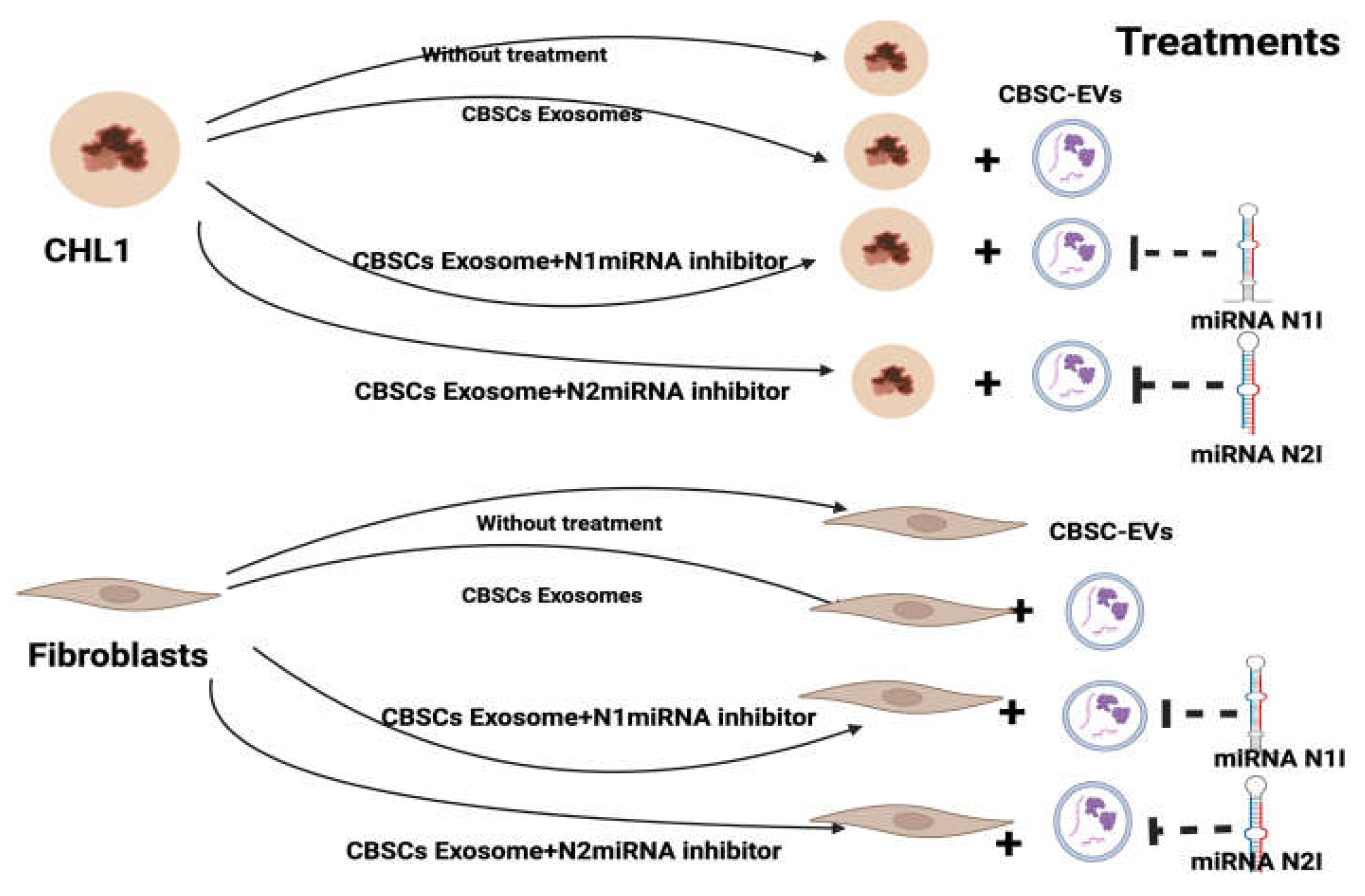
The treatment design for the present study is illustrated in Figure 2. The experimental groups were designed such that one group was treated with exosomes that down-regulated the newly discovered miRNAs, while another group was treated with CBSC-EVs. The treated cells were assessed using various assays including Comet assay to quantify the level of DNA damage, CCK8 to assay cell viability, and RNA sequencing to determine transcript isoforms, gene fusions, single nucleotide variants, and other features without the requirement of any prior knowledge.

Materials and Methods

Cell Culturing and Treatment

Human dermal fibroblasts (HDF) (106-05A, Merck) and a melanoma human cell line (CHL-1) (CRL-3619, ATCC) were cultured in Dulbecco's Modified Eagle Medium (DMEM) in low and high glucose formulations from Thermo-Fisher, supplemented with 10% fetal bovine serum. The cells were incubated at 37°C in a humidified atmosphere with 5% CO2 to maintain optimal growth conditions.
To assess cell viability, the Cell Counting Kit-8 (CCK-8) assay from Abcam (ab228554) was used. In brief, 3000 cells were seeded into each well of a 96-well plate and exposed to different treatments, including CBSCs exosomes (12%), Novel 1 miRNA inhibitor (N1) (5 nM), Novel 2 miRNA inhibitor (N2) (5 nM), a combination of CBSCs exosomes and N1 (12% + 5nM), as well as N2, NC (negative control, untreated cells), and PC (positive control, treated with 80 µM H2O2) (Figure 2). The cells were then cultured for 1 hour, 24 hours, and 48 hours. Subsequently, the CCK-8 reagent was added to each culture, and the plate was incubated for 1-2 hours at 37°C. The absorbance (OD value) was then measured at a wavelength of 450 nm.
For the Comet assay, cells at 80% confluence were treated with CBSCs exosomes (12%), Novel 1 miRNA inhibitor (N1) (5 nM), Novel 2 miRNA inhibitor (N2) (5 nM), a combination of CBSCs exosomes and N1 (12% + 5nM), as well as N2, NC (negative control, untreated cells), and PC (positive control, treated with 80 µM H2O2) (Figure 2). After 24 hours of treatment, cells were harvested and washed with cold Phosphate-buffered saline (PBS). Subsequently, 40 μl of cell suspension was mixed with 0.5% low melting point agarose and spread onto a slide pre-coated with 1% melting point agarose, following the comet assay protocol outlined by Andrew et al. in 2008. Following 24 hours of cell lysis at 4°C, electrophoresis was conducted at 25 V and 300 mA for 30 minutes at 4°C. The slides were then stained with ethidium bromide (20 μg/mL), and 100 nuclei per slide were randomly assessed using a fluorescent microscope at 20X magnification equipped with a CCD camera and Komet 6 software (Andor Technology Ltd, Belfast). Human dermal fibroblasts (HDF) (106-05A, Merck) and a melanoma human cell line (CHL-1) (CRL-3619, ATCC) were cultured in Dulbecco's Modified Eagle Medium (DMEM) in low and high glucose formulations from Thermo-Fisher, supplemented with 10% fetal bovine serum. The cells were incubated at 37°C in a humidified atmosphere with 5% CO2 to maintain optimal growth conditions.

RNA Extraction

The experiment involved the use of QIAGEN RNA isolation kits to isolate RNA from human dermal fibroblasts (HDF) (106-05A, Merck) and a melanoma human cell line (CHL-1) (CRL-3619, ATCC) that were subjected to various treatments, including CBSCs exosomes (12%), Novel 1 miRNA inhibitor (N1) (5 nM), Novel 2 miRNA inhibitor (N2) (5 nM), a combination of CBSCs exosomes and N1 (12% + 5nM), N2, NC (negative control, cells without treatment), and PC (positive control, 80 µM H2O2) (Figure 2).
The sample processing involved the lysis of 5 x 10 pelleted cells following the QIAamp RNA Blood Mini Handbook procedure. To achieve this, 350 µl volume of RNA lysis buffer (RLT) was added to the pelleted cells and mixed thoroughly until no cell clumps were visible. The resulting lysate was then directly transferred into a QIA shredder spin column, placed in a 2 ml collection tube, and centrifuged for 2 minutes at maximum speed to ensure homogenisation.
Following this, one volume of 70% ethanol was added to the homogenised lysate, and the mixture was carefully pipetted into a new QIAamp spin column in a 2 ml collection tube. The QIAamp spin column was then centrifuged for 15 seconds at the highest speed (>8000 x g). The QIAamp membranes were washed with RNA wash buffer (RW1) and centrifuged for 15 seconds at high speed.
The second RNA wash buffer containing ethanol (RPE) was then used to wash the QIAamp membranes, which were centrifuged at full speed (20,000 x g) for 3 minutes. Finally, RNA extraction was performed by adding 30-50 µl of RNase-free water directly onto the QIAamp membrane and centrifuging for 1 minute at high speed (> 8000 x g). The concentration of the eluted RNA was determined using the BioDrop uLite+ Spectrophotometer.

RNA/Transcriptome Sequencing

Library Preparation for Transcriptome Sequencing

mRNA was purified using magnetic beads. First and second-strand cDNA was synthesised using random hexamer primers, followed by end repair, A-tailing, adapter ligation, size selection, amplification, and purification. USER Enzyme Digestion was also performed for the directional library.

Clustering and Sequencing

Samples were clustered according to the manufacturer’s instructions and then sequenced on an Illumina platform to generate paired-end reads. Novogene Ltd. performed the data analysis.

Results

Cell Survival Rate (CCK8)

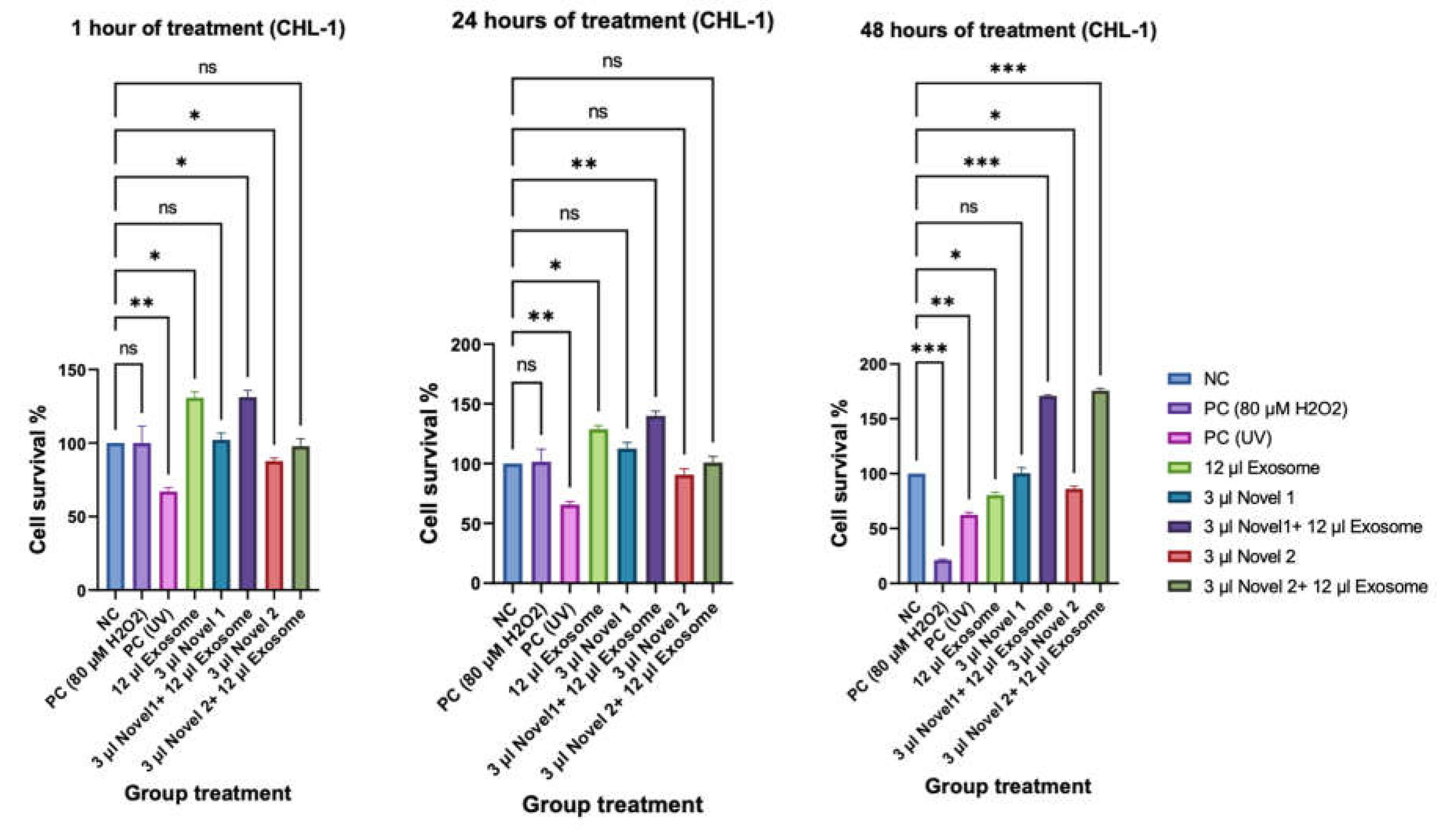
The present study aimed to evaluate the impact of extracellular vesicles (EVs) derived from cord blood stem cells (CBSCs) on the cell viability of human dermal fibroblasts and HCL1 melanoma cells. The CCK-8 assay was conducted to measure the cell viability of both cell types. The results indicated that CBSC-EVs treatment significantly decreased the level of cell viability after 48 hours of treatment (p < 0.001) compared to the untreated cells. However, in CHL1 cells, the knockdown of Novel 1 miRNA by transfecting the cells with the N1 miRNA inhibitor increased cell survival by two fold (p < 0.0001) following CBSC-EVs treatment. The same pattern was observed after treatment with CBSC-EVs and knockdown of Novel 2 miRNA by N2 miRNA inhibitor (Figure 2).
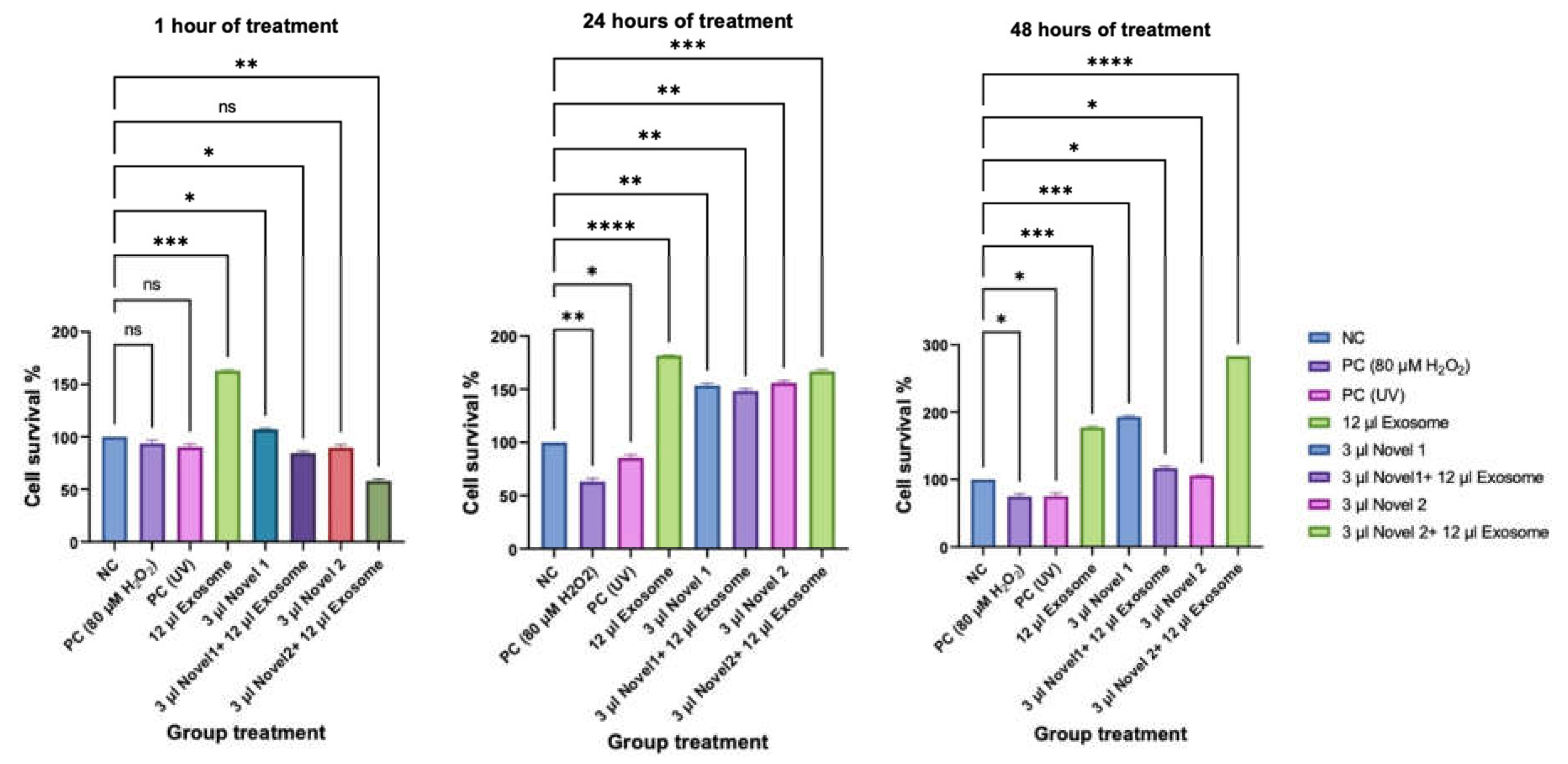
Furthermore, CBSC-EVs treatment significantly increased the level of fibroblasts compared to the untreated cells (p < 0.001). However, after transfecting the fibroblasts with the N1 miRNA inhibitor, the cell survival decreased significantly compared to the untreated cells after 48 hours (p < 0.01) (Figure 3). Conversely, the N2 miRNA inhibitors, after transfecting the CBSC-EVs-treated fibroblasts, improved cell growth and proliferation compared to the untreated cells (p < 0.0001). Overall, these findings suggest that CBSC-EVs treatment has a significant impact on cell viability and growth, which can be modulated by Novel 1 and Novel 2 miRNAs. These results provide valuable insights into the potential use of CBSC-EVs in various therapeutic applications.
Figure 3. The cell survival of CHL1 cells at 1h, 24h and 48h following treatment with CBSCs exosomes, Novel 1 miRNA inhibitor (N1), Novel 2 miRNA inhibitor (N2), NC (negative control, cells without treatment); PC (positive control, 80 µM H2O2); ns stands for non-significant. The number of asterisks denotes the degree of significance between results: * = p < 0.05; ** = p < 0.0016; *** = p< 0.0002; **** = p<0.0001. Errors bars represent the standard error of the mean (SEM).
Preprints 104124 g003
Figure 4. The cell survival of human fibroblast cells) at 1h, 24h and 48h following treatment with CBSCs exosomes, Novel 1 miRNA inhibitor (N1), Novel 2 miRNA inhibitor (N2), NC (negative control, cells without treatment); PC (positive control, 80 µM H2O2); ns stands for non-significant. The number of asterisks denotes the degree of significance between results: * = p < 0.05; ** = p < 0.0016; *** = p< 0.0002; **** = p<0.0001. Errors bars represent the standard error of the mean (SEM).
Preprints 104124 g004

Comet Assay

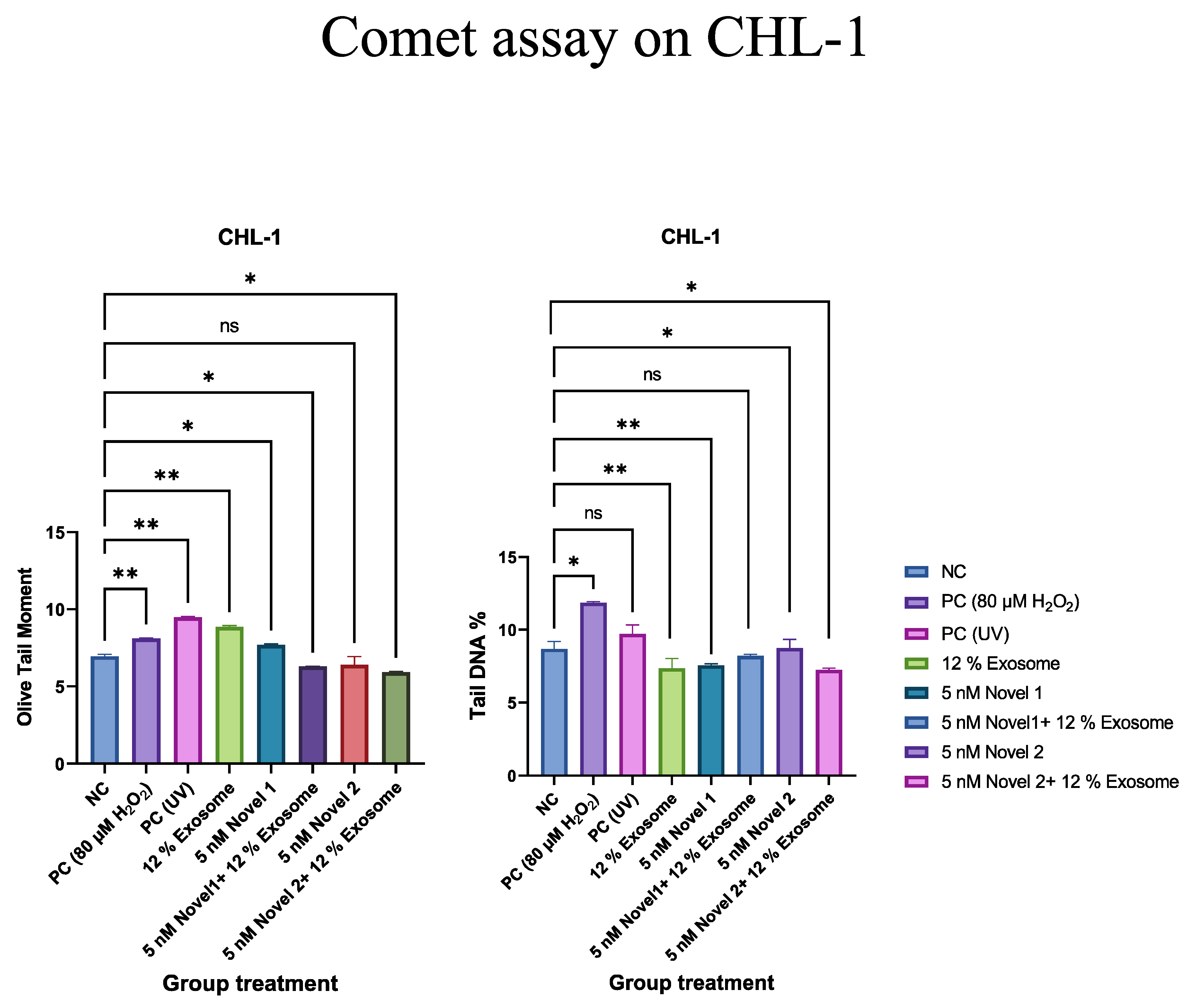
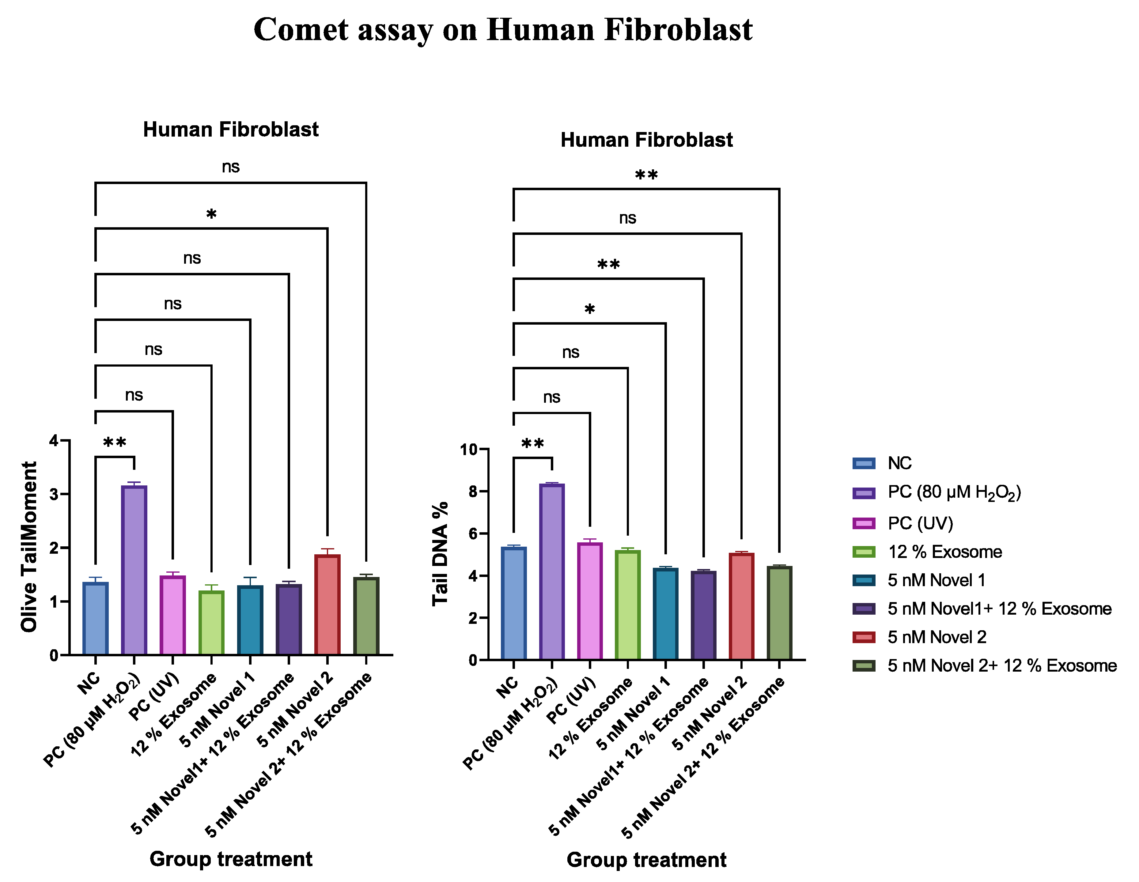
Upon reaching 80% confluence, the cultured fibroblasts and CHL1 cells were subjected to centrifugation, following which, 40µL of the cell suspension were treated with various agents to assess their impact on DNA damage under induced oxidative stress conditions. The treatment agents included 900µL of DMEM as a negative control and 80 µM of H2O2 as a positive control. Moreover, the cells were exposed to UVA (320-400 nm) (1.53 ± 0.01). Statistical analysis showed a statistically significant difference between the negative control and PC (p < 0.0001 and p < 0.001, respectively) (Figure 5 and Figure 6). The findings of the study indicated that the presence of CBSC-EVs resulted in a significant increase in the level of DNA damage in CHL1 cells compared to the untreated cells and the positive control (p<0.01). Moreover, the level of DNA damage in CHL1 cells treated with CBSC-EVs and having knockdown of N1 miRNA and that of N2 miRNA were lower than those treated with CBSC-EVs alone (p<0.001) (Figure 5). Upon conducting 3 times experiments on fibroblasts, it was observed that treatment with CBSC-EVs resulted in a notable reduction in the level of DNA damage. This reduction was found to be statistically significant when compared to the positive control group (p<0.001). However, the reduction was not statistically significant when compared to the cells without treatment. Further, no significant differences were observed between the group of cells treated with CBSC-EVs and the group in which the N1 and N2 miRNAs were knocked down (Figure 6).
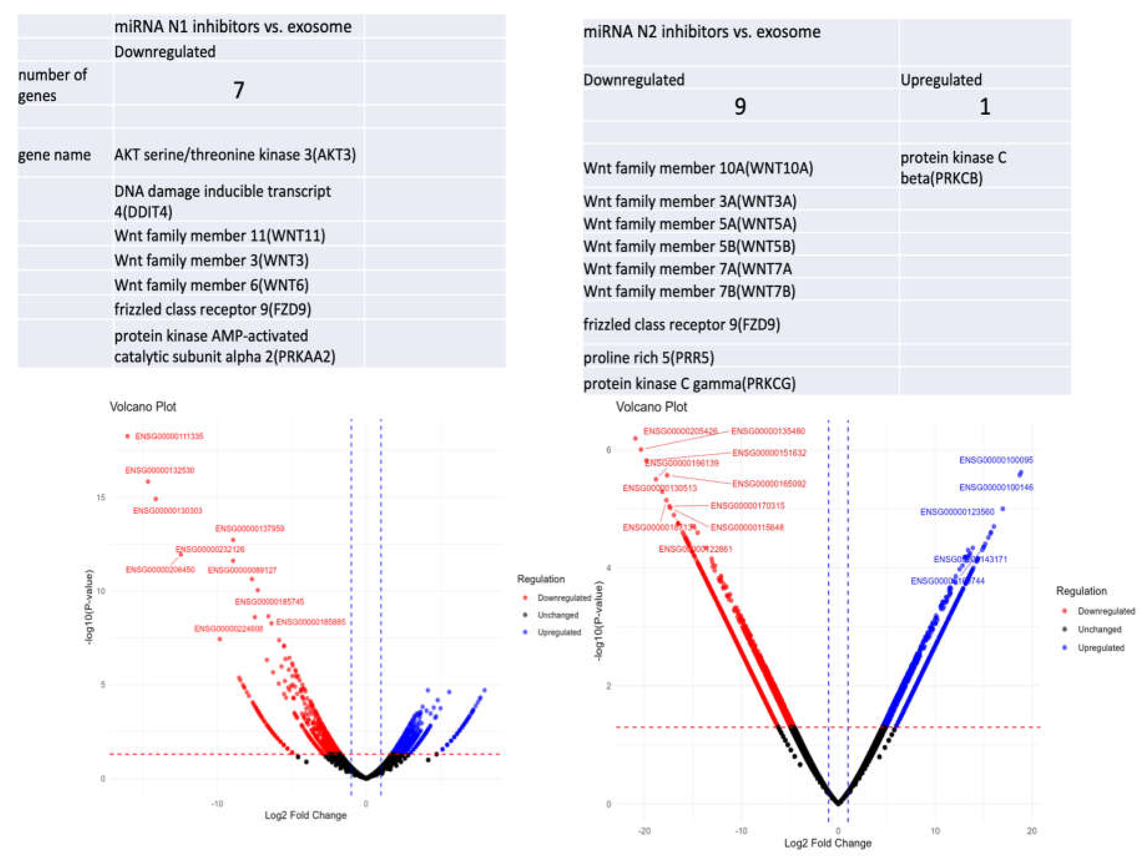
Based on the results obtained from RNA sequencing, it has been identified that a list of genes underwent downregulation, while another list of genes underwent upregulation as a consequence of N1 miRNA. Furthermore, a series of gene expression alterations were noticed due to N2 miRNA. These findings are represented in Figure 7.

Discussion

Over a decade ago, the use of miRNAs as a diagnostic tool was introduced (Kim and Croce, 2023). The therapeutic potential of miRNAs has recently been a subject of investigation. In the context of cancer, research has demonstrated that miRNA-based therapy, which involves the inhibition of oncomiRs or the induction of tumour suppressors, has proven to be an effective approach to cancer treatment (Menon et al., 2022). The presented data indicates that the CHL1 malignant melanoma cell line exhibited significant differences in behaviour when treated with CBSC-EVs, as compared to cells treated with CBSC-EVs and transfected with Novel miRNAs to suppress their functions (Figure 2). The CCK8 results indicate that the cell viability and cell survival percentage reduced after administering CBSC-EVs for 48 hours. However, after transfecting Novel 1 miRNA or Novel 2 miRNA to the CHL1 cells treated with CBSC-EVs, the level of cell survival increased significantly (p<0.001) (refer to Figure 3) which suggests that the Novel miRNAs have a cytotoxic impact on the CHL1 cells. In human fibroblast cell lines, CBSC-EVs demonstrated a significant increase in cell survival (p<0.001). Notably, the novel miRNA inhibitors did not exhibit any adverse effects on cell growth or viability within the first hour, 24 hours, and 48 hours (as illustrated in Figure 4). Moreover, melanoma is a type of cancer that is characterised by mutations in homologous recombination (HR) and DNA damage response (DDR) genes. Additionally, high replicative stress in melanoma cells can lead to an increase in endogenous DNA damage, which in turn triggers the activation of DDR (Maresca et al., 2022). Our study found that CHL1 cells, upon treatment with CBSC-EVs 12%, exhibited a significant increase in DNA damage compared to untreated cells (p<0.01); however, the presence of novel miRNA inhibitors resulted in a significant reduction in the level of DNA damage (Figure 5). This was determined using Comet assay, a highly sensitive diagnostic tool for detecting single-strand DNA breakages (Dunkenberger et al., 2022). The effects of CBSC-EVs on human fibroblasts were found to be contrary, as they caused a reduction in the levels of DNA damage in the cells. This effect was observed even after the suppression of novel miRNAs through the addition of their inhibitors, as shown in Figure 6. These results suggest that CBSC-EVs and the novel miRNAs may have a potential therapeutic application in reducing DNA damage by which CBSC-EVs exert their effects on cellular DNA repair pathways. Furthermore, the PI3K/AKT/mTOR signalling pathway has been identified as a key mediator of BRAF inhibitor (BRAFi) resistance mechanisms. In resistant melanoma tumours, mutations in genes such as AKT1, AKT3, PIK3CA, PIK3CG, PIK3R2, or PHLPP1, as well as PTEN loss, or the overexpression of multiple receptor tyrosine kinases, including epidermal growth factor receptors (EGFRs), insulin-like growth factor 1 receptors, platelet-derived growth factor receptors α and β, or fibroblast growth factor receptor 3 have been reported (Caporali et al., 2016). The study examined the impact of CBSC-EVs on CHL1 cells. Group 1 was treated with CBSC-EVs, group 2 received CBSC-EVs plus miRNA novel1 knockdown, and group 3 was treated with CBSC-EVs plus miRNA novel 2 knockdown. The cells were subjected to RNA sequencing to analyse the changes in gene expression. The results indicated that the genes involved in the mTOR pathways were significantly suppressed and downregulated in group 1 when compared to groups 2 and 3 (Figure 7). The present study investigated the potential of CBSC-EVs and their newly identified miRNAs as a therapeutic strategy for melanoma. Our results demonstrated that CBSC-EVs can significantly decrease cell viability, induce DNA damage, and downregulate the expression of genes involved in mTOR pathways in CHL1 melanoma cells (Figure 7) without causing similar effects in human fibroblasts. This promising data suggests that CBSC-EVs could serve as a novel therapeutic component in the management of melanoma. These findings contribute to our understanding of the potential of CBSC-EVs in cancer therapy and warrant further investigation in preclinical and clinical settings.

Author Contributions

Mojgan Najafzadeh: Project’s Principal Investigator, data analysing, first author. Shohreh Jafarinejad: Second author, the primary researcher and postdoctoral who conducted the study. Sajad Falsafi Zadeh: The research assistant who conducted the bioinformatic assessment. Adi Baumgartner: The expert on cellular DNA damage and cytotoxicity. Mohammad Isreb: The pharmaceutic expert contributor on this project. Pouria Akhbari: The expert on miRNA extraction and cell transfection. Nader Ghaderi: The NHS consultant in charge of collecting blood samples from melanoma patients involved in this study. Farshid Sefat: The expert who was involved in Cord Blood Stem Cells-derived exosomes extraction. Saeed Heidari Keshel: The expert who was involved in Cord Blood Stem Cells-derived exosomes extraction. Rojan Ghaderi: The individual responsible for the review and editing of the text. Diana Anderson: One of the key advisors in this study. Andrew Wright: The main NHS consultant in charge of collecting blood samples from melanoma patients involved in this study.

Funding

The presented study has been funded by the University of Bradford, UK.

Data Availability Statement

Should the need arise, the non-confidential data and accompanying material will be made available for access.

Acknowledgments

The authors express their gratitude to all the healthcare professionals associated with the Dermatology Department at St. Luke's Hospital who assisted in the sample collection process. Their support enabled us to carry out this study.

Conflicts of Interest

It is to be noted that there exists no conflict of interest regarding the publication of the data presented in the article.

Ethics Approval and Consent to Participate

The use of lymphocytes from healthy controls and patients diagnosed with malignant melanoma received ethical approval from the NRES Committee Yorkshire & The Humber - Leeds, with the REC number 12/YH/0464.

Consent for Publication

All participants involved in the project gave their informed consent for the publication of anonymised data.

References

  1. Caporali, S.; Alvino, E.; Lacal, P.M.; Levati, L.; Giurato, G.; Memoli, D.; Caprini, E.; Cappellini, G.C.A.; D’atri, S. Targeting the PI3K/AKT/mTOR pathway overcomes the stimulating effect of dabrafenib on the invasive behavior of melanoma cells with acquired resistance to the BRAF inhibitor. Int. J. Oncol. 2016, 49, 1164–1174. [Google Scholar] [CrossRef]
  2. Debela, D.T.; Muzazu, S.G.; Heraro, K.D.; Ndalama, M.T.; Mesele, B.W.; Haile, D.C.; Kitui, S.K.; Manyazewal, T. New approaches and procedures for cancer treatment: Current perspectives. SAGE Open Med. 2021, 9. [Google Scholar] [CrossRef] [PubMed]
  3. DUNKENBERGER, L. , REISS, K. & DEL VALLE, L. 2022. Comet Assay for the Detection of Single and Double-Strand DNA Breaks. Methods Mol Biol, 2422, 263-269.
  4. Goydel, R.S.; Rader, C. Antibody-based cancer therapy. Oncogene 2021, 40, 3655–3664. [Google Scholar] [CrossRef]
  5. Ha, T.-Y. The Role of MicroRNAs in Regulatory T Cells and in the Immune Response. Immune Netw. 2011, 11, 11–41. [Google Scholar] [CrossRef] [PubMed]
  6. HART, M. , NICKL, L., WALCH-RUECKHEIM, B., KRAMMES, L., RHEINHEIMER, S., DIENER, C., TAENZER, T., KEHL, T., SESTER, M., LENHOF, H. P., KELLER, A. & MEESE, E. 2020. Wrinkle in the plan: miR-34a-5p impacts chemokine signaling by modulating CXCL10/CXCL11/CXCR3-axis in CD4(+), CD8(+) T cells, and M1 macrophages. J Immunother Cancer, 8.
  7. Kalluri, R.; LeBleu, V.S. The biology, function, and biomedical applications of exosomes. Science 2020, 367, eaau6977. [Google Scholar] [CrossRef] [PubMed]
  8. Kim, T.; Croce, C.M. MicroRNA: trends in clinical trials of cancer diagnosis and therapy strategies. Exp. Mol. Med. 2023, 55, 1314–1321. [Google Scholar] [CrossRef] [PubMed]
  9. Lykke-Andersen, S.; Brodersen, D.E.; Jensen, T.H. Origins and activities of the eukaryotic exosome. J. Cell Sci. 2009, 122, 1487–1494. [Google Scholar] [CrossRef] [PubMed]
  10. Mao, J.J.; Ismaila, N.; Bao, T.; Barton, D.; Ben-Arye, E.; Garland, E.L.; Greenlee, H.; Leblanc, T.; Lee, R.T.; Lopez, A.M.; et al. Integrative Medicine for Pain Management in Oncology: Society for Integrative Oncology–ASCO Guideline. J. Clin. Oncol. 2022, 40, 3998–4024. [Google Scholar] [CrossRef] [PubMed]
  11. Maresca, L.; Stecca, B.; Carrassa, L. Novel Therapeutic Approaches with DNA Damage Response Inhibitors for Melanoma Treatment. Cells 2022, 11, 1466. [Google Scholar] [CrossRef] [PubMed]
  12. Menon, A.; Abd-Aziz, N.; Khalid, K.; Poh, C.L.; Naidu, R. miRNA: A Promising Therapeutic Target in Cancer. Int. J. Mol. Sci. 2022, 23, 11502. [Google Scholar] [CrossRef] [PubMed]
  13. Naeem, P.; Baumgartner, A.; Ghaderi, N.; Sefat, F.; Alhawamdeh, M.; Heidari, S.; Shahzad, F.; Swaminathan, K.; Akhbari, P.; Isreb, M.; et al. Anticarcinogenic impact of extracellular vesicles (exosomes) from cord blood stem cells in malignant melanoma: A potential biological treatment. J. Cell. Mol. Med. 2022, 27, 222–231. [Google Scholar] [CrossRef] [PubMed]
  14. O'Brien, J.; Hayder, H.; Zayed, Y.; Peng, C. Overview of MicroRNA Biogenesis, Mechanisms of Actions, and Circulation. Front. Endocrinol. 2018, 9, 402. [Google Scholar] [CrossRef] [PubMed]
  15. Ratajczak, M.Z.; Kim, C.H.; Abdel-Latif, A.; Schneider, G.; Kucia, M.; Morris, A.J.; Laughlin, M.J.; Ratajczak, J. A novel perspective on stem cell homing and mobilization: review on bioactive lipids as potent chemoattractants and cationic peptides as underappreciated modulators of responsiveness to SDF-1 gradients. Leukemia 2011, 26, 63–72. [Google Scholar] [CrossRef]
  16. Ribas, A.; Wolchok, J.D. Cancer immunotherapy using checkpoint blockade. Science 2018, 359, 1350–1355. [Google Scholar] [CrossRef] [PubMed]
  17. Rupaimoole, R.; Slack, F.J. MicroRNA therapeutics: towards a new era for the management of cancer and other diseases. Nat. Rev. Drug Discov. 2017, 16, 203–222. [Google Scholar] [CrossRef] [PubMed]
  18. STERNER, R. C. & STERNER, R. M. 2021. CAR-T cell therapy: current limitations and potential strategies. Blood Cancer J, 11, 69.
  19. Zaborowski, M.P.; Balaj, L.; Breakefield, X.O.; Lai, C.P. Extracellular Vesicles: Composition, Biological Relevance, and Methods of Study. Bioscience 2015, 65, 783–797. [Google Scholar] [CrossRef] [PubMed]
  20. Zhang, H.; Deng, T.; Liu, R.; Bai, M.; Zhou, L.; Wang, X.; Li, S.; Wang, X.; Yang, H.; Li, J.; et al. Exosome-delivered EGFR regulates liver microenvironment to promote gastric cancer liver metastasis. Nat. Commun. 2017, 8, 15016. [Google Scholar] [CrossRef] [PubMed]
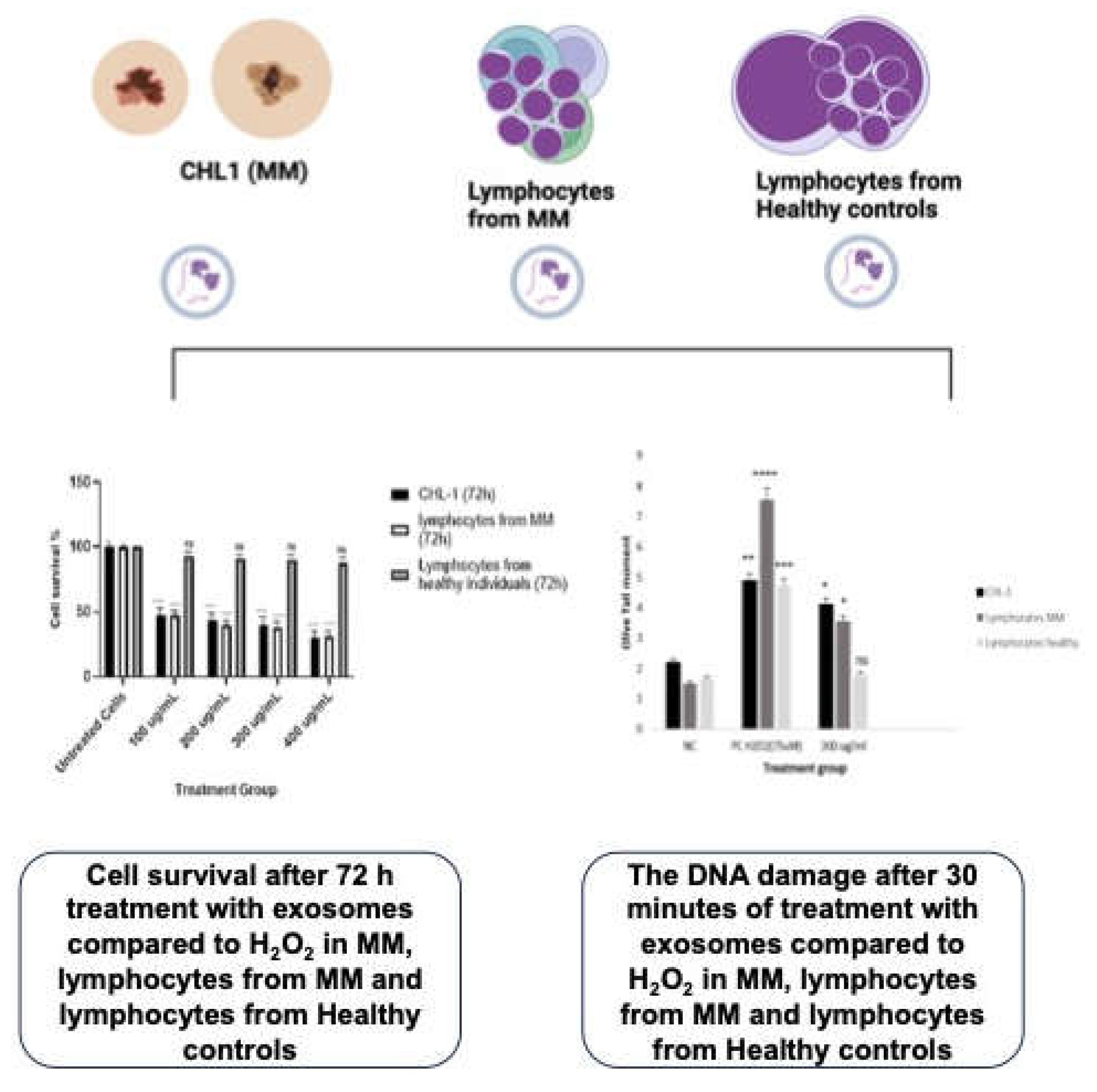
Figure 1. The therapeutic/anti-carcinogenic impact of cord blood stem cells-derived exosomes in malignant melanoma.
Preprints 104124 g001
Figure 2. An illustration of the study treatment design.
Preprints 104124 g002
Figure 5. The impact of CBSCs exosomes, miRNA Novel1 inhibitor (N1), and miRNA Novel 2 inhibitor (N2) on CHL1 (malignant melanoma) cell lines after 24 hours of treatment. The measurement was taken using the OTM in the Comet assay. NC (negative control without treatment); PC (positive control 80µl H2O2); CBSCs exosomes (120µl); CBSCs Exosomes and N1(120µl+3µl); ns stands for non-significant. The number of asterisks denotes the degree of significance between results: (* = p <0.05; ** = p <0.01; *** = p <0.001; **** = p<0.0001; ns = non-significant) errors bars represent the standard error of the mean (SEM).
Preprints 104124 g005
Figure 6. The impact of CBSCs exosomes, miRNA Novel1 inhibitor (N1), and miRNA Novel 2 inhibitor (N2) on human fibroblasts after 24 hours of treatment. The measurement was taken using the OTM in the Comet assay. NC (negative control without treatment); PC (positive control 80µl H2O2); CBSCs exosomes (120µl); CBSCs Exosomes and N1(120µl+3µl); ns stands for non-significant. The number of asterisks denotes the degree of significance between results: (* = p <0.05; ** = p <0.01; *** = p <0.001; **** = p<0.0001; ns = non-significant) errors bars represent the standard error of the mean (SEM).
Preprints 104124 g006
Figure 7. The RNA seq results of CHL1 melanoma cell lines show a significant decrease in gene expression levels in response to blocking the Novel miRNAs N1 and N2 treatment compared to CBSC-EVs. This decrease is particularly noteworthy in genes related to the mTOR pathway. Furthermore, treated cells with CBSC-EVs show a similar trend. The comparison between treated cells with CBSC-EVs and knocked-down novel miRNAs N1 and N2 in CBSC-EVs reveals a clear negative correlation. The study revealed that treating melanoma cells with CBSC-EV/Novel miRNAs exhibits promising potential therapeutic benefits by regulating the mTOR pathway.
Preprints 104124 g007
Disclaimer/Publisher’s Note: The statements, opinions and data contained in all publications are solely those of the individual author(s) and contributor(s) and not of MDPI and/or the editor(s). MDPI and/or the editor(s) disclaim responsibility for any injury to people or property resulting from any ideas, methods, instructions or products referred to in the content.
Copyright: This open access article is published under a Creative Commons CC BY 4.0 license, which permit the free download, distribution, and reuse, provided that the author and preprint are cited in any reuse.
Prerpints.org logo

Preprints.org is a free preprint server supported by MDPI in Basel, Switzerland.

Subscribe

© 2024 MDPI (Basel, Switzerland) unless otherwise stated