2.1. A Superconductivity in Microtubules
Superconductivity was discovered at a low temperature of 4 K a bit more than 100 years ago [
1]. It is a phenomenon, with which a scientist can touch the infinity in the experiment, because electrical conductance becomes infinite, and the resistance reaches absolute zero. It is not surprising that for a long time it was no consensus on the nature of superconductivity [
2], apart that it is a quantum phenomenon. A realistic quantum model of superconductivity came 46 years after its discovery [
3]. This model is very successful in application to metals at low temperatures but not linked to organic or biological matter.
However, straight after the discovery explanations were different. Albert Einstein considered it to be related to supercurrents “carried through closed molecular chains where electrons undergo continuous cyclic exchanges”, Joseph John Thompson to fluctuating electric dipole chains, and Kamerlingh Onnes to superconducting filaments emphasising its one-dimensional (1D) nature [
2]. These intuitive explanations could directly be applied to organic and biological materials. Moreover, in a seminal paper “Possibility of synthesizing an organic superconductor”, W. A. Little [
4] predicted superconductivity at very high temperatures in a 1D organic chain of molecules interacting with periodically spaced molecular complexes. Furthermore, E. H. Halperin and A. A. Wolf suggested searching for superconductivity in biological systems, specifically in low-entropy DNA molecules, brain, and nerve cells [
5]. An experiment confirmed weak induced superconductivity in DNA strands, but not at ambient conditions and only in a contact with another superconductor [
6].
The progress in quantum processing of information, which led to the development of powerful low-temperature superconducting quantum computers [
7], added a new incentive in the search for room-temperature superconductivity. Taking into account the enormous computational power of the brain, it could be inferred that it processes information quantum-mechanically using superconductivity too, but at room temperature. To identify molecular structures in the brain, which could use superconductivity for quantum processing of information, it is useful to review progress in developing superconducting materials with high critical temperature (T
c), i.e., temperature, below which materials become superconducting. Historically, first, elemental metals were checked for their T
c observing that highest T
c of 9.2 K was in Nb. Later, complexity was enhanced adding other chemical elements to Nb and increasing T
c to about 23 K. After that, there were no progress in increasing T
c for few years. The situation changed dramatically when it was found that reducing dimensionality on the molecular level it is possible to have superconductivity in CuO
2 planes at much higher temperatures [
8,
9]. Logically, one could try to enhance complexity even further by packing more elements in a periodical unit and reducing dimensionality to one. Ironically, it was already done naturally on a massive scale in biological life forms, specifically in microtubules which are quasi-one-dimensional structures composed of helically arranged tubulin proteins containing about 3000 atoms [
10,
11].
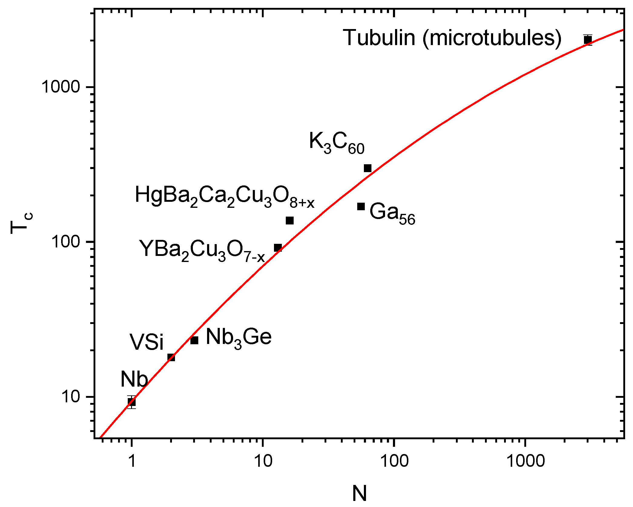
Figure 1 summarizes the increase in T
c with increase of the number of elements (N) in a periodical unit, presented in double logarithmic scale. In the plot, the maximum reported T
c has been used. The points with highest T
c can still be disputed. Therefore, for Ga
56 an K
3C
60, the references are given [
12,
13], and in the following paragraphs, key experimental observations for the highest point will be presented.
First evidences of superconductivity came from electrical transport measurements of the slices of brain, which have a particularly high density of microtubules [
14]. The measurements were done
ex vivo, on the brain slices fixed in formalin. Before the measurements, slices were exposed to a water solution of graphene nano-flakes to provide electrical connection to microtubules. It is important that graphene nano-flakes behave quantum-mechanically at room temperature due to their sub-nanometre thickness, which gives possibility to assess quantum properties of microtubules. It was expected that for the chains of organic molecules, it would not be possible to prove superconductivity by recording temperature dependence of resistance, as predicted T
c is so high [
4] that the chains would be destroyed long before T
c is reached. However, at room temperature, at which the chains are stable, electrical measurements should give superconductor-like current-voltage characteristics with a dissipation-free current up to its critical value and a transition to a normal resistance state. These current-voltage characteristics were measured in [
14] in a special case when potential leads were not suppressing superconductivity by the proximity effect in a delicate network of microtubules.
Additionally, it was found that transition from the superconducting to resistive state at the increase of current takes place by characteristic jumps of voltage equal to the minimum energy necessary to destroy superconductivity, namely 2Δ/e, where Δ is energy gap of superconductor and e is charge of electron. When superconductivity is suppressed close to potential leads, dissipation-free current is absent, but the conductance-voltage characteristics show increase in conductance at the voltage of 2Δ/e for the suppression at both leads or Δ/e at one of them. This allowed measuring directly 2Δ/e, which was found to be 615 ± 48 mV and estimating
using standard relation [
15]:
where k is the Boltzmann constant and Δ(0) is the energy gap at zero temperature. This relation is applicable to all superconductors with a coefficient on the order of unity. The estimated T
c appeared to be very high, 2022 ± 157 K and close to that predicted by Little [
4]. Another feature in current-voltage characteristics confirming superconductivity was a strong decrease of non-linearity at superconducting quantum conductance of
, where h is Planck constant [
16]. Still, this work assessed only large statistical ensembles of microtubules. Individual microtubules were measured by Sahu et al. [
17] who registered unusually high conductivity of microtubules with 1000 times increase when water was present inside them. It was also reported that at certain resonance frequencies, microtubules conducted electrical signals with almost no resistance.
These observations of low resistance in combination with measured energy gap and registration of a feature linked to the superconducting quantum conductance are strong arguments in favour of superconductivity. However, for the completeness, proof of Meissner effect [
15], or expulsion of the magnetic flux from the microtubules in the field-cooled regime, and the evidence of flux quantisation are also necessary. This was done by nano-technique of magnetic force microscopy (MFM) [
18].
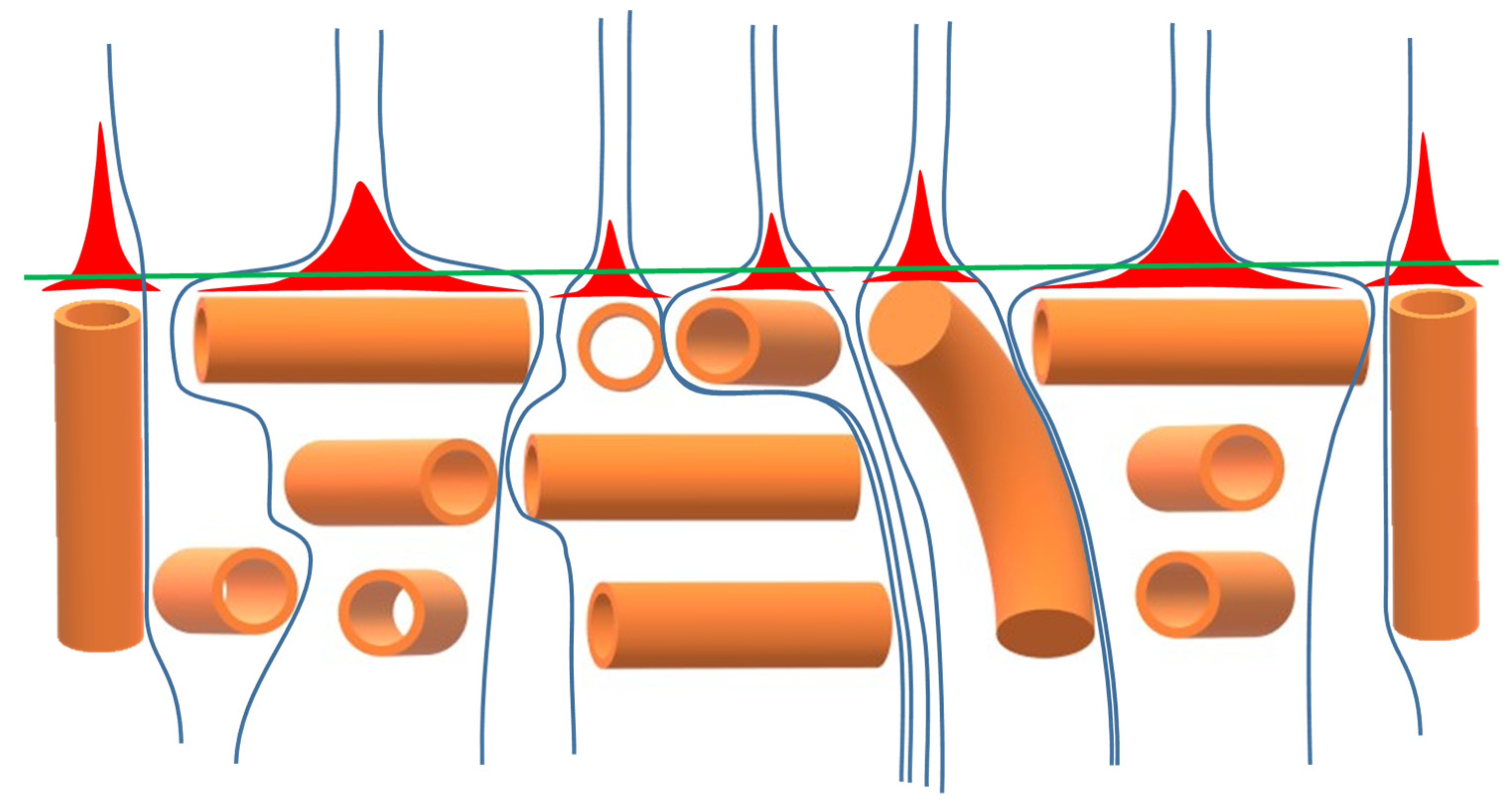
The MFM allows measuring magnetic properties of the surfaces with the resolution of a few nanometres. Still, measurements of the individual microtubules are very challenging because magnetic flux lines are expected to be distorted on distances comparable with the diameter of microtubule, which is 25 nm. Schematically, the distortion of magnetic field lines close to the surface by microtubules is shown in
Figure 2. To detect flux expulsion from them (red areas in
Figure 2), magnetic probe should be very close to the surface, therefore, its roughness should be at least comparable with diameter of microtubules. This condition was satisfied by the extraction of tubulin protein from a brain slice rinsing it with water solution of graphene and setting a drop of the solution on a smooth glass slide. With the evaporation of water, the concentration of tubulin protein reaches a critical value, and the microtubules start self-assembling on the smooth surface of drop. Exploring different areas, both in-plane (middle area in
Figure 2) and perpendicular to the surface (edges in
Figure 2) microtubules has been imaged. When moving probe close to the surface (green line in
Figure 2), it is consequentially coming to the areas of screened magnetic flux (red in
Figure 2). In these areas, the attraction to permanent magnet below the sample is lost, and the magnetic probe start moving up initiating a positive feedback loop due to reduction of van der Waals force, which is strong at these distances too. This creates rather strong signal in MFM.
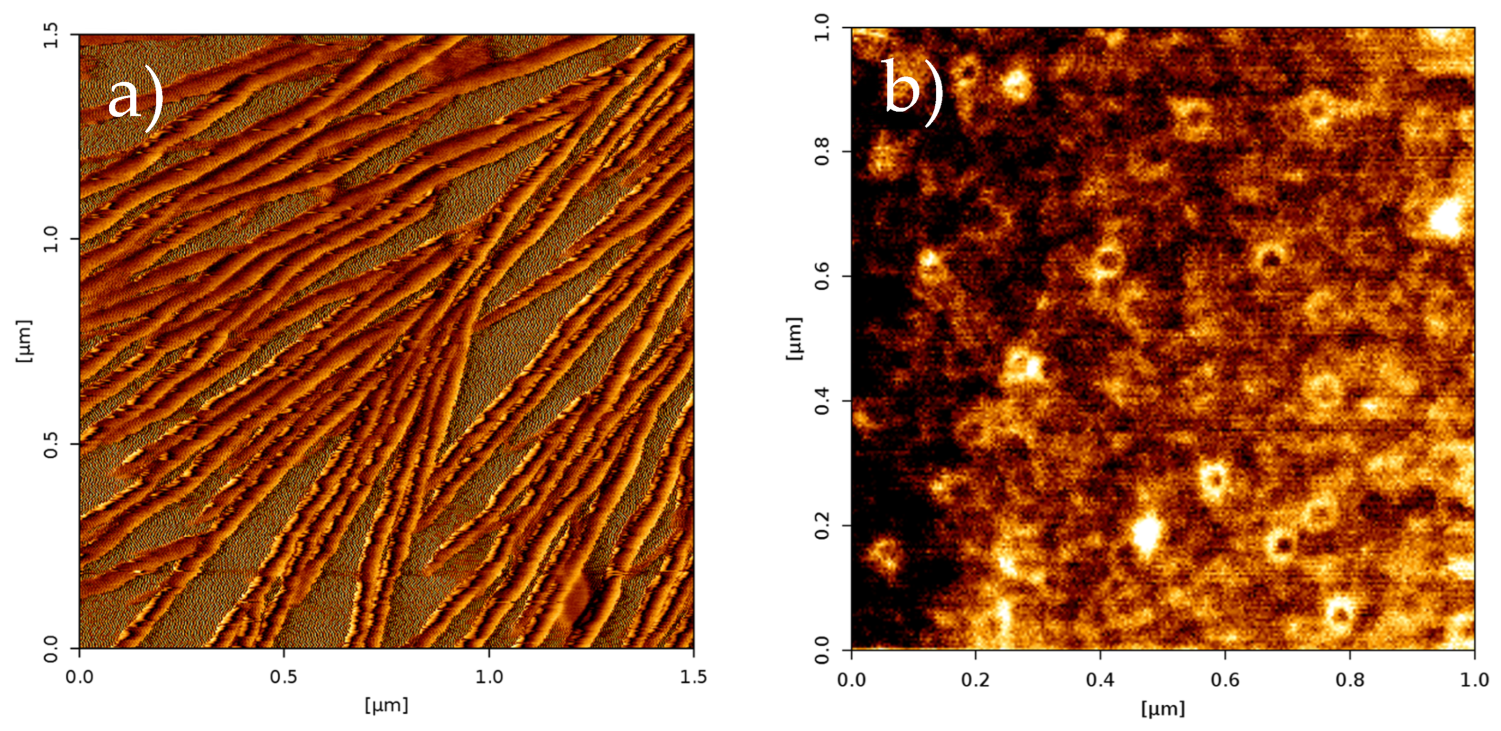
Figure 3 shows examples of MFM images for the in-plane (a) and out-of-plane (b) microtubules. The in-plane microtubules are seen as the lines growing and spreading on the surface of the drop. The out-of-plane view gives cross-sections of the microtubules. The magnetic state of the microtubules was examined via phase shift of the probe oscillations, and it was found that they fully expel magnetic flux [
19]. Since the formation of microtubules takes place in the presence of magnetic field, it is the Meissner effect on the nanometre length scale.
Moreover, it was found that when microtubules are close to each other, there are superconducting correlations between them, which transforms superconductivity from quasi 1D to 2D, and even 3D one. In this case, it is possible to trace position of magnetic field lines shown by blue in
Figure 2, which will experience effect of flux quantisation. The simplest configuration for such experiment is when microtubules are aligned perpendicular to the surface, as in
Figure 3b. Then magnetic flux enters between microtubules and is easy to identify. In the phase shift of MFM, at a certain distance of a few nanometres from the surface, when admixture of van der Waals force is minimal, the areas containing magnetic flux were found as dark spots on phase shift map with magnetic flux equal to superconducting flux quantum (individual vortices) [
20]. Additionally to Meissner effect, confirmed flux quantisation is a very strong argument in favour to superconductivity, and in general to quantum-mechanical behaviour of microtubules at room temperature.
Identifying superconducting flux vortices allowed reconstructing distribution of magnetic field around them and calculating magnetic penetration depth λ (12.8±0.63) [
20]. The measurement of energy gap of superconductor [
14], on the other hand, gives the value of coherence length ξ (1 ± 0.08 nm). The ratio of these parameters is Ginzburg-Landau parameter k = λ/ ξ, which is found to be 12.8±2.6. Since it is bigger than
[
15], this ensures that superconductor is of type II, i.e., promotes quantisation of the magnetic flux, which was found in the experiment.
Another important parameter of superconductor is wavelength of the Cooper pairs
, i.e., the wavelength of united electrons. In a superconductor, it is related to the superconducting gap energy
and can be calculated using the following formula [
15]:
where:
is the mass of the electron. Substituting all parameters into Equation (2), one can find that
is equal to 2.2±0.003 nm. It is half of the size of the tubulin protein in the microtubule, i.e., a resonant condition is satisfied, in which two wavelengths of Cooper pairs are confined in each periodical unit of the microtubule. This fits well to the intuitive suggestion of A. Einstein about “molecular chains where electrons undergo continuous cyclic exchanges” and further supports quantum-mechanical behaviour of microtubules at room temperature. To summarize, parameters of room-temperature superconductor are presented in
Table 1.
2.2. B Josephson Radiation
As it is seen from
Figure 3a, the straight fragments of microtubules are not very long. They are frequently coming in contact with each other and stop growing after that. Although they are longer than in
Figure 3a in neurons, their length rarely exceeds tens of micrometres, while the length of neurons typically varies from a few millimetres to tens of centimetres. This means that individual microtubules are linked with each other along the length of neuron [
21,
22]. It is well established that microtubules have positive (growing) and negative (stable, blocked with very thin capping layer) ends. The plus-minus connections between the microtubules [
22] are through the capping layer. From the point of view of superconductivity they are Josephson junctions. The junctions are of primary importance for quantum processing of information and therefore for functioning of living organisms [
23]. They preserve superconducting correlation between microtubules but cause oscillation of supercurrent when a voltage is applied to it, with the emission of Josephson radiation [
15]. The frequency
of Josephson radiation is proportional to the applied voltage
and equals to:
where coefficient 2 accounts for paired electrons. The wavelength of radiation is correspondingly:
where
is the speed of light.
One of the most important facts established in molecular biology is the propagation of action potential along the membrane of the neuron (or its axon) [
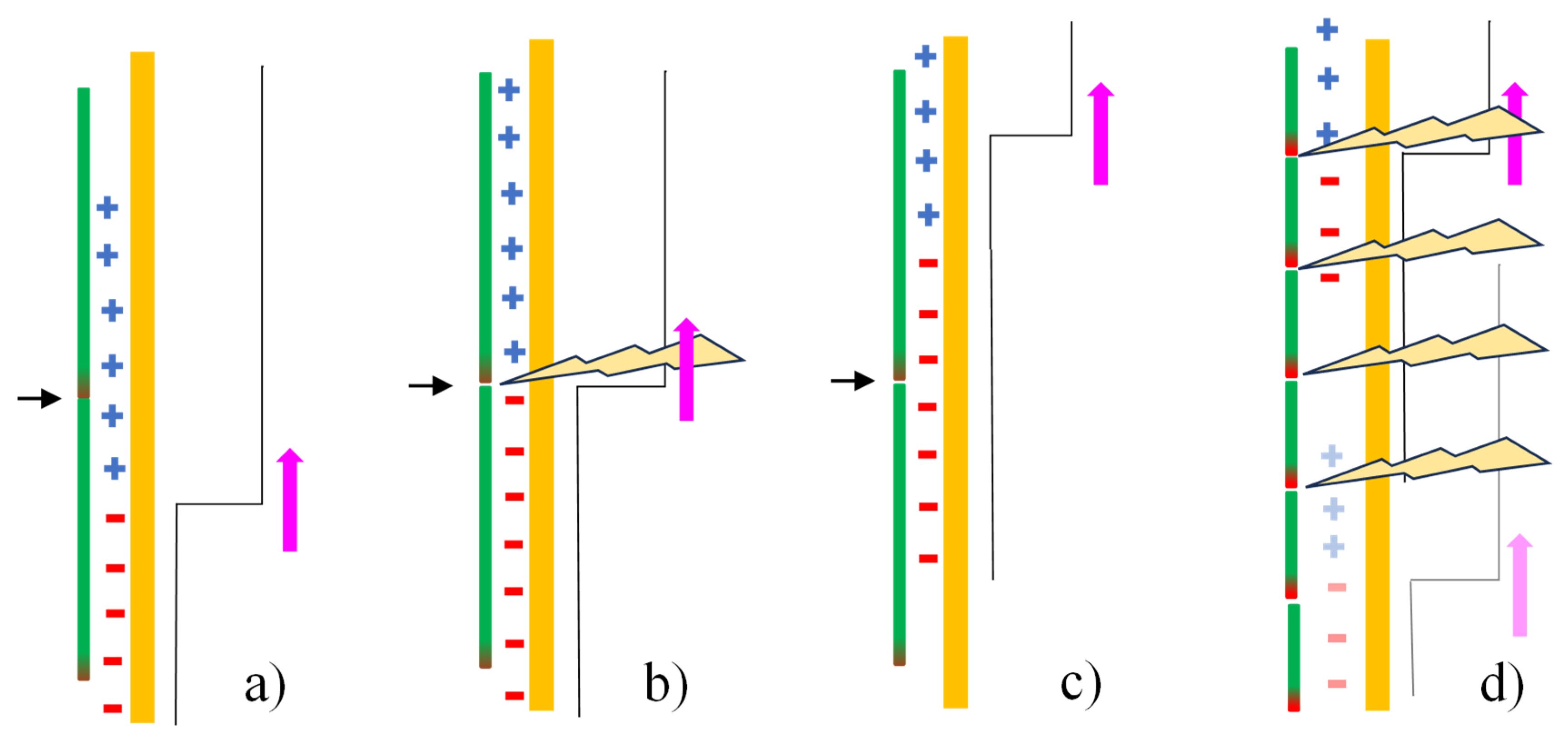
24]. Here it is stated that this propagation results in something really remarkable – generation of coherent Josephson radiation in the connections of microtubules which are close to the membrane. The mechanism of the generation is presented in
Figure 4.
In the images in
Figure 4a–c, position of Josephson contacts between the microtubules (green vertical lines with capped negative end in red) is marked by the horizontal black arrows. The membrane is shown in orange, and the action potential is a step-like line to the right with the corresponding distribution of ion charges along the membrane represented by blue pluses and red minuses. The magenta vertical arrows show direction of the propagation of action potential. In a) and c), there is no potential difference on the Josephson contact, while it is present for a short time in b) and produces a pulse of radiation. The pulses from different contacts join coherently during the propagation of action potential, as it is shown in image d). In the latter image, initial position of the action potential and charge distributions are shown in a weaker contrast. The generation is similar to the emission of coherent light in a solid-state quantum cascade laser [
25].
Since the value of action potential propagating along membrane is well known, typically being close to 70 mV [
25], it is not difficult to calculate the frequency and wavelength of the coherent Josephson radiation coming from contacts of microtubules using Equations (3) and (4), respectively. They appear to be about 33.8 THz and 8.8 µm. The chains of connected superconducting microtubules act as ideal transducers of voltage to the frequency of coherent radiation. Each set of microtubules, which are close to the membrane, generates the same radiation. Each neuron generates similar radiation with slight differences defined by the small differences in the action potential. Altogether, hundreds of billions of neurons create a symphony like a music in a symphony orchestra. However, in contrast to the orchestra, these are not mechanical vibrations on kHz frequencies, but electromagnetic waves on the frequencies some 10 orders of magnitudes higher. There are about 300 strings of the instruments in a typical symphony orchestra, while the number of “strings” in the brain, which produce coherent radiation could be as big as 10000000000000. This radiation has very specific spectrum for every alive organism but still in the same range of frequencies close to 34 THz. It is streamed inside and outside of the bodies. Inside, it synchronises functions of organism, outside it is used for communications.
The radiation resonates on all anatomical structures, which are frequently equal to integer numbers of wavelengths of the radiation. The ideal resonators are neuron and blood vessel walls, and also red blood cells whose diameter is exactly one wavelength of the radiation. The radiation is in the window of transparency of the Earth atmosphere similar to the window for the visible light, but the transparency is not ideal, which keeps most of radiation inside the bodies in the form of standing electromagnetic waves. These standing waves are our perceptions, emotions, memory, nearly everything that we are. It is correct that we are made of atoms, but we are electromagnetic waves. It is not a general statement. The frequencies and wavelengths of these electromagnetic waves are known, but difficult to access experimentally. They are about 34 THz and 9 µm, respectively. Due to subtle anatomical differences, there are no identical brains, bodies, and identical spectra of electromagnetic waves. There could be a majestic symphony or irregular dissonance of the strings inside the organism. Decoding the symphony is outside the abilities on our current technological level due to the enormous number of strings and resonators. In spite of the superconductivity in the microtubules, the generation of radiation is dissipative with considerable amount of heat needed to be removed from the brain and other parts of organisms. Still, it is a very elegant process of functioning on the quantum-mechanical level with direct access to quantum features of the environment around and direct quantum-mechanical processing of information [
23].
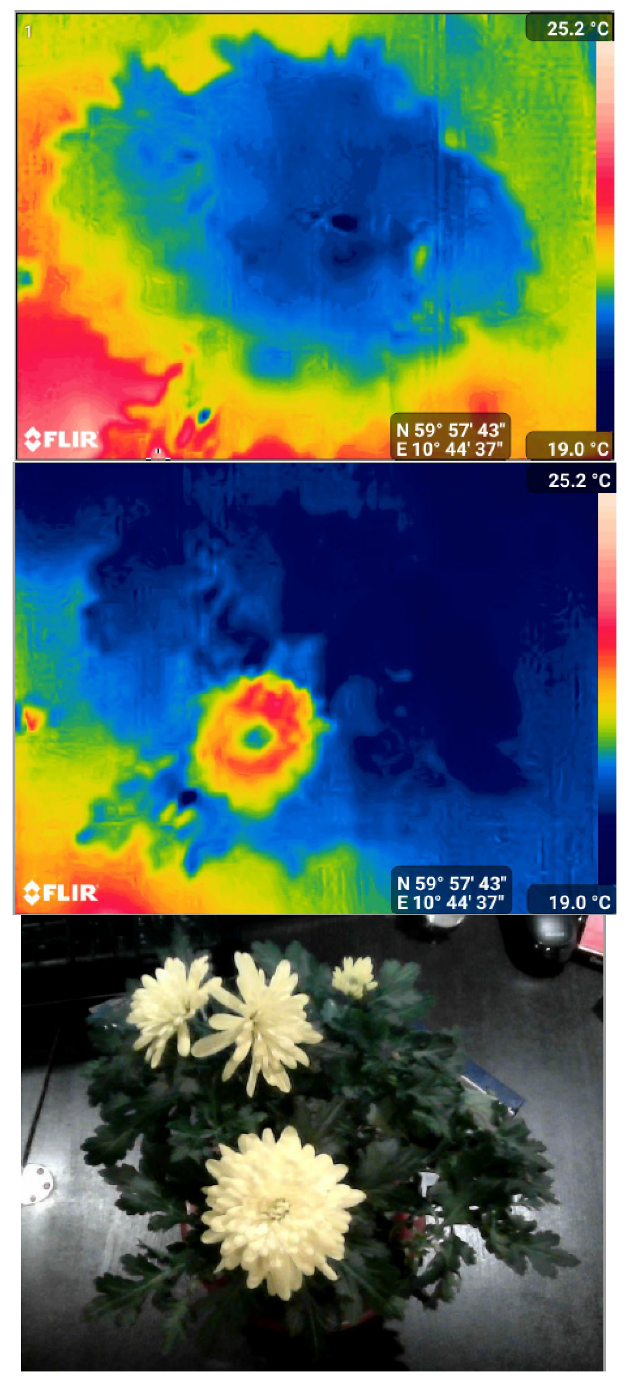
Without spectral resolution, coherent Josephson radiation is readily accessible by thermal cameras like Flir One Pro
TM, although with some difficulties of separating it from the blackbody radiation [
26]. The Flir One Pro camera measures radiation in the range of the wavelengths from 8 to 15 µm, which is outside of the peak of blackbody radiation for typical objects, for example human body at the temperature of 36.7 °C (16 µm), but close to the peak of the coherent Josephson radiation emitted by alive organisms. Analysing radiation detected by this and similar cameras could be an exciting research direction in tracing the functioning of living organisms and following their quantum-mechanical behaviour. An example is activation of a flower by the radiation coming from the palm of hand kept for half of minute at a distance of about one centimetre shown in
Figure 5.
In
Figure 5 a), the thermal image of the plant before the activation is shown. The plant is seen in blue having temperature slightly below the temperature of the background. In b), thermal image of the activated flower is seen in red and yellow. The image c) is the optical image of the plant. In b), one could see that additionally to the biggest flower, the flower above is also partially activated. The activation lasts a short time, and the influence of the coherent radiation still needs to be separated from the effect of incoherent thermal heating in this experiment. From the point of view of quantum mechanics, the processes in in the flower plant are not much different from the processes in human body. They have the same microtubules, but in different quantity and connected in a different way. Both, however, are parts of the same tree of life and linked with each other, also by coherent Josephson radiation.
The radiation, and an active response to it, are features of all living organisms, which have nearly identical microtubules and similar ways of using them for quantum-mechanical processing of information, synchronisation of functions and communication on THz frequencies. The possible practical applications of this concept are straightforward. On an advanced level, via decoding spectra of radiation, the information could be channelled into the brain directly on THz without need of monitors, speakers, or other devices. It could give complete simulation of reality nearly undistinguishable from our everyday experiences, and perhaps create a majestic symphony in the brain. However, even without decoding of spectra, THz radiation could be used for healing, relaxation, harmonisation, and improvement of wellbeing.
