Preprint
Review

Nuclear miRNAs: gene regulation activities

Altmetrics

Downloads

96

Views

34

Comments

0

A peer-reviewed article of this preprint also exists.

Submitted:

29 April 2024

Posted:

30 April 2024

You are already at the latest version

Alerts
Abstract
MicroRNAs are small non-coding RNAs, which contribute to the regulation of many physiolog-ical and pathological processes. Conventionally, miRNAs perform their activity in the cyto-plasm, where they regulate gene expression by interacting in a sequence-specific manner with mature messenger RNAs. Recent studies point to the presence of mature miRNAs in the nucleus. This review summarizes current findings regarding the molecular activities of nuclear miRNAs. These molecules can regulate gene expression at the transcriptional level by directly binding DNA on the promoter or the enhancer of regulated genes. MiRNAs recruit to these regions dif-ferent protein complexes, resulting both in activation or repression of transcription, through a number of molecular mechanisms. Haematopoiesis is presented as a paradigmatic biological process whereby nuclear miRNAs possess a relevant regulatory role. Nuclear miRNAs may af-fect gene expression also acting on nuclear mRNA processing and on the biogenesis of miRNA themselves by regulating pri-miRNA maturation. Overall, nuclear miRNAs are biologically ac-tive molecules that can be critical for the fine tuning of gene expression and deserve further studies in a number of physiological and pathological conditions
Keywords: 
Subject: Biology and Life Sciences  -   Biochemistry and Molecular Biology

1. Introduction

MiRNAs are classically regarded as negative regulators of messenger RNA (mRNA) function. This activity is mainly obtained through both the repression of mRNA translation and induction of mRNA degradation, upon the binding of miRNAs to the 3’ untranslated region (3’ UTR) of mRNAs [1,2]. These fu
nctions of microRNAs take place within cellular cytoplasm, through their binding to mature mRNAs [3]. However, a number of reports indicate that miRNAs may possess wider activities, that include the direct regulation of chromatin structure and transcription [4,5]. This type of regulatory activity could be activating or repressing, depending on the protein complexes that miRNAs drive to chromatin DNA. Therefore, miRNAs are active within the nucleus, where they must remain or be transported back to the nuclear side of the nuclear membrane.
This review briefly summarizes current evidences indicating such non - canonical nuclear activities of miRNAs, highlighting their biological roles in the regulation of haematopoiesis.

2. Biogenesis of MicroRNAs

The biogenesis of miRNAs takes place in the nucleus and cytoplasm in several steps. (Figure 1). They are typically transcribed by RNA polymerases II or III as long primary miRNAs (pri-miRNAs) with an internal loop structure containing a double-stranded stem region and an apical loop. The pri-miRNAs are transformed into precursor miRNAs (pre-miRNAs), structures of approximately 70 nucleotides (nt), by means of a protein complex consisting of the RNase III enzyme Drosha and the DGCR8 protein (Di George syndrome critical region 8 gene) that binds double-stranded RNA (Figure 1(1)) [6,7,8]. The pre-miRNAs are further processed after being translocated into the cytoplasm. The export mechanism involves a protein complex consisting of Exportin5 (XPO5), a member of the karyopherin β family, and a GTPase (Ran GTP) [9,10] (Figure 1(1)). Pre-miRNAs in the cytoplasm are converted into mature, 22 nt duplex miRNAs by hairpin cleavage by the enzyme DICER [11]. The ds-miRNA molecule is then loaded onto the miRNA induced silencing complex (miRISC), a ribonucleoprotein complex consisting of the Argonaute (AGO1-4), DICER, TRBP and TNRC6A proteins. TRBP identifies the 'guide' and 'passenger' strands in the molecule. TRBP loads the ds-miRNAs in the correct orientation onto the AGO proteins based on their thermodynamic properties and, once it recognizes the strand with the least stable 5' end, the AGO protein duplexes and removes the passenger strand to form the mature miRNA molecule [12,13] (Figure 1(2)). The AGO protein family contains several members, among them AGO2 possesses the activity of cutting RNA molecules. AGO2 is important for miRNA maturation and for gene silencing mechanisms regulated by small RNAs [14,15]. Several previous studies have found that miRISC is imported into the nucleus of mammalian cells and that the nuclear RISC (a complex of ~158 kDa) is about 20fold smaller than its cytoplasmic counterpart (a complex of nearly 3 MDa). Western blotting analysis showed that nuclear RISC consists of various proteins, such as AGO and GW182/TNRC6 [16,17]. In the study by Kalantari et al [18] semi-quantitative mass spectrometry confirms that the association of AGO2 with GW182/TNRC6 and AGO3 is well conserved and more stable in both nucleus and cytoplasm [19]. In contrast, AGO2 interactions with DICER and TRBP are limited to the cytoplasm. The import process of miRNAs into the nucleus involves various nuclear transport receptor proteins [20,21]. It has been discovered that the nuclear and cytoplasmic movement of miRNAs is driven by importin 8 and exportin 1. These are two members of the importin family that recognize the nuclear localization sequences in proteins and perform their active transport through the nuclear pore complex [22,23,24,25,26].
To enter the nucleus, miRNAs must first form a complex with the AGO protein, which binds to importin 8 and TNRC6A [20,26] (Figure 1(3)). TNRC6A can move from the nucleus to the cytoplasm and back again as it possesses a nuclear localization signal (NLS) and a nuclear export signal (NES) [25,26]. Consequently, by interacting with TNRC6A, AGO2-miRNA complex enters into the nucleus. TNRC6A guides the miRNA AGO complex to the target gene, in its promoter region complementary to seed region. Thus, TNRC6A is a central protein involved in miRNA-mediated gene transcription regulation [27,28]. Furthermore, AGO-TNRC6 complex can move to the cytoplasm interacting with exportin 1 (Figure 1(4)) [29]. In contrast, DICER and TRBP enter the nucleus without being linked to RISC.

3. Evidence of miRNAs in the Nucleus

Meister et al., by studying Hela cells, demonstrated that mature miR-21 can be found in the cytoplasm and in the nucleus. This discovery suggested that a number of mature miRNAs may be transported back into the nucleus, where they may exert a functional activity [30,31]. In 2009, Földes-Papp et al. used femtosecond laser microscopy to assess the association between molecular beacon probes and human single-stranded cellular targets of miR-122 [32]. They demonstrated that mature miR-122 from the cytoplasm enters the nucleus of human hepatocytes. In recent years, more and more studies have highlighted the presence of mature nuclear miRNAs in most mammalian cells thanks to high-throughput analyses [4,33]. Several studies have studied the distribution of microRNA in the cytoplasm and nucleus, taking advantage of high performance next-generation sequencing and microarrays [34,35]. A sequencing study of nuclear and cytoplasmic fractions deriving from endothelial cells subjected to hypoxia highlighted an increased localization of miR-210-3p in the nucleus. By using confocal microscopy, we studied miR-223 localization during retinoic acid-induced granulopoiesis in HL60 cells and primary blasts. In these conditions mature miR-223 translocates to the nucleus and shows an increasing nuclear pattern. Furthermore, in situ hybridization experiments showed that, following treatment with retinoic acid, miR-223 shows the ability to bind metaphase chromosomes, where RNA transcription is absent or minimal [36]. The study of the sequence of nuclear miRNAs has highlighted the existence of a nuclear localization signal (AGUGUU) at their 3′ terminal sequence [33]. Some nuclear miRNAs such as miR-193b, miR-19, miR30b, miR-30c, miR-590-5p, miR-374 and miR-374b contain very similar nuclear localisation motifs in their sequence with small variations, including UGUGUU, ACUGUU, AGAGUUU, AGUCUUU, AGUGAU, AGUGUA, AGNGUN [4,37]. It is hypothesized that these specific sequences targeting miRNAs to the nucleus are recognized by the nuclear pore structures, allowing their entry. Interestingly, the nuclear localization of a specific miRNA varies based on the tissue type. For example, miR-29b has a nuclear localization in HeLa cells but not in other cell lines [38], suggesting a tissue-specific functional role of nuclear localization. Overall, the significant number of recent data showing the nuclear localization of miRNAs, supports their functional role into the nucleus.

4. Functions of miRNAs in the Nucleus

Many functions of nuclear miRNAs have been reported in recent years, among them very important is their ability to bind complementary sequences located on gene promoters or enhancers. The action of nuclear miRNAs associates with mechanisms of epigenetic modifications of DNA or chromatin status at the targeted genomic sites, leading to transcriptional activation or repression of target genes [94]. Nuclear miRNA activity depends also on the genomic location of binding sequences and on the epigenetic status at these sites (such as presence of TATA box motifs or CpG island regions) [39]. In addition, miRNA target regions can be located far away from the transcription start site (TSS) [40].

5.1. Transcriptional Activation

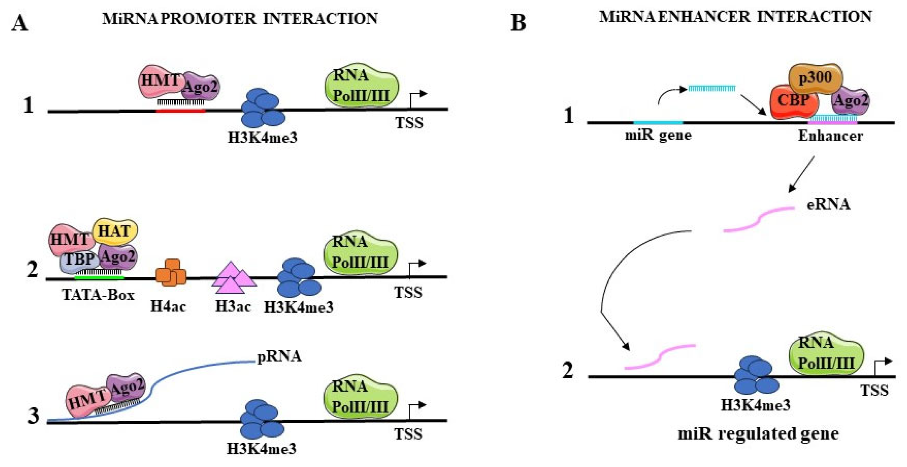
Numerous studies show how miRNAs can mediate transcriptional gene activation (TGA) by interacting with the promoters (Figure 2A), or with enhancers (Figure 2B), of their target genes.

5.1.1. Interaction with Promoters

One mechanism by which miRNAs activate gene transcription could be the association with promoters and their epigenetic regulation. MiR-589 targets the promoter RNA of cyclooxygenase 2 (COX-2) by recruiting AGO2 and GW182 (TNRC6A) to form a complex. This complex can modify histones through association with WDR5, a protein that stimulates histone methyltransferase activity. Consequently, in the COX-2 promoter H3K4me3 and H4 acetylation levels increase, and gene transcription is activated [48] (Figure 2(A1)). FOXO3, an important gene in the regulation of ovarian follicular development and atresia, is regulated by miR-195-5p. This miRNA enters the cellular nucleus, associates with AGO2 and interacts with the FOXO3 promoter by recognizing a complementary sequence in the TATA-box (Figure 2(A2)). Consequently, histone acetylation, hypomethylation, and transcriptional activation take place [41]. Transcriptional activation markers such as acetylated H3 and H4 and H3K4me2 are enriched in the promoter of the interleukin (IL) tumor suppressor genes IL24 and IL32, at specific sites bound by miR-205. As a consequence, RNA polymerase II (Pol II) is recruited and, subsequently, transcribes the IL24 and IL32 genes [42,43]. MiR-744 and miR-1186 increase transcription of the cyclin B1 gene (Ccnb1), by interacting with sequences on this gene promoter, which are highly complementary to their respective seed regions. Moreover, miR-744 association with AGO1 causes an enrichment of Pol II and H3K4me3 marks at the Ccnb1 TSS [44].
Overall, three types of interactions have been hypothesized between miRNAs and the promoters of the genes that are regulated at the transcriptional level. One type of interaction (Figure 2(A1)) leads to the formation of a direct miRNA-DNA double helix complex. In this case, miRNA binds the promoter in association with AGO proteins and recruits histone modifiers that increase the levels of activating markers, such as H3K4me3, while decreasing the inhibitory histone marks [47]. In a second model the miRNA-AGO complex interacts directly with the TATA box motif region or sites on the promoter also bound by transcription factors (Figure 2(A2)). This binding event leads to the recruitment of TATA box-binding protein (TBP), Pol II and histone-modifying proteins such as methyltransferases or acetyltransferases, resulting in gene transcriptional activation [45,46]. Among miRNAs that act accordingly to this model there are miR-181d, let-7i and miR-138, which activate transcription of c-myc, IL-2 and insulin genes, respectively by interacting with the TATA-box on the promoter of these genes. In a last model, miRNAs bind to the promoter-associated sense or antisense RNA (pRNA), transcribed by the target gene promoter. Again, as in the other models, this interaction recruits transcription factors, histone modifiers and Pol II to transcriptionally regulate gene expression [48,49,50] (Figure 2(A3)).

5.1.2. Interaction with Enhancers

MiRNA may mediate transcriptional activation through the regulation of enhancers. In this case, miRNA binding loci are located in the proximity of enhancer regions displaying the typical histone marks of functional enhancer regions, such as H3K27ac (Figure 2B). miRNAs are transcribed and produced. Subsequently, they form a complex with AGO2 and p300/CBP, which is able to increase the levels of H3K27Ac and H3K4me1 and to decrease those of H3K27me3 on the enhancer region surrounding the miRNA locus. These histone modifications induce the interaction of Pol II with the enhancer, the recruitment of transcription factors and the expression of enhancer RNA (eRNA) (Figure 2(B1)). Once transcribed, the eRNA interacts with the promoter of nearby located genes to promote their transcription (Figure 2(B2)). The gene locus of miR-26a-1 is located near to the genes coding for the ITGA9, CTDSPL, VILL and PLCD1 proteins [51,52]. It has been shown that when miR-26a-1 is overexpressed, there is a transcriptional activation of the ITGA9 and VILL genes. The seed sequence of miR-26a-1 and the enhancer sequence of these genes shows complementarity. When the miRNA seed or the enhancer sequence of the two genes is deleted or mutated, no transcriptional activation is obtained. Similarly, an enrichment of H3K27ac, a marker of enhancer regions, is detectable nearby the genomic locus of miR-3179, inducing transcriptional activation of adjacent PKD1P and ABCC6 genes [53]. In breast cancer cells, Liang et al. observed that miR-339 interacts with the enhancer of the neighbouring gene GPER resulting in its upregulation [54]. A similar mechanism was observed for miR-24-1, that interacts with the enhancers of the neighbouring genes FBP1 and FANCC. In association with AGO2, it promotes the transcription of these genes [53]. In this model, the transcribed eRNA is ready to interact with specific sites in the promoters of the target genes. Using microarrays, several enhancers were identified that exhibit sequence complementarity with the seed region of miR-24-1. The ectopic overexpression of miR-24-1 activates distant target genes, such as the KDM6B gene, by increasing the transcription of their enhancers [53]. Thus, miRNA-induced transcriptional activation of target genes via enhancer regulation can affect the expression of genes residing on the same genomic locus as well as distantly located genes.

5.2. Transcriptional Repression

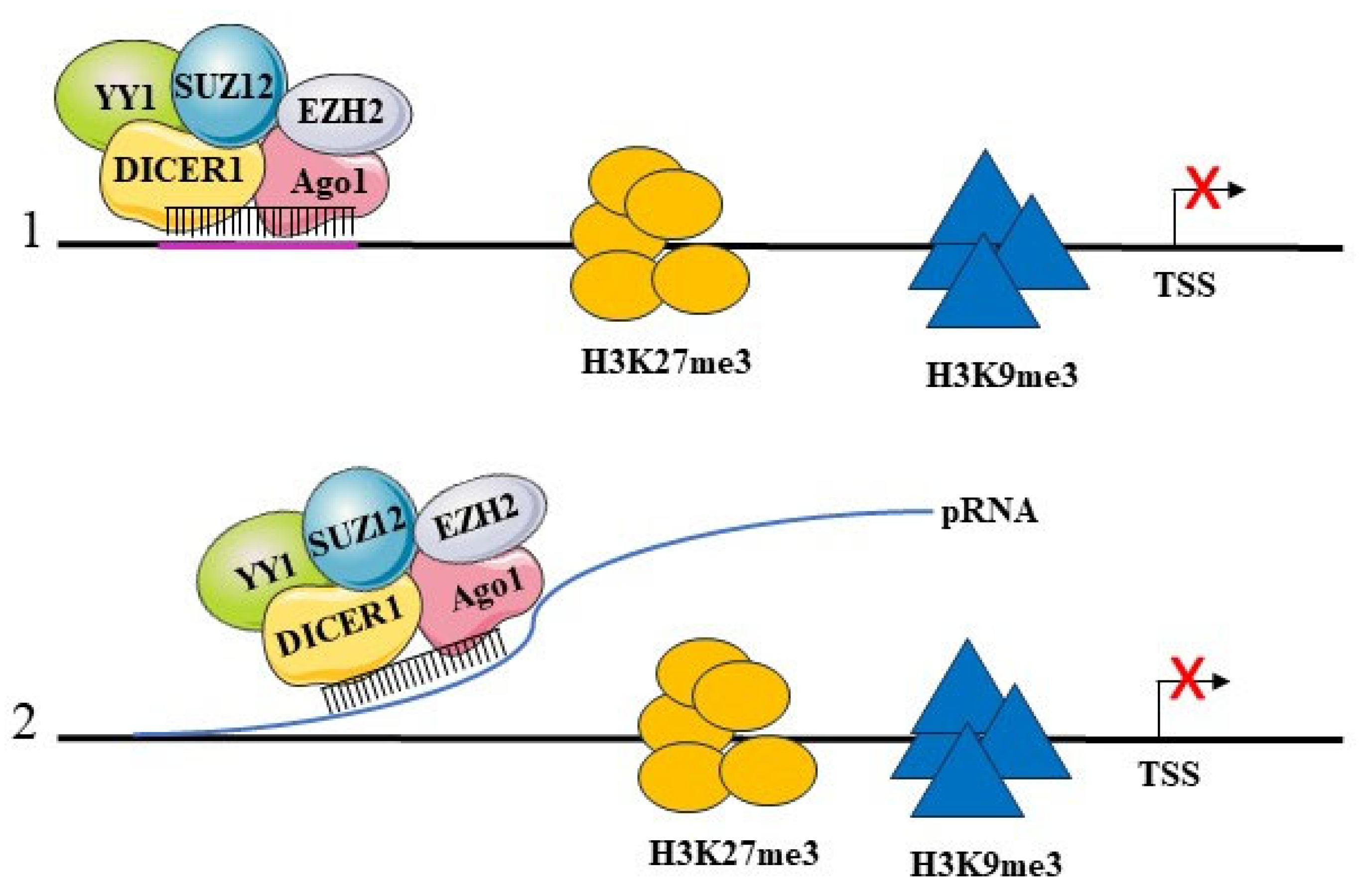
Many studies show that mature nuclear miRNAs can repress the transcription of their target genes. Among the known mechanisms that are used for this regulation is their direct interaction with the promoter through the recognition of DNA sequences highly complementary with the miRNAs seed region. We have shown that this mechanism is used by miR-223 to regulate NFI-A gene transcription during granulopoiesis. We showed that miR-223 localizes in the nucleus and binds to two evolutionarily conserved regions in the NFI-A gene promoter, which are complementary to the miR-223 seed sequence. MiR-223 recruits to that genomic region a protein complex consisting of AGO1, DICER1 and Polycomb proteins (PcG) YY1 and SUZ12, which are responsible for the trimethylation of H3K27 [55,56]. Thus, miR-223 generates an inactive chromatin state on the NFI-A gene promoter, which in turn blocks its expression. This event is required to direct the granulocytic differentiation of hematopoietic progenitors [57]. Benhamed et al. demonstrated that nuclear miRNAs of the let-7 family, in particular by let-7f, acts on the promoters of genes involved in cellular senescence and induces their transcriptional gene silencing [58]. The CDC2 and CDCA8 are genes of the RB1/E2F complex and are repressed during cellular senescence. The sequence of their promoters shows sites complementary to let-7f, where the association of miRNA with a protein complex consisting of AGO2, RB1 and E2F occurs. This results in the recruitment at these sites of histone methyltransferases (HMTs) and histone deacetylases (HDACs), which induce an increase in repressive H3K27me3 and H3K9me2 histone marks, paralleling a decrease in the activating H3K4me3 marks and leading to a repression of the expression of genes involved in cellular senescence. Our unpublished data on acute promyelocytic leukemia cells induced into myeloid differentiation by treatment with retinoic acid show an increased expression of let-7c, which upon this treatment translocates into the nucleus of differentiated cells. In addition, whole-genome chromatin IP sequencing identifies a number of genes that are bound by let-7c and are relevant for myeloid differentiation. These genes can be transcriptionally activated or repressed, following an accumulation in their chromatin of activating H3K4me3 or repressing H3K27me3 marks, respectively. These data further confirm that nuclear miRNAs can perform activatory and repressive transcriptional functions. In gastric cancer, miR-584-3p interacts with the promoter of MMP14 (matrix metalloproteinase 14), at an upstream region of the TSS, and represses its transcription. This regulation occurs as a result of the recruitment of a protein complex consisting of AGO2, EZH2 and EHMT2 to the promoter. This results in increased levels of H3K27me3 and H3K9me2 characteristic of inactive chromatin [59] (Figure 3(1)). Genes involved in cardiac hypertrophy are transcriptionally repressed by the interaction of miR-208b with the promoters and recruitment of EZH2. As a consequence of EZH2 binding, the chromatin of these promoters is enriched with inhibitory markers (H3K27me3) [60]. In addition, miR-320 interacts with the gene promoter of the RNA polymerase III subunit D (POLR3D), where it recruits the EZH2 and AGO1 proteins. This results in transcriptional silencing of the POLR3D gene following the accumulation of the inhibitory marker H3K27me3 [61]. A peculiar mechanism of repression is operated by miR-126-5p to support endothelial integrity and counteract atherosclerosis. MiR-126-5p in a complex with AGO2 is transported to the nucleus by means of its association with Mex3a, an RNA binding protein. Within the nucleus, AGO2 is released from miR-126-5p, which then is found associated with caspase-3. The association blocks the activity of caspase-3 since it impairs its ability to dimerize, leading to apoptosis inhibition. Thus, in this mechanism, a nuclear miRNA is able to block the function of a protein through the formation of a riboprotein complex. Biologically, this leads to the protection of endothelial cells from apoptosis in conditions of shear stress during the process of atherosclerosis [62]. Some miRNAs appear to function as transcriptional and posttranscriptional regulators of the same target gene. As described above, this concept applies to let-7 family miRNAs [58] and to the regulation of the expression of the NFI-A gene during granulopoiesis by miR-223 [57] (see below). MiR-552 also has a dual regulatory effect at both the transcriptional and post-transcriptional levels. This miRNA directly binds the CYP2E1 gene promoter, but this interaction occurs through a region of the miRNA that is not the seed region. At the same time, miR-552 performs a classical posttranscriptional regulation through the binding of the 3' UTR of CYP2E1 mRNA [63]. As mentioned above, miRNAs may regulate transcription by interacting with RNA produced by promoter sequences. Some promoters are transcribed into an antisense strand, producing a pRNA, containing sequences complementary to miRNAs [64]. The miRNA interaction with the antisense pRNA transcript occurs through the seed sequence and results in transcriptional repression of the target gene. Following this binding, an inhibitory protein complex consisting of EZH2, YY1, AGO, Dicer1 and SUZ12 is recruited to the promoter, resulting in chromatin modification and an increase in the repressor markers H3K27me3 and H3K9me2 (Figure 3(2)). An example of this regulatory pattern is the activity of miR-423-5p on the antisense transcript of the promoter of the gene coding for the progesterone receptor (PR). It has been observed that as a result of this interaction, miR-423-5p recruits AGO2 and histone modifiers that increase the level of histone H3K9me2 in the PR promoter leading to transcriptional silencing of the gene [65].

6. Nuclear miRNAs in Hematopoiesis

Many studies have related the differential expression and activity of miRNAs with the regulation of hematopoietic differentiation [66,67,68,69,70]. In particular, differentiation factors, cell cycle regulators or transcription factors that are involved in hemopoiesis appear to be targets of miRNAs. By studying the expression profiles of miRNAs at different stages of hematopoietic differentiation, some of the key regulators have been identified [71,72,73,74,75]. Among these miRNAs, some show a predominantly nuclear localization. However, there is limited work evaluating the nuclear role of these miRNAs and their involvement in hematopoietic differentiation. We showed that miR-223 performs a dual regulation on the NFI-A gene, by virtue of both its cytoplasmic and nuclear component. The cytoplasmic component has a major regulatory role in hematopoiesis. MiR-223 transcription is repressed by NFI-A, whereas it is increased by the transcription factor cEBPalpha, which induces myeloid differentiation of granulocyte-monocyte progenitors. In turn, cytoplasmic miR-223 represses NFI-A translation [76]. Besides these activities, as described above in this review, we demonstrated that miR-223 localizes in the nucleus of hematopoietic cells, where it transcriptionally represses NFI-A gene expression to promote granulocyte differentiation [36]. In fact, the levels of miR-223 are finely regulated during hematopoiesis [78]; the transcriptional repression of miR-223 expression by the oncogenic fusion protein AML1/ETO, triggers the development of leukemia [78]. A study of the nuclear expression profiles of miRNAs during mouse granulopoiesis identified an accumulation of miR-690 in the nucleus. This miRNA targets the CEBPalpha gene; when miR-690 is overexpressed CEBPalpha is downregulated. The retention of miR-690 in the nucleus decreases the share of cytoplasmic miR-690, with consequent increased expression of CEBPalpha and myeloid differentiation. MiR-706 is also enriched in the nucleus of myeloid cells, with a decrease in its cytoplasmic localization in granulocytes compared to promyelocytes. This miRNA would appear to negatively regulate the STAT1 transcription factor, which is important for myeloid differentiation [79,80]. Thus, the accumulation of miR-706 in the nucleus would decrease its activity as a negative regulator of STAT1 in the cytoplasm, thus favouring myeloid differentiation. It has also been reported that miR-709 binds with perfect complementarity to pri-miR-15a and pri-miR-16-1 and inhibits the production of mature miRNAs [81]. Therefore, nuclear miRNAs can regulate the expression of other miRNAs through a novel interaction with their immature precursors. MiR-709 increases its expression and nuclear localization in granulocytes compared to promyelocytes. Several studies also show that miR-709 post-transcriptionally represses c-myc, whose expression levels are downregulated during myeloid cell differentiation [82]. Overall, the analysis of miR-690, miR-706 and miR-709 reveals new important mechanisms of functioning in the process of granulopoiesis. A first mechanism is the interaction and blockade at the nuclear level of the primary transcripts of other miRNAs. This results in the repression of the miRNAs and increased expression of their target genes. The other rather novel and peculiar mechanism is that miRNAs can be retained in the nucleus to decrease their cytoplasmic levels. As a consequence, the expression of its cytoplasmic target genes is increased [82].

7. Non Transcriptional Activities of Nuclear miRNAs

Nuclear miRNAs are reported to exert posttranscriptional regulation of RNA. Several data show the interaction between miRNAs and mRNA in the nucleus, where miRNAs recognise and bind to miRNAs responsive elements (MRE) sequences in the 3' or 5' UTR of the mRNA [83]. This interaction leads to the degradation of the mRNA, often involving AGO2 [84], but the mechanism of this effect has yet to be defined. MiRNAs can also target long non-coding RNAs (lncRNAs) within the nucleus. For example, miR-210 is reported to interact with a specific sequence present on XIST [84], a long non-coding RNA involved in mammalian X-chromosome inactivation during early female embryogenesis. XIST is also associated with cancer. In particular, its inappropriate expression/localisation on active X-chromosome is associated with breast cancer where miR-210 is highly expressed [85,86,87,88], further suggesting a regulatory activity of this miRNA on XIST. MiRNAs also locate within the nucleolus, where they may exert their activity by regulating the pri-miRNA levels. In particular, miR-122 localises in the nucleolus and binds to specific sequences on pri-miR-21. This interaction suppresses the miR-21 maturation induced by the Drosha complex and DGCR8 [89]. Other nucleolar miRNAs have been shown to interact with rRNAs. An example is miR-92a-2-3p, which recognises and binds a site on the 28S rRNA [90]. Other authors show an effect on the biogenesis of rRNAs, since the suppression of a minimal RISC formed by AGO2 and miRNAs leads to an increase in the amount of ribosomal RNA, suggesting a repressor role of miRNAs on rRNAs [91,92]. Lastly, it has been observed that miRNAs interact with their target mRNA already in the nucleolus. Here, they exert an initial pre-repression on the mRNA that will be completed in the cytoplasm [93]. In conclusion, the data suggest that nuclear miRNAs may act in part by regulating RNA processing.

8. Conclusion and Future Perspectives

It is well established that miRNAs play a critical role in many biological processes such as cell differentiation, lineage specification, cell cycle and immune response. In the common view, the activity of miRNAs is mostly restricted to the cytoplasm, where they regulate mRNA stability and translation. Our review highlights current evidence showing that miRNAs possess alternative mechanisms of action, that involve a nuclear activity. It is now clear that miRNAs localise in the nucleus, where they regulate gene expression through complex molecular mechanisms, including the regulation of transcription. MiRNAs can promote both activation and inhibition of transcription of their target genes, which are defined by miRNA-complementary sequences in their genomic loci. Thus, alteration of the miRNA biogenesis mechanism and miRNA expression levels may influence biological processes at multiple levels, including gene transcription. Indeed, the complexity of chromatin remodelers and miRNA-associated machineries contribute to dynamically modify the modality of genomic interaction, according to cell type, cell differentiation programs and cell metabolic status, requiring further focused studies. This generates a wide range of pathological consequences, including cell degeneration and neoplastic transformation. In this context, the activity of nuclear miRNAs needs further investigation in both physiological and pathological settings, especially in the definition of their role in tumorigenesis.

Author Contributions

Conceptualization, F.-G. and M.-B, writing—original draft preparation, F-G. and M.-B., writing—review-editing, C.-N., E.-D. and M.-G.; funding acquisition, F.-G. and C.-N.

Funding

This research was funded by the Italian Ministry of University and Research PRIN grant number 2017WWB99Z, and The APC was funded by PRIN grant number 2017WWB99Z.

Institutional Review Board Statement

Not applicable.

Informed Consent Statement

Not applicable.

Data Availability Statement

Not applicable.

Conflicts of Interest

The authors declare no conflicts of interest.

References

  1. Ambros, V. The functions of animal microRNAs. Nature 2004, 431, 350–355. [Google Scholar] [CrossRef] [PubMed]
  2. Bartel, D.P. MicroRNAs: genomics, biogenesis, mechanism, and function. Cell 2004, 116, 281–297. [Google Scholar] [CrossRef] [PubMed]
  3. Bartel, D.P. MicroRNAs: target recognition and regulatory functions. Cell 2009, 136, 215–233. [Google Scholar] [CrossRef]
  4. Jeffries, C.D. , Fried, H.M; Perkins, D.O. Nuclear and cytoplasmic localization of neural stem cell microRNAs. RNA 2011, 17, 675–686. [Google Scholar] [CrossRef] [PubMed]
  5. Stavast, C. , Erkeland, S. The non-canonical aspects of MicroRNAs: many roads to gene regulation. Cells, 2019; 8, 1465. [Google Scholar] [CrossRef]
  6. Lee, Y. , Ahn, C., Han, J., Choi, H., Kim, J., Yim, J., Lee, J., Provost, P., Rådmark, O., Kim, S. and Kim, V.N. The nuclear RNase III Drosha initiates microRNA processing. Nature, 2003; 425, 415–419. [Google Scholar] [CrossRef]
  7. Partin, A. C. Zhang, K. Jeong, B. C. Herrell, E. Li, S. Chiu, W. et al Cryo-EM structures of human Drosha and DGCR8 in complex with primary MicroRNA. Mol Cell 2020, 78, 411–422. [Google Scholar] [CrossRef] [PubMed]
  8. Rice, G.M. , Shivashankar V., Ma E.J., Baryza J.L., Nutiu R. Functional Atlas of Primary miRNA Maturation by the Microprocessor. Mol Cell 2020, 80, 892–902. [Google Scholar] [CrossRef]
  9. Haase, A.D.; Jaskiewicz, L.; Zhang, H.; Laine, S.; Sack, R.; Gatignol, A.; Filipowicz, W. TRBP, a regulator of cellular PKR and HIV-1 virus expression, interacts with Dicer and functions in RNA silencing. EMBO Rep 2005, 6, 961–967. [Google Scholar] [CrossRef] [PubMed]
  10. Peng, T; He, Y. ; Wang, T.; Yu, J.; Ma, X.; Zhou, Z.; Sheng, Y.; Li, L.; Peng, H.; Li, S. et al.; Discovery of a Novel Small-Molecule Inhibitor Disrupting TRBP-Dicer Interaction against Hepatocellular Carcinoma via the Modulation of microRNA Biogenesis. J Med Chem 2022, 65, 11010–11033. [Google Scholar] [CrossRef]
  11. Yi, R.; Qin, Y.; Macara, I.G. and Cullen, B.R. Exportin-5 mediates the nuclear export of pre-microRNAs and short hairpin RNAs. Gene Dev 2003, 17, 3011–3016. [Google Scholar] [CrossRef]
  12. Chendrimada, T.P.; Gregory, R.I.; Kumaraswamy, E.; Norman, J.; Cooch, N.; Nishikura, K.; Shiekhattar, R. TRBP recruits the Dicer complex to Ago2 for microRNA processing and gene silencing. Nature 2005, 436, 740–744. [Google Scholar] [CrossRef]
  13. Lund, E.; Güttinger, S.; Calado, A.; Dahlberg, J.E.; Kutay, U. Nuclear export of microRNA precursors. Science 2004, 303, 95–98. [Google Scholar] [CrossRef]
  14. Hock, J.; Meister, G. The Argonaute protein family. Genome Biol 2008, 9, 210. [Google Scholar] [CrossRef]
  15. Leitao, A. L.; Enguita, F. J. A structural view of miRNA biogenesis and function. Non coding RNA 2022, 10. [Google Scholar] [CrossRef]
  16. Liu, H; Lei, C. ; He, Q.; Pan, Z.; Xiao, D. and Tao, Y.. Nuclear functions of mammalian microRNAs in gene regulation, immunity and cancer. Mol Cancer 2018, 17, 64. [Google Scholar] [CrossRef] [PubMed]
  17. Gagnon, K.T. , Li, L., Chu, Y., Janowski, B.A. and Corey, D.R. RNAi factors are present and active in human cell nuclei. Cell Rep 2014, 6, 211–221. [Google Scholar] [CrossRef]
  18. Kalantari, R.; Hicks, J.A.; Li, L.; Gagnon, K.T.; Sridhara, V.; Lemoff, A.; Mirzaei, H.; and Corey, D.R. Stable association of RNAi machinery is conserved between the cytoplasm and nucleus of human cells. RNA 2016, 22, 1085–1098. [Google Scholar] [CrossRef] [PubMed]
  19. La Rocca. G.; Cavalieri. V. Roles of the Core Components of the Mammalian miRISC in Chromatin Biology. Genes (Basel) 2022, 13, 414. [Google Scholar] [CrossRef] [PubMed]
  20. Weinmann, L. , Höck, J., Ivacevic, T., Ohrt, T., Mutze, J., Schwille, P., Kremmer, E., Benes, V., Urlaub, H.; and Meister, G. Importin 8 is a gene silencing factor that targets argonaute proteins to distinct mRNAs. Cell 2009, 136, 496–507. [Google Scholar] [CrossRef]
  21. 21. Shuaib M, Parsi KM, Thimma M, Adroub SA, Kawaji H, Seridi L, Ghosheh Y, Fort A, Fallatah B, Ravasi T, et al. Nuclear AGO1 Regulates Gene Expression by Affecting Chromatin Architecture in Human Cells. Cell Systems. [CrossRef]
  22. Wei, Y.; Li, L.; Wang, D.; Zhang, C.Y. and Zen, K. Importin 8 regulates the transport of mature microRNAs into the cell nucleus. J Biol Chem 2014, 289, 10270–10275. [Google Scholar] [CrossRef]
  23. Azmi, A.S.; Uddin, M.H. and Mohammad, R.M. The nuclear export protein XPO1-from biology to targeted therapy. Nat Rev Clin Oncol 2021, 18, 152–169. [Google Scholar] [CrossRef]
  24. Azizian, N.G.; Li, Y. XPO1-dependent nuclear export as a target for cancer therapy. J Hematol Oncol 2020, 13, 61. [Google Scholar] [CrossRef]
  25. Nishi, K.; Nishi, A.; Nagasawa, T. and Ui-Tei, K. Human TNRC6A is an argonaute-navigator protein for microRNA-mediated gene silencing in the nucleus. RNA, 2013; 17, 17–35. [Google Scholar] [CrossRef]
  26. Perconti, G.; Rubino, P.; Contino, F.; Bivona, S.; Bertolazzi, G.; Tumminello, M.; Feo, S.; Giallongo, A.; Coronnello, C. RIP-Chip analysis supports different roles for AGO2 and GW182 proteins in recruiting and processing microRNA targets. BMC Bioinformatics 2019, 20, 120. [Google Scholar] [CrossRef]
  27. Nishi, K.; Takahashi, T.; Suzawa, M.; Miyakawa, T.; Nagasawa, T.; Ming, Y.; Tanokura, M. and Ui-Tei, K. Control of the localization and function of a miRNA silencing component TNRC6A by argonaute protein. Nucleic Acids Res 2015, 43, 9856–9873. [Google Scholar] [CrossRef] [PubMed]
  28. Hicks, J.A.; Li, L.; Matsui, M.; Chu, Y.; Volkov, O.; Johnson, K.C. and Corey, D.R. Human GW182 paralogs are the central organizers for RNA-Mediated control of transcription. Cell Rep 2017, 20, 1543–1552. [Google Scholar] [CrossRef] [PubMed]
  29. Castanotto, D.; Lingeman, R.; Riggs, A.D. and Rossi, J.J. CRM1 medi- ates nuclear-cytoplasmic shuttling of mature microRNAs. Proc Natl Acad Sci USA 2009, 106, 21655–21659. [Google Scholar] [CrossRef] [PubMed]
  30. Meister, G.; Landthaler, M.; Patkaniowska, A.; Dorsett, Y.; Teng, G.; Tuschl, T. Human Argonaute2 mediates RNA cleavage targeted by miRNAs and siRNAs. Mol Cell 2004, 15, 185–97. [Google Scholar] [CrossRef] [PubMed]
  31. Toshinari Ishikawa, Ko Sugawara, Junwei Zhang, Takashi Funatsu, Kohki Okabe Direct observation of cytoskeleton-dependent trafficking of miRNA visualized by the introduction of pre-miRNA iScience 2024, 27, 108811. [CrossRef]
  32. Földes-Papp. Z.; König, K.; Studier, H.; Bückle, R.; Breunig, H.G.; Uchugonova, A. et al. Trafficking of mature miRNA-122 into the nucleus of live liver cells. Curr Pharm Biotechnol 2009, 10, 569–78. [Google Scholar] [CrossRef] [PubMed]
  33. Liao, J.Y.; Ma, L.M.; Guo, Y.H.; Zhang, Y.C.; Zhou, H.; Shao, P. Deep sequencing of human nuclear and cytoplasmic small RNAs reveals an unexpectedly complex subcellular distribution of miRNAs and tRNA 3’ trailers. PloS One 2010, 5, e10563. [Google Scholar] [CrossRef] [PubMed]
  34. Politz, J.C.R.; Hogan, E.M.; Pederson, T. MicroRNAs with a nucleolar location. RNA 2009, 15, 1705–15. [Google Scholar] [CrossRef]
  35. Wong, J.J.; Ritchie, W.; Gao, D.; Lau, K.A.; Gonzalez, M.; Choudhary, A.; Taft, R.J.; Rasko, J.E.; Holst, J. Identification of nuclear-enriched miRNAs during mouse granulopoiesis. J Hematol Oncol 2014, 7, 42. [Google Scholar] [CrossRef]
  36. Zardo, G. Ciolfi, A. Vian, L. Starnes, L.M. Billi, M. Racanicchi, S. Maresca, C. Fazi, F. Travaglini, L. Noguera, N. Mancini, M. Nanni, M. Cimino, G. Lo-Coco F, Grignani F, Nervi C. Polycombs and microRNA-223 regulate human granulopoiesis by transcriptional control of target gene expression. Blood, 2012; 119, 4034–46. [Google Scholar] [CrossRef]
  37. Hwang, H.W.; Wentzel, E.A.; Mendell, J.T. Nucleotide motifs providing localization elements and methods of use. Geneva: World Intellectual Property Organization 2007, Patent No. WO 2007/149521 A2.
  38. Hwang, H.W.; Wentzel, E.A.; Mendell, J.T. A hexanucleotide element directs microRNA nuclear import. Science 2007, 315, 97–100. [Google Scholar] [CrossRef] [PubMed]
  39. Yang, J.; Cheng, M.; Gu, B.; Wang, J.; Yan, S.; Xu, D. ; CircRNA_09505 aggravates inflammation and joint damage in collagen-induced arthritis mice via miR-6089/ AKT1/NF-κB axis. Cell Death Dis 2020, 11, 1–13. [Google Scholar] [CrossRef] [PubMed]
  40. Wang, Y.; Zheng, F.; Gao, G.; Yan, S.; Zhang, L.; Wang, L.; Cai, X.; Wang, X.; Xu, D.; Wang, J. MiR-548a-3p regulates inflammatory response via TLR4/NF-κB signaling pathway in rheumatoid arthritis. J Cell Biochem 2019, 120, 1133–1140. [Google Scholar] [CrossRef] [PubMed]
  41. Bai, Y.; Pan, B.; Zhan, X.; Silver, H. and Li, J. MicroRNA 195-5p targets foxo3 promoter region to regulate its expression in granulosa cells. Int J Mol Sci 2021, 22, 6721. [Google Scholar] [CrossRef] [PubMed]
  42. Majid, S.; Dar, A.A.; Saini, S.; Yamamura, S.; Hirata, H.; Tanaka, Y.; Deng, G. MicroRNA-205-directed transcriptional activation of tumor suppressor genes in prostate cancer. Cancer 2010, 116, 5637–5649. [Google Scholar] [CrossRef]
  43. Chauhan, N.; Manojkumar, A.; Jaggi, M.; Chauhan, S.C.; Yallapu, M.M. MicroRNA-205 in prostate cancer: Overview to clinical translation. Biochim Biophys Acta Rev Cancer 2022, 1877, 188809. [Google Scholar] [CrossRef]
  44. Shuaib, M.; Place, R.F.; Portnoy, V.; Wang, J.; Qi, Z.; Jia, Z.; et al. Upregulation of cyclin B1 by miRNA and its implications in cancer. Nucleic Acids Res 2012, 40, 1695–707. [Google Scholar] [CrossRef]
  45. Zhang, Y.; Fan, M.; Zhang, X.; Huang, F.; Wu, K.; Zhang, J.; Liu, J.; Huang, Z.; Luo, H.; Tao, L.; et al. Cellular microRNAs up-regulate transcription via interaction with promoter TATA-box motifs. RNA, 2014; 20, 1878–1889. [Google Scholar] [CrossRef]
  46. Liu, H.; Lei, C.; He, Q.; Pan, Z.; Xiao, D. , Tao Y. Nuclear functions of mammalian MicroRNAs in gene regulation, immunity and cancer. Mol Cancer 2018, 17. [Google Scholar] [CrossRef]
  47. Toscano-Garibay, J.D.; Aquino-Jarquin, G. Transcriptional regulation mechanism mediated by miRNA-DNA*DNA triplex structure stabilized by Argonaute. Biochim Biophys Acta 2014, 1839, 1079–1083. [Google Scholar] [CrossRef]
  48. Matsui, M.; Chu, Y.; Zhang, H.; Gagnon, K.T.; Shaikh, S.; Kuchimanchi, S.; Manoharan, M.; Corey, D.R.; Janowski, B.A. Promoter RNA links transcriptional regulation of inflammatory pathway genes. Nucleic Acids Res 2013, 41, 10086–10109. [Google Scholar] [CrossRef]
  49. Chellini L, Frezza V and Paronetto MP: Dissecting the transcrip- tional regulatory networks of promoter-associated noncoding RNAs in development and cancer. J Exp Clin Cancer Res, 2020; 39, 51. [CrossRef]
  50. Li H, Zhan J, Zhao Y, Fan J, Yuan S, Yin Z, Dai B, Chen C, Wang DW. Identification of ncRNA-Mediated Functions of Nucleus-Localized miR-320 in Cardiomyocytes. Mol Ther Nucleic Acids 2020, 19, 132–143. [Google Scholar] [CrossRef] [PubMed]
  51. Odame, E.; Chen, Y.; Zheng, S.; Dai, D.; Kyei, B.; Zhan, S.; Cao, J.; et al. Enhancer RNAs: transcriptional regulators and workmates of NamiRNAs in myogenesis. Cell Mol Biol Lett 2021, 26. [Google Scholar] [CrossRef]
  52. Ding, M.; Liu, Y.; Liao, X.; Zhan, H.; Liu, Y.; Huang, W. Enhancer RNAs (eRNAs): new insights into gene transcription and disease treatment. J Cancer 2018, 9, 2334–2340. [Google Scholar] [CrossRef]
  53. Xiao, M.; Li, J.; Li, W.; Wang, Y.; Wu, F.; Xi, Y.; Zhang, L.; et al. MicroRNAs activate gene transcription epigenetically as an enhancer trigger. RNA Biol 2017, 14, 1326–1334. [Google Scholar] [CrossRef]
  54. Liang, Y.; Lu, Q.; Li, W.; Zhang, D.; Zhang, F.; Zou, Q.; et al. Reactivation of tumour suppressor in breast cancer by enhancer switching through NamiRNA network. Nucleic Acids Res 2021, 49, 8556–72. [Google Scholar] [CrossRef]
  55. Zardo, G.; Cimino, G.; Nervi, C. Epigenetic plasticity of chromatin in embryonic and hematopoietic stem/progenitor cells: therapeutic potential of cell reprogramming. Leukemia 2008, 22, 1503–1518. [Google Scholar] [CrossRef]
  56. Schuettengruber, B.; Chourrout, D.; Vervoort, M.; Leblanc, B.; Cavalli, G. Genome regulation by polycomb and trithorax proteins. Cell 2007, 128, 735–745. [Google Scholar] [CrossRef]
  57. Zardo, G.; Ciolfi, A.; Vian, L.; Starnes, L.M.; Billi, M.; Racanicchi, S.; Maresca, C, et al. Polycombs and microRNA-223 regulate human granulopoiesis by transcriptional control of target gene expression. Blood 2012, 119, 4034–4046. [Google Scholar] [CrossRef]
  58. Benhamed, M.; Herbig, U.; Ye, T.; Dejean, A.; Bischof, O. Senescence is an endogenous trigger for microRNA-directed transcriptional gene silencing in human cells. Nat Cell Biol 2012, 14, 266–75. [Google Scholar] [CrossRef] [PubMed]
  59. Zheng, L.; Chen, Y.; Ye, L.; Jiao, W.; Song, H.; Mei, H.; Li, D.; Yang, F. , Li, H., Huang, K., Tong, Q. miRNA-584-3p inhibits gastric cancer progression by repressing Yin Yang 1- facilitated MMP-14 expression. Scientific reports 2017, 7, 8967. [Google Scholar] [CrossRef] [PubMed]
  60. Mathiyalagan, P.; Okabe, J.; Chang, L.; Su, Y.; Du, X.J.; El-Osta, A. The primary microRNA-208b interacts with Polycomb-group protein, Ezh2, to regulate gene expression in the heart. Nucleic Acids Res 2014, 42, 790–803. [Google Scholar] [CrossRef]
  61. Kim, D.H.; Saetrom, P.; Snøve, O. Jr.; Rossi, J.J. MicroRNA-directed transcriptional gene silencing in mammalian cells. Proc. Natl. Acad. Sci. USA 2008, 105, 16230–16235. [Google Scholar] [CrossRef]
  62. Santovito, D.; Egea, V.; Bidzhekov, K.; Natarelli, L.; Mourão, A.; Blanchet, X.; et al. Noncanonical inhibition of caspase-3 by a nuclear microRNA confers endothelial protection by autophagy in atherosclerosis. Sci Transl Med 2020, 12. [Google Scholar] [CrossRef]
  63. Miao, L.; Yao, H.; Li, C.; Pu, M.; Yao, X.; Yang, H.; Qi, X.; et al. A dual inhibition: microRNA-552 suppresses both transcription and translation of cytochrome P450 2E1. Biochim Biophys Acta (BBA)-Gene Regulatory Mech 2016, 1859, 650–66. [Google Scholar] [CrossRef]
  64. Younger, S.T.; Pertsemlidis, A.; Corey, D.R. Predicting potential miRNA target sites within gene promoters. Bioorg Med Chem Lett 2009, 19, 3791–3794. [Google Scholar] [CrossRef]
  65. Younger, S.T.; Corey, D.R. Transcriptional gene silencing in mammalian cells by miRNA mimics that target gene promoters. Nucleic Acids Res 2011, 39, 5682–5691. [Google Scholar] [CrossRef]
  66. Chen, C.Z.; Li, L.; Lodish, H.F.; Bartel, D.P. MicroRNAs modulate hematopoietic lineage differentiation. Science 2004, 303, 83–6. [Google Scholar] [CrossRef]
  67. Nikhat, S.; Yadavalli, A.D.; Prusty, A.; Narayan, P.K.; Palakodeti, D.; Murre, C.; Pongubala, J.M.R. A regulatory network of microRNAs confers lineage commitment during early developmental trajectories of B and T lymphocytes. Proc Natl Acad Sci U S A 2021, 118, e2104297118. [Google Scholar] [CrossRef]
  68. Ng, A.; Lovat, F.; Shih, A.J.; Ma, Y.; Pekarsky, Y.; DiCaro, F.; Crichton, L.; Sharma, E.; Yan, X.J.; Sun, D.; Song, T.; Zou, Y.R.; Will, B.; Croce, C.M.; Chiorazzi, N. Complete miRNA-15/16 loss in mice promotes hematopoietic progenitor expansion and a myeloid-biased hyperproliferative state. Proc Natl Acad Sci U S A 2023, 120, e2308658120. [Google Scholar] [CrossRef]
  69. Nassiri, S.M.; Ahmadi Afshar, N.; Almasi, P. Insight into microRNAs' involvement in hematopoiesis: current standing point of findings. Stem Cell Res Ther 2023, 14, 282. [Google Scholar] [CrossRef]
  70. Li, L.; Ni, R.; Li, Z.; Ming, Y.; Liu, L.; Peng, D.; Cai, Y.; Wu, Y.; Jiang, T.; Li, Y.; Liu, Y. Insights into Regulatory Factors in Megakaryocyte Development and Function: Basic Mechanisms and Potential Targets. Front Biosci (Landmark Ed) 2022, 27, 313. [Google Scholar] [CrossRef]
  71. Liang, H.; Zhang, J.; Zen, K.; Zhang, C.Y.; Chen, X. Nuclear microRNAs and their unconventional role in regulating non-coding RNAs. Protein Cell 2013, 4, 325–30. [Google Scholar] [CrossRef]
  72. Jafari, M.; Ghadami, E.; Dadkhah, T.; Akhavan-Niaki, H.J. PI3k/AKT signaling pathway: Erythropoiesis and beyond. Cell Physiol 2019, 234, 2373–2385. [Google Scholar] [CrossRef]
  73. Attaway, M.; Chwat-Edelstein, T.; Vuong, B.Q. Regulatory Non-Coding RNAs Modulate Transcriptional Activation During B Cell Development. Front Genet 2021, 12, 678084. [Google Scholar] [CrossRef]
  74. Nath, A.; Rayabaram, J.; Ijee, S.; Bagchi, A.; Chaudhury, A.D.; Roy, D.; Chambayil, K.; Singh, J.; Nakamura, Y.; Velayudhan, S.R. Comprehensive Analysis of microRNAs in Human Adult Erythropoiesis. Cells 2021, 10, 3018. [Google Scholar] [CrossRef]
  75. Olson, W.J.; Derudder, E. The miR-142 miRNAs: Shaping the naive immune system. Immunol Lett 2023, 261, 37–46. [Google Scholar] [CrossRef]
  76. Fazi, F.; Rosa, A.; Fatica, A.; Gelmetti, V.; De Marchis, M.L.; Nervi, C.; Bozzoni, I. A minicircuitry comprised of microRNA-223 and transcription factors NFI-A and CEBPalpha regulates human granulopoiesis. Cell 2005, 123, 819–31. [Google Scholar] [CrossRef]
  77. Vian, L.; Di Carlo, M.; Pelosi, E.; Fazi, F.; Santoro, S.; Cerio, A.M.; Boe, A.; Rotilio, V.; Billi, M.; Racanicchi, S.; Testa, U.; Grignani, F.; Nervi, C. Transcriptional fine-tuning of microRNA-223 levels directs lineage choice of human hemaopoietic progenitors. Cell Death Differ 2014, 21, 290–301. [Google Scholar] [CrossRef]
  78. Fazi, F.; Racanicchi, S.; Zardo, G.; Starnes, L.M.; Mancini, M.; Travaglini, L.; Diverio, D.; Ammatuna, E.; Cimino, G.; Lo-Coco, F.; Grignani, F.; Nervi, C. Epigenetic silencing of the myelopoiesis regulator microRNA-223 by the AML1/ETO oncoprotein. Cancer Cell 2007, 12, 457–66. [Google Scholar] [CrossRef]
  79. O'Connell, R. M.; Zhao, J. L.; Rao, D. S. MicroRNA function in myeloid biology. Blood 2011, 118, 2960–2969. [Google Scholar] [CrossRef]
  80. Bhatnagar, B.; Garzon, R. Clinical Applications of MicroRNAs in Acute Myeloid Leukemia: A Mini-Review. Front Oncol 2021, 11, 679022. [Google Scholar] [CrossRef]
  81. Tang, R.; Li, L.; Zhu, D.; Hou, D.; Cao, T.; Gu, H.; Zhang, J.; Chen, J.; Zhang, C.Y.; Zen, K. Mouse miRNA-709 directly regulates miRNA-15a/16-1 biogenesis at the posttranscriptional level in the nucleus: evidence for a microRNA hierarchy system. Cell Res 2011, 22, 504–15. [Google Scholar] [CrossRef]
  82. (34) Wong, J.J.; Ritchie, W.; Gao, D.; Lau, K.A.; Gonzalez, M.; Choudhary, A.; Taft, R.J.; Rasko, J.E.; Holst, J. Identification of nuclear-enriched miRNAs during mouse granulopoiesis. J Hematol Oncol 2014, 7, 42. [Google Scholar] [CrossRef]
  83. Ørom, U.A.; Nielsen, F.C.; Lund, A.H. MicroRNA-10a binds the 5′UTR of ribosomal protein mRNAs and enhances their translation. Mol Cell 2008, 30, 460–471. [Google Scholar] [CrossRef]
  84. Jo, M.H.; Shin, S.; Jung, S.R.; Kim, E.; Song, J.J.; Hohng, S. Human argonaute 2 has diverse reaction pathways on target RNAs. Mol Cell 2015, 59, 117–124. [Google Scholar] [CrossRef]
  85. Fasanaro, P.; Greco, S.; Lorenzi, M.; Pescatori, M.; Brioschi, M.; Kulshreshtha, R.; Banfi, C.; et al. An integrated approach for experimental target identification of hypoxia-induced miR-210. J Biol Chem 2009, 284, 35134–35143. [Google Scholar] [CrossRef]
  86. Turunen, T.A.; Roberts, T.C.; Laitinen, P.; Vaananen, M.A.; Korhonen, P.; Malm, T.; Yla-Herttuala, S.; Turunen, M.P. Changes in nuclear and cytoplasmic microRNA distribution in response to hypoxic stress Sci Rep 2019, 9, 10332; [CrossRef]
  87. Foekens, J. A.; Sieuwerts, A. M.; Smid, M.; Look, M. P.; de Weerd, V.; Boersma, A. W.; Klijn, J. G.; Wiemer, E. A.; Martens, J. W. Four miRNAs associated with aggressiveness og linfonode-negative, estrogen receptor positive human breast cancer. Proc. Natl. Acad. Sci. U.S.A. 2008, 105, 13021–13026. [Google Scholar] [CrossRef]
  88. Camps, C.; Buffa, F. M.; Colella, S.; Moore, J.; Sotiriou, C.; Sheldon, H.; Harris, A.L.; Gleadle, J.M. Ragoussis. Hsa-miR-210 is induced by hypoxia and is an independent prognostic factor in breast cancer. J Clin Cancer Res 2008, 14, 1340–13477. [Google Scholar] [CrossRef]
  89. Wang, D.; Sun, X.; Wei, Y.; Liang, H.; Yuan, M.; Jin, F.; Chen, X.; et al. Nuclear miR-122 directly regulates the biogenesis of cell survival oncomiR miR21 at the post-transcriptional level. Nucleic Acids Res 2018, 46, 2012–2029. [Google Scholar] [CrossRef]
  90. Bai, B.; Liu, H.; Laiho, M. Small RNA expression and deep sequencing analyses of the nucleolus reveal the presence of nucleolus-associated microRNAs. FEBS Open Bio 2014, 4, 441–44. [Google Scholar] [CrossRef]
  91. Atwood, B.L.; Woolnough, J.L.; Lefevre, G.M.; Saint Just Ribeiro, M.; Felsenfeld, G.; Giles, KE. Human argonaute 2 is tethered to ribosomal RNA through MicroRNA interactions. J Biol Chem 2016, 291, 17919–17928. [Google Scholar] [CrossRef]
  92. Böğürcü-Seidel, N.; Ritschel, N.; Acker, T.; Németh, A. Beyond ribosome biogenesis: noncoding nucleolar RNAs in physiology and tumor biology. Nucleus 2023, 14, 2274655. [Google Scholar] [CrossRef]
  93. Reyes-Gutierrez, P.; Ritland Politz, J.C.; Pederson, T. A. mRNA and cognate microRNAs localize in the nucleolus. Nucleus 2014, 5, 636–642. [Google Scholar] [CrossRef]
  94. 94. Pu M, Chen J, Tao Z, Miao L, Qi X, Wang Y, Ren J. Regulatory network of miRNA on its target: coordination between transcriptional and post-transcriptional regulation of gene expression. Cell Mol Life Sci, 2019; 76, 441–451. [CrossRef]
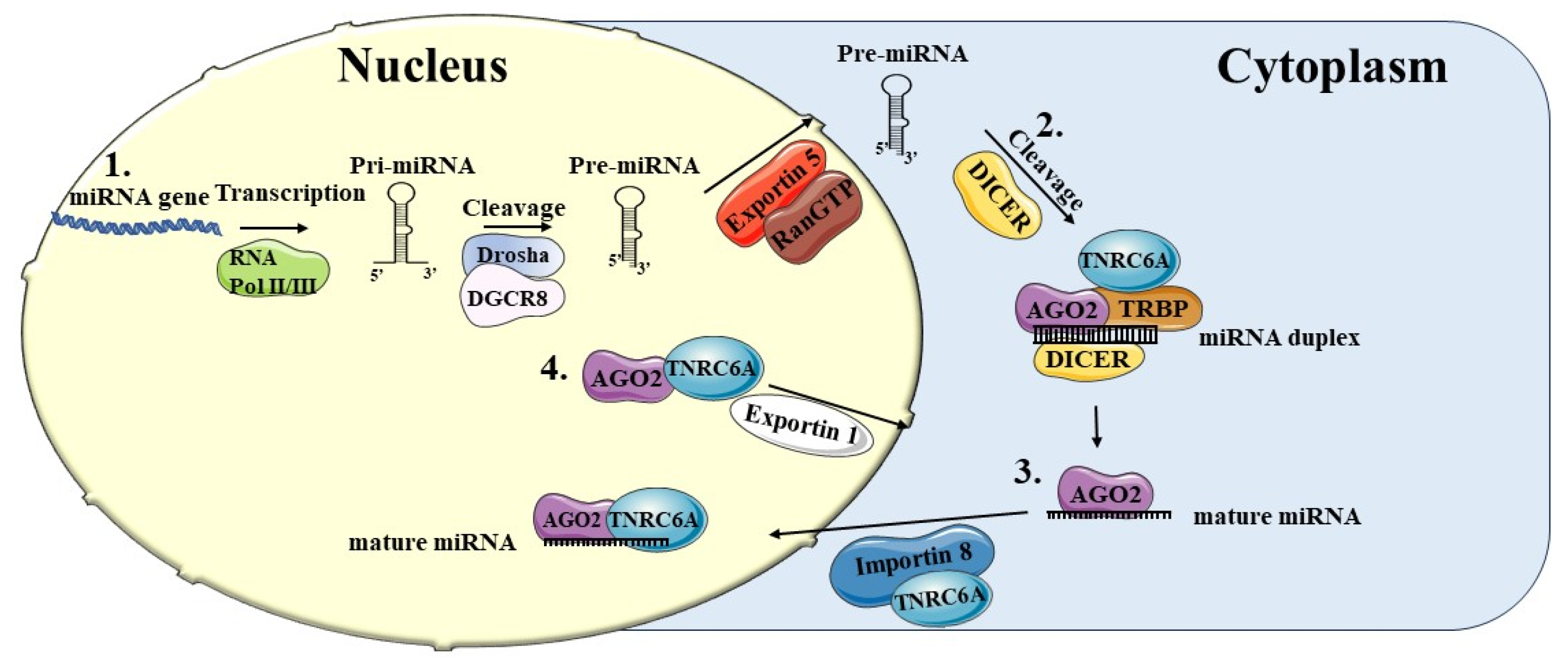
Figure 1. Biogenesis of miRNAs and their return to the nucleus. The biogenesis of miRNAs is mediated by several steps. (1) miRNAs genes are transcribed by RNA Pol II or Pol III into primary miRNAs (pri-miRNAs) and then cleaved by Drosha and DGCR8 into precursor miRNAs (pre-miRNAs). They are then exported into the cytoplasm with the help of Exportin-5 and RanGTP. (2) The pre-miRNAs are cleaved into miRNA duplexes by DICER1 in the cytoplasm and associated with the Argonaute proteins (AGO), forming the mature miRNA. (3) Part of the mature miRNAs are translocated into the nucleus with the help of Importin-8 (IPO8) and TNRC6. (4) AGO2 and TNRC6 can be transported into the cytoplasm via Exportin 1 (also called XPO1 or chromosomal region maintenance 1 (CRM1). miRNA, microRNA; RNAPolII/III, RNA Polymerase II/III; pri-miRNA, miRNA primary transcript; pre-miRNA, miRNA precursor-; AGO2, Argonaute 2; TNRC6A, trinucleotide repeat containing 6; TRBP, transactivation response-RNA-binding protein.
Preprints 105212 g001
Figure 2. The activating function of nuclear miRNAs. (A) miRNA-mediated gene promoter regulation. (1) Direct interaction between miRNAs and complementary sequences on target genes promoters with the presence of AGO2. This interaction allows an activator protein complex to be located in the vicinity of the targeted promoter region, whose chromatin structure is enriched with activator markers such as H3K4me3. RNA Pol II is recruited and the gene is transcribed. (2) the miRNA-AGO-TBP complex interacts directly with the TATA box motif present on the promoter recruiting HMT and HAT. Chromatin is modified, activating markers such as H3K4me3, H3ac and H4ac are increased, and RNA Pol II is recruited to initiate transcription. (3) RNA Pol II transcribes the promoter into promoter-associated RNA (pRNA). AGO2 mediates miRNA binding to pRNA, recruits HMT by producing active histone modifications, such as H3K4me3 with RNA polymerase II enrichment. (B) Transcription regulation through interaction with enhancers.(1) A miRNA is transcribed from its gene located near enhancer loci. The mature miRNA forms a complex with AGO2 and p300/CBP, inducing active chromatin markers in the enhancer regions, and RNA polymerase II call. Thus, the enhancer is transcribed. (2) Next, the eRNA binds to p300 and other proteins to activate the target gene promoter. miRNAs, microRNAs; RNA Pol II, RNA polymerase II; H3K, histone H3; AGO2, Argonaute 2; TBP, TATA-BOX-binding protein; eRNA enhancer RNA; HAT, histone acetyltransferase; HMT, histone methyltransferase; pRNA, promoter-associated RNA.
Preprints 105212 g002
Figure 3. The suppressive function of nuclear miRNAs. MiRNAs can inhibit gene expression at transcriptional level. The miRNA-AGO1 complex recruits a protein complex consisting of YY1, DICER1, SUZ12, EZH2 that promotes inhibitory cromatin modifications with increased H3K27me3 and H3K9me3 markers. This can occur through direct binding to the promoter via sites complementary to the seed region (1) or through binding to the pRNA, a promoter associated non-coding transcript (2). In this way, miRNA leads to a decrease in RNA Pol II and the suppression of target genes. YY1, Yin Yang 1 transcription factor; EZH2, Enhancer of Zeste Homolog 2; SUZ12, Suppressor of Zeste 12 protein homolog; TSS, transcription start site, pRNA, a promoter-associated RNA.
Preprints 105212 g003
Disclaimer/Publisher’s Note: The statements, opinions and data contained in all publications are solely those of the individual author(s) and contributor(s) and not of MDPI and/or the editor(s). MDPI and/or the editor(s) disclaim responsibility for any injury to people or property resulting from any ideas, methods, instructions or products referred to in the content.
Copyright: This open access article is published under a Creative Commons CC BY 4.0 license, which permit the free download, distribution, and reuse, provided that the author and preprint are cited in any reuse.
Prerpints.org logo

Preprints.org is a free preprint server supported by MDPI in Basel, Switzerland.

Subscribe

© 2024 MDPI (Basel, Switzerland) unless otherwise stated