Preprint
Communication

New Isomeric Dipyridothiazine Dimers — Synthesis, Structural Characterization and Cytotoxicity Screening

Altmetrics

Downloads

81

Views

39

Comments

0

Submitted:

05 May 2024

Posted:

06 May 2024

You are already at the latest version

Alerts
Abstract
New dimers of selected dipyridothiazines with an m-xylene linker have been designed, synthesized and characterized. Their structure was identified using 1H and 13C NMR as well as 2D NMR techniques (COSY, ROESY, HSQC and HMBC experiments) and HR MS spectrometry. The compounds were subjected to preliminary in silico biological profile tests using the Way2Drug server and analysis of the probable cytotoxic effect on various cancer cell lines. Preliminary in vitro cytotoxicity tests were performed against normal cell lines (HaCaT) and five cancer cell lines (SW480, SW620, MDA-MB-231, A-549, LN-229) showing moderate activity. An analysis of the structure-activity relationship was performed.
Keywords: 
Subject: Chemistry and Materials Science  -   Medicinal Chemistry

1. Introduction

Phenothiazines are tricyclic heterocyclic compounds in which two benzene rings have been joined by nitrogen and sulfur atoms to form a central 1,4-thiazine ring. There are completely synthetic compounds that have no precursor of their own in the animate natural world [1,2]. Although phenothiazine was discovered more than 140 years ago by German researcher August Bernthsen to this day the molecule is still very popular in the world of medical, pharmaceutical as well as chemical research [3,4,5].
In the 1950s, phenothiazines containing dialkylaminoalkyl substituents in their structure triggered revolutionary changes in psychiatry by showing phenomenal antipsychotic activity to treat schizophrenia or manic states. These drugs are still used in medical treatment to this day [1]. Scientific research in the area of phenothiazines is constantly being carried out, revealing many further interesting pharmacological activities, including: anticancer [6,7,8,9,10], antimicrobial [11,12], antifungal [13], antiviral [14,15], antioxidant [16] or reversal of multidrug resistance activities [17]. These topics are rich resulting in numerous review papers published each year around the world [18,19,20,21,22]. In 2006, English researcher Mitchell named the phenothiazine “parent molecule”, which fully reflects the application nature of this chemical compound [23].
The structure of phenothiazines is also modified at various levels, which leads to the creation of new, previously undescribed derivatives with interesting biological activities. [24]. Such a group of modified phenothiazines includes dipyridothiazines having two pyridine rings in their structure instead of benzene rings [21]. Dipyridothiazines show valuable anticancer activities involving induction of apoptosis via mitochondrial activation of it as well as partial DNA intercalation [25,26,27]. Dipyridothiazines also showed significant immunosuppressive and anti-inflammatory activity by inhibiting TNF alpha and interleukin 6 levels [28].
Dimeric derivatives of dipyridothiazines in which two dipyridothiazine units were connected by selected organic lines, such as o-xylene, p-xylene and 2,6-dimethylpyridine (lutidine), have recently shown highly promising anticancer activities in relation to colon (SW480) and breast (MCF 7) cancer lines. Additionally, these compounds were characterized by relatively low cytotoxicity in relation to normal myoblast cell (L6) lines [29]. This work is a continuation of this research. The synthesis of further new dimeric systems of dipyridothiazines (1,6-, 1,8-, 2,7- and 3,6-diazaphenothiazines 1-4) has been described, in which two dipirydothiazine units have been linked by an m-xylene linker. The structure of new dimers was clearly confirmed by nuclear magnetic resonance spectroscopy (1H, 13C NMR), two-dimensional COSY, ROESY, HSQC, HMBC spectra and high-resolution mass spectrometry HR MS. In silico analyzes of probable molecular targets were performed using the Way2Drug server. The new derivatives were tested for anticancer activity in relation to cancer cell lines: human primary colon cancer (SW480), human metastatic colon cancer (SW620), human breast adenocarcinoma (MDA-MB-231), human lung carcinoma (A-549), human glioblastoma (LN-229), and to human immortal keratinocyte cell line from adult human skin (HaCaT).

2. Results & Discussion

2.1. Chemistry Part

The preparation of new dipyridothiazine dimers was carried out using previously synthesized, selected dipyridothiazines (10H-1,6-, 1,8-, 2,7- and 3,6-diazaphenothiazines 1-4), for which the promising anticancer activities was determinated [29]. In the next step, selected dipyridothiazines were subjected to alkylation reactions with α,α’-dichloro-m-xylene. The reactions were carried out in anhydrous DMF in the presence of the strong base NaH at room temperature, which led to the formation of the final dimers 6-9 (Scheme 1). The crude solid reaction products were purified using column chromatography. All new compounds were obtained in good yields (79-86%).
It is worth noting that the obtained dimers are specific structural analogues of dimers with the 2,6-dimethylpyridine system, which were described in a previous publication [29].

2.2. Structural Identification Using Spectroscopic Methods

Identification of the structure of new organic compounds is a fundamental problem both in organic chemistry and in the drug design process [30,31]. Due to the fact that chemical reactions may be accompanied by rearrangements or parallel or subsequent reactions, proving the structure of the obtained product is essential. For this purpose, advanced NMR spectroscopic techniques and high-resolution HR MS mass spectrometry are used. The identification of the molecule’s structure was carried out on the basis of 1H, 13C NMR spectra, two-dimensional experiments: 2D NMR correlations: COSY (COrrelation SpectroscopY), ROESY (Rotating frame nuclear Overhauser Effect SpectroscopY), HSQC (Heteronuclear Single Quantum Correlation), HMBC (Heteronuclear Multiple Bond Correlation) and HR MS mass spectrometry.
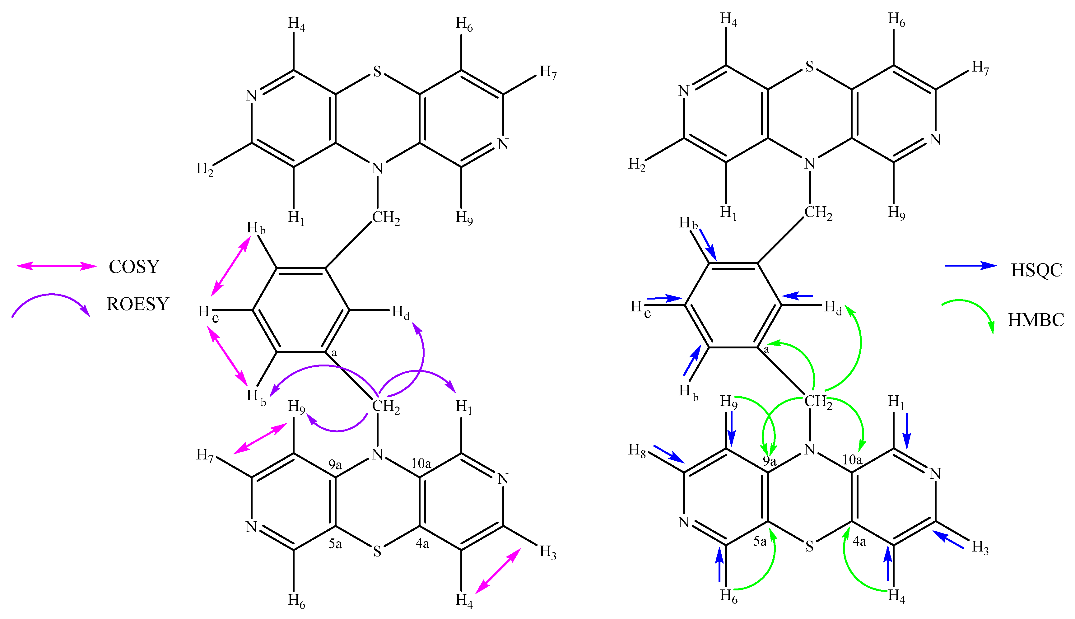
Structural analysis was performed for dimer 8, which contains two 2,7-diazaphenothiazine units connected by an m-xylene liner. The 1H NMR spectrum shows ten signals in the form of singlet (s), doublet (d) and degraded triplet (t) with different intensities, which correspond to proton signals in the molecule. In order to assign individual signals to appropriate protons, two-dimensional COSY and ROESY correlation spectra were performed. In the ROESY spectrum, spatial interactions of the CH2 group with neighboring protons of both the m-xylene system and 2,7-diazaphenothiazine were observed (Figure 1.). The COSY spectrum shows the coupling of the CH2 group at 5.06 ppm with a singlet at 6.94 ppm, which was assigned to the Hd proton of the m-xylene system, and with a doublet at 7.25 ppm with twice the intensity, which was assigned to Hb protons. In the ROESY spectrum, there is a visible correlation of the CH2 group with a singlet at 7.7 ppm, which was assigned to the H1 proton of the 2,7-diazaphenotiazine system and with a doublet at 6.35 ppm, which corrects to the H9 signal. The ROESY spectrum also shows the interaction of the H1 proton with a doublet at 6.97 ppm, which is assigned to the H4 proton, which is coupled to the H3 proton at 8.10 ppm. In the COSY spectrum, it can be observed that the H9 proton is coupled to the H8 proton at 7.96 ppm. In this way, all signals were assigned to the protons present in compound 8. The results of the analysis are summarized in Table 1. Then, the individual carbon signals in the 13C NMR spectrum were assigned, first using the two-dimensional HSQC spectrum showing the carbons directly adjacent to the protons. Then, using the HMBC spectrum, quaternary carbons were assigned. The results are summarized in Table 1. The molecular mas and purity of compound were confirmed by high-resolution mass spectrometry (HR MS).

2.3. In Silico Target Prediction

The new dimers 6-9 were analyzed in silico using the Way2Drug server, which includes the PASS (Prediction of Activity Spectra for Substances) application, which allows predicting the potential biological activities of new molecules and probability of cytotoxicity toward cancer cell lines [32,33]. The obtained results are summarized in Table 2 and Table 3.
The results indicated that dimers 6-9 have a high probability of biological activity aimed at the anticancer potential associated with mechanism of stimulation of histone deacetylase, inhibition of glycosylphosphatidylinositol phospholipase D, which may directly lead to activation of the mitochondrial apoptosis pathway. Probability of action in neurodegenerative diseases, antipsychotic and antiallergic activity as well as cytochrome 450 inhibition has been demonstrated.
More importantly, in addition to the probable mechanisms of biological activity, the PASS program indicated the likelihood of cytotoxic effects on breast, kidney, colon, pancreatic and ovarian cancer cell lines, as well as on non-small cell lung cancer.

2.4. Biological Part – Analysis of Cytotoxicity towards Cancer and Normal Cells

Taking into account the results of in silico simulations and the previously promising anticancer activities and low cytotoxicity of the obtained dimers with an o-, p-xylene and 2,6-dimetylpyridine (lutidine) linker [29], the new derivatives 6-9 were tested for anticancer activity. The cytotoxicity and antiproliferative activity of new dimers was determined in vitro in relation to the following five cancer cell lines: human primary colon cancer (SW480), human metastatic colon cancer (SW620), human breast adenocarcinoma (MDA-MB-231), human lung carcinoma (A-549), human glioblastoma (LN-229), and to the normal line: human immortal keratinocyte cell line from adult human skin (HaCaT) using the MTT assay. The study used doxorubicin and cisplatin as reference drugs. Data are expressed as mean SD, IC50 (µM) – the half-maximal growth inhibitory concentration after cultured the cells for 72 h with the individual compound and the SI (Selectivity Index), which was calculated using formula: SI = IC50 for normal cell line/IC50 cancer cell line.
The results obtained are summarized in the Table 4. The results showed that the compounds 6-9 were not toxic to the normal keratinocyte HaCaT cell line in the range of tested concentration. The analysis of anticancer activity showed that the tested derivatives were mostly inactive (IC50> 100 µM). The derivative 6 showed poor activity in relation to SW480, MDA-MB-231 and A-549 cell lines (IC50> 81-92 µM) and the derivative 7 in relation to MDA-MB-231 the cell line (IC50> 67 µM).
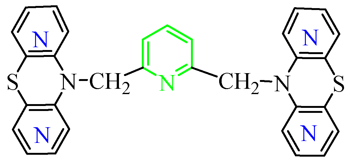
The obtained cytotoxicity results were surprising when analyzed in relation to the previously obtained results of anticancer activity of structural analogues containing the 2,6-dimethylpyridine (lutidine) system Figure 2 [29]. These compounds showed activity in the range of IC50= 0.1-20 µM towards MCF and IC50= 4.5-25 µM SW480 cell lines. This led us to consider the reason for the deactivation of the obtained derivatives 6-9.
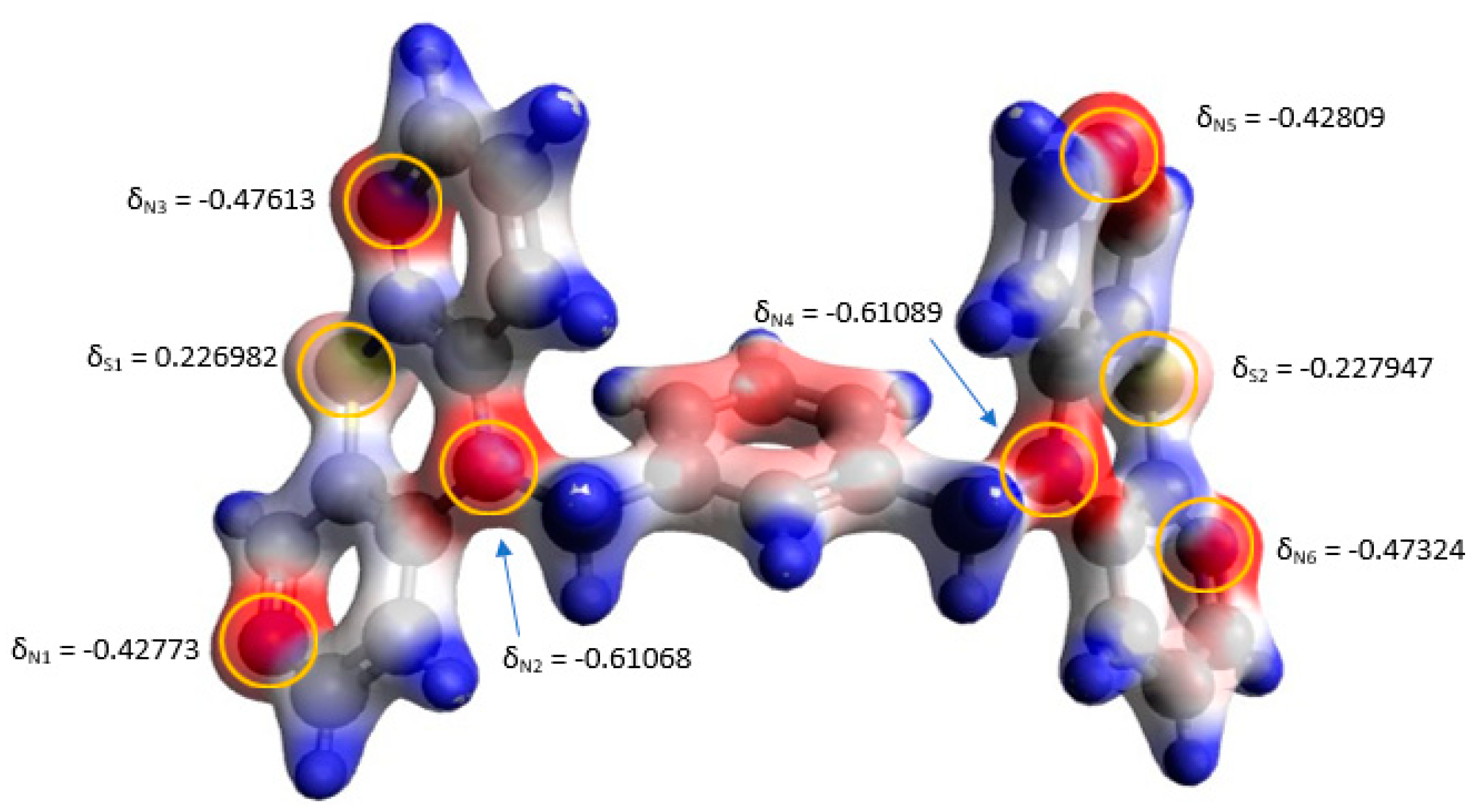
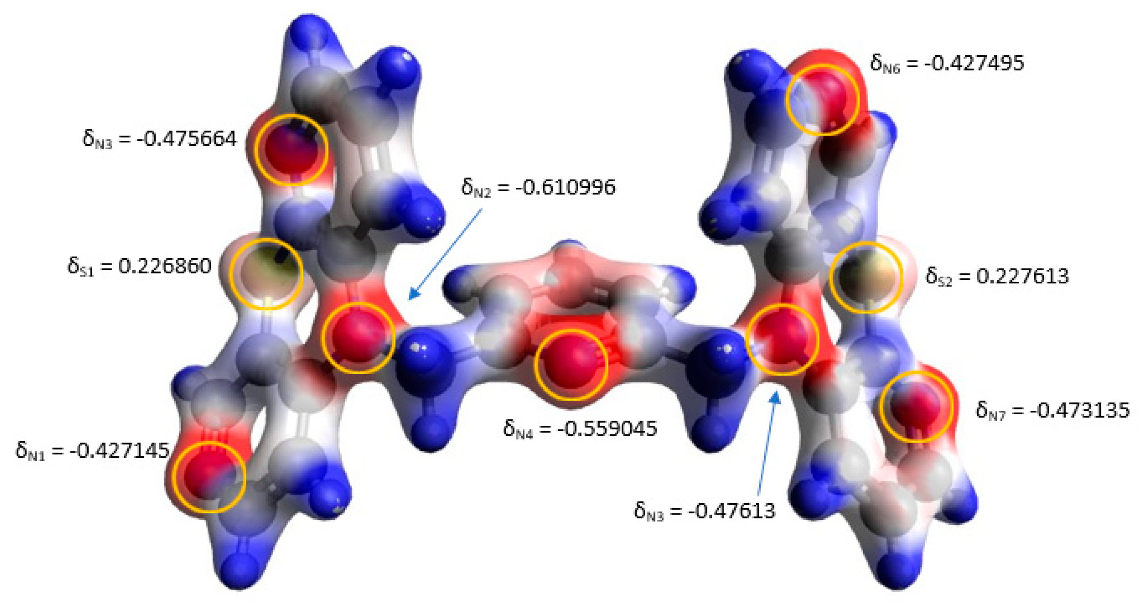
For this purpose, measurements of the electron densities of the selected dimer 8 containing 2,7-diazaphenothiazine in its structure and its analogue with the lutidine system were performed using the Gaussian program [34]. A visualization of the obtained results is presented in Figure 3 and Figure 4 using Avogadro program [35]. These measurements showed that the presence of the lutidine system causes a differentiation of electron charges on thiazine nitrogen atoms in 1,4-thiazine rings directly connected to the CH2 moiety (Figure 4). Additionally, the lutidine system contains a basic nitrogen atom with a lone electron pair, which causes the entire molecule to be enriched with electrons. This situation is not observed in dimer 8 with m-xylene linker (Figure 3).
Additionally, bond length measurements were made, with particular attention paid to the connecting linkers. In the dimer containing lutidine, there are CH2-Clutidine bonds with a length of 1.516 Å and a characteristic Clutidine-Nlutidine bond with a length of 1.362 Å. These bonds are essentially shorter than the analogous bonds found in dimer 8 (CH2-Cm-xylene a length of 1.521 Å, Cm-xylene - Cm-xylene bond with a length of 1.405 Å) (Supplementary materials). The performed structural analysis explains the weak anticancer activity of the obtained dimers with the m-xylene system, while also showing that subtle differences in bond lengths and electron density have a significant impact on the biological activity of the new compounds.

3. Materials and Methods

3.1. Chemistry

The NMR spectra were recorded on Bruker Advance spectrometers (1H at 600 MHz, 13C at 150 MHz) in CDCl3 and DMSOd6. Two-dimensional COSY, ROESY, HSQC and HMBC spectra of compound 8 were recorded on a Bruker Avance spectrometer at 600 MHz. The HR MS spectra (ESI – Electro Spray Ionisation) was run on a Brucker Impact II. The thin layer chromatography was performed on silica gel 60 F254 (Merck 1.05735) with CHCl3-EtOH (10:1 v/v) and on Al2O3 60 F254 neutral (type E) (Merck 1.05581) with CHCl3-EtOH (10:1 v/v) as eluents. Melting points were determined in open capillary tubes on a Boetius melting point apparatus.
The parent starting dipyridothiazines 1-4 (Scheme 1) were prepared according to reference [29].

3.1.1. General Procedure for Synthesis of Compounds (6-9)

To a solution of selected diazaphenothiazine (1-4) (0.201 mg, 1 mmol) in dry DMF (10 mL), NaH (0.028 g, 1.2 mmol, 60% NaH in mineral oil was washed out with hexane) was added. The reaction mixture was stirred at room temperature for 1 h and then linker α,α`-dichloro-m-xylene (0.5 mmol) was added and the stirring was continued for 24 h. The mixture was poured into water (15 ml), extracted with CHCl3 (3 × 10 ml) and dried using Na2SO4. The obtained product was purified by column chromatography (aluminum oxide, CHCl3-Ethanol 10:1) to give:.
1,3-bis((10H-dipyrido [2,3-b:2′,3′-e][1,4]thiazin-10-yl)methyl)benzene (6)
(214 mg, 85%) mp = 268-270 oC
1H NMR (CDCl3) δ: 5.16 (s, 2H, CH2), 6.70 (dd, 1H), 6.78 (m, 1H), 6.84 (m, 1H), 6.93 (s, 1H), 7.18 (d, 1H), 7.31 (t, 1H), 7.37 (m, 1H), 7.68 (m, 1H), 7.82 (d, 1H). 13C NMR: 47.58, 79.65, 115.14, 119.10, 121.95, 122.56, 124.59, 125.45, 129.28, 135.11, 137.54, 142.85, 143.17, 145.41, 151.89, 162.77. HR MS (EI) m/z for: [C28H21N6S2 + H] calc. 505.1264, Found: 505.1283 (100%).
1,3-bis((10H-dipyrido[3,2-b:3′,4′-e][1,4]thiazin-10-yl)methyl)benzene (7)
(209 mg, 83%) mp = 262-264 oC
1H NMR (CDCl3) δ: 5.42 (s, 2H, CH2), 6.90 (m, 1H), 7.17 (m, 2H), 7.20 (m, 1H), 7.21 (m, 1H), 7.29 (s, 2H), 8.05 (m, 1H), 8.14 (d, 1H), 8.19 (s, 1H). 13C NMR: 48.02, 111.95, 120.20, 121.59, 125.52, 126.67, 128.23, 130.03, 135.05, 140.91, 143.84, 147.14, 151.13, 152.90. HR MS (EI) m/z for: [C28H21N6S2 + H] calc. 505.1264, Found: 505.1283 (100%).
1,3-bis((10H-dipyrido[3,4-b:3′,4′-e][1,4]thiazin-10-yl)methyl)benzene (8)
(216 mg, 86%) mp = 256-257 oC
1H NMR (CDCl3) δ: 5.06 (s, 2H, CH2), 6.35 (d, 1H, H9), 6.94 (s, 1H, Hd-m-xylen), 6.97 (d, 1H, H4), 7.25 (d, 1H, Hb-m-xylen), 7.45 (t, 1H, Hc-m-xylen), 7.70 (s, 1H, H1), 7.99 (d, 1H, H8), 8.01 (s, 1H, H6), 8.10 (d, 1H, H3). 13C NMR: 51.08 CCH2, 109.41 C9, 117.19 C9a, 121.56 Cd, 123.48 C4, 126. 06 Cb, 130.79 Cc, 133.35 C10a, 134.46 Ca, 135.56 C1, 137.22 C4a, 143.44 C6, 145.70 C3, 147.14 C8, 151.47 C5a. HR MS (EI) m/z for: [C28H21N6S2 + H] calc. 505.1264 Found: 505.1229 (100%).
1,3-bis((10H-dipyrido[2,3-b:4′,3′-e][1,4]thiazin-5-yl)methyl)benzene (9)
(199 mg, 79%), mp = 288-290 oC
1H NMR (CDCl3) δ: 5.04 (s, 2H, CH2), 6.44 (d, 1H), 6.81 (m, 1H), 6.88 (m, 1H), 6.97 (s, 1H), 7.27 (m, 1H), 7.44 (t, 1H), 7.90 (d, 1H), 7.95 (d, 1H), 8.02 (s, 1H). 13C NMR: 50.88, 79.56, 110.32, 123.30, 123.52, 124.36, 126.67, 130.56, 136.07, 142.94, 145.93. HR MS (EI) m/z for: [C28H21N6S2 + H] calc. 505.1264, Found: 505.1299 (100%).

3.2. In Silico Target Prediction

The prediction of biological targets and cytotoxicity towards cancer cell lines was performed using the Way2Drug server [32,33]. This server analyzes the biological activity of an organic drug based on molecular recognition against its database.

3.3. Biological Evaluation

3.3.1. Cell Line and Culture

Human primary colon cancer (SW480) and metastatic colon cancer (SW620), human breast adenocarcinoma (MDA-MB-231), human lung carcinoma (A-549), human glioblastoma (LN-229), and human immortal keratinocyte (HaCaT) cell lines were obtained from the American Type Culture Collection (ATCC) in Rockville, USA. These cell lines were cultured in the recommended medium according to ATCC protocols: SW480 and SW620 in Minimum Essential Medium (MEM), and MDA-MB-231, A-549, LN-229, and HaCaT cells in Dulbecco’s Modified Eagle Medium (DMEM). The culture medium was supplemented with 10% fetal bovine serum (FBS), penicillin (100 U/mL), and streptomycin (100 μg/mL). Cells were maintained in a humidified incubator at 37°C with 5% CO2. Passaging was performed when cells reached 80%–90% confluence using 0.25% trypsin (Gibco Life Technologies, Gibco, Grand, Island, NY, USA) treatment before being utilized for experiments.

3.3.2. MTT Cell Viability Assay

Cell viability was determined through enzymatic conversion of 3-(4,5-dimethylthiazol-2-yl)-2,5-diphenyltetrazolium bromide salt (MTT) to insoluble formazan crystals by mitochondrial dehydrogenases present in viable cells. Cells were seeded into 96-well plates at a density of 1x10^4 cells per well and allowed to adhere for 24 hours at 37°C in a CO2 incubator. Following the 24-hour incubation period, the culture medium was replaced with fresh medium, and cells were exposed to varying concentrations of the compounds (ranging from 10 to 100 μM), then further incubated for 72 hours at 37°C in a CO2 incubator. Untreated cells served as controls. Subsequently, MTT solution (0.5 mg/mL in serum-free medium) was added, and samples were incubated for 4 hours at 37°C in a CO2 incubator. Following this, the medium was removed, and formazan crystals were solubilized by adding an isopropanol-DMSO mixture (1:1). The absorbance of the dissolved crystals was measured at a wavelength of 570 nm using a UVM 340 reader (ASYS Hitech GmbH, Austria). IC50 values were calculated using GraphPad Prism 8 software (GraphPad Software).

3.4. Quantum Mechanical Calculations

Calculation of the electron density and bond lengths were carried out with B3LYP functional and 6-31+G(d,p) basis set, using Gaussian16 software [34] and visualized using the Avogadro program [35].

3.5. The Statistical Analysis

The statistical analysis was performed using GraphPad Prism 9 software (Graph Pad Software, San Diego, CA, USA). The results were reported as mean ± SD from a minimum of three independent experiments. Statistical significance of differences between values was assessed using analysis of variance with Dunnett’s multiple comparison post hoc test, and significance was defined as p < 0.05.

4. Conclusions

An effective synthesis of new dimers of selected dipyridothiazines with an m-xylene linker was presented. The structure of new derivatives has been identified using 1H and 13C NMR and advanced techniques 2D NMR (COSY, ROESY, HSQC and HMBC experiments). The compounds were subjected to preliminary in silico screening using the Way2Drug server to determine molecular targets and the probability of cytotoxicity towards various types of cancer cells. In the next step cytotoxicity studies have been performed against normal cell lines (HaCaT) and five cancer cell lines (SW480, SW620, MDA-MB-231, A-549, LN-229). The compounds showed weak anticancer activity and low cytotoxicity compared to previously described structural analogues with the 2,6-dimethylpyridine (lutidine) system [29]. The obtained results of cytotoxicity of new dimers are in opposition to the results of in silico tests, which confirms the validity of performing experimental tests. To explain the low activity of the obtained derivatives, quantum mechanical calculations regarding electron densities and bond lengths were performed to explain structural differences, which directly affect the conformations of the molecule, as well as interactions with molecular targets.

Supplementary Materials

The following supportinginformation can be downloaded at the website of this paper posted on Preprints.org. The following supporting information of spectra analysis and in silico analysis are downloaded.

Author Contributions

Conceptualization, B.M.-M. and E.M.; methodology, E.M., P.S., M. S., K.H, A.K; software, E.M., B.M.-M. and M.J., P.S., M. S., K.H, A.K,; validation, E.M.; formal analysis, E.M., B.M.-M., M.J., M.J., P.S., M. S., K.H, A.K; investigation, E.M., B.M.-M.; resources, E.M., B.M.-M., M.J., P.S., M. S., K. H, A.K; data curation, E.M., B.M.-M.; writing—original draft preparation, E.M. and B.M.-M.; writing—review and editing, B.M.-M., E.M., M. J., visualization, B.M.-M.; supervision, E.M. and B.M.-M.; project administration, B.M.-M.; funding acquisition, E.M., B.M.-M. All authors have read and agreed to the published version

Funding

Emilia Martula was supported by a research subsidy from the Medical University of Silesia in Katowice (grant BNW-2-015/N/3/F); APC was funded by the Metropolis of Upper Silesia and Zagłebie Basin as part of the implementation of the project entitled “Support for scientific activity of doctoral students and employees at the doctoral level of the Silesian Medical University in Katowice”, which was implemented within the framework of the Metropolitan Science Support Fund program for 2022–2024 (grants agreement no. RW/27/20243, PCTT/621/2024).

Institutional Review Board Statement

Not applicable.

Informed Consent Statement

Not applicable.

Data Availability Statement

Data is contained within the article and Supplementary Materials.

Conflicts of Interest

The authors declare no conflict of interest.

References

  1. López-Muñoz, F.; Alamo, C.; Cuenca, E.; Shen, W.W.; Clervoy, P.; Rubio, G. History of the Discovery and Clinical Introduction of Chlorpromazine. Annals of Clinical Psychiatry 2005, 17, 113–135. [Google Scholar] [CrossRef]
  2. Jaszczyszyn, A.; Gąsiorowski, K.; Świątek, P.; Malinka, W.; Cieślik-Boczula, K.; Petrus, J.; Czarnik-Matusewicz, B. Chemical structure of phenothiazines and their biological activity. Pharmacological Reports 2012, 64, 16–23. [Google Scholar] [CrossRef]
  3. Otręba, M.; Stojko, J.; Rzepecka-Stojko, A. Phenothiazine derivatives and their impact on the necroptosis and necrosis processes. A review Toxicology 2023, 492, 1–9. [Google Scholar] [CrossRef]
  4. Kumar, A.; Vigato, C.; Boschi, D.; Lolli, M.L.; Kumar, D. Phenothiazines as anti-cancer agents: SAR overview and synthetic strategies. European Journal of Medicinal Chemistry 2023, 254, 115337. [Google Scholar] [CrossRef]
  5. Swoboda, D.; Nycz, J.E.; Karaush-Karmazin, N.; Minaev, B.; Książek, M.; Kusz, J.; Podsiadły, R. Synthesis and Spectroscopic Characterization of Selected Phenothiazines and Phenazines Rationalized Based on DFT Calculation. Molecules 2022, 27, 7519–1. [Google Scholar] [CrossRef]
  6. Li, J.J. Heterocyclic Chemistry in Drug Discovery; Wiley: Hoboken, 2013; Volume 52, pp. 1–16. [Google Scholar] [CrossRef]
  7. Otręba, M.; Stojko, J.; Rzepecka-Stojko, A. The role of phenothiazine derivatives in autophagy regulation: A systematic review. Journal of Applied Toxicology 2023, 43, 474–489. [Google Scholar] [CrossRef]
  8. Lopes, R.M.; Souza, A.C.; Otręba, M.; Rzepecka-Stojko, A.; Tersariol, I.; Rodrigues, T. Targeting autophagy by antipsychotic phenothiazines: potential drug repurposing for cancer therapy. Biochemical Pharmacology 2024, 222, 116075. [Google Scholar] [CrossRef]
  9. Xu, F.; Xi, H.; Liao, M.; Zhang, Y.; Ma, H.; Wu, M.; Xue, Q.; Sun, H.; Zhang, Y.; Xia, Y. Repurposed antipsychotic chlorpromazine inhibits colorectal cancer and pulmonary metastasis by inducing G2/M cell cycle arrest, apoptosis, and autophagy. Cancer Chemotherapy and Pharmacology 2022, 89, 331–346. [Google Scholar] [CrossRef]
  10. Li, A.; Chen, X.; Jing, Z.; Chen, J. Trifluoperazine induces cellular apoptosis by inhibiting autophagy and targeting NUPR1 in multiple myeloma. FEBS Open Bio 2020, 10, 2097–2106. [Google Scholar] [CrossRef]
  11. Posso, M.C.; Domingues, F.C.; Ferreira, S.; Silvestre, S. Development of Phenothiazine Hybrids with Potential Medicinal Interest: A Review. Molecules 2022, 27, 276. [Google Scholar] [CrossRef]
  12. Rineh, A.; Dolla, N.K.; Ball, A.R.; Magana, M.; Bremner, J.B.; Hamblin, M.R.; Tegos, G.P.; Kelso, M.J. Attaching the NorA Efflux Pump Inhibitor INF55 to Methylene Blue Enhances Antimicrobial Photodynamic Inactivation of Methicillin-Resistant Staphylococcus aureus in Vitro and in Vivo. ACS Infectious Diseases 2017, 3, 756–766. [Google Scholar] [CrossRef]
  13. Brilhante, R.S.N.; Gotay, W.J.P.; Pereira, V.S.; de Oliveira, J.S.; Pereira-Neto, W.A.; Castelo-Branco, D.d.S.C.M.; de Aguiar Cordeiro, R.; Sidrim, J.J.C.; Rocha, M.F.G. Antifungal Activity of Promethazine and Chlorpromazine Against Planktonic Cells and Biofilms of Cryptococcus neoformans/Cryptococcus gattii Complex Species. Medical Mycology 2020, 58, 906–912. [Google Scholar] [CrossRef]
  14. Piccini, L.E.; Castilla, V.; Damonte, E.B. Inhibition of dengue virus infection by trifluoperazine. Archives of Virology 2022, 167, 2203–2212. [Google Scholar] [CrossRef]
  15. Simanjuntak, Y.; Liang, J.J.; Lee, Y.L.; Lin, Y.L. Repurposing of prochlorperazine for use against dengue virus infection. Journal of Infectious Diseases 2015, 211, 394–404. [Google Scholar] [CrossRef]
  16. Egbujor, M.C.; Tucci, P.; Buttari, B.; Nwobodo, D.C.; Marini, P.; Saso, L. Phenothiazines: Nrf2 activation and antioxidant effects. Journal of Biochemical and Molecular Toxicology 2024, 38, 23661. [Google Scholar] [CrossRef]
  17. Takács, D.; Csonka, Á.; Horváth, Á.; Windt, T.; Gajdács, M.; Riedl, Z.; Hajós, G.; Amaral, L.; Molnár, J.; Spengler, G. Reversal of ABCB1-related Multidrug Resistance of Colonic Adenocarcinoma Cells by Phenothiazines. Anticancer Research 2015, 35, 3245–3251. [Google Scholar]
  18. Mosnaim, A.D.; Ranade, V.V.; Wolf, M.E.; Puente, J.; Valenzuela, M.A. Phenothiazine molecule provides the basic chemical structure for various classes of pharmacotherapeutic agents. American Journal of Therapeutics 2006, 13, 261–273. [Google Scholar] [CrossRef]
  19. Ohlow, M.; Moosmann, B. Phenothiazine: The seven lives of pharmacology’s first lead structure. Drug Discovery Today 2011, 16, 119–131. [Google Scholar] [CrossRef]
  20. Pluta, K.; Morak-Młodawska, B.; Jeleń, M. Synthesis and properties of diaza-, triaza- and tetraazaphenothiazines. Journal of Heterocyclic Chemistry 2009, 46, 355–391. [Google Scholar] [CrossRef]
  21. Morak-Młodawska, B.; Pluta, K.; Jeleń, M. Phenothiazines Modified with the Pyridine Ring as Promising Anticancer Agents. Life 2021, 11, 206. [Google Scholar] [CrossRef]
  22. Kumar, A.; Vigato, C.; Boschi, D.; Lolli, M.L.; Kumar, D. Phenothiazines as anti-cancer agents: SAR overview and synthetic strategies. European Journal of Medicinal Chemistry 2023, 254, 115337. [Google Scholar] [CrossRef]
  23. Mitchell, S.C. Phenothiazine: the parent molecule. Current Drug Targets 2006, 7, 1181–1189. [Google Scholar] [CrossRef]
  24. Gupta, R.R.; Kumar, M. Synthesis, properties and reactions of phenothiazines. Phenothiazine and 1,4-Benzothiazines—Chemical and Biological Aspect. Elsevier 1988, 108, 1–161. [Google Scholar]
  25. Zhang, J.; Chen, M.; Wenzhi, Z.; Okechukwu Nwabueze, P.; Morak-Młodawska, B.; Pluta, K.; Jeleń, M.; Md Akim, A.; Ang, K.P.; Ooi, K.K. 10H-3,6-Diazaphenothiazines Induce G2/M Phase Cell Cycle Arrest, Caspase-dependent Apoptosis and Inhibits Cell Invasion of A2780 Ovarian Carcinoma Cells through Regulation on NF-κB and [BIRC6-XIAP] Complexes. Drug Design, Development and Therapy 2017, 11, 3045–3063. [Google Scholar] [CrossRef]
  26. Skonieczna, M.; Kasprzycka, A.; Jeleń, M.; Morak-Młodawska, B. Tri- and Pentacyclic Azaphenothiazine as Pro-Apoptotic Agents in Lung Carcinoma with a Protective Potential to Healthy Cell Lines. Molecules 2022, 27, 5255. [Google Scholar] [CrossRef]
  27. Sochacka, J.; Pacholczyk, M.; Jeleń, M.; Morak-Młodawska, B.; Pluta, K. Interaction of new tri-, tetra-, and pentacyclic azaphenothiazine derivatives with calf thymus DNA: Spectroscopic and molecular docking studies. Spectrochimica Acta, Part A: Molecular and Biomolecular Spectroscopy 2021, 262. [Google Scholar] [CrossRef]
  28. Morak-Młodawska, B.; Pluta, K.; Zimecki, M.; Jeleń, M.; Artym, J.; Kocięba, M. Synthesis and selected immunological properties of 10-substituted 1,8-diazaphenothiazines. Medicinal Chemistry Research 2015, 24, 1408–1418. [Google Scholar] [CrossRef]
  29. Martula, E.; Morak-Młodawska, B.; Jeleń, M.; Okechukwu, P.; Balachandran, A.; Tehirunavukarasu, P.; Anamalay, K.; Ulaganathan, V. Synthesis and structural characterization of novel dimers of dipyridothiazine as promising antiproliferative agents. Molecules 2023, 28, 7662. [Google Scholar] [CrossRef]
  30. Kiemle, D.; Webster, F.X.; Silverstein, R.M.; Bryce, D.L. Spectrometric Identification of Organic Compounds, 8th Edition; John Wiley & Sons Inc, 2014; ISBN 978-0-470-61637-6. [Google Scholar]
  31. Hermann, C.h.K.F.; Morrill, T.C.; Shriner, R.L.; Fuson, R.C. Systematic Identification of Organic Compounds, Ninth Edition; Wiley, 2023; ISBN 9781119799665. [Google Scholar]
  32. Way2Drug Understanding Chemical-Biological Interactions, Predictive Services. Available online: www.way2drug.com/passonline/index.php (accessed on 18 December 2023).
  33. Druzhilovskiy, D.S.; Rudik, A.V.; Filimonov, D.A.; Gloriozova, T.A.; Lagunin, A.A.; Dmitriev, A.V.; Pogodin, P.V.; Dubovskaya, V.I.; Ivanov, S.M.; Tarasova, O.A.; Bezhentsev, V.M.; Murtazalieva, K.A.; Semin, M.I.; Maiorov, I.S.; Gaur, A.S.; Sastry, G.N.; Poroikov, V.V. Computational platform Way2Drug: from the prediction of biological activity to drug repurposing. Russian Chemical Bulletin 2017, 66, 1832–1841. [Google Scholar] [CrossRef]
  34. Gaussian 16, Revision C.01; Gaussian, Inc.: Wallingford, CT, 2019.
  35. Avogadro: an open-source molecular builder and visualization tool. Version 1.2.0. Available online: http://avogadro.cc/ (accessed on 12 March 2024).
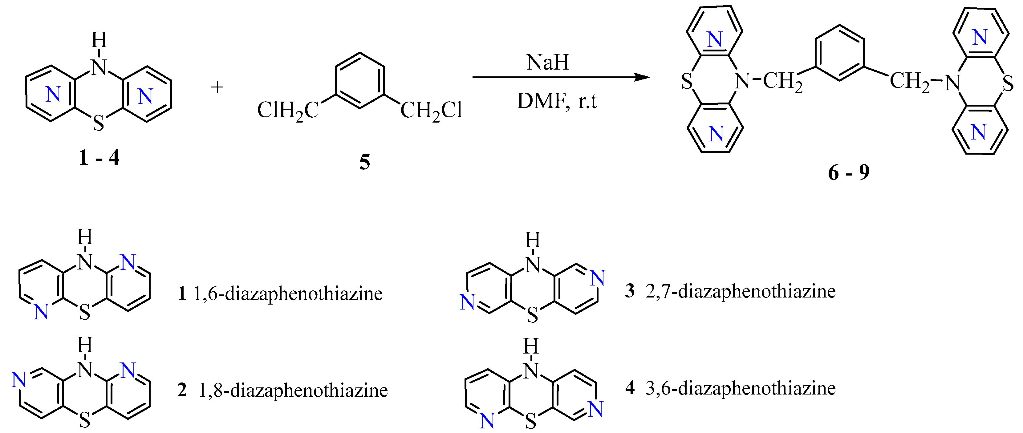
Scheme 1. Synthesis of isomeric dipyridothiazine dimers 6-9.
Preprints 105650 sch001
Figure 1. Assignment of 1H and 13C NMR and interactions of 1H-1H (COSY and ROESY) and 1H-13C (HSQC and HMBC) in compound 8 with particular emphasis on the interactions of the CH2 group.
Preprints 105650 g001
Figure 2. Structure of the previously described isomeric anti-cancer active dimers with the 2,6-dimethylpyridine (lutidine) system [29].
Preprints 105650 g002
Figure 3. Visualization of the electron density of the compound 8 (electron density was calculated in the Gaussian program, visualized in the Avogadro program). Electron density is represented by a color gradient, with red representing higher electron density and dark blue representing lower electron density.
Preprints 105650 g003
Figure 4. Visualization of the electron density of the structural analogue of compound 8 with a pyridine ring as a linker (electron density was calculated in the Gaussian program, visualized in the Avogadro program). Electron density is represented by a color gradient, with red representing higher electron density and dark blue representing lower electron density.
Preprints 105650 g004
Table 1. Tabulated list of 1H-1H and 1H-13C correlations of a compound 8.
1H NMR (ppm) ROESY COSY 13C NMR (ppm) HSQC HMBC
CH2 5.06
H9 6.35
Hd 6.94
H4 6.97
Hb 7.25
Hc 7.45
H1 7.70
H8 7.98
H6 8.01
H3 8.10
5.06-7.70
5.06-6.35
5.06-7.25
5.06-6.94
6.35-8.01
7.70-6.97
6.97-8.10
5.06-6.94
5.06-7.25
7.25-7.45
6.35-7.96
6.97-8.10
C CH2 51.21
C9 109.49
C9a 117.19
Cd 121.74
C4 123.39
Cb 126.06
Cc 130.79
C10a 133.35
Ca 134.46
C1 135.56
C4a 137.22
C6 143.44
C3 145.05
C8 147.14
C5a 151.47
5.06-51.21
H9 6.35-109.53
Hd 6.94-121.74
H4 6.97-123.39
Hb 7.25-126.06
Hc 7.45-130.79
H1 7.70-135.56
H8 7.98-147.14
H6 8.01-143.44
H3 8.10-145.05
Hc 7.45-Ca 134.46
CH2 5.06-C5a 151.47
CH2 5.06- C4a 137.22
H3 8.10- C10a 133.35
H8 7.98- C9a 117.19
Table 2. Probability (%) of activities spectrum of dimers (6-9) using PASS program.
No. Probability of Activity Spectrum
6 (28%) Histone deacetylase stimulant (77%) Glycosylphosphatidylinositol phospholipase D inhibitor (47%) Transcription factor inhibitor (33%) Alzheimer’s disease treatment (46%) Antiallergic
7 (29%) Mitochondrial processing peptidase inhibitor (83%) Glycosylphosphatidylinositol phospholipase D inhibitor (18%) Antipsychotic (30%) Cytochrome P450 inhibitor (10%) MAP3K8 inhibitor
8 (75%) Neurodegenerative diseases treatment (84%) Glycosylphosphatidylinositol phospholipase D inhibitor (65%) Histone deacetylase stimulant (42%) Alzheimer’s disease treatment (41%) Cytochrome P450 inhibitor
9 (76%) Histone deacetylase stimulant (66%) Antiallergic (60%) Neurodegenerative diseases treatment (58%) Alzheimer’s disease treatment (59%) Antiasthmatic
Table 3. The probable cytotoxic effect (%) on various cancer cell lines of compound 6-9.
No. Probability of Cytotoxicity towards Cancer Cell Lines
6 Breast cancer MDA-MB-468 (62%) Melanoma
UACC-257(51%)
Non-small cell lung carcinoma NCI-H322M (49%) Colon carcinoma HCT-116 (42%) Pancreatic carcinoma YAPC (43%)
7 Breast cancer MDA-MB-468 (59%) Melanoma
UACC-257 (55%)
Renal carcinoma 786 (45%) Colon carcinoma HCT-116 (39%) Pancreatic carcinoma YAPC (44%)
8 Breast cancer MDA-MB-468 (39%) Melanoma
UACC-257 (54%)
Renal carcinoma 786-0 (49%) Colon carcinoma HCT-116 (55%) Ovarian adenocarcinoma OVCAR-4 (43%)
9 Breast cancer MDA-MB-468 (44%) Melanoma
UACC-257 (49%)
Renal carcinoma 786-0 (44%) Colon carcinoma HCT-116 (54%) Ovarian adenocarcinoma OVCAR-4 (37%)
Table 4. Cytotoxic activity (IC50, µM) of studied compounds estimated by the MTT assay.
No. Cancer Cells   Normal Cells
SW480 SW620 MDA-MB-231 A-549 LN-229 HaCaT
IC50 SI IC50 SI IC50 SI IC50 SI IC50 SI IC50
6 92.6 ± 5.31 1.08 >100 1 83.2 ± 3.38 1.20 81.5 ± 5.71 1.22 >100 1 >100
7 >100 0.93 >100 0.93 67.3 ± 4.13 1.38 >100 0.93 >100 0.93 92.7 ± 5.00
8 >100 1 >100 1 >100 1 >100 1 >100 1 >100
9 >100 1 >100 1 96.5 ± 1.37 1.03 >100 1 >100 1 >100
Doxorubicin 0.7 ± 0.1 0.4 0.3 ± 0.1 1.0 1.6 ± 0.23 0.19 0.2 ± 0.09 1.5 1.1 ± 0.12 0.27 0.3 ± 0.1
Cisplatin 10.4 ± 0.9 0.6 6.7 ± 1.1 0.9 7.8 ± 0.98 0.81 3.2 ± 1.24 5.08 2.6 ± 0.15 2.42 6.3 ± 0.7
Disclaimer/Publisher’s Note: The statements, opinions and data contained in all publications are solely those of the individual author(s) and contributor(s) and not of MDPI and/or the editor(s). MDPI and/or the editor(s) disclaim responsibility for any injury to people or property resulting from any ideas, methods, instructions or products referred to in the content.
Copyright: This open access article is published under a Creative Commons CC BY 4.0 license, which permit the free download, distribution, and reuse, provided that the author and preprint are cited in any reuse.
Prerpints.org logo

Preprints.org is a free preprint server supported by MDPI in Basel, Switzerland.

Subscribe

© 2024 MDPI (Basel, Switzerland) unless otherwise stated