Submitted:
02 May 2024
Posted:
07 May 2024
You are already at the latest version
Abstract
Keywords:
1. Introduction
2. Materials and Methods
2.1. Case Selection
- -
- Inclusion criteria: Endodontically treated mandibular molar with its root growth completed, has to be fully included in CBCT’s imaging range, while its clear and complete images available.
- -
- Exclusion criteria: Presence of periodontitis at least in a moderate stage. Diagnosis of conditions other than pulpal or periapical lesions (such as fibro-osseous lesions, benign neoplasms, oral cancer, etc.). Difficulty in radiographic interpretation due to metal artifacts associated with the tooth.
2.2. CBCT Analysis
2.2.1. An Anatomical Factor: Root Canal Morphology Classification
2.2.2. Treatment Factors: Intra- and Post-Operative Factors


2.3. Statistical Analysis
3. Results
3.1. An Anatomical Factor: The Root Canal Morphology
3.2. Intra- and Post-Operative Treatment Factors: Missing Canals, Obturation Length, Leaky Canals, Iatrogenic Problems, and Coronal Restoration
3.3. The Correlation between an Anatomical Factor and Treatment Factors
4. Discussion
Author Contributions
Funding
Institutional Review Board Statement
Informed Consent Statement
Data Availability Statement
Acknowledgments
Conflicts of Interest
References
- Chugal, N.M.; Clive, J.M.; Spangberg, L.S. Endodontic infection: some biologic and treatment factors associated with outcome. Oral Surg Oral Med Oral Pathol Oral Radiol Endod 2003, 96, 81-90. [CrossRef]
- Vertucci, F.J. Root canal morphology and its relationship to endodontic procedures. Endodontic Topics 2005, 10, 3-29. [CrossRef]
- Lloyd, A.; Uhles, J.P.; Clement, D.J.; Garcia-Godoy, F. Elimination of intracanal tissue and debris through a novel laser-activated system assessed using high-resolution micro-computed tomography: a pilot study. J Endod 2014, 40, 584-587. [CrossRef]
- Ng, Y.L.; Mann, V.; Rahbaran, S.; Lewsey, J.; Gulabivala, K. Outcome of primary root canal treatment: systematic review of the literature -- Part 2. Influence of clinical factors. Int Endod J 2008, 41, 6-31. [CrossRef]
- Tronstad, L.; Asbjornsen, K.; Doving, L.; Pedersen, I.; Eriksen, H.M. Influence of coronal restorations on the periapical health of endodontically treated teeth. Endod Dent Traumatol 2000, 16, 218-221. [CrossRef]
- Baruwa, A.O.; Martins, J.N.R.; Meirinhos, J.; Pereira, B.; Gouveia, J.; Quaresma, S.A.; Monroe, A.; Ginjeira, A. The Influence of Missed Canals on the Prevalence of Periapical Lesions in Endodontically Treated Teeth: A Cross-sectional Study. J Endod 2020, 46, 34-39 e31. [CrossRef]
- Fan, B.; Cheung, G.S.; Fan, M.; Gutmann, J.L.; Bian, Z. C-shaped canal system in mandibular second molars: Part I--Anatomical features. J Endod 2004, 30, 899-903. [CrossRef]
- Melton, D.C.; Krell, K.V.; Fuller, M.W. Anatomical and histological features of C-shaped canals in mandibular second molars. J Endod 1991, 17, 384-388. [CrossRef]
- Kato, A.; Ziegler, A.; Higuchi, N.; Nakata, K.; Nakamura, H.; Ohno, N. Aetiology, incidence and morphology of the C-shaped root canal system and its impact on clinical endodontics. Int Endod J 2014, 47, 1012-1033. [CrossRef]
- Grocholewicz, K.; Lipski, M.; Weyna, E. Endodontic and prosthetic treatment of teeth with C-shaped root canals. Ann Acad Med Stetin 2009, 55, 55-59.
- Nair, P.N. On the causes of persistent apical periodontitis: a review. Int Endod J 2006, 39, 249-281. [CrossRef]
- Ng, Y.L.; Mann, V.; Rahbaran, S.; Lewsey, J.; Gulabivala, K. Outcome of primary root canal treatment: systematic review of the literature - part 1. Effects of study characteristics on probability of success. Int Endod J 2007, 40, 921-939. [CrossRef]
- Kotoku, K. [Morphological studies on the roots of Japanese mandibular second molars]. Shikwa Gakuho 1985, 85, 43-64.
- Carlsen, O. Root complex and root canal system: a correlation analysis using one-rooted mandibular second molars. Scand J Dent Res 1990, 98, 273-285. [CrossRef]
- Fan, W.; Fan, B.; Gutmann, J.L.; Cheung, G.S. Identification of C-shaped canal in mandibular second molars. Part I: radiographic and anatomical features revealed by intraradicular contrast medium. J Endod 2007, 33, 806-810. [CrossRef]
- Karabucak, B.; Bunes, A.; Chehoud, C.; Kohli, M.R.; Setzer, F. Prevalence of Apical Periodontitis in Endodontically Treated Premolars and Molars with Untreated Canal: A Cone-beam Computed Tomography Study. J Endod 2016, 42, 538-541. [CrossRef]
- Song, M.; Park, M.; Lee, C.Y.; Kim, E. Periapical status related to the quality of coronal restorations and root fillings in a Korean population. J Endod 2014, 40, 182-186. [CrossRef]
- Siqueira Junior, J.F.; Rocas, I.D.N.; Marceliano-Alves, M.F.; Perez, A.R.; Ricucci, D. Unprepared root canal surface areas: causes, clinical implications, and therapeutic strategies. Braz Oral Res 2018, 32, e65. [CrossRef]
- Jara, C.M.; Hartmann, R.C.; Bottcher, D.E.; Souza, T.S.; Gomes, M.S.; Figueiredo, J.A.P. Influence of apical enlargement on the repair of apical periodontitis in rats. Int Endod J 2018, 51, 1261-1270. [CrossRef]
- Maggiore, C.; Gallottini, L.; Resi, J.P. Mandibular first and second molar. The variability of roots and root canal system. Minerva Stomatol 1998, 47, 409-416.
- Ahn, H.R.; Moon, Y.M.; Hong, S.O.; Seo, M.S. Healing outcomes of root canal treatment for C-shaped mandibular second molars: a retrospective analysis. Restor Dent Endod 2016, 41, 262-270. [CrossRef]
- Barbakow, F.H.; Cleaton-Jones, P.; Friedman, D. An evaluation of 566 cases of root canal therapy in general dental practice. 2. Postoperative observations. J Endod 1980, 6, 485-489. [CrossRef]
- Siqueira, J.F., Jr.; Rocas, I.N.; Lopes, H.P. Patterns of microbial colonization in primary root canal infections. Oral Surg Oral Med Oral Pathol Oral Radiol Endod 2002, 93, 174-178. [CrossRef]
- Ricucci, D.; Siqueira, J.F., Jr. Biofilms and apical periodontitis: study of prevalence and association with clinical and histopathologic findings. J Endod 2010, 36, 1277-1288. [CrossRef]
- Ricucci, D.; Siqueira, J.F., Jr.; Bate, A.L.; Pitt Ford, T.R. Histologic investigation of root canal-treated teeth with apical periodontitis: a retrospective study from twenty-four patients. J Endod 2009, 35, 493-502. [CrossRef]
- Versiani, M.A.; Martins, J.; Ordinola-Zapata, R. Anatomical complexities affecting root canal preparation: a narrative review. Aust Dent J 2023, 68 Suppl 1, S5-S23. [CrossRef]
- Lin, L.M.; Skribner, J.E.; Gaengler, P. Factors associated with endodontic treatment failures. J Endod 1992, 18, 625-627. [CrossRef]
- Costa, F.; Pacheco-Yanes, J.; Siqueira, J.F., Jr.; Oliveira, A.C.S.; Gazzaneo, I.; Amorim, C.A.; Santos, P.H.B.; Alves, F.R.F. Association between missed canals and apical periodontitis. Int Endod J 2019, 52, 400-406. [CrossRef]
- Spili, P.; Parashos, P.; Messer, H.H. The impact of instrument fracture on outcome of endodontic treatment. J Endod 2005, 31, 845-850. [CrossRef]
- Crump, M.C.; Natkin, E. Relationship of broken root canal instruments to endodontic case prognosis: a clinical investigation. J Am Dent Assoc 1970, 80, 1341-1347. [CrossRef]
- McGuigan, M.B.; Louca, C.; Duncan, H.F. The impact of fractured endodontic instruments on treatment outcome. Br Dent J 2013, 214, 285-289. [CrossRef]
- Ray, H.A.; Trope, M. Periapical status of endodontically treated teeth in relation to the technical quality of the root filling and the coronal restoration. Int Endod J 1995, 28, 12-18. [CrossRef]
- Jang, Y.E.; Kim, Y.; Kim, S.Y.; Kim, B.S. Predicting early endodontic treatment failure following primary root canal treatment. BMC Oral Health 2024, 24, 327. [CrossRef]
- Walid, N. The use of two pluggers for the obturation of an uncommon C-shaped canal. J Endod 2000, 26, 422-424. [CrossRef]
- Jin, G.C.; Lee, S.J.; Roh, B.D. Anatomical study of C-shaped canals in mandibular second molars by analysis of computed tomography. J Endod 2006, 32, 10-13. [CrossRef]
- Jafarzadeh, H.; Wu, Y.N. The C-shaped root canal configuration: a review. J Endod 2007, 33, 517-523. [CrossRef]
- Fernandes, M.; de Ataide, I.; Wagle, R. C-shaped root canal configuration: A review of literature. J Conserv Dent 2014, 17, 312-319. [CrossRef]
- Patel, S.; Durack, C.; Abella, F.; Shemesh, H.; Roig, M.; Lemberg, K. Cone beam computed tomography in Endodontics - a review. Int Endod J 2015, 48, 3-15. [CrossRef]
- Kim, Y.; Lee, S.J.; Woo, J. Morphology of maxillary first and second molars analyzed by cone-beam computed tomography in a korean population: variations in the number of roots and canals and the incidence of fusion. J Endod 2012, 38, 1063-1068. [CrossRef]
- Neelakantan, P.; Subbarao, C.; Subbarao, C.V. Comparative evaluation of modified canal staining and clearing technique, cone-beam computed tomography, peripheral quantitative computed tomography, spiral computed tomography, and plain and contrast medium-enhanced digital radiography in studying root canal morphology. J Endod 2010, 36, 1547-1551. [CrossRef]
- Chan, F.; Brown, L.F.; Parashos, P. CBCT in contemporary endodontics. Aust Dent J 2023, 68 Suppl 1, S39-S55. [CrossRef]
- de Chevigny, C.; Dao, T.T.; Basrani, B.R.; Marquis, V.; Farzaneh, M.; Abitbol, S.; Friedman, S. Treatment outcome in endodontics: the Toronto study--phase 4: initial treatment. J Endod 2008, 34, 258-263. [CrossRef]

| factors | description | |
| Obturation density [1] | Good | Homogeneous radiopaque material and no visible space. No more than 2 small voids (<1mm) |
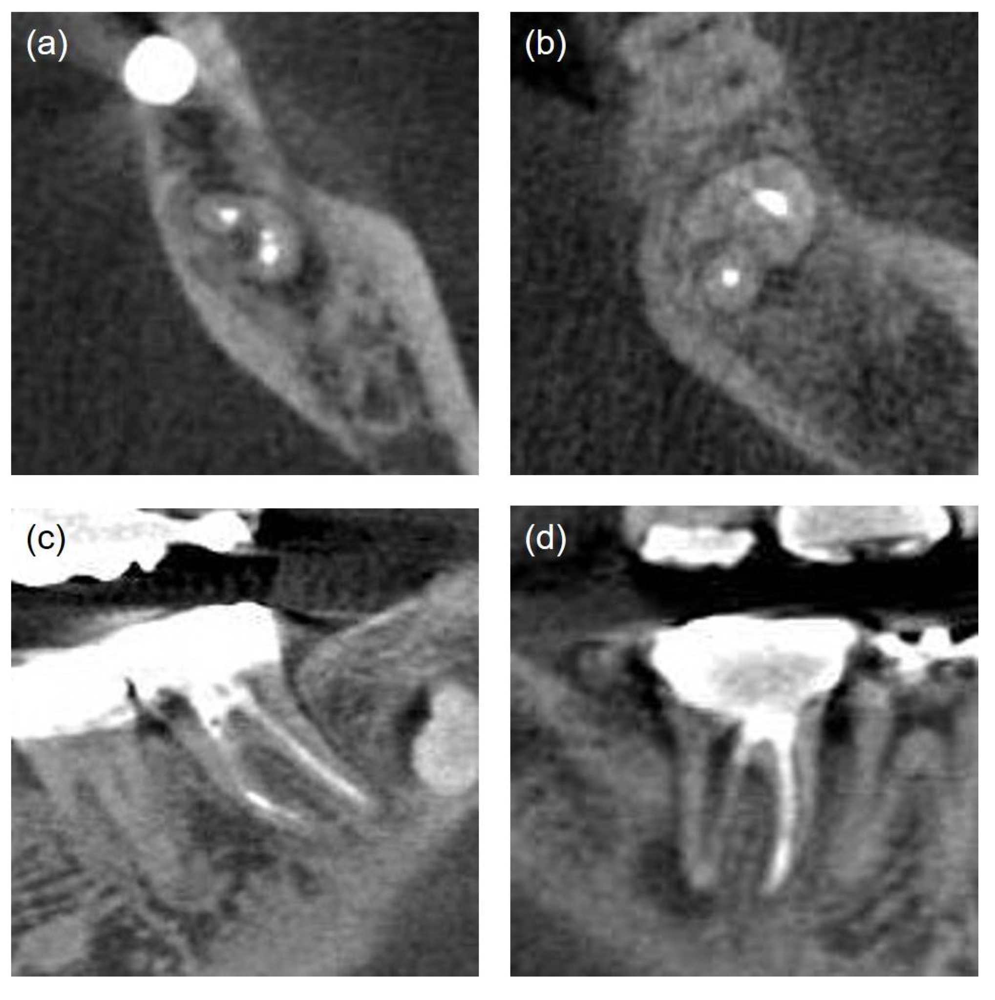
| Poor | Non-uniform radiodensity, with the canal space visible laterally and apically. Isthmus area that had not been treated (Figure 3-a) | |
| Missed canal [17] |
Unfilled canals appearing from cemento-enamel junction to apex including canals splitting from a main canal at coronal, mid, or apical third (Figure 3-b) | |
| Obturation level [1] | Obturation level of a filling material was measured in millimeters relative to the radiographic apex | |
| Normal | Fillings within 2mm short of the radiographic apex | |
| improper | Fillings more than 2mm short of the radiographic apex / Excess root filling, Sealer extrusion | |
| Iatrogenic problem | File separation, Perforation (present/absent) (Figure 3-c, d). | |
| Coronal restoration [17] | Adequate | Any permanent restoration that appeared intact radiographically and had no comment on the clinical examination record |
| Inadequate | Any permanent restoration with detectable radiographic sings of overhangs, open margins, or recurrent caries or comments such as “ill-fitting margin” or “secondary dental caries” on the clinical examination record. | |
| C-shape N=77 (100%) |
Non C-shape N=73 (100%) |
Total N=150 |
|
|---|---|---|---|
| Success | 20 (26%) | 30 (41.1%) | 50 |
| Failure | 57 (74%) | 43 (58.9%) | 100 |
| χ2(p) | 3.856 (p=0.05) | ||
| Treatment factors | N | Endodontic outcome | |
| Success (N=50) |
Failure (N=100) |
||
| Missing canal | |||
| Present | 46 | 8 (17.4%) | 38 (82.6%) |
| Absent | 104 | 42 (40.4%) | 62 (59.6%) |
| Obturation length | |||
| Adequate | 107 | 44 (41.1%) | 63 (58.9%) |
| Inadequate | 42 | 6 (14.3%) | 36 (85.7%) |
| Obturation density | |||
| Poor | 80 | 18 (22.5%) | 62 (77.5%) |
| Good | 70 | 32 (45.7%) | 38 (54.3%) |
| Iatrogenic problem | |||
| Present | 9 | 3 (33.3%) | 6 (66.7%) |
| Absent | 141 | 47 (33.3%) | 94 (66.7%) |
| Coronal restoration | |||
| Adequate | 75 | 35 (46.7%) | 40 (53.3%) |
| Inadequate | 71 | 15 (21.1%) | 56 (78.9%) |
| Endodontic Failure | |||||
| B | S.E. | Wald | p | Exp (B) | |
| C-configuration | 0.115 | 0.504 | 0.053 | 0.819 | 1.122 |
| Missing canal | 1.132 | 0.478 | 5.606 | 0.018* | 3.103 |
| Obturation length | 1.068 | 0.533 | 4.019 | 0.045* | 2.909 |
| Obturation density | 0.572 | 0.508 | 1.267 | 0.260 | 1.772 |
| Iatrogenic events | -0.110 | 0.819 | 0.018 | 0.893 | 0.896 |
| Coronal leakage | 1.117 | 0.405 | 7.619 | 0.006* | 3.057 |
| C-shape | Non C-shape | X2 | p value | |
| Missing canal (N=46) | 25 (54.3%) | 21 (45.7%) | 0.241 | 0.623 |
| Inadequate obturation length (N=42) |
26 (61.9%) | 16 (38.1%) | 2.450 | 0.118 |
| Poor obturation density (N=80) |
65 (81.3%) | 15 (18.7%) | 61.416 | <0.001* |
| Iatrogenic problem (N=9) |
3 (33.3%) | 6 (66.7%) | 0.318 † |
Disclaimer/Publisher’s Note: The statements, opinions and data contained in all publications are solely those of the individual author(s) and contributor(s) and not of MDPI and/or the editor(s). MDPI and/or the editor(s) disclaim responsibility for any injury to people or property resulting from any ideas, methods, instructions or products referred to in the content. |
© 2024 by the authors. Licensee MDPI, Basel, Switzerland. This article is an open access article distributed under the terms and conditions of the Creative Commons Attribution (CC BY) license (http://creativecommons.org/licenses/by/4.0/).


