Preprint
Review

A GIBAC-based selectivity strategy for the design of PDE5 inhibitors to minimize visual disturbances

Altmetrics

Downloads

603

Views

36

Comments

0

Wei Li  *

This version is not peer-reviewed

Submitted:

21 May 2024

Posted:

21 May 2024

You are already at the latest version

Alerts
Abstract
Phosphodiesterase type 5 (PDE5) inhibitors are widely used in the treatment of various conditions, including erectile dysfunction and pulmonary hypertension. Despite their clinical efficacy, these drugs quite often cause visual disturbances due to off-target effects on Phosphodiesterase type 5 (PDE6) in the retina. This review explores the application of a general intermolecular binding affinity calculator (GIBAC)-based binding selectivity strategy in the design of PDE5 inhibitors, aiming to enhance binding selectivity and minimize visual side effects. This GIBAC strategy integrates computational structural biological approaches to iteratively refine drug-target binding affinities, thereby improving target specificity and the structural biophysical limit for the efficacy-safety balance of PDE5 inhibitors. Through detailed analysis of PDE5’s and PDE6’s biological role and the molecular mechanisms underlying visual disturbances, this article underscores the necessity of target binding selectivity in PDE5 inhibitor design in future. Additionally, this article discusses the practical applications of GIBAC in computational drug discovery and design, along with the future directions and the potential for GIBAC to transform PDE5 inhibitor development, ultimately enhancing therapeutic outcomes and patient safety. Finally, this article calls for a GIBAC-based paradigm shift in computational drug discovery and design towards the continued development of the pharmaceutical industry.
Keywords: 
Subject: Biology and Life Sciences  -   Biophysics

An Introduction to Phosphodiesterase Type 5 (PDE5) and Phosphodiesterase Type 6 (PDE6)

Phosphodiesterase type 5 (PDE5) and phosphodiesterase type 6 (PDE6) are crucial enzymes involved in cellular signaling pathways with significant implications in human health and disease [1,2,3]. PDE5, predominantly found in smooth muscle cells of the vasculature and corpus cavernosum, plays a pivotal role in the regulation of cyclic guanosine monophosphate (cGMP) levels [4]. PDE5 inhibitors cause vasodilation in the penis and lung by blocking the breakdown of cyclic guanosine monophosphate (cGMP) which results in prolongation of the action of mediators of vasodilation including nitric oxide (NO) [5]. Clinically, inhibitors of PDE5, such as sildenafil, tadalafil, and vardenafil, are widely prescribed for the treatment of erectile dysfunction, pulmonary hypertension, and other cardiovascular conditions [6]. However, despite their therapeutic efficacy, PDE5 inhibitors are associated with a notable side effect profile, notably visual disturbances, attributed to off-target effects on PDE6 [7]. PDE6, primarily expressed in rod and cone photoreceptor cells of the retina, serves as a key mediator in the visual transduction cascade [8].
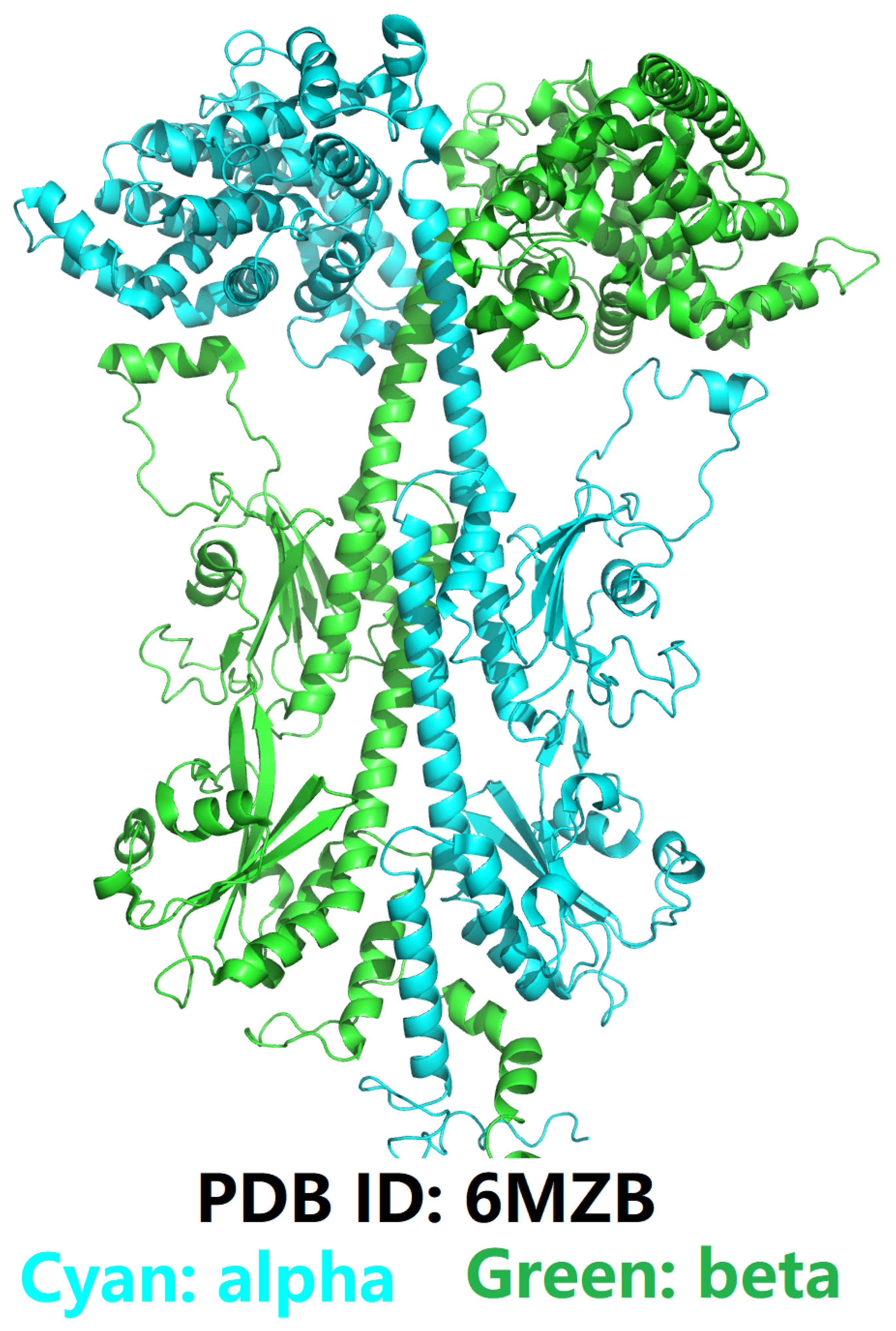
In general, the essential biological function of phosphodiesterase (PDE) type enzymes is to regulate the cytoplasmic levels of intracellular second messengers, 3’,5’-cyclic guanosine monophosphate (cGMP) and/or 3’,5’-cyclic adenosine monophosphate (cAMP) [7]. PDE targets have 11 isoenzymes. Of these enzymes [7], PDE5 (Figure 1) has attracted a special attention over the years after its recognition as being the target enzyme in treating erectile dysfunction. Due to the amino acid sequence and the secondary structural similarity of PDE6 and PDE11 with the catalytic domain of PDE5, first-generation PDE5 inhibitors (i.e. sildenafil and vardenafil) are also competitive inhibitors of PDE6 and PDE11 [9]. Thus, inhibition of PDE6 by PDE5 inhibitors can disrupt phototransduction processes, leading to transient visual changes, including altered color perception and sensitivity to light [9]. Understanding the distinct roles of PDE5 and PDE6 in cellular signaling pathways is essential for elucidating the mechanisms underlying both the therapeutic effects and adverse reactions associated with PDE5 inhibitors [8]. Moreover, it underscores the importance of developing strategies to enhance binding selectivity towards PDE5 while minimizing off-target interactions with PDE6, thereby mitigating visual disturbances and improving the overall safety profile of these drugs [8].
Specifically, the amino acid sequence of the catalytic domain of PDE5 is listed in italics in fasta format as below,
>3SHY_1|Chain A|cGMP-specific 3’,5’-cyclic phosphodiesterase|Homo sapiens (9606)
MGSSHHHHHHSSGLVPRGSHMEETRELQSLAAAVVPSAQTLKITDFSFSDFELSDLETALCTIRMFTDLNLVQNFQMKHEVLCRWILSVKKNYRKNVAYHNWRHAFNTAQCMFAALKAGKIQNKLTDLEILALLIAALSHDLDHRGVNNSYIQRSEHPLAQLYCHSIMEHHHFDQCLMILNSPGNQILSGLSIEEYKTTLKIIKQAILATDLALYIKRRGEFFELIRKNQFNLEDPHQKELFLAMLMTACDLSAITKPWPIQQRIAELVATEFFDQGDRERKELNIEPTDLMNREKKNKIPSMQVGFIDAICLQLYEALTHVSEDCFPLLDGCRKNRQKWQALAEQQ
and the amino acid sequence of the entire PDE5 is listed in italics in fasta format as below,
>PDE5A_(UniProtKB ID: O76074)_cGMP-specific 3’,5’-cyclic phosphodiesterase
MERAGPSFGQQRQQQQPQQQKQQQRDQDSVEAWLDDHWDFTFSYFVRKATREMVNAWFAERVHTIPVCKEGIRGHTESCSCPLQQSPRADNSAPGTPTRKISASEFDRPLRPIVVKDSEGTVSFLSDSEKKEQMPLTPPRFDHDEGDQCSRLLELVKDISSHLDVTALCHKIFLHIHGLISADRYSLFLVCEDSSNDKFLISRLFDVAEGSTLEEVSNNCIRLEWNKGIVGHVAALGEPLNIKDAYEDPRFNAEVDQITGYKTQSILCMPIKNHREEVVGVAQAINKKSGNGGTFTEKDEKDFAAYLAFCGIVLHNAQLYETSLLENKRNQVLLDLASLIFEEQQSLEVILKKIAATIISFMQVQKCTIFIVDEDCSDSFSSVFHMECEELEKSSDTLTREHDANKINYMYAQYVKNTMEPLNIPDVSKDKRFPWTTENTGNVNQQCIRSLLCTPIKNGKKNKVIGVCQLVNKMEENTGKVKPFNRNDEQFLEAFVIFCGLGIQNTQMYEAVERAMAKQMVTLEVLSYHASAAEEETRELQSLAAAVVPSAQTLKITDFSFSDFELSDLETALCTIRMFTDLNLVQNFQMKHEVLCRWILSVKKNYRKNVAYHNWRHAFNTAQCMFAALKAGKIQNKLTDLEILALLIAALSHDLDHRGVNNSYIQRSEHPLAQLYCHSIMEHHHFDQCLMILNSPGNQILSGLSIEEYKTTLKIIKQAILATDLALYIKRRGEFFELIRKNQFNLEDPHQKELFLAMLMTACDLSAITKPWPIQQRIAELVATEFFDQGDRERKELNIEPTDLMNREKKNKIPSMQVGFIDAICLQLYEALTHVSEDCFPLLDGCRKNRQKWQALAEQQEKMLINGESGQAKRN
and the amino acid sequences of four (, , and ) subunits of PDE6 (Figure 2) are also listed in italics in fasta format as below:
>PDE6A_(UniProtKB ID: P16499)_Human Retinal Rod cGMP-specific 3’,5’-cyclic phosphodiesterase subunit alpha
MGEVTAEEVEKFLDSNIGFAKQYYNLHYRAKLISDLLGAKEAAVDFSNYHSPSSMEESEIIFDLLRDFQENLQTEKCIFNVMKKLCFLLQADRMSLFMYRTRNGIAELATRLFNVHKDAVLEDCLVMPDQEIVFPLDMGIVGHVAHSKKIANVPNTEEDEHFCDFVDILTEYKTKNILASPIMNGKDVVAIIMAVNKVDGSHFTKRDEEILLKYLNFANLIMKVYHLSYLHNCETRRGQILLWSGSKVFEELTDIERQFHKALYTVRAFLNCDRYSVGLLDMTKQKEFFDVWPVLMGEVPPYSGPRTPDGREINFYKVIDYILHGKEDIKVIPNPPPDHWALVSGLPAYVAQNGLICNIMNAPAEDFFAFQKEPLDESGWMIKNVLSMPIVNKKEEIVGVATFYNRKDGKPFDEMDETLMESLTQFLGWSVLNPDTYESMNKLENRKDIFQDIVKYHVKCDNEEIQKILKTREVYGKEPWECEEEELAEILQAELPDADKYEINKFHFSDLPLTELELVKCGIQMYYELKVVDKFHIPQEALVRFMYSLSKGYRKITYHNWRHGFNVGQTMFSLLVTGKLKRYFTDLEALAMVTAAFCHDIDHRGTNNLYQMKSQNPLAKLHGSSILERHHLEFGKTLLRDESLNIFQNLNRRQHEHAIHMMDIAIIATDLALYFKKRTMFQKIVDQSKTYESEQEWTQYMMLEQTRKEIVMAMMMTACDLSAITKPWEVQSQVALLVAAEFWEQGDLERTVLQQNPIPMMDRNKADELPKLQVGFIDFVCTFVYKEFSRFHEEITPMLDGITNNRKEWKALADEYDAKMKVQEEKKQKQQSAKSAAAGNQPGGNPSPGGATTSKSCCIQ
>PDE6B_(UniProtKB ID: P35913)_Human Retinal Rod cGMP-specific 3’,5’-cyclic phosphodiesterase subunit beta
MSLSEEQARSFLDQNPDFARQYFGKKLSPENVAAACEDGCPPDCDSLRDLCQVEESTALLELVQDMQESINMERVVFKVLRRLCTLLQADRCSLFMYRQRNGVAELATRLFSVQPDSVLEDCLVPPDSEIVFPLDIGVVGHVAQTKKMVNVEDVAECPHFSSFADELTDYKTKNMLATPIMNGKDVVAVIMAVNKLNGPFFTSEDEDVFLKYLNFATLYLKIYHLSYLHNCETRRGQVLLWSANKVFEELTDIERQFHKAFYTVRAYLNCERYSVGLLDMTKEKEFFDVWSVLMGESQPYSGPRTPDGREIVFYKVIDYVLHGKEEIKVIPTPSADHWALASGLPSYVAESGFICNIMNASADEMFKFQEGALDDSGWLIKNVLSMPIVNKKEEIVGVATFYNRKDGKPFDEQDEVLMESLTQFLGWSVMNTDTYDKMNKLENRKDIAQDMVLYHVKCDRDEIQLILPTRARLGKEPADCDEDELGEILKEELPGPTTFDIYEFHFSDLECTELDLVKCGIQMYYELGVVRKFQIPQEVLVRFLFSISKGYRRITYHNWRHGFNVAQTMFTLLMTGKLKSYYTDLEAFAMVTAGLCHDIDHRGTNNLYQMKSQNPLAKLHGSSILERHHLEFGKFLLSEETLNIYQNLNRRQHEHVIHLMDIAIIATDLALYFKKRAMFQKIVDESKNYQDKKSWVEYLSLETTRKEIVMAMMMTACDLSAITKPWEVQSKVALLVAAEFWEQGDLERTVLDQQPIPMMDRNKAAELPKLQVGFIDFVCTFVYKEFSRFHEEILPMFDRLQNNRKEWKALADEYEAKVKALEEKEEEERVAAKKVGTEICNGGPAPKSSTCCIL
>PDE6G_(UniProtKB ID: P18545)_Human Retinal rod rhodopsin-sensitive cGMP 3’,5’-cyclic phosphodiesterase subunit gamma
MNLEPPKAEFRSATRVAGGPVTPRKGPPKFKQRQTRQFKSKPPKKGVQGFGDDIPGMEGLGTDITVICPWEAFNHLELHELAQYGII
>PDE6D_(UniProtKB ID: O43924)_Human Retinal rod rhodopsin-sensitive cGMP 3’,5’-cyclic phosphodiesterase subunit delta
MSAKDERAREILRGFKLNWMNLRDAETGKILWQGTEDLSVPGVEHEARVPKKILKCKAVSRELNFSSTEQMEKFRLEQKVYFKGQCLEEWFFEFGFVIPNSTNTWQSLIEAAPESQMMPASVLTGNVIIETKFFDDDLLVSTSRVRLFYV
To further account for the molecular and structural characterizations of PDE6, the amino acid sequences of , and subunits of PDE6 are also listed in italics in fasta format as below:
>6MZB_1|Chain A[auth B]|Rod cGMP-specific 3’,5’-cyclic phosphodiesterase subunit beta|Bos taurus (9913)
MSLSEGQVHRFLDQNPGFADQYFGRKLSPEDVANACEDGCPEGCTSFRELCQVEESAALFELVQDMQENVNMERVVFKILRRLCSILHADRCSLFMYRQRNGVAELATRLFSVQPDSVLEDCLVPPDSEIVFPLDIGVVGHVAQTKKMVNVQDVMECPHFSSFADELTDYVTRNILATPIMNGKDVVAVIMAVNKLDGPCFTSEDEDVFLKYLNFGTLNLKIYHLSYLHNCETRRGQVLLWSANKVFEELTDIERQFHKAFYTVRAYLNCDRYSVGLLDMTKEKEFFDVWPVLMGEAQAYSGPRTPDGREILFYKVIDYILHGKEDIKVIPSPPADHWALASGLPTYVAESGFICNIMNAPADEMFNFQEGPLDDSGWIVKNVLSMPIVNKKEEIVGVATFYNRKDGKPFDEQDEVLMESLTQFLGWSVLNTDTYDKMNKLENRKDIAQDMVLYHVRCDREEIQLILPTRERLGKEPADCEEDELGKILKEVLPGPAKFDIYEFHFSDLECTELELVKCGIQMYYELGVVRKFQIPQEVLVRFLFSVSKGYRRITYHNWRHGFNVAQTMFTLLMTGKLKSYYTDLEAFAMVTAGLCHDIDHRGTNNLYQMKSQNPLAKLHGSSILERHHLEFGKFLLSEETLNIYQNLNRRQHEHVIHLMDIAIIATDLALYFKKRTMFQKIVDESKNYEDRKSWVEYLSLETTRKEIVMAMMMTACDLSAITKPWEVQSKVALLVAAEFWEQGDLERTVLDQQPIPMMDRNKAAELPKLQVGFIDFVCTFVYKEFSRFHEEILPMFDRLQNNRKEWKALADEYEAKVKALEEDQKKETTAKKVGTEICNGGPAPRSSTCRIL
>6MZB_2|Chain B[auth A]|Rod cGMP-specific 3’,5’-cyclic phosphodiesterase subunit alpha|Bos taurus (9913)
MGEVTAEEVEKFLDSNVSFAKQYYNLRYRAKVISDLLGPREAAVDFSNYHALNSVEESEIIFDLLRDFQDNLQAEKCVFNVMKKLCFLLQADRMSLFMYRARNGIAELATRLFNVHKDAVLEECLVAPDSEIVFPLDMGVVGHVALSKKIVNVPNTEEDEHFCDFVDTLTEYQTKNILASPIMNGKDVVAIIMVVNKVDGPHFTENDEEILLKYLNFANLIMKVFHLSYLHNCETRRGQILLWSGSKVFEELTDIERQFHKALYTVRAFLNCDRYSVGLLDMTKQKEFFDVWPVLMGEAPPYAGPRTPDGREINFYKVIDYILHGKEDIKVIPNPPPDHWALVSGLPTYVAQNGLICNIMNAPSEDFFAFQKEPLDESGWMIKNVLSMPIVNKKEEIVGVATFYNRKDGKPFDEMDETLMESLTQFLGWSVLNPDTYELMNKLENRKDIFQDMVKYHVKCDNEEIQTILKTREVYGKEPWECEEEELAEILQGELPDADKYEINKFHFSDLPLTELELVKCGIQMYYELKVVDKFHIPQEALVRFMYSLSKGYRRITYHNWRHGFNVGQTMFSLLVTGKLKRYFTDLEALAMVTAAFCHDIDHRGTNNLYQMKSQNPLAKLHGSSILERHHLEFGKTLLRDESLNIFQNLNRRQHEHAIHMMDIAIIATDLALYFKKRTMFQKIVDQSKTYETQQEWTQYMMLDQTRKEIVMAMMMTACDLSAITKPWEVQSKVALLVAAEFWEQGDLERTVLQQNPIPMMDRNKADELPKLQVGFIDFVCTFVYKEFSRFHEEITPMLDGITNNRKEWKALADEYETKMKGLEEEKQKQQAANQAAAGSQHGGKQPGGGPASKSCCVQ
>6MZB_3|Chains C, D|Retinal rod rhodopsin-sensitive cGMP 3’,5’-cyclic phosphodiesterase subunit gamma|Bos taurus (9913)
MNLEPPKAEIRSATRVMGGPVTPRKGPPKFKQRQTRQFKSKPPKKGVQGFGDDIPGMEGLGTDITVICPWEAFNHLELHELAQYGII

Background on Currently Available PDE5 Inhibitors and Their Action Mechanisms

Phosphodiesterase type 5 (PDE5) inhibitors represent a class of drugs with diverse clinical applications and profound therapeutic significance [1,2,3]. Primarily known for their efficacy in the management of erectile dysfunction (ED), PDE5 inhibitors, including sildenafil (Viagra, Figure 1), tadalafil (Cialis), and vardenafil (Levitra), exert their effects by selectively inhibiting the enzymatic activity of PDE5. By blocking the degradation of cyclic guanosine monophosphate (cGMP), PDE5 inhibitors prolong the vasodilatory effects initiated by nitric oxide (NO) release upon sexual stimulation [7]. This results in enhanced blood flow to the corpus cavernosum of the penis, facilitating erectile function [5]. Beyond their use in ED, PDE5 inhibitors have demonstrated efficacy in the treatment of pulmonary arterial hypertension (PAH) by promoting vasodilation and inhibiting pulmonary vascular remodeling [15]. Furthermore, emerging research suggests potential applications of PDE5 inhibitors in various other conditions, including benign prostatic hyperplasia (BPH) and Raynaud’s phenomenon [16].
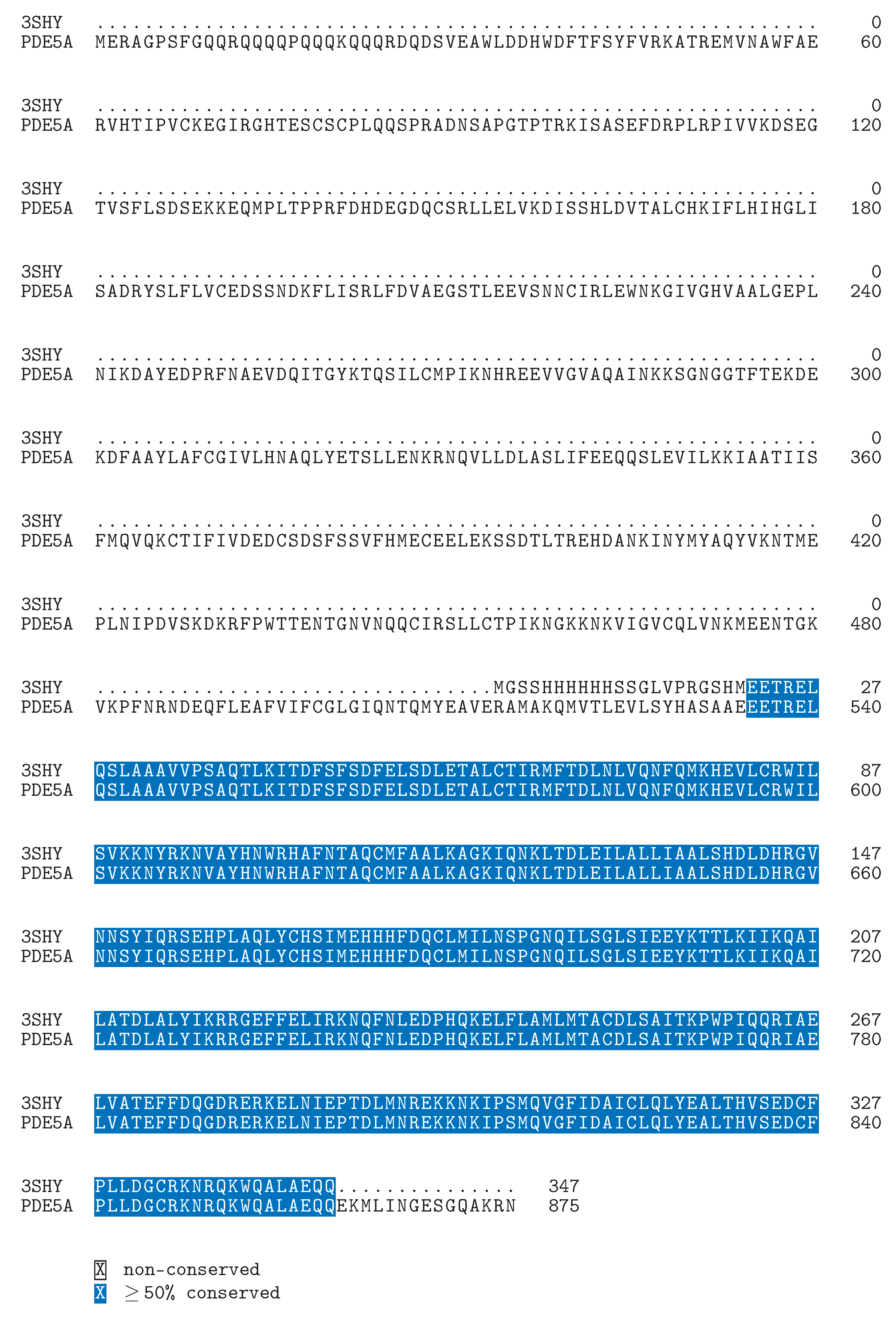
At the molecular level, PDE5 inhibitors bind directly to the catalytic site of PDE5 (Figure 1), preventing the hydrolysis of cGMP to guanosine monophosphate (GMP). From the amino acid sequence point of view, the binding between PDE5 inhibitor (e.g., Sildenafil) and PDE5’s catalytic site is located between sequence position number 536 and 860 (Figure 3). This binding between PDE5 inhibitor (e.g., Sildenafil) leads to an accumulation of cGMP within smooth muscle cells, resulting in prolonged relaxation and vasodilation. Additionally, the inhibition of PDE5 in other tissues, such as the pulmonary vasculature, contributes to the therapeutic effects observed in PAH [7]. However, the non-selective nature of some PDE5 inhibitors can lead to off-target interactions with other phosphodiesterase isoforms, notably PDE6 in the retina, resulting in visual disturbances. Understanding the molecular mechanisms underlying PDE5 inhibitor action is crucial for optimizing their therapeutic benefits while minimizing adverse effects [19,20].

Challenges in the Design of PDE5 Inhibitors: Balancing Efficacy and Safety

Challenges in PDE5 Inhibitor Design encompass a spectrum of factors, notably the prevalence of common side effects, particularly visual disturbances, and the imperative for enhanced selectivity in drug design [5,20]. Among the various adverse reactions associated with PDE5 inhibitors, visual disturbances represent a significant concern. These disturbances primarily arise due to off-target effects on phosphodiesterase type 6 (PDE6) in the retina [21]. PDE6, a crucial enzyme involved in phototransduction, is responsible for the hydrolysis of cyclic guanosine monophosphate (cGMP) in photoreceptor cells. By inhibiting PDE6 activity, PDE5 inhibitors disrupt the delicate balance of cGMP levels in the retina, leading to alterations in visual perception [9]. The mechanism underlying visual disturbances involves perturbations in the phototransduction cascade, affecting processes such as light sensitivity and color discrimination. Moreover, the non-selective nature of some PDE5 inhibitors exacerbates this issue, as they may interact with other phosphodiesterase isoforms, further compromising visual function [22]. Thus, the challenge lies in designing PDE5 inhibitors with improved selectivity profiles, minimizing off-target interactions with PDE6 while maintaining therapeutic efficacy. Addressing these challenges is paramount for optimizing the safety and tolerability of PDE5 inhibitors, thereby enhancing their clinical utility in the treatment of erectile dysfunction and other related conditions [5].

Defining the Challenge in PDE5 Inhibitor Design with a Structural and Biophysical Perspective

As is known, ligand-receptor binding affinity is an essential parameter in computer-assisted drug discovery and structure-based drug design [23]. Thanks to the continued development of experimental structural biology and the half-a-century old Protein Data Bank (PDB) [24,25,26,27,28], a comprehensive structural biophysical analysis becomes possible [29,30] for specific ligand-receptor complex structures deposited in PDB, such that our understanding of the structural and biophysical basis of their interfacial stability is able to help us modify the binding affinity of certain drug target and its interacting partners [31,32,33,34,35]. Take PDE5 as an example here, where the structural key is the design of non-PDE6-binding PDE5 inhibitor (abbreviated below as molecule X), ensuring that:
  • molecule X does bind to PDE5, i.e., ∆G (kcal/mol) < 0 ; and
  • molecule X does not bind to PDE6, i.e., ∆G (kcal/mol) = 0 or ∆G (kcal/mol) > 0 ; and
  • in the absence of molecule X, cGMP does bind to PDE5 as usual (∆Gnoelec = -9.07 kcal/mol [10,11] as calculated by Prodigy [36]) and is able to be catalyzed by PDE5; and
  • in the presence of molecule X, cGMP does not bind to PDE5 (i.e., ∆G (kcal/mol) = 0 or ∆G (kcal/mol) > 0 ), such that PDE5 is unable, or at least not as able as in the absence of molecule X, to catalyze cGMP.
To put it simply, the structural key to the design of non-PDE6-binding PDE5 inhibitor (molecule X) here is also described in [37] as the question for a ChatGPT-like chatbot for drug discovery & design in future: can you generate a Kd-ranked list of therapeutic candidates which targets X (e.g., PDE5) but not Y (e.g., PDE6)?

A GIBAC-Based Selectivity Strategy for the Design of Non-PDE6-Binding PDE5 Inhibitors

On August 11, 2022, the concept of a general intermolecular binding affinity calculator (GIBAC) was for the first time proposed in an MDPI preprint [38] and defined as below:
K d = f ( m o l e c u l e s , e n v P a r a )
where m o l e c u l e s represents the molecular system described either in strings (e.g., amino acid sequences, strings of SMILES to represent small molecules [39,40]), or in graphs to describe PTMs (e.g., glycosylated proteins) and PEMs (e.g., insulin icodec of Novo Nordisk [41,42,43]).
On October 19, 2023, the concept of GIBAC (Equation 1) was for the first time updated, including its inception, definition (Equation 1), construction, practical applications, technical challenges and limitations, and future directions [37,44]. As defined in [37], a real GIBAC (Equation 1) is able to meet the criteria listed as below:
  • a real GIBAC needs to take genetic variations into account; and
  • a real GIBAC needs to work even without structural information; and
  • for a real GIBAC, a variety of factors need to be taken into account, such as temperature, pH [45,46], site-specific protonation states (e.g., side chain pKa of protein) [47,48], post-translational modifications (PTMs) [49,50,51], post-expression modifications (PEMs) [52,53], buffer conditions [54], et cetera; and
  • a real GIBAC requires a general forcefield for all types of molecules [55]; and
  • a real GIBAC requires a universal notation system for accurate and flexible description of all molecular types and drug modalities [56,57]; and
  • a real GIBAC is able to be used the other way around, i.e., to be used as a search engine for therapeutic candidate(s). With such a GIBAC-based search engine, a list of therapeutic candidates can be retrieved and ranked according to drug-target Kd value(s), with input parameters including drug target(s) and a desired drug-target Kd value or a range of it.
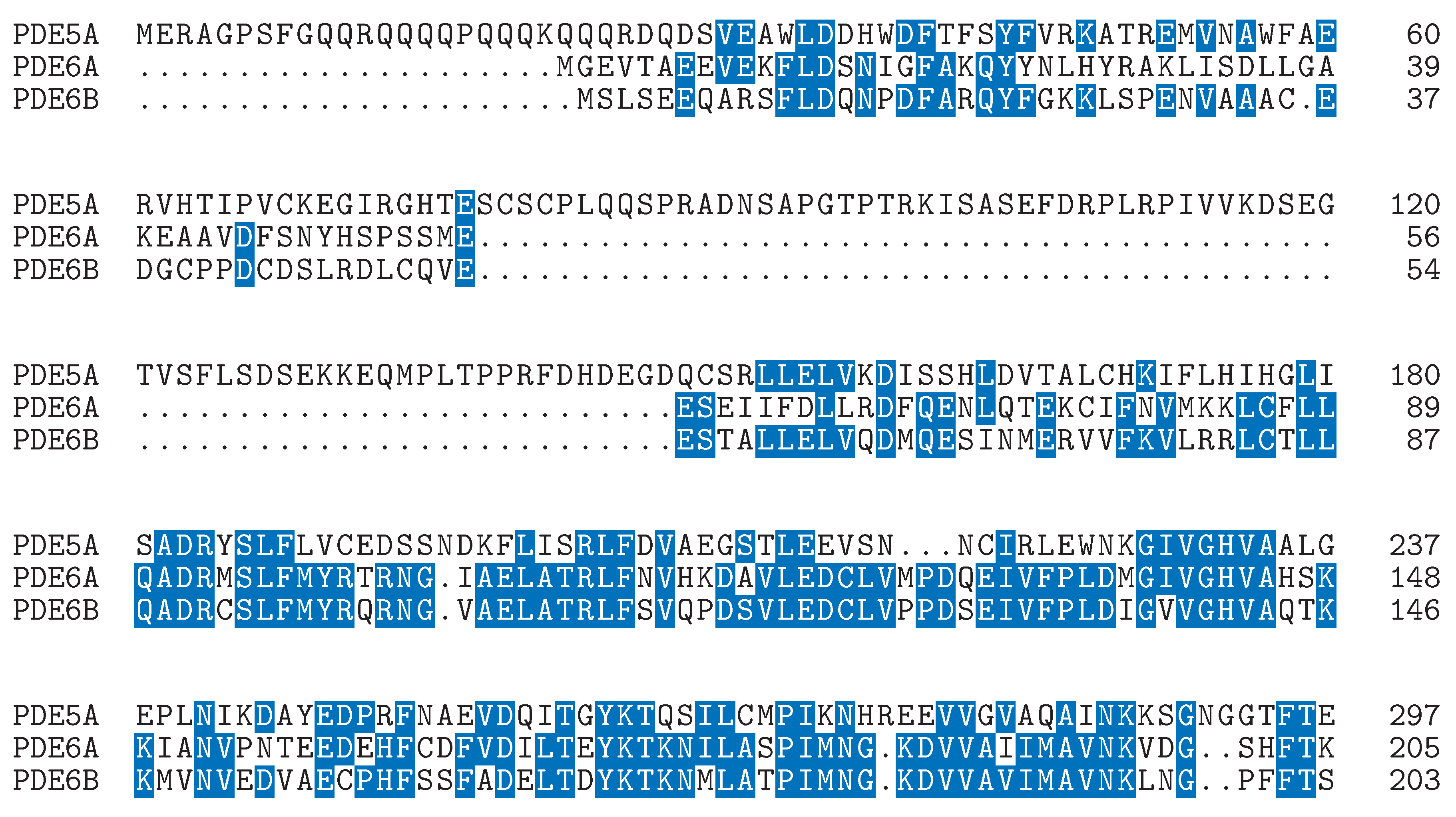
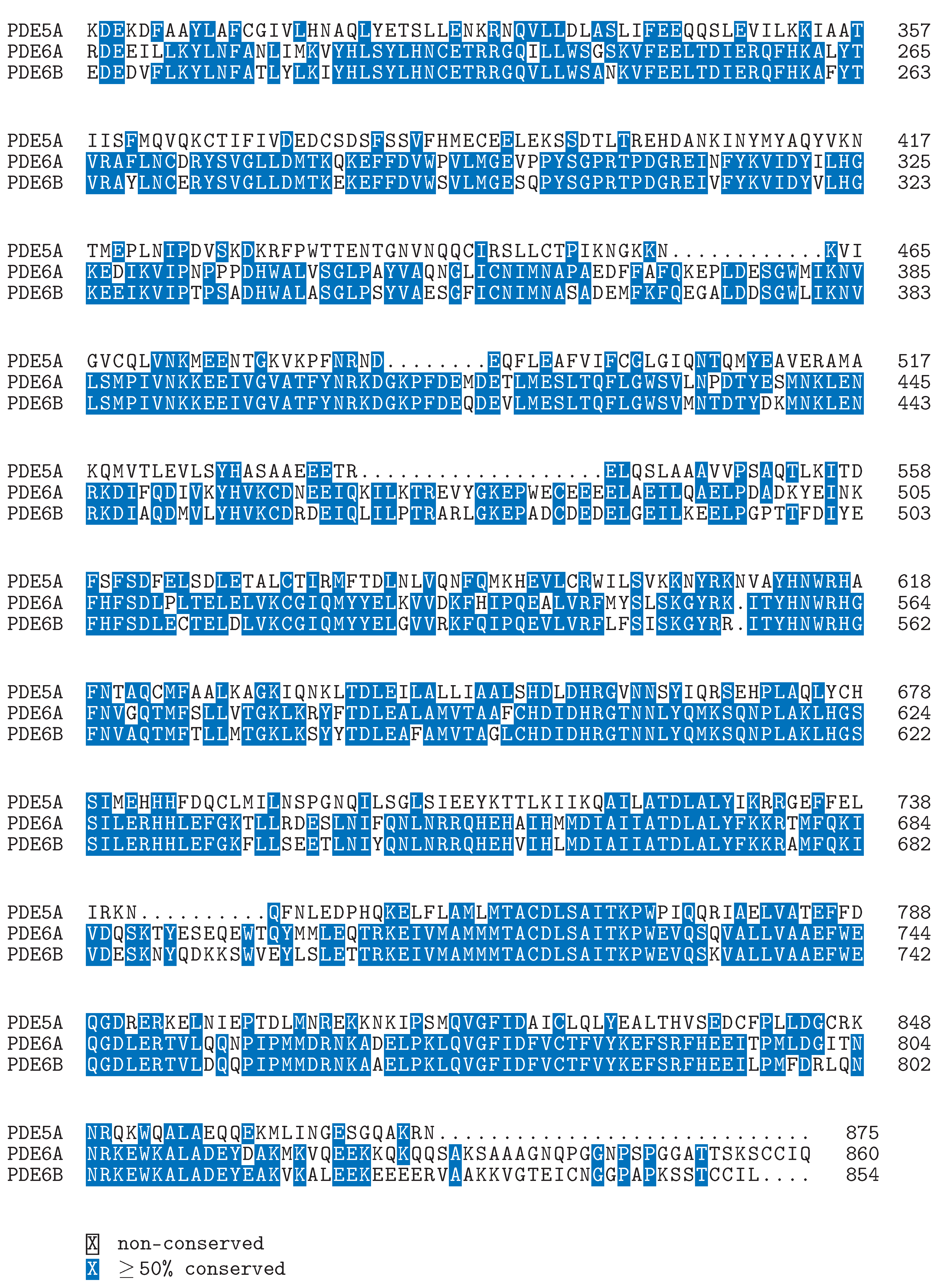
As mentioned above, the binding between PDE5 inhibitor (e.g., sildenafil) and PDE5’s catalytic domain is located between sequence position number 536 and 860 (Figure 3) of PDE5. From Figure 4, it can been seen that the three amino acid sequences (PDE5, PDE6A and PDE6B) do possess a certain degree of sequence homology. Yet, the three sequences are also different from one another, especially for the region which corresponds to the catalytic domain of PDE5, i.e., the region between sequence position number 536 and 860 (Figure 3) of PDE5. Thus, from a structural and biophysical point of view, it is conceivable to design non-PDE6-binding PDE5 inhibitors, which selectively targets PDE5, binds to its binding pocket as shown in Figure 1, and does not target PDE6 at all, i.e., ∆G (kcal/mol) = 0 or ∆G (kcal/mol) > 0 [37].
In practice, the design of non-PDE6-binding PDE5 inhibitors is equivalent to a matter of the construction of a mini GIBAC based on experimental structures of PDE5 and its inhibitor(s) using currently available AI algorithms as listed in [37]. Specifically, the construction of a mini GIBAC based on experimental structures of PDE5 and its inhibitor(s) needs at least four key ingredients: data, algorithm, knowledge and computational power, e.g.,
  • experimental structures of PDE5 and its inhibitor(s);
  • experimental structures of PDE6 and its inhibitor(s);
  • PDE5-related computational structural data from AlphaFold database [?];
  • PDE6-related computational structural data from AlphaFold database [?];
  • synthetic (both apo and complex) PDE5-related structural data generators [?];
  • synthetic (both apo and complex) PDE6-related structural data generators [?];
  • feature extraction using a comprehensive structural biophysical analysis [29,30], including structural biophysics and interfacial geometrics [25,58] underlying the complex structures of PDE5 (or PDE6) and its inhibitors.
  • molecular docking & dynamics simulation tools [??].
  • synthetic Kd data generators [36];
  • side chain placement and energy minimization algorithms [?] to incorporate structural information of PTMs [49,50,51] and PEMs [52,53,59] into structural models.

Future Direction of GIBAC’s Practical Application in Drug Discovery and Design

While drug-target Kd is an essential parameter for drug discovery & design, it is but one of the many aspects of drug discovery & design. Usually, Kd and ∆G has been used to indicate the efficacy of a drug. However, it has been shown that residence time ( R T ) is yet another indicator of efficacy for some systems [60,61]. In biophysics, the relationship between Kd, Kon (association rate constant), and Koff (dissociation rate constant) can be described as K d = K o f f / K o n , with R T representing the average time a molecule spends bound to its target before dissociation.
Essentially, computational drug discovery and design is a matter of a package of parameters focusing on efficacy, safety, et cetera, of the candidates. To this end, a package of biophysical parameters (in addition to Kd) allows a further generalization of the GIBAC originally coined in [38], leading to the concept of a real GIBC (Table 1), which is to be one biophysics-based future direction of GIBAC. Moreover, this article further discusses the potential of GIBAC to act as a ChatGPT-like chatbot for drug discovery & design in future, which is able to accurately, precisely and efficiently handle questions such as:
  • can you generate a Kd-ranked list of therapeutic candidates which targets X of different species, e.g., X of human [35,63], X of cat [64], X of horse [65]?
  • can you generate a Kd-ranked list of therapeutic candidates which targets X, Y and Z? such as vorolanib [66] or retatrutide [67]?
  • can you generate a Kd-ranked list of therapeutic candidates which targets X but not Y or Z?
  • can you generate a list of insulin analogues does not bind to IDE [42,68], but still binds to IR to ensure downstream signal transduction?
  • can you generate a set of mutations into the S protein to stabilize its conformation in the prefusion state for the design of a subunit vaccine of Covid-19 [69,70,71]?

Towards a GIBAC-Based Paradigm Shift in Computational Drug Discovery and Design

Finally, this article argues that the time (May 21, 2024) is now ripe for a GIBAC-based paradigm shift in computational drug discovery and design, and for the construction of such a GIBAC to be on the agenda of the drug discovery & design community, in light of
  • the crucial roles of Kd and ∆G in drug discovery & design; and
  • the availability of a substantial amount of structural and biophysical data, experimental and synthetic; and
  • the continued accumulation of our knowledge of the biophysics [72,73] underlying biological systems; and
  • the availability of a variety of AI algorithms [37] for drug discovery & design; and
  • the democratization of high performance computing since the beginning of this century, and its continued evolution towards scalable quantum computing [74], and perhaps computation beyond silicon [75,76] in future.

Ethical Statement

No ethical approval is required.

Declaration of Generative AI and AI-Assisted Technologies in the Writing Process

During the preparation of this work, the author used OpenAI’s ChatGPT in order to improve the readability of the manuscript, and to make it as concise and short as possible. After using this tool, the author reviewed and edited the content as needed and takes full responsibility for the content of the publication.

Author Contributions

Conceptualization, W.L.; methodology, W.L.; software, W.L.; validation, W.L.; formal analysis, W.L.; investigation, W.L.; resources, W.L.; data duration, W.L.; writing–original draft preparation, W.L.; writing–review and editing, W.L.; visualization, W.L.; supervision, W.L.; project administration, W.L.; funding acquisition, not applicable.

Funding

This research received no external funding.

Acknowledgments

The author is grateful to the communities of structural biology, biophysics, medicinal and computational chemistry and algorithm design, for the continued accumulation of knowledge and data for drug discovery & design, and for the continued development of tools (hardware, software and algorithm) for drug discovery & design.

Conflicts of Interest

The author declares no conflict of interest.

References

  1. Das, A.; Durrant, D.; Salloum, F.N.; Xi, L.; Kukreja, R.C. PDE5 inhibitors as therapeutics for heart disease, diabetes and cancer. Pharmacology & Therapeutics 2015, 147, 12–21. [Google Scholar] [CrossRef]
  2. Ahmed, W.S.; Geethakumari, A.M.; Biswas, K.H. Phosphodiesterase 5 (PDE5): Structure-function regulation and therapeutic applications of inhibitors. Biomedicine & Pharmacotherapy 2021, 134, 111128. [Google Scholar] [CrossRef] [PubMed]
  3. Hemnes, A.R.; Champion, H.C. Sildenafil, a PDE5 inhibitor, in the treatment of pulmonary hypertension. Expert Review of Cardiovascular Therapy 2006, 4, 293–300. [Google Scholar] [CrossRef] [PubMed]
  4. Samidurai, A.; Xi, L.; Das, A.; Kukreja, R.C. Beyond Erectile Dysfunction: cGMP-Specific Phosphodiesterase 5 Inhibitors for Other Clinical Disorders. Annual Review of Pharmacology and Toxicology 2023, 63, 585–615. [Google Scholar] [CrossRef] [PubMed]
  5. Andersson, K. PDE5 inhibitors – pharmacology and clinical applications 20 years after sildenafil discovery. British Journal of Pharmacology 2018, 175, 2554–2565. [Google Scholar] [CrossRef]
  6. Giovannoni, M.; Vergelli, C.; Graziano, A.; Dal Piaz, V. PDE5 Inhibitors and their Applications. Current Medicinal Chemistry 2010, 17, 2564–2587. [Google Scholar] [CrossRef] [PubMed]
  7. Kayık, G.; Tüzün, N.S.; Durdagi, S. Investigation of PDE5/PDE6 and PDE5/PDE11 selective potent tadalafil-like PDE5 inhibitors using combination of molecular modeling approaches, molecular fingerprint-based virtual screening protocols and structure-based pharmacophore development. Journal of Enzyme Inhibition and Medicinal Chemistry 2017, 32, 311–330. [Google Scholar] [CrossRef] [PubMed]
  8. Bischoff, E. Potency, selectivity, and consequences of nonselectivity of PDE inhibition. International Journal of Impotence Research 2004, 16, S11–S14. [Google Scholar] [CrossRef] [PubMed]
  9. Cote, R.H. Characteristics of Photoreceptor PDE (PDE6): similarities and differences to PDE5. International Journal of Impotence Research 2004, 16, S28–S33. [Google Scholar] [CrossRef]
  10. Sung, B.J.; Yeon Hwang, K.; Ho Jeon, Y.; Lee, J.I.; Heo, Y.S.; Hwan Kim, J.; Moon, J.; Min Yoon, J.; Hyun, Y.L.; Kim, E.; Jin Eum, S.; Park, S.Y.; Lee, J.O.; Gyu Lee, T.; Ro, S.; Myung Cho, J. Structure of the catalytic domain of human phosphodiesterase 5 with bound drug molecules. Nature 2003, 425, 98–102. [Google Scholar] [CrossRef]
  11. Sung, B.J.; Lee, J.; Heo, Y.S.; Kim, J.; Moon, J.; Yoon, J.; Hyun, Y.L.; Kim, E.; Eum, S.; Lee, T.; Cho, J.; Park, S.Y.; Lee, J.O.; Jeon, Y.; Hwang, K.; Ro, S. Crystal structure of Human Phosphodiesterase 5 complexed with Sildenafil(Viagra). 2004. [Google Scholar] [CrossRef]
  12. DeLano, W.L. Pymol: An open-source molecular graphics tool. CCP4 Newsletter On Protein Crystallography 2002, 40, 82–92. [Google Scholar]
  13. Gulati, S.; Palczewski, K.; Engel, A.; Stahlberg, H.; Kovacik, L. Cryo-EM structure of phosphodiesterase 6 reveals insights into the allosteric regulation of type I phosphodiesterases. Science Advances 2019, 5. [Google Scholar] [CrossRef] [PubMed]
  14. Gulati, S.; Palczewski, K. Cryo-EM structure of phosphodiesterase 6, 2019. [Google Scholar] [CrossRef]
  15. Kim, K.H.; Kim, H.K.; Hwang, I.C.; Cho, H.J.; Je, N.; Kwon, O.M.; Choi, S.J.; Lee, S.P.; Kim, Y.J.; Sohn, D.W. PDE 5 inhibition with udenafil improves left ventricular systolic/diastolic functions and exercise capacity in patients with chronic heart failure with reduced ejection fraction; A 12-week, randomized, double-blind, placebo-controlled trial. American Heart Journal 2015, 169, 813–822.e3. [Google Scholar] [CrossRef]
  16. Maltez, N.; Maxwell, L.J.; Rirash, F.; Tanjong Ghogomu, E.; Harding, S.E.; Tingey, P.C.; Wells, G.A.; Tugwell, P.; Pope, J. Phosphodiesterase 5 inhibitors (PDE5i) for the treatment of Raynaud’s phenomenon. Cochrane Database of Systematic Reviews 2023, 2023. [Google Scholar] [CrossRef] [PubMed]
  17. Chen, T.; Chen, T.; Xu, Y. Crystal structure of the PDE5A1 catalytic domain in complex with novel inhibitors. 2011. [Google Scholar] [CrossRef]
  18. Xu, Z.; Liu, Z.; Chen, T.; Chen, T.; Wang, Z.; Tian, G.; Shi, J.; Wang, X.; Lu, Y.; Yan, X.; Wang, G.; Jiang, H.; Chen, K.; Wang, S.; Xu, Y.; Shen, J.; Zhu, W. Utilization of Halogen Bond in Lead Optimization: A Case Study of Rational Design of Potent Phosphodiesterase Type 5 (PDE5) Inhibitors. Journal of Medicinal Chemistry 2011, 54, 5607–5611. [Google Scholar] [CrossRef] [PubMed]
  19. Azzouni, F.; Abu samra, K. Are Phosphodiesterase Type 5 Inhibitors Associated with Vision-Threatening Adverse Events? A Critical Analysis and Review of the Literature. The Journal of Sexual Medicine 2011, 8, 2894–2903. [Google Scholar] [CrossRef] [PubMed]
  20. Aliferis, K.; Petropoulos, I.; Farpour, B.; Matter, M.; Safran, A. Should Central Serous Chorioretinopathy Be Added to the List of Ocular Side Effects of Phosphodiesterase 5 Inhibitors? Ophthalmologica 2011, 227, 85–89. [Google Scholar] [CrossRef] [PubMed]
  21. Laties, A.; Sharlip, I. Ocular Safety in Patients Using Sildenafil Citrate Therapy for Erectile Dysfunction. The Journal of Sexual Medicine 2006, 3, 12–27. [Google Scholar] [CrossRef]
  22. Saikia, Q.; Hazarika, A.; Mishra, R. A Review on the Pharmacological Importance of PDE5 and Its Inhibition to Manage Biomedical Conditions. Journal of Pharmacology and Pharmacotherapeutics 2022, 13, 246–257. [Google Scholar] [CrossRef]
  23. Fuji, H.; Qi, F.; Qu, L.; Takaesu, Y.; Hoshino, T. Prediction of Ligand Binding Affinity to Target Proteins by Molecular Mechanics Theoretical Calculation. Chemical and Pharmaceutical Bulletin 2017, 65, 461–468. [Google Scholar] [CrossRef] [PubMed]
  24. Berman, H.; Henrick, K.; Nakamura, H. Announcing the worldwide Protein Data Bank. Nature Structural & Molecular Biology 2003, 10, 980–980. [Google Scholar]
  25. Li, W. Half-a-century Burial of ρ, θ and φ in PDB 2021. φ. [CrossRef]
  26. Li, W. Visualising the Experimentally Uncharted Territories of Membrane Protein Structures inside Protein Data Bank 2020.
  27. Li, W. A Local Spherical Coordinate System Approach to Protein 3D Structure Description 2020.
  28. Li, W. Structurally Observed Electrostatic Features of the COVID-19 Coronavirus-Related Experimental Structures inside Protein Data Bank: A Brief Update 2020.
  29. Li, W. How do SMA-linked mutations of SMN1 lead to structural/functional deficiency of the SMA protein? PLOS ONE 2017, 12, e0178519. [Google Scholar] [CrossRef] [PubMed]
  30. Li, W. Extracting the Interfacial Electrostatic Features from Experimentally Determined Antigen and/or Antibody-Related Structures inside Protein Data Bank for Machine Learning-Based Antibody Design 2020.
  31. Li, W. Structural Identification of the Electrostatic Hot Spots for Severe Acute Respiratory Syndrome Coronavirus Spike Protein to Be Complexed with Its Receptor ACE2 and Its Neutralizing Antibodies 2020.
  32. Li, W. Calcium Channel Trafficking Blocker Gabapentin Bound to the α-2-δ-1 Subunit of Voltage-Gated Calcium Channel: A Computational Structural Investigation 2020.
  33. Li, W. Inter-Molecular Electrostatic Interactions Stabilizing the Structure of the PD-1/PD-L1 Axis: A Structural Evolutionary Perspective 2020.
  34. Li, W. Structural and Functional Consequences of the SMA-Linked Missense Mutations of the Survival Motor Neuron Protein: A Brief Update. In Novel Aspects on Motor Neuron Disease; IntechOpen, 2019.
  35. Li, W. Designing Nerve Growth Factor Analogues to Suppress Pain Signal Transduction Mediated by the p75NTR-NGF-TrkA Complex: A Structural and Biophysical Perspective 2024. [CrossRef]
  36. Vangone, A.; Bonvin, A.M. Contacts-based prediction of binding affinity in protein-protein complexes. eLife 2015, 4. [Google Scholar] [CrossRef] [PubMed]
  37. Li, W.; Vottevor, G. Towards a Truly General Intermolecular Binding Affinity Calculator for Drug Discovery & Design 2023.
  38. Li, W. Towards a General Intermolecular Binding Affinity Calculator 2022.
  39. Lee, J.; Pilch, P.F. The insulin receptor: structure, function and signaling. American Journal of Physiology-Cell Physiology 1994, 266, C319–C334. [Google Scholar] [CrossRef] [PubMed]
  40. Rahuel-Clermont, S.; French, C.A.; Kaarsholm, N.C.; Dunn, M.F. Mechanisms of Stabilization of the Insulin Hexamer through Allosteric Ligand Interactions. Biochemistry 1997, 36, 5837–5845. [Google Scholar] [CrossRef] [PubMed]
  41. Nishimura, E.; Pridal, L.; Glendorf, T.; Hansen, B.F.; Hubálek, F.; Kjeldsen, T.; Kristensen, N.R.; Lützen, A.; Lyby, K.; Madsen, P.; Pedersen, T.Å.; Ribel-Madsen, R.; Stidsen, C.E.; Haahr, H. Molecular and pharmacological characterization of insulin icodec: a new basal insulin analog designed for once-weekly dosing. BMJ Open Diabetes Research & Care 2021, 9, e002301. [Google Scholar]
  42. Li, W. Designing Insulin Analogues with Lower Binding Affinity to Insulin Receptor than That of Insulin Icodec 2024. [CrossRef]
  43. Li, W. How Structural Modifications of Insulin Icodec Contributes to Its Prolonged Duration of Action: A Structural and Biophysical Perspective 2023. [CrossRef]
  44. Wong, C.H.; Siah, K.W.; Lo, A.W. Estimation of clinical trial success rates and related parameters. Biostatistics 2018, 20, 273–286. [Google Scholar] [CrossRef]
  45. Yang, A.S.; Honig, B. On the pH Dependence of Protein Stability. Journal of Molecular Biology 1993, 231, 459–474. [Google Scholar] [CrossRef]
  46. Harris, T.K.; Turner, G.J. Structural Basis of Perturbed pKa Values of Catalytic Groups in Enzyme Active Sites. IUBMB Life (International Union of Biochemistry and Molecular Biology: Life) 2002, 53, 85–98. [Google Scholar] [CrossRef]
  47. Li, W. Gravity-driven pH adjustment for site-specific protein pKa measurement by solution-state NMR. Measurement Science and Technology 2017, 28, 127002. [Google Scholar] [CrossRef]
  48. Hansen, A.L.; Kay, L.E. Measurement of histidine pKa values and tautomer populations in invisible protein states. Proceedings of the National Academy of Sciences 2014, 111, E1705–E1712. [Google Scholar] [CrossRef]
  49. Herget, S.; Ranzinger, R.; Maass, K.; Lieth, C.W. GlycoCT—a unifying sequence format for carbohydrates. Carbohydrate Research 2008, 343, 2162–2171. [Google Scholar] [CrossRef]
  50. Foster, J.M.; Moreno, P.; Fabregat, A.; Hermjakob, H.; Steinbeck, C.; Apweiler, R.; Wakelam, M.J.O.; Vizcaíno, J.A. LipidHome: A Database of Theoretical Lipids Optimized for High Throughput Mass Spectrometry Lipidomics. PLoS ONE 2013, 8, e61951. [Google Scholar] [CrossRef]
  51. Sud, M.; Fahy, E.; Subramaniam, S. Template-based combinatorial enumeration of virtual compound libraries for lipids. Journal of Cheminformatics 2012, 4. [Google Scholar] [CrossRef]
  52. Li, W. Strengthening Semaglutide-GLP-1R Binding Affinity via a Val27-Arg28 Exchange in the Peptide Backbone of Semaglutide: A Computational Structural Approach. Journal of Computational Biophysics and Chemistry 2021, 20, 495–499. [Google Scholar] [CrossRef]
  53. Weiss, M. Design of ultra-stable insulin analogues for the developing world. Journal of Health Specialties 2013, 1, 59. [Google Scholar] [CrossRef]
  54. Olsson, M.H.M.; Søndergaard, C.R.; Rostkowski, M.; Jensen, J.H. PROPKA3: Consistent Treatment of Internal and Surface Residues in Empirical pKa Predictions. Journal of Chemical Theory and Computation 2011, 7, 525–537. [Google Scholar] [CrossRef]
  55. Hofmann, D.W.M.; Kuleshova, L.N. A general force field by machine learning on experimental crystal structures. Calculations of intermolecular Gibbs energy with iFlexCryst. Acta Crystallographica Section A Foundations and Advances 2023, 79, 132–144. [Google Scholar] [CrossRef]
  56. Wang, T.; He, X.; Li, M.; Shao, B.; Liu, T.Y. AIMD-Chig: Exploring the conformational space of a 166-atom protein Chignolin with ab initio molecular dynamics. Scientific Data 2023, 10. [Google Scholar] [CrossRef]
  57. Müller, C.E.; Hansen, F.K.; Gütschow, M.; Lindsley, C.W.; Liotta, D. New Drug Modalities in Medicinal Chemistry, Pharmacology, and Translational Science. ACS Pharmacology & Translational Science 2021, 4, 1712–1713. [Google Scholar]
  58. Li, W. A Reversible Spherical Geometric Conversion of Protein Backbone Structure Coordinate Matrix to Three Independent Vectors of ρ, θ and φ 2024. [CrossRef]
  59. Li, W. An Exhaustive Exploration of the Semaglutide-GLP-1R Sequence Space towards the Design of Semaglutide Analogues with Elevated Binding Affinity to GLP-1R 2024. [CrossRef]
  60. Costa, B.; Pozzo, E.D.; Giacomelli, C.; Barresi, E.; Taliani, S.; Settimo, F.D.; Martini, C. TSPO ligand residence time: a new parameter to predict compound neurosteroidogenic efficacy. Scientific Reports 2016, 6. [Google Scholar] [CrossRef]
  61. Copeland, R.A. The drug-target residence time model: a 10-year retrospective. Nature Reviews Drug Discovery 2015, 15, 87–95. [Google Scholar] [CrossRef]
  62. Rahnama, R.; Kizerwetter, M.; Yang, H.; Christodoulou, I.; Fearnow, A.; Vorri, S.; Spangler, J.; Bonifant, C. Poster: AML-496 Study of Affinity Tuning in AML-Targeted CAR-NK Cells. Clinical Lymphoma Myeloma and Leukemia 2022, 22, S136. [Google Scholar] [CrossRef]
  63. Zimmermann, A.; Sutter, A.; Shooter, E.M. Monoclonal antibodies against beta nerve growth factor and their effects on receptor binding and biological activity. Proceedings of the National Academy of Sciences 1981, 78, 4611–4615. [Google Scholar] [CrossRef]
  64. Enomoto, M.; Mantyh, P.W.; Murrell, J.; Innes, J.F.; Lascelles, B.D.X. Anti-nerve growth factor monoclonal antibodies for the control of pain in dogs and cats. Veterinary Record 2019, 184, 23–23. [Google Scholar] [CrossRef]
  65. Kalnins, G. mature recombinant horse NGF, 2020.
  66. Sheng, X.; Ye, D.; Zhou, A.; Yao, X.; Luo, H.; He, Z.; Wang, Z.; Zhao, Y.; Ji, Z.; Zou, Q.; He, C.; Guo, J.; Tu, X.; Liu, Z.; Shi, B.; Liu, B.; Chen, P.; Wei, Q.; Hu, Z.; Zhang, Y.; Jiang, K.; Zhou, F.; Wu, D.; Fu, C.; Li, X.; Wu, B.; Wang, L.; Qin, S.; Li, G.; Liu, Y.; Guo, H.; Chen, K.; Zhang, D.; Wang, G.; Ding, L.; Wang, Y.; Yuan, X.; Guo, J. Efficacy and safety of vorolanib plus everolimus in metastatic renal cell carcinoma: A three-arm, randomised, double-blind, multicentre phase III study (CONCEPT). European Journal of Cancer 2023, 178, 205–215. [Google Scholar] [CrossRef]
  67. Urva, S.; O’Farrell, L.; Du, Y.; Loh, M.T.; Hemmingway, A.; Qu, H.; Alsina-Fernandez, J.; Haupt, A.; Milicevic, Z.; Coskun, T. The novel GIP, GLP-1 and glucagon receptor agonist retatrutide delays gastric emptying. Diabetes, Obesity and Metabolism 2023, 25, 2784–2788. [Google Scholar] [CrossRef]
  68. Fakhrai-Rad, H. Insulin-degrading enzyme identified as a candidate diabetes susceptibility gene in GK rats. Human Molecular Genetics 2000, 9, 2149–2158. [Google Scholar] [CrossRef]
  69. Li, W. Delving deep into the structural aspects of a furin cleavage site inserted into the spike protein of SARS-CoV-2: A structural biophysical perspective. Biophysical Chemistry 2020, 264, 106420. [Google Scholar] [CrossRef]
  70. Shi, W.; Cai, Y.; Zhu, H.; Peng, H.; Voyer, J.; Rits-Volloch, S.; Cao, H.; Mayer, M.L.; Song, K.; Xu, C.; Lu, J.; Zhang, J.; Chen, B. Cryo-EM structure of SARS-CoV-2 postfusion spike in membrane. Nature 2023, 619, 403–409. [Google Scholar] [CrossRef] [PubMed]
  71. Heo, L.; Feig, M. Modeling of Severe Acute Respiratory Syndrome Coronavirus 2 (SARS-CoV-2) Proteins by Machine Learning and Physics-Based Refinement 2020.
  72. Noble, D.; Blundell, T.L.; Kohl, P. Progress in biophysics and molecular biology: A brief history of the journal. Progress in Biophysics and Molecular Biology 2018, 140, 1–4. [Google Scholar] [CrossRef] [PubMed]
  73. Becker, O.M.; MacKerell Jr, A.D.; Roux, B.; Watanabe, M. Computational biochemistry and biophysics; CRC Press,, 2001. [Google Scholar]
  74. Kendon, V. Quantum computing using continuous-time evolution. Interface Focus 2020, 10, 20190143. [Google Scholar] [CrossRef] [PubMed]
  75. Cookson, C. Taking bold bets: new UK agency prepares to fund breakthrough technologies, 2023. Accessed: (August 30, 2023).
  76. Zhou, S.F.; Zhong, W.Z. Drug Design and Discovery: Principles and Applications. Molecules 2017, 22, 279. [Google Scholar] [CrossRef] [PubMed]
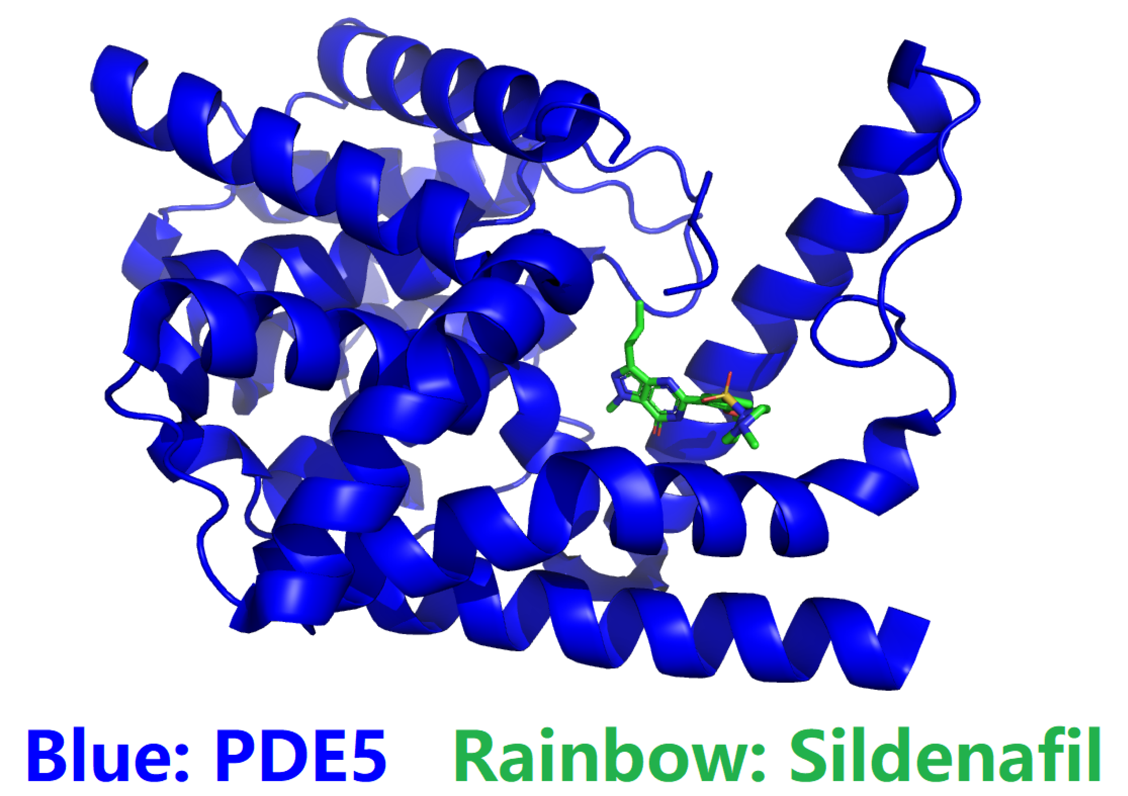
Figure 1. Crystal structure of human phosphodiesterase 5 (PDE5) complexed with sildenafil (PDB ID: 1UDT) [10,11]. In this figure, the blue cartoon represents the catalytic domain of PDE5, while the small molecule in rainbow sticks represents sildenafil. This figure was prepared with PyMol [12]
Preprints 107052 g001
Figure 2. Cryo-EM structure of phosphodiesterase 6 (PDB ID: 6MZB) [13,14]. In this figure, cyan represents the subunit of PDE6, while green represents the subunit of PDE6. This figure was prepared with PyMol [12]
Preprints 107052 g002
Figure 3. Sequence alignment (https://www.genome.jp/tools-bin/clustalw) of human cGMP-specific 3’,5’-cyclic phosphodiesterase (PDE5, UniProtKB ID: O76074) and its catalytic domain whose structure is experimentally determined (PDB ID: 3SHY [17,18]).
Preprints 107052 g003
Figure 4. Sequence alignment (https://www.genome.jp/tools-bin/clustalw) of human cGMP-specific 3’,5’-cyclic phosphodiesterase (PDE5, UniProtKB ID: O76074), PDE6A_(UniProtKB ID: P16499)_Human Retinal Rod cGMP-specific 3’,5’-cyclic phosphodiesterase subunit alpha (UniProtKB ID: P16499) and PDE6B_(UniProtKB ID: P35913)_Human Retinal Rod cGMP-specific 3’,5’-cyclic phosphodiesterase subunit beta (UniProtKB ID: P35913).
Preprints 107052 g004aPreprints 107052 g004b
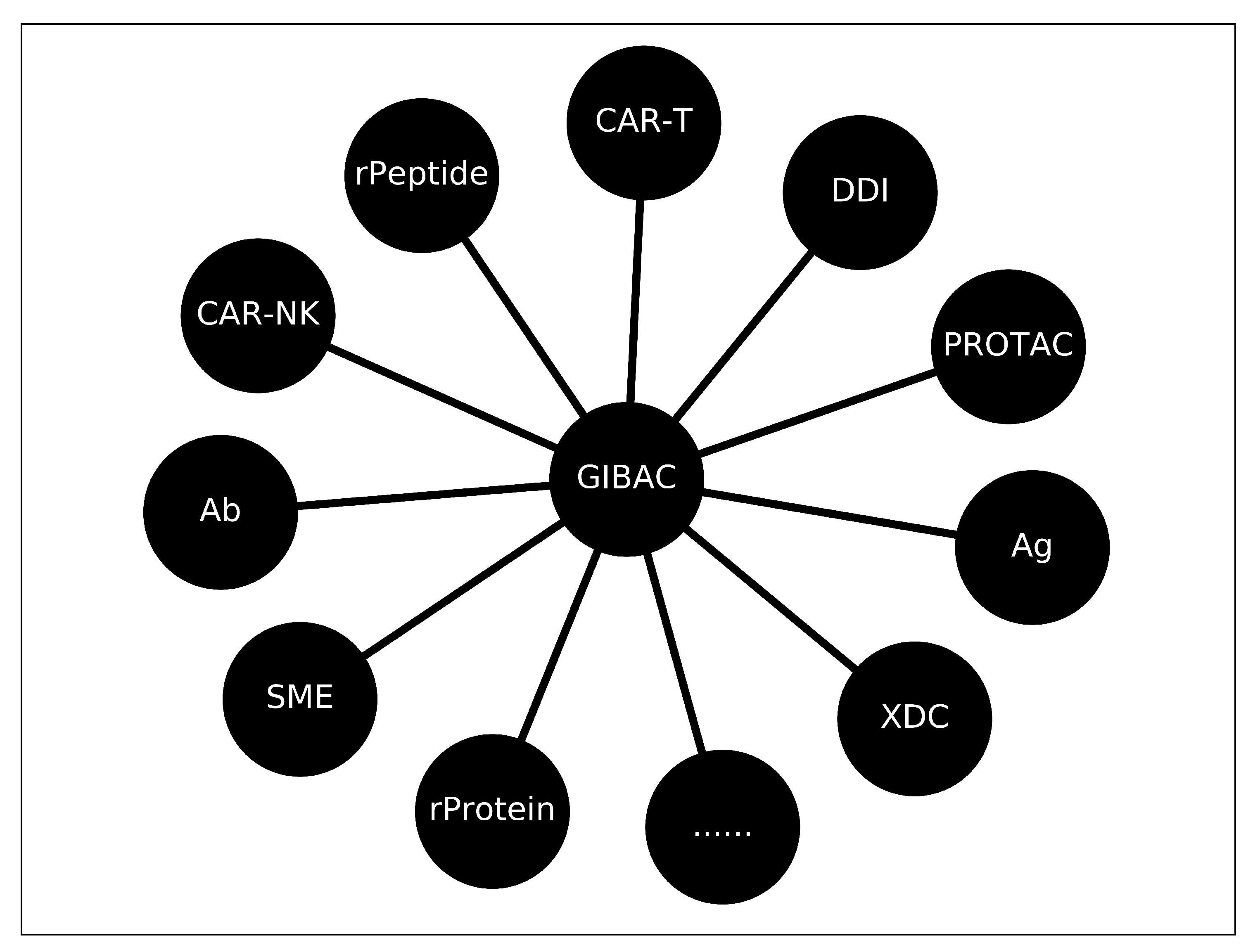
Figure 5. Areas where GIBAC are applicable and involved. SME, Antibody, Antigen, XDC, ApDC: Aptamer drug conjugate ADC: Ab drug conjugate, rPeptide, rProtein, PROTAC, DDI, Purify, Chimeric antigen receptor (CAR) T + CAR-NK [62] cell therapy relies on T cells that are guided by synthetic receptors to target and lyse cancer cells. CARs bind to cell surface antigens through an scFv (binder), the affinity of which is central to determining CAR T cell function and therapeutic success [37].
Preprints 107052 g005
Table 1. A tabular description of a general intermolecular biophysics calculator (GIBC).
Input 1 Input 2 Output
m o l A s t r i n g , m o l B s t r i n g , ... e n v P a r a Kd, Kon, Koff, RT, pKa
m o l A g r a p h , m o l B g r a p h , ... e n v P a r a Kd, Kon, Koff, RT, pKa
Disclaimer/Publisher’s Note: The statements, opinions and data contained in all publications are solely those of the individual author(s) and contributor(s) and not of MDPI and/or the editor(s). MDPI and/or the editor(s) disclaim responsibility for any injury to people or property resulting from any ideas, methods, instructions or products referred to in the content.
Copyright: This open access article is published under a Creative Commons CC BY 4.0 license, which permit the free download, distribution, and reuse, provided that the author and preprint are cited in any reuse.
Prerpints.org logo

Preprints.org is a free preprint server supported by MDPI in Basel, Switzerland.

Subscribe

© 2024 MDPI (Basel, Switzerland) unless otherwise stated