Preprint
Article

Carbazolyl-Modified Neutral Ir(III) Complexes for Efficient Detection of Picric Acid in Aqueous Media

Altmetrics

Downloads

149

Views

21

Comments

0

A peer-reviewed article of this preprint also exists.

This version is not peer-reviewed

Submitted:

21 May 2024

Posted:

23 May 2024

You are already at the latest version

Alerts
Abstract
Based on the electron-deficient property of picric acid (PA), two neutral Ir(III) complexes 1 and 2 modified with the electron-rich carbazolyl groups were synthesized and characterized. Both 1 and 2 exhibit aggregation-induced phosphorescence emission (AIPE) properties in THF/H2O. Among them, 2 is extremely sensitive for detecting PA with a limit of detection of 0.15 μM in THF/H2O. Furthermore, the selectivity for PA is significantly higher compared to other analytes, enabling efficient detection of PA in four common water samples. The density functional theory calculations and the spectroscopic results confirm that the sensing mechanism is photo-induced electron transfer.
Keywords: 
Subject: Chemistry and Materials Science  -   Electronic, Optical and Magnetic Materials

1. Introduction

Since Tang et al. firstly stated the concept of aggregation-induced emission (AIE) in 2001 [1], numerous AIE materials have been reported [2,3,4,5]. The restriction of intramolecular motion (RIM) is the widely accepted AIE mechanism. According to RIM, when aggregates are formed, intramolecular rotation and/or vibration of the corresponding molecules are restricted. This restriction reduces the amount of thermal energy that the molecule consumes as it decays from the electronically excited state to the ground state. Consequently, the energy is released in the form of radiative transitions, which enhances the luminescence of the molecules [6]. Over the past two decades, AIE materials have undergone significant scientific and technological development, leading to a continuously expanding variety of AIE luminogens (AIEgens). The family of AIEgens has grown to include heteroatom compounds, pure hydrocarbons, macromolecules, as well as metallic-organic compounds. Among them, metal-organic complexes with AIE properties have been widely used in various fields, including organic light-emitting diodes [7], bio-imaging [8], disease treatment [9], and explosives detection [10,11].
Designing the structures of ligands to modulate the properties of metal complexes is a useful method in the development of novel functional materials. Carbazolyl, as a kind of electron-rich moiety with excellent hole-transporting ability and a large conjugation plane, is common for modulating molecular structures [12,13,14]. The carbazolyl group could enhance the interaction between Ir(III) complexes and electron-deficient nitroaromatic explosives. For example, Su and co-workers reported two Ir(III) complexes ((PFBHC)2Ir(acac) and FIrPicOMPBHCz) containing carbazolyl groups for detecting nitroaromatic explosives in CH2Cl2, and the limits of detection (LODs) for PA were 29 μM and 10 μM, respectively. The authors suggested that the introduction of the electron-rich carbazolyl group facilitate the excited-state electron-transfer process [15,16]. Furthermore, Ir(III) complexes featuring carbazolyl groups exhibited significantly enhanced solubility and photostability. In 2023, Di et al. reported two carbazolyl-modified Ir(III) complexes IrCzPh and IrPhCz. Compared to Ir(ppy)2(acac), the photostability of both complexes was significantly improved and their AIE properties were activated in MeCN/H2O [17].
2,4,6-Trinitrophenol, often referred to as picric acid (PA), has been utilized in paints, dyes, pharmaceuticals, and aerospace applications due to its excellent explosive property [18,19,20]. However, PA is highly toxic and non-biodegradable, and can lead to liver and kidney dysfunction, conjunctivitis, skin allergic ulcers, and other serious diseases, if it accumulates in the body for a prolonged period [21,22,23,24]. Leakage and misuse of PA could result in serious contamination of soil and water. As a result, it is critical to develop simple, highly sensitive and selective methods to detect PA, especially in aqueous media. Scientists widely prefer photoluminescence spectroscopy for its exceptional sensitivity, cost-effectiveness, ease of operation, and practical applicability. Therefore, photoluminescence for the detection of PA has attracted much attention.
Our group possesses an enduring interest in investigating the relationship between the structural composition and functional properties of cyclometalating Pt(II) and Ir(III) complexes [25,26,27,28,29]. Recently, we have successfully applied cationic Ir(III) complexes with AIPE properties for the detection of PA in aqueous media with high sensitivity [30,31]. However, the performances of AIPE-active neutral Ir(III) complexes for detecting PA still need to be further explored. In this work, we synthesized two carbazolyl-modified neutral Ir(III) complexes with 2-phenylpyridine derivatives as cyclometalating ligands and acetylacetone as the auxiliary ligand. The corresponding structures of the complexes are shown in Figure 1. Their luminescent properties and performance of 2 in the detection of PA in the aqueous media have been investigated in detail.

2. Experimental Section

2.1. Reagents and Instruments

All raw materials from commercial suppliers were utilized without additional purification. The 1H NMR and 13C NMR spectra were acquired utilizing a 400 MHz Varian Unity Inova spectrophotometer. Mass spectra were obtained employing a Bruker Ultraflexetreme MALDI TOF mass spectrometer. Photoluminescence quantum yields (ФPL) were measured using [Ir(ppy)2(acac)] as a standard (ФPL = 0.34 in CH2Cl2, under deoxygenated conditions). Phosphorescence lifetimes were measured employing an Edinburgh FLS920 spectrometer in a degassed CH2Cl2 solution. UV-Vis absorption spectra were acquired utilizing an Agilent Cary 100 UV-Vis spectrophotometer. Emission spectra were recorded utilizing a Hitachi F-7100 fluorescence spectrophotometer, with luminescence characteristics of 1 and 2 observed under identical instrumental parameters. Density functional theory (DFT) calculations were measured using the B3LYP floods. The 6-31G basis sets were applied for C, H, and O atoms, while the LanL2DZ basis set was utilized for iridium atoms. All computations were executed using Gaussian 16.

2.2. Synthesis and Characterizations of 1 and 2

The Ir(III) complexes were synthesized according to a two-step method reported in the literature [25]. Firstly, IrCl3·3H2O (0.20 mmol, 70.52 mg) and the cyclometalating ligands (0.50 mmol) were added to 12 mL of anhydrous EtOC2H4OH/H2O (3:1, V/V). The mixture was stirred for 24 h at 110°C under N2 to get a dichloro-bridged intermediate. The dichloro-bridge intermediate without further purification, K2CO3 (1 mmol, 138.21 mg), and acetylacetone (1.00 mmol, 103 μL) were reacted in 12 mL of anhydrous EtOC2H4OH under stirring and refluxing at 120°C in N2 for 24 h. After reaction, the synthesized complexes were subjected to purification via silica gel column chromatography, employing a solvent system consisting of n-hexane and CH2Cl2 in a 1:1 ratio as the eluting agent, resulting in the complexes 1 and 2.

2.3. Sample Preparation and Sensing of PA

WARNING! The nitroaromatic compounds used in emission spectroscopic studies are highly explosive and must be used safely in small quantities.
Stock solutions of 1 and 2 (100 μM) in THF were prepared firstly. For each suspension of the complexes in THF/H2O, 300 μL of the stock solution was mixed with appropriate volume of THF and deionized water to formulate 3 mL (10 μM) suspensions with various water fractions. The emission spectra and UV-Vis absorption spectra of the samples were each documented. A suspension of 2 (10 μM) with a 90% water fraction was prepared in a 200 mL volumetric flask. Then, 3 mL of each suspension was withdrawn and added into a quartz cuvette, and the emission spectra for each of 11 blank suspensions were recorded for the calculation of the standard deviation (Figure S7). Solutions of PA were prepared in THF/H2O (fw = 90%) at concentrations ranging from 0.1 to 50 mM. PA solutions (30 μL) with various concentrations were introduced into cuvettes containing 3 mL of the complex suspension, and the emission spectra were recorded. For selectivity experiments on PA detection, various analytes (30 μL, 30 mM) were added, including nitrobenzene (NB), 1,3-dinitrobenzene (1,3-DNB), nitromethane (NM), p-Cresol, m-Cresol, Phenol, and 4-methoxyphenol (MEHQ). Ion interference experiments were conducted to assess the effects of various ionic compounds (30 μL, 30 mM each of CaCl2, MnCl2, FeCl2, NiCl2, ZnCl2, CoCO3, NaHCO3, MgSO4, CuSO4, KF, KBr, and CH3COONa) on the suspensions of 2, where 3 mL of the suspension was used. In competition experiments, PA solutions were added to suspensions of 2 containing the ionic compounds and other analytes, respectively. To assess the real application capacity of 2, various water samples (tap water from the State Key Laboratory of Fine Chemicals, river water from the Malan River in Dalian, rainwater from Dalian University of Technology, and seawater from Qixianling in Dalian) were used instead of deionized water to formulate suspensions of 2. Subsequently, 30 μL of 30 mM PA solutions were added to the suspensions respectively, and the corresponding emission spectra were recorded.

3. Results and Discussion

3.1. Photophysical and AIPE Properties

The UV-Vis absorption spectra and normalized emission spectra at room temperature of complexes 1 and 2 in THF solution are shown in Figure S3. The photophysical data of both complexes can be found in Table S1. Similar to most Ir(III) complexes, both 1 and 2 exhibit strong absorption bands below 300 nm, which belong to the typical ligand-centered (1LC(1π-π*)) transitions. The lower energy absorption between 300-430 nm is attributed to a combination of metal-to-ligand charge transitions (1MLCT/3MLCT), ligand-to-ligand charge transitions (1LLCT/3LLCT), and ligand-centered (3LC(3π-π*)) leaps. The normalized emission spectra indicate that the maximum emission wavelength and peak shape of 2 remain largely unchanged compared to those of 1. The phosphorescence lifetimes of 1 and 2 are in the range of 1.77-2.18 μs at room temperature (Figure S4). The phosphorescence quantum yields of 1 and 2 were measured in degassed CH2Cl2 using Ir(ppy)2(acac) (0.34) as a standard [32]. The results indicate that phosphorescence quantum yields of 0.18 for 1 and 0.27 for 2, respectively. Incorporating an electron-donating group (5-CH3) onto the pyridine ring of the complex 2 resulted in an increased phosphorescence quantum yield.
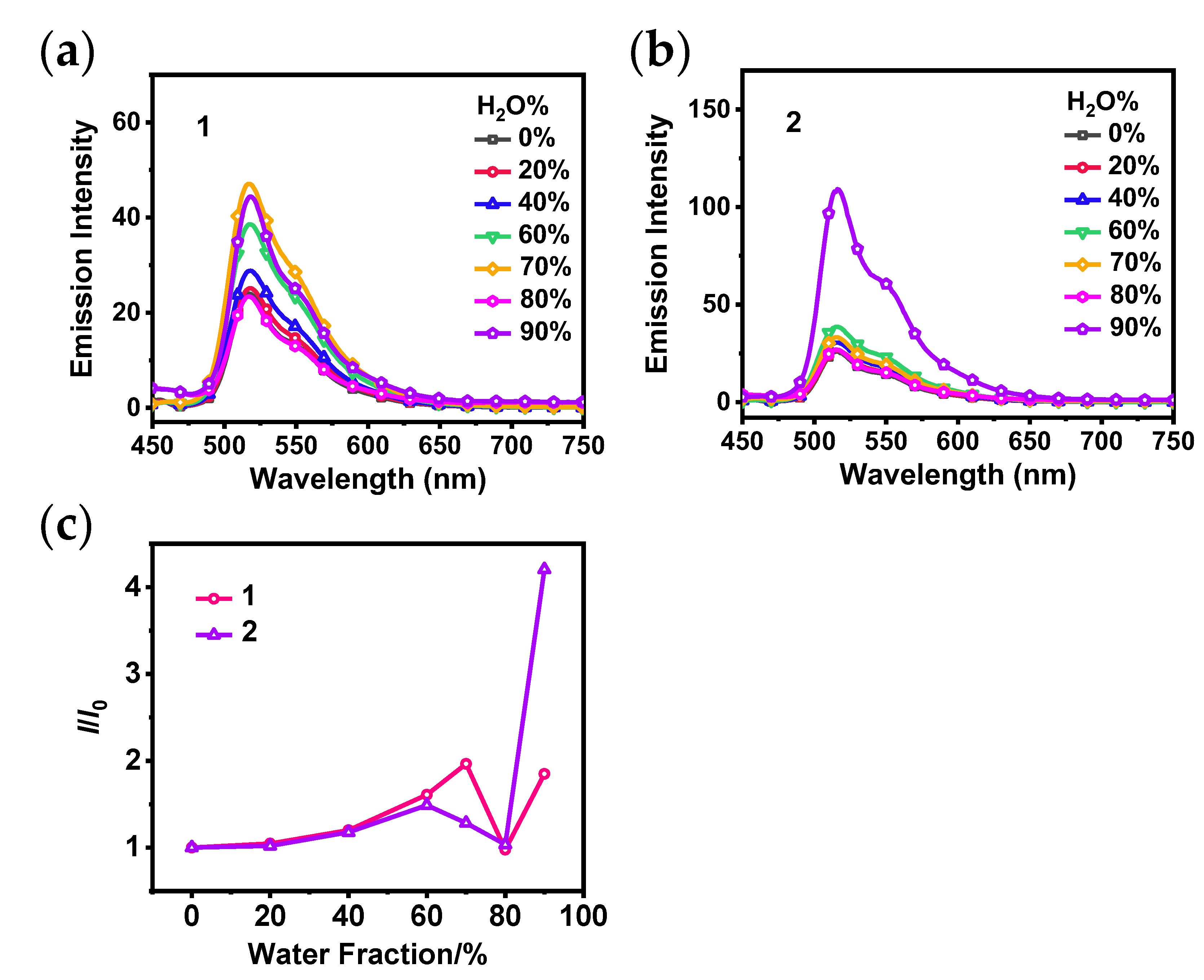
Due to the large conjugation plane of carbazolyl group, molecular motion may be greatly restricted when close stacking occurs. As a result, there is a high probability that 1 and 2 will exhibit AIPE property. To confirm this conjecture, suspensions of 1 and 2 in THF/H2O with various water fractions were prepared, and their emission spectra and UV-vis absorption spectra were recorded as shown in Figure S5. Figure 2a,b show that the emission intensity of 1 or 2 increases gradually as the water fraction increases from 0 to 60%. Further increasing the water fraction resulted in the maximum emission intensity of 1 and 2 at 70% and 90%, respectively, indicating a significant AIPE phenomenon. The ratio plot (Figure 2c) shows that 2 exhibits a higher AIPE property than 1 under the same experimental conditions, which is similar to the findings reported by Yang et al. in 2011 [33]. The introduction of a methyl group into the complex is believed to change the arrangement of 2 in the aggregated state, resulting in tighter intermolecular interaction to restrict intramolecular movement efficiently. This leads to enhanced luminescence of 2 at high water fractions.

3.2. Detection of PA

2 exhibits impressive AIPE property in THF/H2O, suggesting its potential for detection of PA in aqueous media. We further conducted the luminescence quenching experiments of 2. As shown in Figure 3a,b, the emission intensity of 2 decreased progressively as the concentration of PA increased. The quenching efficiency of 2 was 28% when the concentration of PA reached 10 μM. The quenching efficiency of 2 increased significantly to 98% after the addition of PA solution with a concentration of 300 μM (30 equiv.).
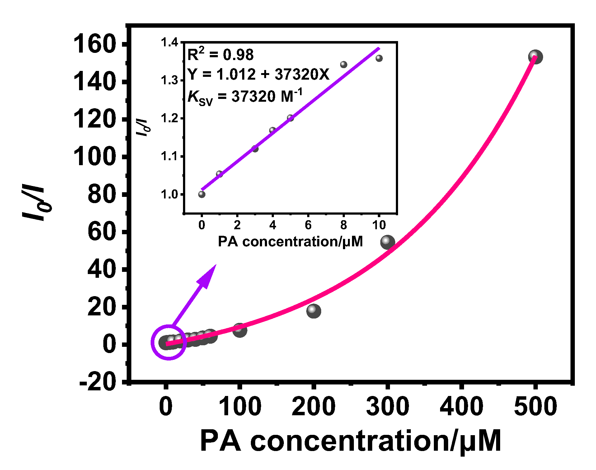
The quenching constant (KSV) is usually used as an index to the sensitivity of a sensor to detect PA. To obtain the KSV of 2 for PA, a Stern-Volmer plot of 2 was constructed using the ratio of the luminescence intensity without PA (I0) to the luminescence intensity with PA (I) vs. the concentration of PA (Figure 4). The Stern-Volmer plot displays excellent linearity within the concentration of PA range from 0-10 μM, which is indicative of static quenching [34,35]. Within the concentration of PA range from 0-500 μM, there is a clear non-linear relationship due to the combination of static and dynamic quenching [36,37]. The sensitivity of 2 for PA is assessed utilizing the Stern-Volmer equation: I0/I = KSV[Q] + 1 [38]. In the concentration range from 0-10 μM, a linear fit is used to calculate the KSV of 2 to be 37320 M-1. This value is higher than those of previously reported neutral Ir(III) complexes with carbazolyl groups for PA detection [15,16]. In addition, the LOD of 2 for PA can be calculated using the formula LOD = 3σ/K, where σ represents the standard deviation of 11 blank samples of 2 (Table 1), and K represents the slope of the linear relationship between the luminescence intensity I and PA concentration [39].
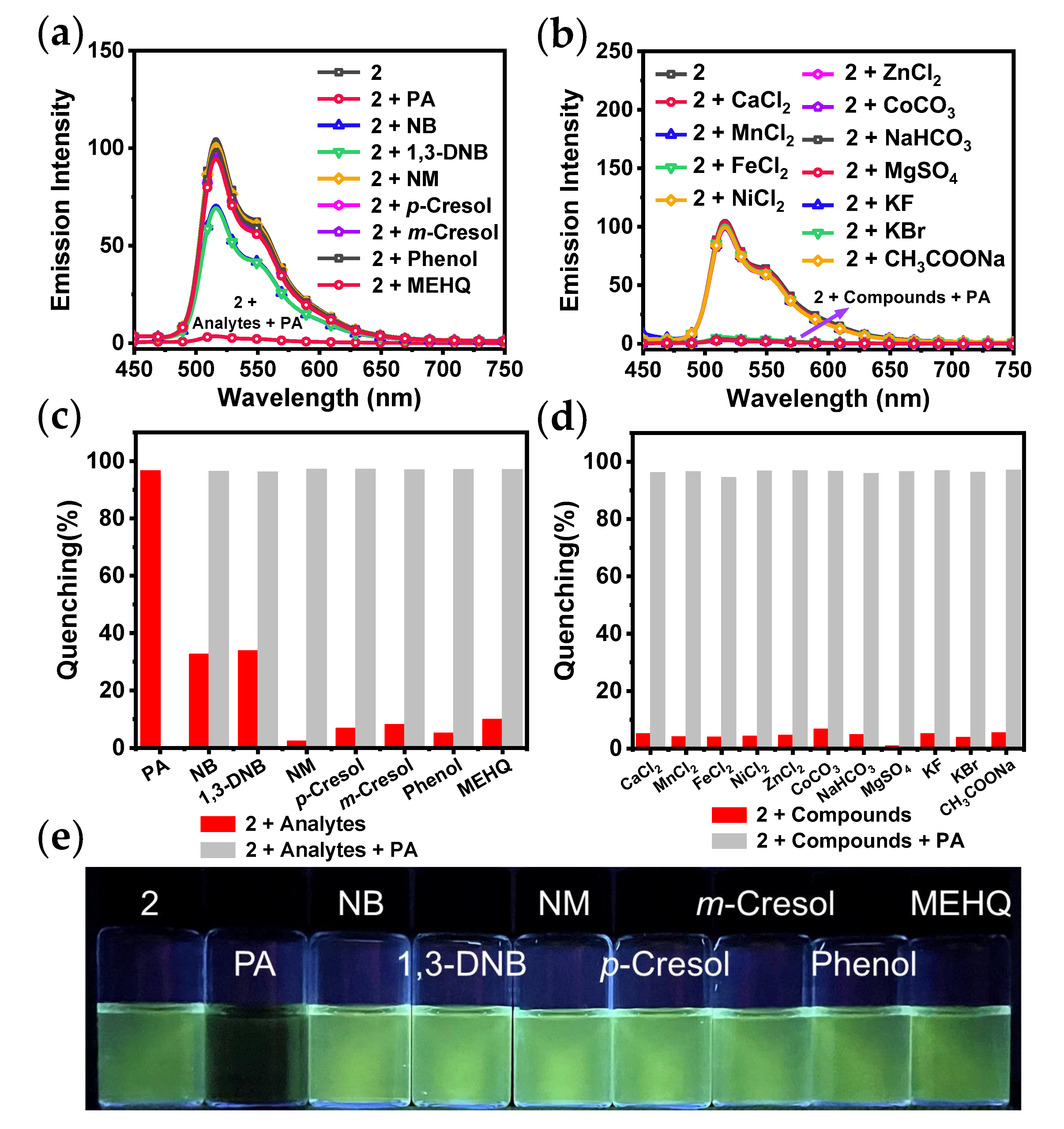
Selectivity is deemed a key factor influencing the probe performance. Therefore, the luminescence response studies on several common analytes were conducted, including NB, 1,3-DNB, NM, p-Cresol, m-Cresol, Phenol, and MEHQ. The emission spectra can be seen in Figure 5a. Figure 5c shows that when PA was added, the luminescence quenching efficiency of 2 was higher than that of the other analytes, demonstrating excellent selectivity.
In order to further explore selectivity and anti-interference capacity of 2 and to expand scope of its application for the detection of PA, various ionic compounds were used for the emission spectroscopic studies, including CaCl2, MnCl2, FeCl2, NiCl2, ZnCl2, CoCO3, NaHCO3, MgSO4, CuSO4, KF, KBr, and CH3COONa. Adding solutions of various compounds did not lead to noticeable alteration in the emission spectra or emission intensity of 2 (Figure 5b). This suggests that the addition of ions has small impact on the luminescent property of 2 in THF/H2O.
Competition experiments were carried out by introducing 30 equiv. of PA solution into suspensions containing various analytes or ionic compounds. As shown in Figure 5c,d, the luminescence quenching efficiency of 2 by PA was found to be almost unaffected in the presence of various competing compounds. Therefore, 2 shows outstanding anti-interference capacity when used to detect PA.
Water resources represent a crucial component of the ecological environment and are utilized in a multitude of social production and life processes. PA is readily soluble in hot water and has the potential to pollute water sources. Consequently, it is of paramount importance to detect the presence of PA in common water samples. The emission spectra of 2 before and after the addition of PA in various water samples as shown in Figure 6a. In comparison to the luminescence observed within deionised water, the emission spectra of 2 in four common water samples exhibit similar shapes, yet distinct emission intensity. The quenching efficiency of 2 for detecting PA in different water samples is shown in Figure 6b, and the results did not differ significantly. The spectral analysis indicates that efficiency of luminescence quenching of 2 by PA is considerably higher than that of the impurities present in four common water samples.

3.1. Sensing Mechanism

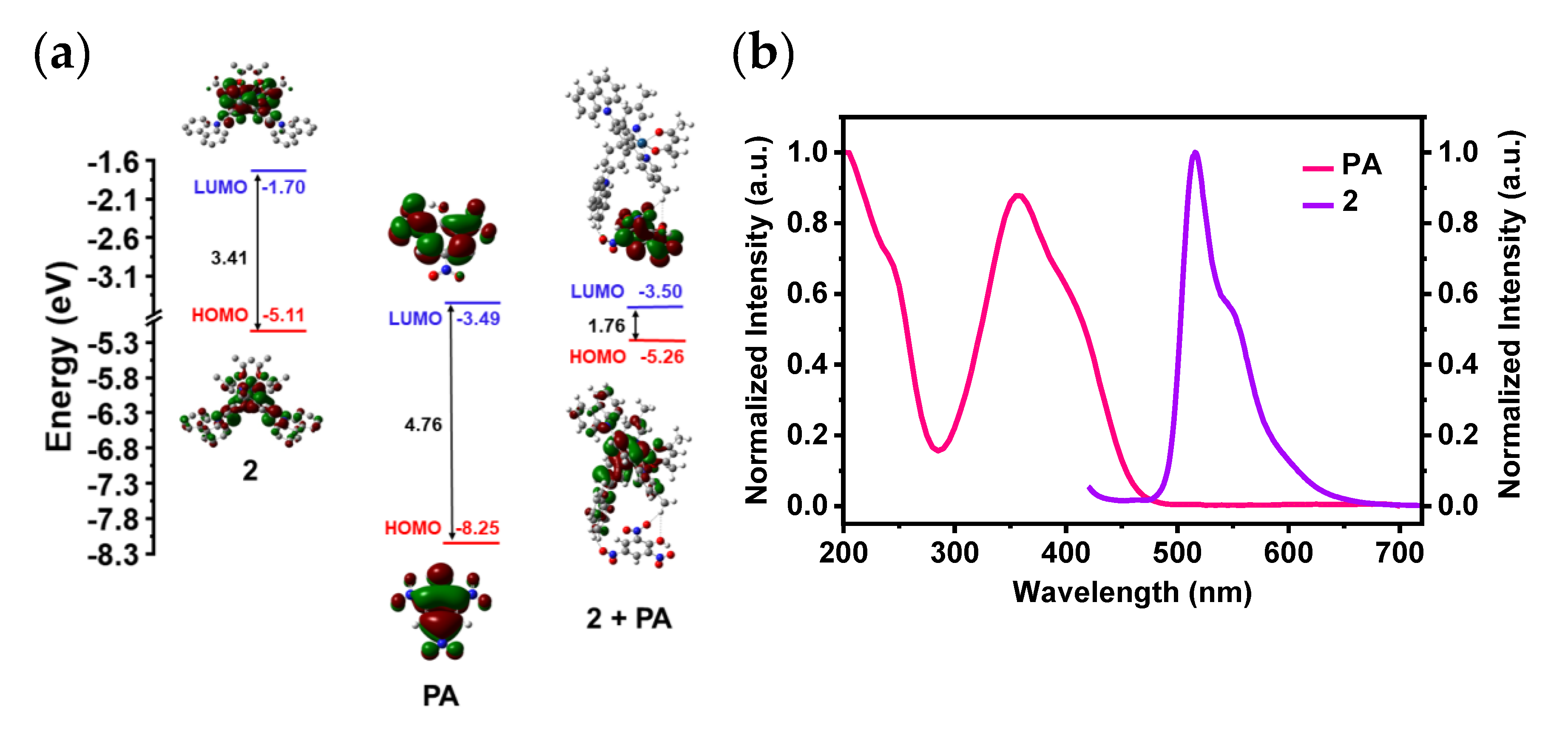
To investigate the sensing mechanism of 2 for detecting PA, we calculated the LUMOs and highest occupied molecular orbitals (HOMOs) of 2, adduct (2 + PA), and PA. Figure 7a shows that the LUMO energy of PA is -3.49 eV, which is lower than that of 2. This implies that 2 and PA are capable of undergoing PET. The excited-state electrons in the LUMO of 2 are easily transferred to the LUMO of PA rather than returning directly to its own HOMO, resulting in the quenching of the luminescence of 2. The energy gap of 2 is higher than that of adduct (2 + PA), indicating the adduct is much more stable. Förster Resonance Energy Transfer (FRET) does not occur during sensing because there is no overlap between the emission spectrum of 2 and the absorption spectrum of PA (Figure 7b). Additionally, the normalized emission spectra of 2 remain almost unchanged after the addition of PA at different concentrations (Figure S10), proving that no other emitting species are formed during the quenching process [15]. Thus, the sensing mechanism of 2 for PA is proposed to be PET.

4. Conclusions

In summary, two carbazolyl-modified neutral Ir(III) complexes 1 and 2 were synthesized and fully characterized. 2 with a methyl group at the 5-position of the pyridyl ring exhibits a much stronger AIPE property than 1 in THF/H2O. Additionally, 2 exhibits excellent sensitivity and selectivity for detecting PA in aqueous media, with the KSV of 37320 M-1 and a LOD of 0.15 μM. Furthermore, 2 shows high quenching efficiency for PA in various common water samples. Spectroscopic studies and DFT calculations indicate that the sensing mechanism is PET. This work offers renewed insights for the rational structural design of neutral Ir(III) complexes for detecting PA in aqueous media.

Supplementary Materials

The following supporting information can be downloaded at: www.mdpi.com/xxx/s1, Figure S1: Synthesis routes of the cyclometalating ligands L1 and L2; Figure S2: Synthetic routes for complexes 1 and 2; Figure S3: UV-Vis absorption and normalized emission spectra of 1 and 2; Figure S4: Phosphorescence decay traces of (ppy)2Ir(acac) (a), 1 (b) and 2 (c) in deoxygenated CH2Cl2 (10 μM); Figure S5: UV-Vis absorption spectra of 1 and 2 at 10 μM in THF/H2O with various water fractions; Figure S6: Calculated energy level diagrams of 1 and 2; Figure S7: The emission spectra of 2 in 11 blank samples in THF/H2O (fw = 90%, 10 μM); Figure S8: The linear graph of the emission intensity of 2 vs. the concentrations of PA; Figure S9: Photos of the mixtures of 2 in THF/H2O with various ionic compounds under 365 nm UV light; Figure S10: The normalized emission spectra of 2 in THF/H2O after addition of different concentrations of PA; Figure S11: 1H NMR spectrum of 1 in CDCl3; Figure S12: 13C NMR spectrum of 1 in CDCl3; Figure S13: 1H NMR spectrum of 2 in CDCl3; Figure S14: 13C NMR spectrum of 2 in CDCl3; Figure S15: The HRMS of 1; Figure S16: The HRMS of 2; Table S1: Photophysical data of complexes 1 and 2.

Author Contributions

Experimental measurements, J.X. and L.Z.; data curation, writing-original draft preparation, J.X.; writing-review and editing, Y.S. and C.L.; supervision, C.L. All authors have read and agreed to the published version of the manuscript.

Funding

The authors thank the financial support from the National Natural Science Foundation of China (21978042) and the Fundamental Research Funds for the Central Universities (DUT22LAB610).

Institutional Review Board Statement

Not applicable.

Informed Consent Statement

Not applicable.

Data Availability Statement

Data are contained within the article.

Conflicts of Interest

The authors declare no conflicts of interest.

References

  1. Luo, J.; Xie, Z.; Lam, J.; Cheng, L.; Chen, H.; Qiu, C.; Kwok, H.; Zhan, X.; Liu, Y.; Zhu, D.; et al. Aggregation-induced emission of 1-methyl-1,2,3,4,5-pentaphenylsilole. Chem. Commun. 2001, 18, 1740–1741. [Google Scholar] [CrossRef] [PubMed]
  2. Dong, Z.; Cui, H.; Wang, Y.; Wang, C.; Li, Y.; Wang, C. Biocompatible AIE material from natural resources: Chitosan and its multifunctional applications. Carbohydr. Polym. 2020, 227, 115338. [Google Scholar] [CrossRef] [PubMed]
  3. Wagalgave, S.; Birajdar, S.; Malegaonkar, J.; Bhosale, S. Chapter eight-patented AIE materials for biomedical applications. Prog. Mol. Biol. Transl. 2021, 185, 199–223. [Google Scholar]
  4. Guan, W.; Chen, J.; Liu, J.; Shi, B.; Yao, H.; Zhang, Y.; Wei, T.; Lin, Q. Macrocycles-assembled AIE supramolecular polymer networks. Coord. Chem. Rev. 2024, 507, 215717. [Google Scholar] [CrossRef]
  5. Hao, Y.; Ji, F.; Li, T.; Tian, M.; Han, X.; Chai, F. Portable smartphone platform utilizing AIE-featured carbon dots for multivariate visual detection for Cu2+, Hg2+ and BSA in real samples. Food Chem. 2024, 446, 138843. [Google Scholar] [CrossRef] [PubMed]
  6. Chen, J.; Xie, Z.; Lam, J.; Law, C.; Tang, B. Silole-containing polyacetylenes. Synthesis, thermal stability, tight emission, nanodimensional aggregation, and restricted intramolecular rotation. Macromolecules 2003, 36, 1108–1117. [Google Scholar] [CrossRef]
  7. Baldo, M.; O’Brien, D.; You, Y.; Shoustikov, A.; Sibley, S.; Thompson, M.; Forrest, S. Highly efficient phosphorescent emission from organic electroluminescent devices. Nature 1998, 395, 151–154. [Google Scholar] [CrossRef]
  8. Caminade, A.; Hameau, A.; Turrin, C.; Laurent, R.; Majoral, J. Dendritic metal complexes for bioimaging. Coord. Chem. Rev. 2021, 430, 213739. [Google Scholar] [CrossRef]
  9. Huynh, M.; Vinck, R.; Gibert, B.; Gasser, G. Strategies for the nuclear delivery of metal complexes to cancer cells. Adv. Mater. 2024, 36, 2311437. [Google Scholar] [CrossRef]
  10. Wen, L.; Hou, X.; Shan, G.; Song, W.; Zhang, S.; Sun, H.; Su, Z. Rational molecular design of aggregation-induced emission cationic Ir(III) phosphors achieving supersensitive and selective detection of nitroaromatic explosives. J. Mater. Chem. C 2017, 5, 10847–10854. [Google Scholar] [CrossRef]
  11. Sainaba, A.; Saha, R.; Venkateswarulu, M.; Zangrando, E.; Mukherjee, P. Pt(II) tetrafacial barrel with aggregation-induced emission for sensing. Inorg. Chem. 2024, 63, 508–517. [Google Scholar] [CrossRef] [PubMed]
  12. Brunner, K.; Dijken, A.; Börner, H.; Bantiaansen, J.; Kiggen, N.; Langeveld, B. Carbazole compounds as host materials for triplet emitters in organic light-emitting diodes:  Tuning the HOMO level without influencing the triplet energy in small molecules. J. Am. Chem. Soc. 2004, 126, 6035–6042. [Google Scholar] [CrossRef] [PubMed]
  13. Zhao, K.; Shan, G.; Fu, Q.; Su, Z. Tuning emission of AIE-Active organometallic Ir(III) complexes by simple modulation of strength of donor/acceptor on ancillary ligands. Organometallics 2016, 35, 3996–4001. [Google Scholar] [CrossRef]
  14. Mao, H.; Yang, Y.; Zhao, K.; Duan, Y.; Song, W.; Shan, G.; Su, Z. Fine-tuning emission color of aggregation-induced emission-active Ir(III) phosphors through simple ligand modification. Dyes Pigm. 2021, 192, 109439. [Google Scholar] [CrossRef]
  15. Ma, Q.; Dong, W.; Ma, Z.; Lv, X.; Li, Y.; Duan, Q. Synthesis of phosphorescent iridium(III) complex containing carbazole and its sensing property towards nitro-aromatic compounds. Mater. Lett. 2019, 249, 120–123. [Google Scholar] [CrossRef]
  16. Dong, W.; Ma, Q.; Ma, Z.; Duan, Q.; Lü, X.; Qiu, N.; Fei, T.; Su, Z. Phosphorescent iridium(III) complex based photoluminescence sensor for sensitive and selective detection of picric acid. Dyes Pigm. 2020, 172, 107799. [Google Scholar] [CrossRef]
  17. Di, L.; Xing, Y.; Yang, Z.; Qiao, C.; Xia, Z. Photostable aggregation-induced emission of iridium(III) complex realizing robust and high-resolution imaging of latent fingerprints. Sensor. Actuat. B Chem. 2023, 375, 132898. [Google Scholar] [CrossRef]
  18. Wang, H.; Chen, C.; Liu, Y.; Wu, Y.; Yuan, Y.; Zhou, Q. A highly sensitive and selective chemosensor for 2,4,6-trinitrophenol based on L-cysteine-coated cadmium sulfide quantum dots. Talanta 2019, 198, 242–248. [Google Scholar] [CrossRef] [PubMed]
  19. Xu, M.; Wang, L.; Chen, G.; Chen, Y.; Liu, D.; Liao, J. A red-emitting COF ionic exchanged with green-emitting Tb(III) complex anion: Synthesis, characterization, ratiometric emission sensing, and removal of picric acid. Front. Chem. 2022, 10, 865304. [Google Scholar] [CrossRef] [PubMed]
  20. Liu, X.; Han, Y.; Shu, Y.; Wang, J.; Qiu, H. Fabrication and application of 2,4,6-trinitrophenol sensors based on fluorescent functional materials. J. Hazard. Mater. 2022, 425, 127987. [Google Scholar] [CrossRef] [PubMed]
  21. James, T.; Sandanayake, K.; Shinkai, S. Chiral discrimination of monosaccharides using a fluorescent molecular sensor. Nature 1995, 374, 345–347. [Google Scholar] [CrossRef]
  22. Lachance, B.; Robidoux, P.; Hawari, J.; Ampleman, G.; Thiboutot, S.; Sunahara, G. Cytotoxic and genotoxic effects of energetic compounds on bacterial and mammalian cells in vitro. Mutat. Res. Fund. Mol. Mech. Mutagen. 1999, 444, 25–39. [Google Scholar] [CrossRef] [PubMed]
  23. Ferrand, Y.; Crump, M.; Davis, A. A synthetic lectin analog for biomimetic disaccharide recognition. Science 2007, 318, 619–622. [Google Scholar] [CrossRef]
  24. Wang, M.; Zhang, H.; Guo, L.; Cao, D. Fluorescent polymer nanotubes as bifunctional materials for selective sensing and fast removal of picric acid. Sensor. Actuat. B Chem. 2018, 274, 102–109. [Google Scholar] [CrossRef]
  25. Liu, C.; Rao, X.; Lv, X.; Qiu, J.; Jin, Z. Substituent effects on the photophysical and electrochemical properties of iridium(III) complexes containing an arylcarbazolyl moiety. Dyes Pigm. 2014, 109, 13–20. [Google Scholar] [CrossRef]
  26. Chen, Y.; Shi, Y.; Gao, Z.; Wang, L.; Tang, Y.; Liu, J.; Liu, C. Supramolecular copolymers under kinetic, thermodynamic, or pathway-switching control. Angew. Chem. Int. Ed. 2023, 62, e202302581. [Google Scholar] [CrossRef] [PubMed]
  27. Lin, Y.; Chen, Y.; Cai, R.; Zhang, H.; Liu, C. Controllable 1D, 2D and 3D supramolecular assemblies of Ir(III) complexes. Mater. Chem. Front. 2023, 7, 5915–5923. [Google Scholar] [CrossRef]
  28. Chen, Y.; Wan, Q.; Shi, Y.; Tang, B.; Che, C.; Liu, C. Three-component multiblock 1D supramolecular copolymers of Ir(III) complexes with controllable sequences. Angew. Chem. Int. Ed. 2023, 62, e202312844. [Google Scholar] [CrossRef] [PubMed]
  29. Yan, Y.; Jia, W.; Cai, R.; Liu, C. An AIPE-active fluorinated cationic platinum(II) complex for efficient detection of picric acid in aqueous media. Chin. Chem. Lett. 2024, 35, 108819. [Google Scholar] [CrossRef]
  30. He, P.; Chen, Y.; Li, X.; Yan, Y.; Liu, C. Aggregation-induced emission-active Ir(III) complexes for sensing picric acid in water. Chemosensors 2023, 11, 177. [Google Scholar] [CrossRef]
  31. He, P.; Chen, Y.; Li, X.; Yan, Y.; Liu, C. AIPE-active cationic Ir(III) complexes for efficient detection of 2,4,6-trinitrophenol and oxygen. Dalton Trans. 2023, 52, 128–135. [Google Scholar] [CrossRef]
  32. Tamayo, A.; Alleyne, B.; Djurovich, P.; Lamansky, S.; Tsyba, I.; Ho, N.; Bau, R.; Thompson, M. Synthesis and characterization of facial and meridional tris-cyclometalated iridium(III) complexes. J. Am. Chem. Soc. 2003, 125, 7377–7387. [Google Scholar] [CrossRef] [PubMed]
  33. Yang, G.; Li, S.; Wang, S.; Li, Y. Emissive properties and aggregation-induced emission enhancement of excited-state intramolecular proton-transfer compounds. C. R. Chimie 2011, 14, 789–798. [Google Scholar] [CrossRef]
  34. Luo, Z.; Liu, B.; Si, S.; Lin, Y.; Luo, C.; Pan, C.; Zhao, C.; Wang, L. A fluorescent chemosensor based on nonplanar donor-acceptor structure for highly sensitive and selective detection of picric acid in water. Dyes Pigm. 2017, 143, 463–469. [Google Scholar] [CrossRef]
  35. Wang, N.; Yang, J.; Chen, L.; Li, J.; An, Y.; Lü, C.; Tian, Y. Efficient synthesis of diethyl benzo[c]cinoline-3, 8-dicarboxylate for fluorescence quenching materials. New J. Chem. 2017, 41, 2786–2792. [Google Scholar] [CrossRef]
  36. Ju, P.; Zhang, E.; Jiang, L.; Zhang, Z.; Hou, X.; Zhang, Y.; Yang, H.; Wang, J. A novel microporous Tb-MOF fluorescent sensor for highly selective and sensitive detection of picric acid. RSC Adv. 2018, 8, 21671–21678. [Google Scholar] [CrossRef] [PubMed]
  37. Li, Z.; Yao, Z.; Feng, R.; Sun, M.; Shan, X.; Su, Z.; Li, W.; Bu, X. A highly stable terbium metal-organic framework for efficient detection of picric acid in water. Chin. Chem. Lett. 2021, 32, 3095–3098. [Google Scholar] [CrossRef]
  38. Chu, Z.; Fan, Z.; Zhang, X.; Tan, X.; Li, D.; Chen, G.; Zhao, Q. A comparison of ACQ, AIE and AEE-based polymers loaded on polyurethane foams as sensors for explosives detection. Sensors 2018, 18, 1565. [Google Scholar] [CrossRef] [PubMed]
  39. Jiang, J.; Li, Z.; Wu, Z.; Huang, X. A soluble porous coordination polymer for fluorescence sensing of explosives and toxic anions under homogeneous environment. Sensors 2023, 23, 9719. [Google Scholar] [CrossRef]
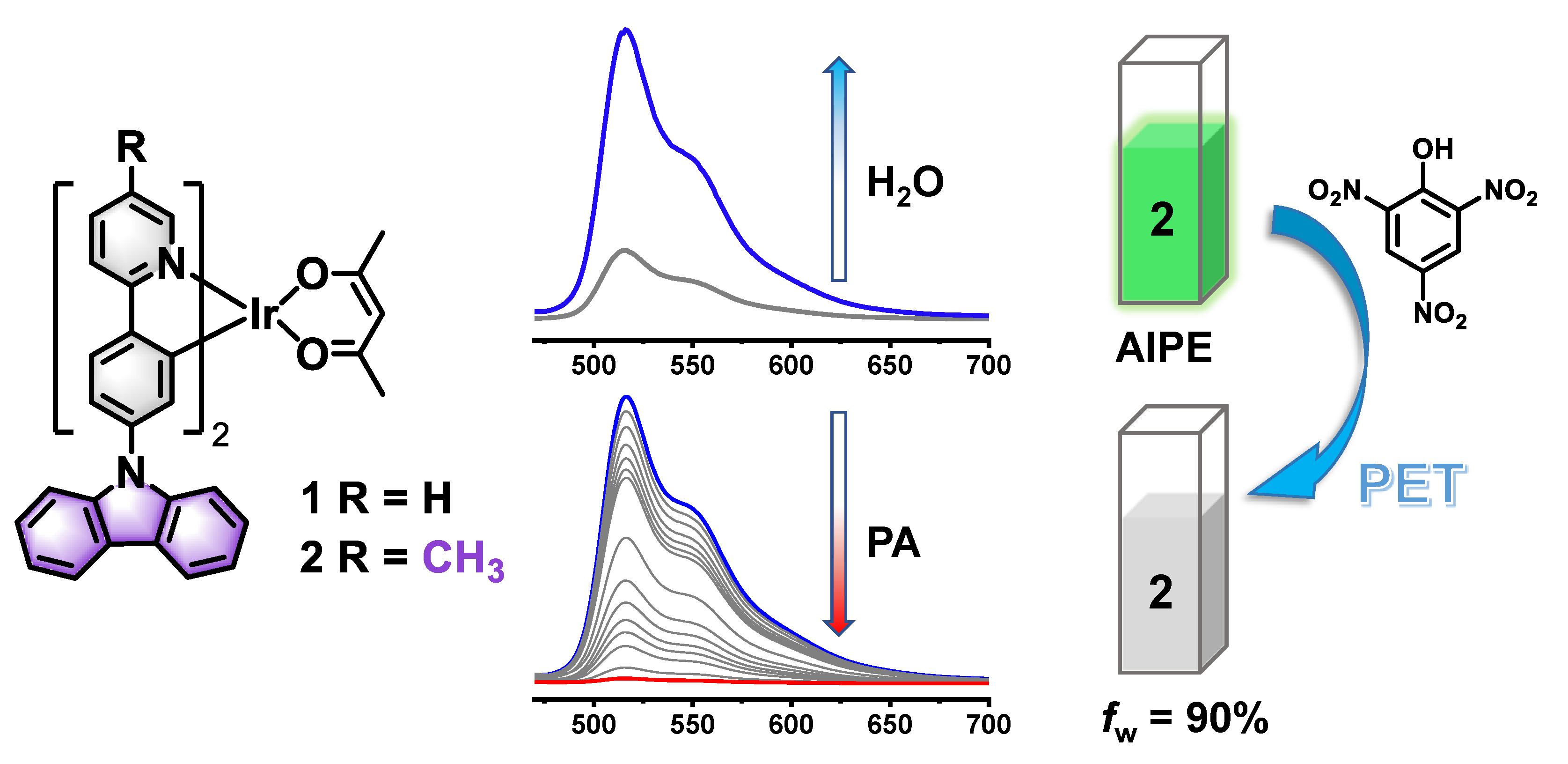
Figure 1. The structures of 1 and 2 and luminescent property of 2 in in THF/H2O, and the schematic diagram for the detection of PA.
Preprints 107051 g001
Figure 2. Emission spectra of 1 (a) and 2 (b) in THF/H2O with various water fractions (0-90%) (c = 10 μM, λex = 400 nm); (c) Line plots of the ratio of the maximum emission intensity (I) of 1 and 2 in THF/H2O at various water fractions to the emission intensity of their monomers (I0).
Preprints 107051 g002
Figure 3. (a) Emission spectra of 2 after addition of PA at various concentrations. Insert: The photographs of 2 with PA at a concentration of 0 or 300 μM under 365 nm UV light (c = 10 μM, λex = 400 nm); (b) The quenching percentages of 2 after addition of PA at various concentrations.
Preprints 107051 g003
Figure 4. The Stern-Volmer plot of 2 for detecting PA. Insert: The linear part of Stern-Volmer plot in the concentration of PA ranging from 0 to 10 μM.
Preprints 107051 g004
Figure 5. Emission spectra of 2 in the presence of various analytes (a), ionic compounds (b) (c = 10 μM, λex = 400 nm); Quenching percentages of 2 with various analytes (c), ionic compounds (d) before (red) and after (gray) adding PA; (e) Photos of the mixtures of 2 in THF/H2O with various analytes under 365 nm UV light (c = 10 μM).
Preprints 107051 g005
Figure 6. (a) Emission spectra of 2 in THF/H2O using common water samples (c = 10 μM, λex = 400 nm) with or without PA; (b) Quenching percentages of 2 towards PA using common water samples.
Preprints 107051 g006
Figure 7. (a) Calculated energy level diagram of 2, PA and adduct (2 + PA); (b) The normalized UV-Vis absorption spectrum of PA (pink) and normalized emission spectrum of 2 (purple) (c = 10 μM, λex = 400 nm).
Preprints 107051 g007
Table 1. The emission intensity of 2 at 516 nm in 11 blank samples in THF/H2O (fw = 90%, 10 μM).
Complex 2 Intensity
X1 101.59
X2 101.52
X3 101.33
X4 101.37
X5 101.33
X6 101.22
X7 101.20
X8 101.39
X9 101.21
X10 101.49
X11 101.28
X 101.36
σ 0.125
1 Xi (i = 1, 2, 3…11) represents the emission intensity of each blank sample, X represents the mean value of the emission intensity, n represents the number of blank samples and the formula for σ is described in the supporting information.
Disclaimer/Publisher’s Note: The statements, opinions and data contained in all publications are solely those of the individual author(s) and contributor(s) and not of MDPI and/or the editor(s). MDPI and/or the editor(s) disclaim responsibility for any injury to people or property resulting from any ideas, methods, instructions or products referred to in the content.
Copyright: This open access article is published under a Creative Commons CC BY 4.0 license, which permit the free download, distribution, and reuse, provided that the author and preprint are cited in any reuse.
Prerpints.org logo

Preprints.org is a free preprint server supported by MDPI in Basel, Switzerland.

Subscribe

© 2024 MDPI (Basel, Switzerland) unless otherwise stated