1. Introduction
Physical training in boxers and kickboxers often results in muscle damage causing soreness and swelling, which leads to post-traumatic scarring and consequently reduces their performance and muscle strength. This condition is frequently a result of inadequate rest and biological regeneration necessary for proper muscle preparation for the next training session [
1]. When recovery from the damage caused by physical activity is insufficient, the risk of injury increases, and difficulties arise in performing subsequent physical tasks [
2], creating a barrier to continued physical engagement [
3].
Achieving post-exercise muscle relaxation, associated with the removal of harmful by-products of energy metabolism and the restoration of motor potential, is a highly desirable and essential element of post-exercise recovery.
Currently, various methods are used in high-level sports to regenerate tired and damaged muscles during exercise. Properly selected biological regeneration shortens the individual phases and accelerates the restitution process, allowing for greater exercise capacity due to the hypercompensation phase of the body's motor potential.
Vibrations, as a factor inducing strong mechanical stimuli in the neuromuscular system, skeletal tissues, and muscles, have been studied in medicine [
3]. It is now known that whole body vibration (WBV) is used in sports training to increase muscle strength, as demonstrated by research from various scientists [
4,
5,
6]. Authors [
7,
8,
9,
10,
11] have also shown in their publications that vibrations are effective in alleviating muscle pain caused by physical exertion. Unfortunately, current studies provide limited information on how the reduction of muscle pain, especially from short-term intense physical exertion, correlates with the recovery and potential hypercompensation of muscle strength. The use of vibrations as an innovative post-exercise recovery method is becoming an important topic that is gaining increasing popularity [
12].
Few authors who have applied vibration therapy after physical exercise report positive effects on muscle recovery [
13], although these studies did not involve high-level athletes. On the other hand, there is information suggesting that using vibrations within 24 hours post-exercise may be detrimental to muscle strength loss and biological regeneration during this period. During vibration therapy, small muscle contractions occur, involving muscle lengthening and shortening. It is possible that the additional work required by the muscles during WBV may have prolonged the damage to the muscles, consequently increasing the loss of muscle strength [
14]. This theory is supported by studies suggesting that increased muscle contractions lead to decreased muscle strength [
15]. Other authors report that WBV does not aid muscle recovery post-exercise, as muscle damage changes the effectiveness of WBV as a recovery tool. They suggest that the excitation-contraction coupling is impaired after muscle damage, reducing calcium release, leading to an inability to activate force-generating muscle fiber structures. It has also been suggested that muscle injury from physical exertion reduces the sensitivity to stretch reflex and muscle stiffness, decreasing the mechanisms that enhance muscle strength [
16].
The aim of this experiment was to assess and compare the impact of an original vibration therapy and passive rest on the compensation of upper limb muscle strength and plasma lactate levels after intense anaerobic exercise.
2. Materials and Methods
The study was conducted according to the guidelines of the Declaration of Helsinki, and approved by the Bioethics Committee at the Regional Medical Chamber (No. 287/KBL/OIL/2020).
Participants
Participants in the study, aged 19-32, comprised an 18-member group of boxers and kickboxers, each holding at least a master class and a minimum of 8 years of training experience. The sample size was calculated using G*Power. All recruited athletes were free from upper limb injuries sustained within 6 months prior to the study date and had fully healed from any other injuries that could affect the results. The participants were in the active training phase during the preparatory period. Each participant had up-to-date medical examinations. Before the experiment began, the participants were informed about the study's purpose and procedure, their right to withdraw at any time, and each signed an individual consent form to participate.
Exclusion criteria included injuries in the past 6 months that could significantly affect upper limb functionality, lack of current medical exams, or lack of consent to participate in the study.
The 18 participants engaged in the study twice, serving as both the experimental and control groups, with group assignment determined by the type of biological regeneration applied (vibration therapy – experimental group, passive rest – control group). Fourteen athletes completed all the required trials. The characteristics of the study group are presented in
Table 1.
Study Procedure
The study was conducted in the morning and divided into several stages.
Stage I
Initially, body height was measured to an accuracy of 0.01 m, body weight (accuracy of 0.01 kg), and body fat percentage using a Tanita scale. The resting plasma lactate level was then determined.
Stage II
Following preliminary measurements, a 15-minute individual standard warm-up for boxers and kickboxers was performed. Subsequently, the individual maximum punching strength was measured based on three punches with each upper limb with maximum force, with short breaks to adjust position. Accelerometric sensors attached to a boxing trainer and the participant’s boxing gloves were used for the measurement. The best result for each limb served as the baseline for determining the minimum punching force level for each upper limb in the main experiment.
Stage III
Next, the maximum strength capabilities were measured under isokinetic contraction conditions for the flexors and extensors of the elbow joints of both upper limbs at an angular velocity of ω=300 °s-1, using the Biodex v.4 according to protocol [
17]. These measurements were used to calculate the muscle force moment generation indices for the elbow joint antagonists of both upper limbs.
Stage IV
This stage involved a muscle exertion phase designed to induce muscle fatigue. Boxers and kickboxers performed auxotonic exercises over three rounds, involving alternating 180 straight punches in each round using a boxing trainer over 120 seconds, at a pace set by a metronome, at no less than 80% of the maximum punching force individually determined for each upper limb. Efforts were monitored in real-time via an accelerometer application. Each round was separated by a 1-minute passive rest. Immediately after the physical exertion phase, maximum strength capabilities under isokinetic contraction conditions for the flexors and extensors of the elbow joints of both upper limbs were remeasured.
Stage V
For muscle biological regeneration, a post-exercise vibration massage of the upper limb muscles was conducted for 15 minutes, applying low-amplitude vibration stimuli with frequencies fluctuating between 20 to 50 Hz. These changes followed the algorithm of the LBV (Local Body Vibration) program, which defined the frequencies, amplitudes, and durations of stimuli and breaks. Seven LBV interventions were applied at level I intensity for 1 minute with 30-second breaks, separated by two 2-minute LBV interventions with increasing intensity from level I to II with a 30-second break. The experiment utilized a vibration therapy device designed by Vitberg, dedicated to vibrational impact on upper limb muscles. A vibrating mat with a vibrating module, covering the flexors and extensors of both elbow joints without repositioning during the procedure, was used. The device produced vibrations in the 20 to 50 Hz frequency range with an amplitude <0.5 mm. Participants underwent vibration therapy lying on their backs, in a relaxed position for the upper limb joints (shoulder joint at 0º, elbow joint at 0º, wrist joint in a relaxed slight palmar flexion). Vibration was directed locally to the shoulder flexors and elbow extensors of both upper limbs. Immediately after vibration therapy, isokinetic tests from stage III were repeated.
After a 10-day break, an identical experiment concluded with a 15-minute passive post-exercise rest in a relaxed supine position, similar to the vibration therapy (passive recreation - PR). During both study phases, all participants were tested using the same measurement tools by the same personnel and identical procedures.
Plasma lactate levels were measured three times in each study: after warm-up, immediately post-exercise, and after vibration or passive rest. Fourteen participants completed all the experiments.
After recording and processing the data, a total of 44 biomechanical and physiological variables for four muscle groups (flexors and extensors of the elbow joint) were analyzed. The strength capabilities analysis included variables: PEAK TORQUE (PTQ), PEAK TQ/BW (PTQ/BW), TIME TO PEAK TORQUE (T_PTQ), WORK/BODYWEIGHT (WRK_BW), TOTAL WORK (TOT_WRK), WORK FATIGUE (WRK_FAT), AVG. POWER (AVG_POW), ACCELERATION TIME (T_ACC), DECELERATION TIME (T_DEC), AVG PEAK TQ (AVG_PTQ), AGON/ANTAG RATIO (AGN_ANT_RAT). Lactate levels (LA) were also measured in both groups at three study stages.
Relative values of the Rel. SBFT index [
18] were used to compare loads in the exertion test.
Statistical Analysis Methods
Statistical analysis of the collected data was conducted using Statistica 13.3 (Tibeco). Basic descriptive statistics were calculated: mean, standard deviation, minimum, and maximum. After assessing the normality of variable distributions with the Shapiro-Wilk test and homogeneity of variances with the Levene test, repeated measures analysis of variance was performed to identify significant contrasts between studies (between-group analysis) using Tukey's post-hoc testing. A p-value of <0.05 was considered statistically significant.
3. Results
Peak Torque Dynamics (PTQ)
This section may be divided by subheadings. It should provide a concise and precise description of the experimental results, their interpretation, as well as the experimental conclusions that can be drawn.
3.1. Peak Torque Dynamics (PTQ)
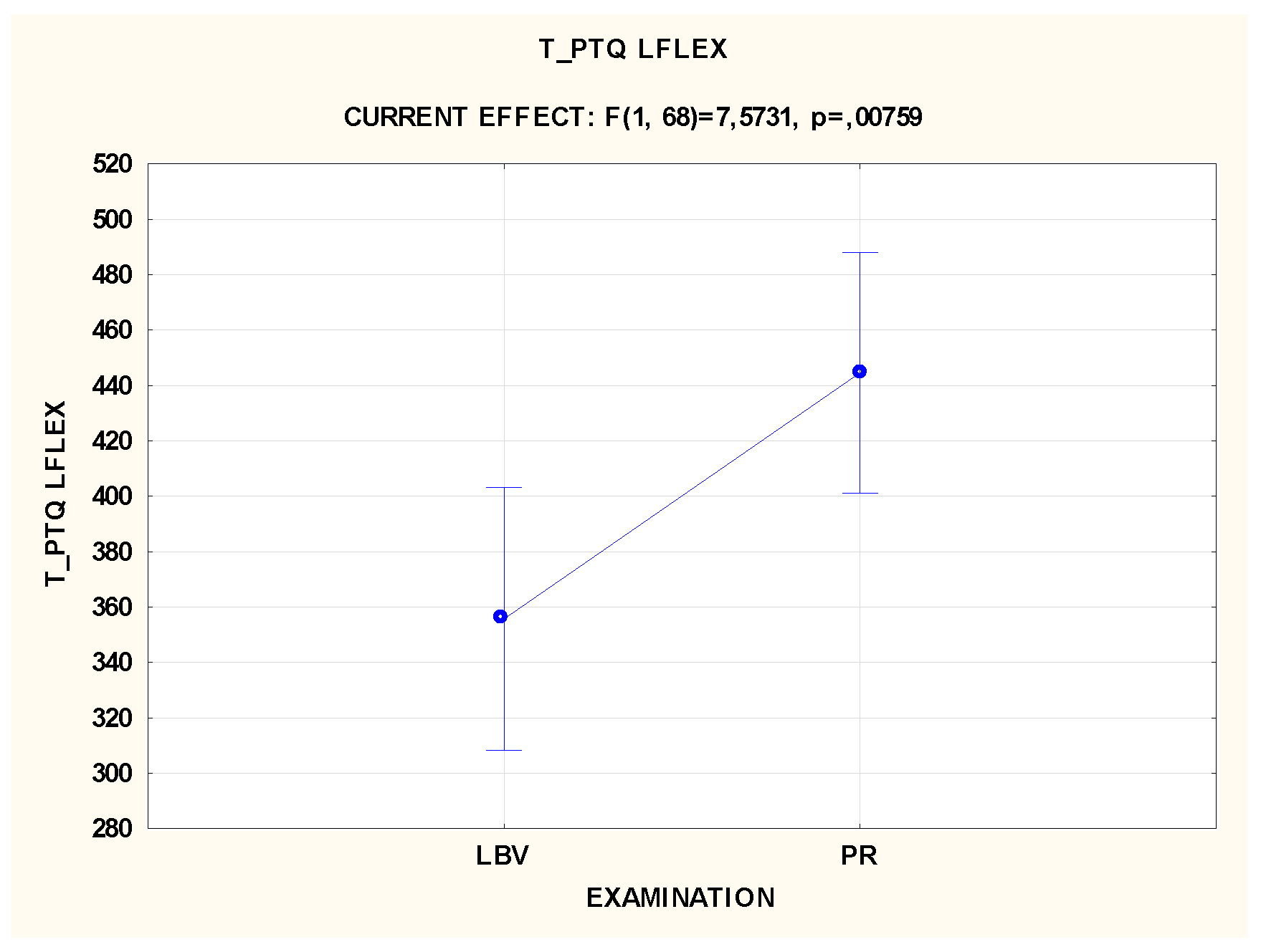
The application of vibration therapy (VT) resulted in improved dynamics of peak torque generation. The VT group achieved peak torque faster than the passive rest (PR) group, indicating quicker recovery of muscle contraction ability (
Table 2,
Figure 1). The mean time to reach peak torque (T_PTQ) was significantly shorter in the VT group, suggesting better muscle performance and faster response to training stimuli.
3.2. Acceleration and Reaction Time
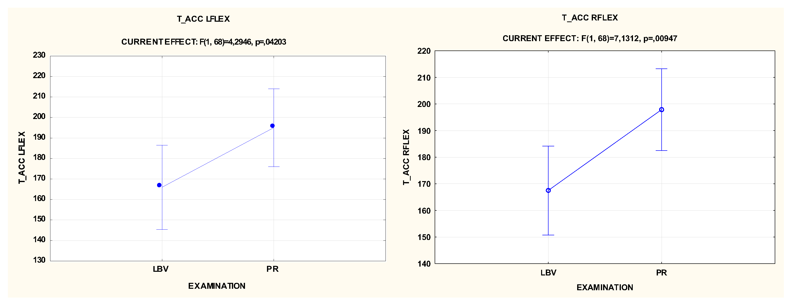
The acceleration time (T_ACC), measuring the speed at which muscles reach a set velocity, was significantly shorter in the VT group. This reflects a faster response and potentially more efficient recruitment of muscle fibers following vibration therapy, which could be crucial in sports requiring rapid muscle reactions (
Table 2,
Figure 2).
3.3. Muscle Work and Fatigue
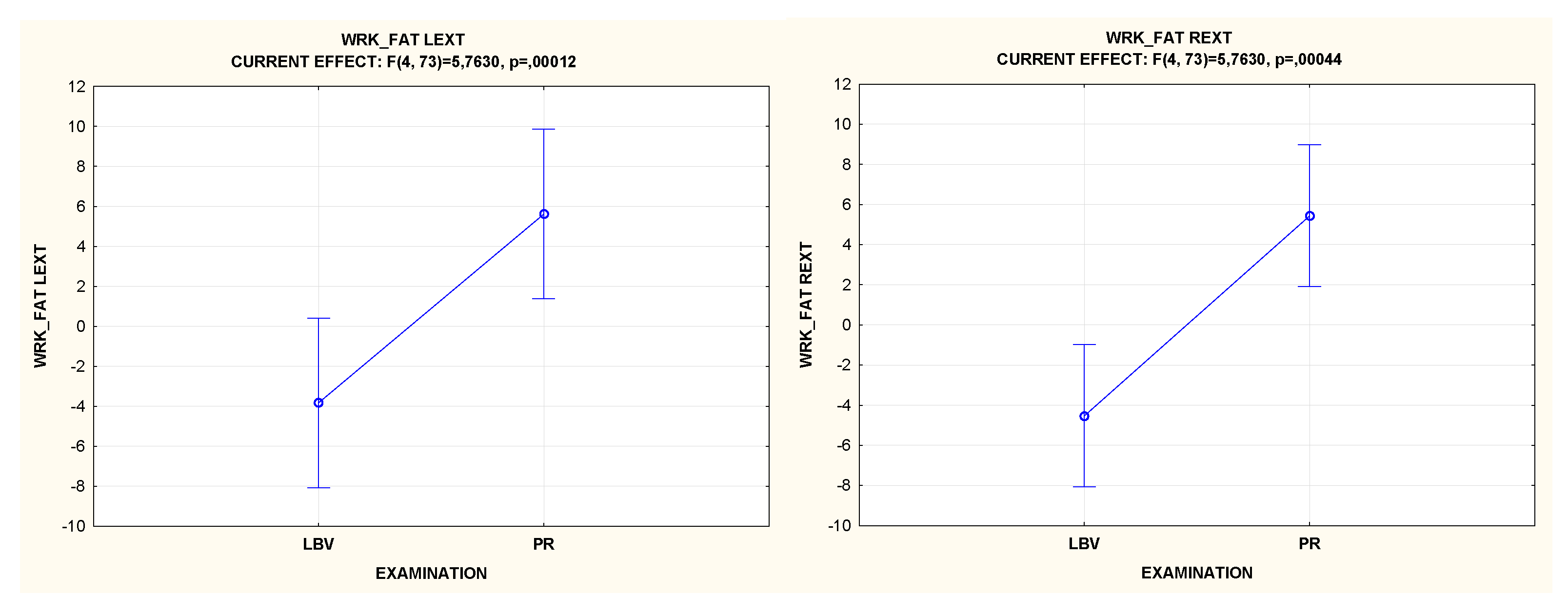
Muscle fatigue indices (WRK_FAT) indicated that the VT group experienced a lower rate of fatigue compared to the PR group. This suggests that VT contributes to better muscle endurance and maintains performance over time, mitigating the effects of fatigue during recovery phases (
Table 2,
Figure 3).
3.4. Muscle Strength Balance
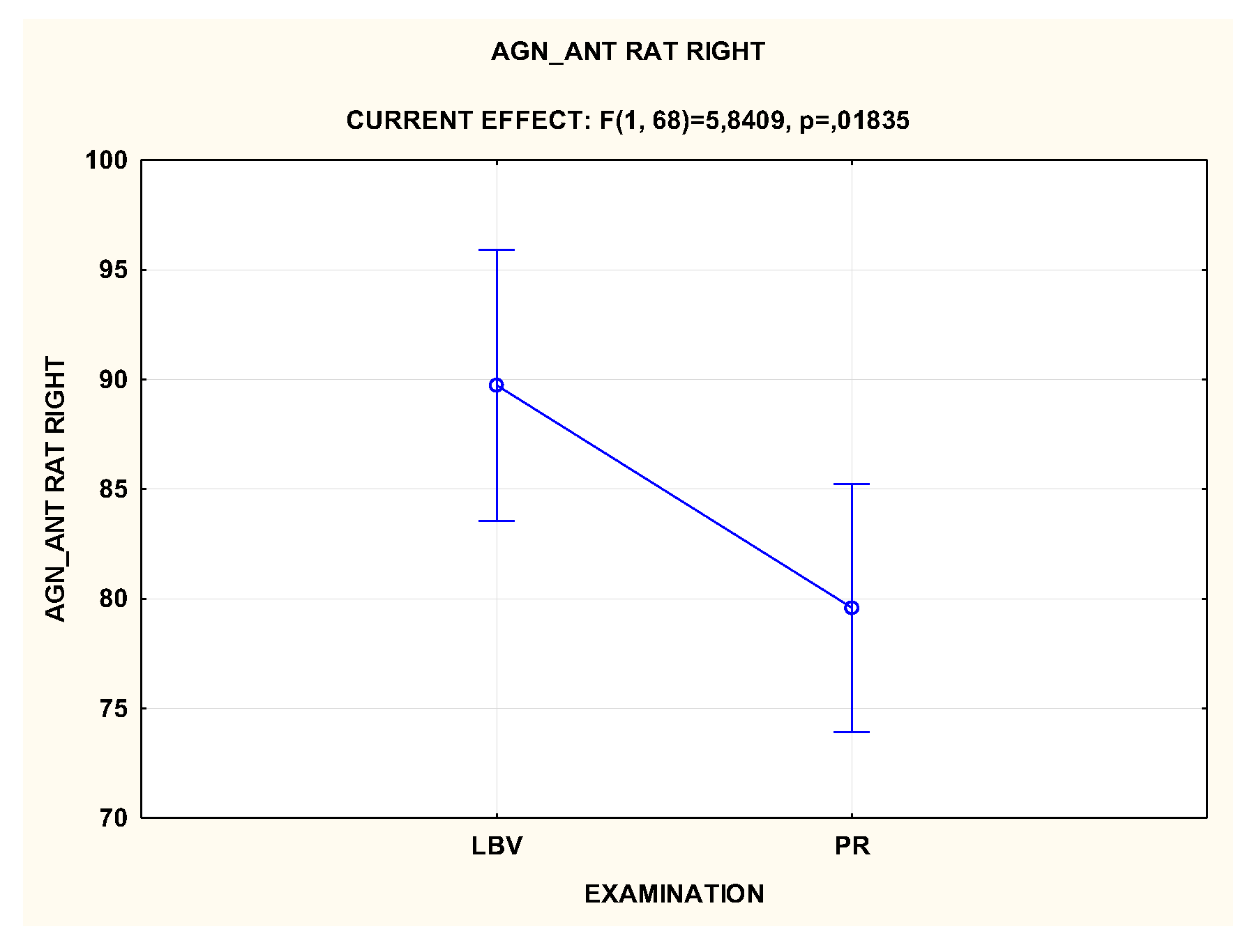
The agonist-to-antagonist ratio (AGN_ANT_RAT) was more favorable in the VT group. The improved ratio suggests a more symmetrical recovery of muscle groups, potentially reducing injury risk by maintaining better joint stability and muscle health (
Table 2,
Figure 4).
3.5. Total Muscle Work
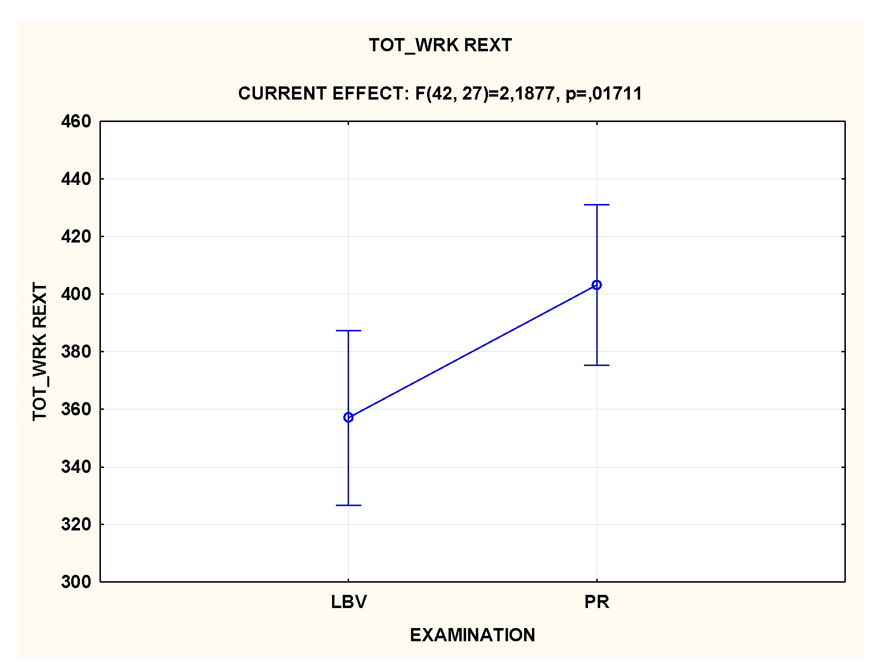
Despite the observed benefits in other variables, the total work (TOT_WRK) performed by the muscles during the tests was slightly higher in the PR group. This may indicate that although VT improves certain aspects of muscle function, it does not necessarily translate into increased total work performed during the recovery phase (
Figure 5).
3.6. Physiological Responses – Lactate Levels
Lactate levels were measured to assess the metabolic response to both recovery methods. Although initial lactate levels post-exercise were comparable between groups, the VT group showed a more significant reduction in lactate levels after recovery, suggesting more efficient removal of metabolic by-products (
Table 2,
Figure 6).
Table 2.
Descriptive statistics of the analyzed variables.
Table 2.
Descriptive statistics of the analyzed variables.
| Variables |
Average |
P |
Min |
Max |
SD |
| LBV |
PR |
LBV |
PR |
LBV |
PR |
LBV |
PR |
| PTQ REXT [Nm] |
56 |
58 |
|
32 |
34 |
85 |
79 |
11.1 |
11.0 |
| PTQ LEXT [Nm] |
58 |
60 |
|
29 |
30 |
88 |
88 |
14.9 |
14.7 |
| PTQ RFLEX [Nm] |
47 |
45 |
|
31 |
29 |
70 |
73 |
9.0 |
10.0 |
| PTQ LFLEX [Nm] |
45 |
46 |
|
28 |
30 |
64 |
69 |
9.0 |
10.1 |
| PTQ_BW REXT [%] |
74 |
77 |
|
50 |
59 |
95 |
96 |
9.9 |
9.4 |
| PTQ_BW LEXT [%] |
76 |
79 |
|
53 |
51 |
100 |
109 |
13.2 |
13.8 |
| PTQ_BW RFLEX [%] |
63 |
61 |
|
43 |
39 |
105 |
90 |
12.3 |
12.4 |
| PTQ_BW LFLEX [%] |
59 |
61 |
|
41 |
38 |
86 |
91 |
10.4 |
11.6 |
| T_PTQ REXT [ ms] |
331 |
363 |
|
10 |
10 |
440 |
460 |
106.1 |
91.6 |
| T_PTQ LEXT [ ms] |
353 |
347 |
|
10 |
160 |
790 |
440 |
116.8 |
66.1 |
| T_PTQ RFLEX [ ms] |
347 |
405 |
|
60 |
190 |
610 |
660 |
137.3 |
126.0 |
| T_PTQ LFLEX [ ms] |
364 |
444 |
0.00759 |
50 |
60 |
650 |
760 |
139.0 |
133.3 |
| WRK_BW REXT [J] |
88 |
96 |
|
61 |
66 |
118 |
127 |
14.0 |
16.5 |
| WRK_BW LEXT [J] |
90 |
96 |
|
62 |
59 |
122 |
130 |
17.5 |
17.9 |
| WRK_BW RFLEX [J] |
85 |
89 |
|
60 |
37 |
119 |
135 |
18.4 |
23.2 |
| WRK_BW LFLEX [J] |
87 |
89 |
|
57 |
41 |
117 |
170 |
18.9 |
24.7 |
| TOT_VRK REXT [J] |
360 |
402 |
0.01711 |
214 |
194 |
535 |
578 |
78.4 |
92.0 |
| TOT_WRK LEXT [J] |
372 |
401 |
|
205 |
234 |
621 |
637 |
107.6 |
103.7 |
| TOT_WRK RFLEX [J] |
364 |
381 |
|
197 |
144 |
553 |
615 |
100.5 |
115.0 |
| TOT_WRK LFLEX [J] |
374 |
377 |
|
199 |
168 |
598 |
596 |
107.0 |
112.5 |
| AVG_POW REXT [W] |
103 |
110 |
|
63 |
62 |
145 |
154 |
20.7 |
21.7 |
| AVG_POW LEXT [W] |
104 |
111 |
|
54 |
68 |
151 |
165 |
26.4 |
25.4 |
| AVG_POW RFLEX [W] |
103 |
99 |
|
58 |
34 |
152 |
157 |
24.8 |
29.6 |
| AVG_POW LFLEX [W] |
103 |
100 |
|
53 |
47 |
157 |
156 |
27.0 |
28.4 |
| AVG_PTQ REXT [Nm] |
51 |
54 |
|
29 |
31 |
80 |
76 |
11.6 |
11.2 |
| AVG_PTQ LEXT [Nm] |
52 |
55 |
|
27 |
28 |
84 |
84 |
14.9 |
14.3 |
| AVG_PTQ RFLEX [Nm] |
44 |
43 |
|
29 |
26 |
65 |
70 |
8.5 |
9.5 |
| AVG_PTQ LFLEX [Nm] |
42 |
43 |
|
25 |
28 |
63 |
64 |
9.2 |
9.3 |
| T_ACC REXT [ms] |
110 |
116 |
|
10 |
80 |
160 |
170 |
25.8 |
21.0 |
| T_ACC LEXT [ms] |
126 |
112 |
|
80 |
80 |
660 |
150 |
91.2 |
17.5 |
| T_ACC RFLEX [ms] |
172 |
197 |
0.00947 |
120 |
120 |
250 |
360 |
36.4 |
56.5 |
| T_ACC LFLEX [ms] |
173 |
195 |
0.04203 |
120 |
120 |
330 |
570 |
40.4 |
72.8 |
| T_DEC REXT [ms] |
238 |
233 |
|
140 |
180 |
430 |
350 |
62.7 |
37.8 |
| T_DEC LEXT [ms] |
246 |
232 |
|
180 |
180 |
460 |
370 |
59.0 |
41.8 |
| T_DEC RFLEX [ms] |
156 |
158 |
|
90 |
90 |
260 |
240 |
42.1 |
36.6 |
| T_DECC LFLEX [ms] |
160 |
169 |
|
100 |
90 |
290 |
260 |
39.6 |
39.3 |
| AGN_ANT RAT RIGHT [%] |
86 |
80 |
0.01835 |
57 |
56 |
136 |
117 |
17.2 |
17.9 |
| AGN_ANT RAT LEFT [%] |
80 |
80 |
|
57 |
49 |
137 |
117 |
17.6 |
15.1 |
| LBV_WRK_FAT REXT [J/J] |
- 4.5 |
5.4 |
0.0044 |
-31.2 |
- 34.2 |
26.0 |
32.4 |
11 |
11 |
| LBV_WRK_FAT LEXT [J/J] |
- 3.8 |
5.6 |
0.0012 |
-37.8 |
- 7.4 |
42.2 |
13.8 |
16 |
6 |
| LBV_WRK_FAT RFLEX [J/J] |
1.8 |
4.8 |
|
-26.8 |
- 13.9 |
20.8 |
24.2 |
10 |
7 |
| LBV_WRK_FAT LFLEX [J/J] |
5.3 |
5.1 |
|
-16.4 |
- 12.9 |
13.0 |
18.3 |
7 |
8 |
| LA [mmol] |
5.2 |
5.3 |
|
0.5 |
0.5 |
14.8 |
12.5 |
4.5 |
4.2 |
| Rel. SBFT Index [bmp/Nkg-1] |
2.42 |
2.42 |
|
2.02 |
1.99 |
3.66 |
3.49 |
0.44 |
0.42 |
The results of lactate level measurements in the blood indicated similar, statistically insignificant average values obtained in both studies, in the LBV and PR groups. However, the serum lactate level differed significantly between individual trials in both studies at the level of p<0.001. The lowest values in both groups were obtained in the initial measurement before exercise. Subsequently, the values increased significantly to above 10 mmol, then returned to minimally above 4 mmol after biological regeneration.
4. Discussion
The results of the studies conducted by Chapman, Barnes, Dabbs, and Chwała & Pogwizd indicate discrepancies in the presented views, allowing the conclusion that the authors did not always achieve improvements in the measured parameters after applying vibration therapy following exercise or during breaks between exercise sets [13-16]. This may be due to the fact that each study used different vibration frequencies and amplitudes. The studies also varied in terms of the duration and location of the vibration application, which undoubtedly could have significantly impacted the final effects of the therapy. However, recent studies have shown positive observations regarding the use of vibration as a superior form of intervention over passive rest in biological regeneration [
11].
Whole-body vibration (WBV) treatments are an innovative form of intervention in the training process, and to date, only a few studies have examined the effect of combining different frequencies and amplitudes of vibration. As indicated by research [
19], beneficial effects can be achieved by smoothly changing the frequency and amplitude of vibration applied during a WBV program. Similar methodologies have been used only in separate treatment cycles, comparing the effects of different frequencies and amplitudes on the achieved results. In most studies, authors utilized a strictly defined vibration stimulus (frequency, amplitude, acceleration) and examined its effect on measured variables.
Therefore, it seems reasonable to apply a vibration stimulus with variable frequency, amplitude, and duration, which will more effectively impact all types of muscle fibers and sensory receptors.
An essential condition for comparing the effects of varied interventions on the speed and effectiveness of biological regeneration is ensuring that the muscles of the study participants are fatigued to the same extent, in this case, during intense anaerobic exercise. When comparing loads in the exercise test, it should be noted that the relative values of the Rel. SBFT index [
18] did not differ significantly between the LBV and PR groups. This indicates a similar level of fatigue in the flexor and extensor muscles of the elbow joints in both groups.
The use of dedicated vibrating mats allowed for localized vibration intervention in a comfortable resting position for the muscles controlling the upper limb joints, which could be significant for the relaxation and regeneration of muscles fatigued by exercise and its superiority over passive rest.
This is confirmed by the significantly shorter time to achieve peak torque (PTQ) for elbow flexor muscles in the LBV group compared to the PR group (p<0.01) (
Figure 1). TIME TO PEAK TORQUE - a measure of the time from the start of a muscular contraction to the point of highest torque development (Peak TQ). This value is an indicator of the muscle's functional ability to produce torque quickly. This variable illustrates the neuromuscular readiness to rapidly develop torque [
20] and achieve maximum contraction [
21]. A short T_PTQ time indicates a high level of proprioception in the joint area being studied.
A similar interpretation applies to the variable acceleration time (T_ACC), measured from the start of the movement to the moment of reaching the set speed during isokinetic contraction. The T_ACC variable for both the right and left limb flexors was significantly shorter in the LBV group compared to the PR group by 25 ms (RFLEX), p<0.01 and 22 ms (LFLEX), p<0.05 (
Figure 2). Lower acceleration time values may indicate better recruitment ability of muscle fibers in the studied skeletal muscles and may be associated with shorter time needed to generate torque [
21,
22,
23].
In our studies, besides faster recruitment of motor units in the muscle group after vibration intervention (LBV) compared to passive rest (PR), more favorable indices of strength endurance and muscle resistance to fatigue were also noted. The work fatigue (WRK_FAT) index, calculated as the ratio of the work performed by the muscles in the first 30% range of motion of all contractions to the work performed during the last 30% range of motion in the entire trial, characterizes the state of muscle fatigue in subsequent repetitions of the isokinetic test. The index value for elbow extensor muscles was significantly higher in the PR group compared to the LBV group - the significance level for contrasts for the right limb p<0.001 and for the left limb p<0.05. Higher WRK_FAT values in the PR group indicate that the work performed in the first 30% of the contraction was significantly higher than in the last 30% range of motion, indicating faster muscle fatigue during this test. In the LBV group, the index values were negative, indicating greater strength endurance of muscles after recovery using vibration interventions compared to passive rest.
As Gillet et al. argue, the risk of injury may be related to the strength balance of antagonistic muscle groups that provide joint stability [
24]. The variable AGN_ANT_RAT (agonist/antagonist ratio), calculated as the ratio of peak PTQ values (maximum torque of agonist muscles divided by the peak torque of antagonist muscles in the joint), is used by researchers to assess muscle balance. This index is important for evaluating muscle function [
25,
26]. Researchers indicate that the value of this index depends on age, sex, and training level [
27]. Comparing the results of our studies regarding this index in the LBV and PR groups, it should be noted that a significantly higher AGN_ANT_RAT index was recorded in the LBV group for the right upper limb, which for all subjects was the dominant limb (p<0.05). The corresponding index for the left upper limb did not show significant differences.
Another variable that showed significant contrasts between the LBV and PR groups was the total work (TOT_WRK) performed by the muscles during all movements in the isokinetic test. The total work value recorded in the PR group for the right elbow extensor was significantly higher by approximately 42 W compared to the analogous variable in the LBV group (p<0.05). As shown above, the elbow extensor muscles performed most of the work in the first 30% of isokinetic contractions, which proved decisive for higher TOT_WRK values. The left elbow extensors also performed more work on average by about 28 W, but the differences were not statistically significant (p<0.05).
Other variables, including the maximum torque levels (PRQ) and their relative values (PRQ_BW), despite some differences between the mean values in both groups, did not show significant contrasts at p<0.05. In summary, it can be concluded that the type of post-exercise muscle recovery used did not significantly affect strength capabilities, but significantly differentiated the speed of motor unit recruitment and strength endurance in the group with proprietary vibration intervention compared to passive rest.
5. Conclusions
The use of post-exercise vibration intervention with variable amplitude and frequency had a more favorable effect on the biological regeneration of elbow joint muscles in the LBV group compared to PR.
The vibration intervention applied after intense anaerobic exercise positively affected the time, acceleration of recruitment, and strength endurance of motor units.
No significant differences were noted in the level of maximum and relative torque between the two groups.
Lactate levels (LA) in both studies were similar at various time points of the experiments, but significantly differed between trials in both groups. After anaerobic exercise, LA significantly increased from baseline values and then significantly decreased following biological regeneration.
Author Contributions
Conceptualization, W.C. and T.A.; methodology, W.C., W.M.; software, W.C.; validation, W.C., W.M. and W.W.; formal analysis, W.C.; investigation, W.C.; resources, W.C., W.M., W.W.; data curation, W.C., Ł.R.; writing—original draft preparation, W.C., T.A., W.W., Ł.R.; writing—review and editing, W.C., T.A., W.W., Ł.R.; visualization, W.C.; supervision, W.C., T.A., Ł.R.; project administration, W.C., W.M., T.A ., W.W., Ł.R.; funding acquisition, W.C., T.A., Ł.R. All authors have read and agreed to the published version of the manuscript.
Funding
The project is funded under the program of the Ministry of Science and Higher Education (Poland) ‘Regional Excellence Initiative’ for the years 2019-2022, project number 022/RID/2018/19, with a budget of 11,919,908 PLN; grant number 40/PB/RID/2022 with funding amount 56,500 PLN
Institutional Review Board Statement
The study was conducted in accordance with the Declaration of Helsinki, and approved by the Bioethics Committee at the at the Regional Medical Chamber (No. 287/KBL/OIL/2020).
Informed Consent Statement
Informed consent was obtained from all subjects involved in the study.
Data Availability Statement
The data presented in this study are available upon request from the corresponding author.
Conflicts of Interest
The authors declare no conflicts of interest.
References
- Dr. Melton. J.L. Perspectives: How many women have osteoporosis now? J. Bone Miner. Res. 1995. 10. 175–177. [CrossRef]
- TUNG. S.; IQBAL. J. Evolution. Aging. and Osteoporosis. Ann. N. Y. Acad. Sci. 2007. 1116. 499–506. [CrossRef]
- Mazzeo. R.S.; Tanaka. H. Exercise Prescription for the Elderly. Sport. Med. 2001. 31. 809–818. [CrossRef]
- Tankisheva. E.; Bogaerts. A.; Boonen. S.; Delecluse. C.; Jansen. P.; Verschueren. S.M.P. Effects of a Six-Month Local Vibration Training on Bone Density. Muscle Strength. Muscle Mass. and Physical Performance in Postmenopausal Women. J. Strength Cond. Res. 2015. 29. 2613–2622. [CrossRef]
- Pamukoff. D.N.; Pietrosimone. B.; Lewek. M.D.; Ryan. E.D.; Weinhold. P.S.; Lee. D.R.; Blackburn. J.T. Whole-Body and Local Muscle Vibration Immediately Improve Quadriceps Function in Individuals With Anterior Cruciate Ligament Reconstruction. Arch. Phys. Med. Rehabil. 2016. 97. 1121–1129. [CrossRef]
- El-Shamy. S. Effect of whole body vibration training on quadriceps strength. bone mineral density. and functional capacity in children with hemophilia: a randomized clinical trial. J. Musculoskelet. Neuronal Interact. 2017. 17. 19–26.
- Bakhtiary. A.H.; Safavi-Farokhi. Z.; Aminian-Far. A.; Rezasoltani. A. Influence of vibration on delayed onset of muscle soreness following eccentric exercise * COMMENTARY. Br. J. Sports Med. 2007. 41. 145–148. [CrossRef]
- Broadbent. S.; Rousseau. J.J.; Thorp. R.M.; Choate. S.L.; Jackson. F.S.; Rowlands. D.S. Vibration therapy reduces plasma IL6 and muscle soreness after downhill running. Br. J. Sports Med. 2010. 44. 888–894. [CrossRef]
- Aminian-Far. A.; Hadian. M.-R.; Olyaei. G.; Talebian. S.; Bakhtiary. A.H. Whole-Body Vibration and the Prevention and Treatment of Delayed-Onset Muscle Soreness. J. Athl. Train. 2011. 46. 43–49. [CrossRef]
- Imtiyaz. S.; Veqar. Z.; Shareef. M.Y. To Compare the Effect of Vibration Therapy and Massage in Prevention of Delayed Onset Muscle Soreness (DOMS). J. Clin. DIAGNOSTIC Res. 2014. [CrossRef]
- Marin. P.J.; Zarzuela. R.; Zarzosa. F.; Herrero. A.J.; Garatachea. N.; Rhea. M.R.; García-López. D. Whole-body vibration as a method of recovery for soccer players. Eur. J. Sport Sci. 2012. 12. 2–8. [CrossRef]
- Chwała. W.; Pogwizd. P.; Rydzik. Ł.; Ambroży. T. Effect of Vibration Massage and Passive Rest on Recovery of Muscle Strength after Short-Term Exercise. Int. J. Environ. Res. Public Health 2021. 18. 11680. [CrossRef]
- Chwała. W.; Pogwizd. P. Effects of vibration and passive resting on muscle stiffness and restitution after submaximal exercise analyzed by elastography. Acta Bioeng. Biomech. 2021. 23. [CrossRef]
- Barnes. M.J.; Perry. B.G.; Mündel. T.; Cochrane. D.J. The effects of vibration therapy on muscle force loss following eccentrically induced muscle damage. Eur. J. Appl. Physiol. 2012. 112. 1189–1194. [CrossRef]
- CHAPMAN. D.W.; NEWTON. M.; MCGUIGAN. M.; NOSAKA. K. Effect of Lengthening Contraction Velocity on Muscle Damage of the Elbow Flexors. Med. Sci. Sport. Exerc. 2008. 40. 926–933. [CrossRef]
- Dabbs. N.C. Effects Of Whole Body Vibration On Vertical Jump Performance Following Exercise Induced Muscle Damage. Int. J. Kinesiol. Sport. Sci. J. Kinesiol. Sport. Sci. J. Kinesiol. Sport. Sci. 2014. 2. 23–30. [CrossRef]
- Dvir. Z. Isokinetics: Muscle Testing. Interpretation and clinical Applications. Edinburgh: Churchill livingstone 2004. Wyd. 2.
- Chwała. W.; Wąsacz. W.; Rydzik. Ł.; Mirek. W.; Snopkowski. P.; Pałka. T.; Ambroży. T. Special Boxing Fitness Test: validation procedure. 2023.
- Padulo. J.; Di Giminiani. R.; Ibba. G.; Zarrouk. N.; Moalla. W.; Attene. G.; M. Migliaccio. G.; Pizzolato. F.; Bishop. D.; Chamari. K. The Acute Effect of Whole Body Vibration on Repeated Shuttle-Running in Young Soccer Players. Int. J. Sports Med. 2013. 35. 49–54. [CrossRef]
- Amaral. G.M.; Marinho. H.V.R.; Ocarino. J.M.; Silva. P.L.P.; Souza. T.R. de; Fonseca. S.T. Muscular performance characterization in athletes: a new perspective on isokinetic variables. Brazilian J. Phys. Ther. 2014. 18. 521–529. [CrossRef]
- Chen. W.-L.; Su. F.-C.; Chou. Y.-L. Significance of Acceleration Period in a Dynamic Strength Testing Study. J. Orthop. Sport. Phys. Ther. 1994. 19. 324–330. [CrossRef]
- Jaric. S. Changes in Movement Symmetry Associated With Strengthening and Fatigue of Agonist and Antagonist Muscles. J. Mot. Behav. 2000. 32. 9–15. [CrossRef]
- van Cingel. R.; Kleinrensink. G.; Stoeckart. R.; Aufdemkampe. G.; de Bie. R.; Kuipers. H. Strength Values of Shoulder Internal and External Rotators in Elite Volleyball Players. J. Sport Rehabil. 2006. 15. 236–245. [CrossRef]
- Gillet. B.; Begon. M.; Diger. M.; Berger-Vachon. C.; Rogowski. I. Shoulder range of motion and strength in young competitive tennis players with and without history of shoulder problems. Phys. Ther. Sport 2018. 31. 22–28. [CrossRef]
- Vieira. A.; Alex. S.; Martorelli. A.; Brown. L.E.; Moreira. R.; Bottaro. M. Lower-extremity isokinetic strength ratios of elite springboard and platform diving athletes. Phys. Sportsmed. 2017. 1–5. [CrossRef]
- Yang. S.; Chen. C.; Du. S.; Tang. Y.; Li. K.; Yu. X.; Tan. J.; Zhang. C.; Rong. Z.; Xu. J.; et al. Assessment of isokinetic trunk muscle strength and its association with health-related quality of life in patients with degenerative spinal deformity. BMC Musculoskelet. Disord. 2020. 21. 827. [CrossRef]
- Ruas. C. V.; Minozzo. F.; Pinto. M.D.; Brown. L.E.; Pinto. R.S. Lower-Extremity Strength Ratios of Professional Soccer Players According to Field Position. J. Strength Cond. Res. 2015. 29. 1220–1226. [CrossRef]
|
Disclaimer/Publisher’s Note: The statements, opinions and data contained in all publications are solely those of the individual author(s) and contributor(s) and not of MDPI and/or the editor(s). MDPI and/or the editor(s) disclaim responsibility for any injury to people or property resulting from any ideas, methods, instructions or products referred to in the content. |
© 2024 by the authors. Licensee MDPI, Basel, Switzerland. This article is an open access article distributed under the terms and conditions of the Creative Commons Attribution (CC BY) license (http://creativecommons.org/licenses/by/4.0/).
