Preprint
Article

Selective Cytotoxicity of Curcumin in Colon Cancer via Talin-Mediated Integrin β1 Activation

Altmetrics

Downloads

125

Views

44

Comments

0

Submitted:

31 May 2024

Posted:

31 May 2024

You are already at the latest version

Alerts
Abstract
Curcumin has attracted attention for its nontoxic chemopreventive effects; however, the specific pathways underlying its anticancer effects in colon cancer remain unclear. This study investigated the potential interplay between curcumin and integrin β1 in colon cancer. HCT116 and fibroblast cells were treated with curcumin, and cell proliferation was assessed using MTT assay and western blotting. We analyzed integrin β1 expression after curcumin treatment of HCT116 cells using western blotting and confocal microscopy. The roles of talin and rab25 in integrin β1 activation by curcumin were investigated using the same methods. Curcumin treatment significantly decreased the survival of HCT 116 cells but did not significantly affect the survival of fibroblast cells. Moreover, curcumin significantly increased integrin β1 expression in HCT 116 cells, which was primarily mediated by talin. In contrast, rab25 expression remained unchanged after curcumin treatment. Using rab25-specific siRNA knockdown experiments, we confirmed that curcumin increased integrin β1 expression even in the absence of rab25. Confocal microscopy revealed a dose-dependent increase in integrin β1 and talin expression, with consistent spatial distribution patterns in response to curcumin. This study highlights the clinical significance of curcumin as a selective anticancer agent in colon cancer by modulating integrin β1 expression through a talin-mediated pathway rather than through rab25.
Keywords: 
Subject: Medicine and Pharmacology  -   Oncology and Oncogenics

1. Introduction

Anticancer drugs are widely used to treat many types of cancer; however, their toxic effects on normal cells remain a concern. In recent years, the efficacy of medicinal herbs in selectively inducing apoptosis in tumor cells and preserving the integrity of healthy organs has received attention [1,2]. Curcumin, a polyphenolic compound derived from turmeric, is a prominent spice and traditional medicinal agent in Indian culture that has received considerable attention for its nontoxic chemopreventive properties against human cancers [3,4]. Curcumin has been shown to interfere with carcinogenesis and disrupt the proliferation of malignant cells by exerting anti-angiogenic effects, promoting apoptosis, and disrupting the cell cycle [5,6,7]. Accumulating evidence suggests that curcumin exerts anticancer effects by regulating various signaling pathways, including the EGFR/PDGFR, AKT/mTOR, NF-kB, MAPK, and STAT pathways [8,9,10,11,12]. Among these pathways involved in the anticancer effects of curcumin, integrin has emerged as an important molecular player capable of modulating various signaling molecules [13,14,15,16].
Integrins, heterodimeric proteins composed of α and β monomers, are a prominent class of cell surface receptors found in many animal species. Alterations in integrin expression or function play important roles in cancer progression [17]. Two main mechanisms regulate the action of integrins: the talin-mediated pathway and the rab-mediated pathway [18]. Talin is a major cytoskeletal actin-binding protein that binds to integrin β tails and co-localizes with activated integrins. Talin plays a critical role in integrin activation by regulating their endocytosis [19]. Rab proteins are members of the Ras superfamily of GTPases and are involved in recycling proteins from the endosome to the plasma membrane. Rab25 promotes integrin β1 activation through a recycling process called trafficking [20]. Several studies have reported that integrins are involved in the anticancer effects of curcumin; however, little is known about the underlying mechanisms, especially in colon cancer.
In this study, we investigated the role of integrin β1 in the anticancer effect of curcumin in colon cancer. In particular, we focused on determining whether talin or rab25 is involved in the integrin β1-mediated anticancer effect of curcumin.

2. Materials and Methods

2.1. Preparation of Cells

The HCT 116 cell line (Cat: CCL-247, ATCC, Manassas, Virginia, USA) was purchased from the Korean Cell Line Bank. HCT 116 cells were cultured in RPMI 1640 medium (Cat: LM 011-01, Welgene, Daegu, Korea) supplemented with 10% fetal bovine serum (Cat: 16000–044, Gibco, Grand Island, NY, USA) and 1% penicillin–streptomycin (Cat: LS 202-02, Welgene) in 90 mm dishes (Cat: 20100, SPL, Pocheon, Korea) in a humidified incubator with 5% CO2 at 37 °C.
The CCD-18Co cell line (Cat: CRL1459, ATCC), which exhibits fibroblast morphology, was purchased from the Korean Cell Line Bank. Cells were cultured in Eagle’s Minimum Essential Medium (Cat: LM007-54, Welgene) supplemented with 10% fetal bovine serum (Cat: 16000–044, Gibco, Grand Island, NY, USA) and 1% penicillin–streptomycin solution (Cat: LS 202-02, Welgene) in 90 mm cell culture dishes (Cat: 20100, SPL, Pocheon, Korea) in a humidified incubator with 5% CO2 at 37 °C.

2.2. MTT Assay

The HCT 116 and CCD-18Co cells were seeded in 96-well plates (Cat: 30096, SPL) and incubated overnight at 37 °C in a humidified 5% CO2 atmosphere. Then, 0.5, 1, 2, 5, 10, 20, and 50 μg/mL of curcumin (Cat: 08511, Sigma-Aldrich, MO, USA) was added to the cells, and the cells were incubated for 24 and 48 h. Thereafter, thiazolyl blue tetrazolium bromide (MTT, Cat: 5655, Sigma-Aldrich, St. Louis, MO, USA) solution (5 mg/mL in phosphate-buffered saline [PBS]) was added to each well, and the cells were incubated for 4 h at 37 °C. The culture medium was then removed from each well, and 200 μL DMSO solution was added to each well for 15 min. Absorbance was measured at 590 nm using an enzyme-linked immunosorbent assay plate reader (SpectraMax ABS Plus, San Jose, CA, USA).

2.3. Protein Purification

The cultured cells were washed twice with cold PBS and treated with 0.05% trypsin–EDTA (Cat: LS015-01, Welgene) for 3 min at 37 °C. A complete medium was added to inactivate trypsin–EDTA, the cells were collected in a tube, centrifuged at 2000 rpm for 5 min, and the supernatant was removed. The pellet was washed with cold PBS, centrifuged at 2000 rpm for 5 min, and the supernatant was removed twice. Harvested cell pellets were treated with 200 µL of pro-prep lysis buffer (Cat: 17081 iNtRON Biotechnology, Korea), incubated on ice for 30 min at -20 °C, centrifuged at 13,000 rpm for 10 min, and the supernatants were transferred to a new tube. Protein concentrations were measured using the Pierce™ BCA Protein Assay Kits (Cat: 23227, Thermo Fisher Scientific), according to the manufacturer’s instructions.

2.4. Immunoblotting

Equal amounts of proteins were separated by 8% or 15% sodium dodecyl sulfate-polyacrylamide gel electrophoresis and transferred onto polyvinylidene fluoride membranes (Cat: 10600021, Cytiva). The membrane was blocked using 5% skim milk for 1 h, incubated with a specific primary antibody for 1 h, and blotted with a horseradish peroxidase (HRP)-linked secondary antibody for 1 h. Labeled proteins were detected using the SuperSignal™ West Atto Ultimate Sensitivity Substrate (Cat: a38554, Thermo Fisher Scientific) on the ChemiDoc Imaging Systems (Bio-Rad Laboratories). Primary antibodies were diluted to 1:1000, whereas secondary antibodies were diluted to 1:5000. The anti-integrin β1 (Cat: 34971), anti-rab25 (Cat: 13081), anti-talin 1 (Cat: 4021), anti-β-actin (Cat: 4970S), HRP-linked anti-mouse IgG (Cat: 7076), and HRP-linked anti-rabbit IgG (Cat: 7074) antibodies were purchased from Cell Signaling Technology (Danvers, MA, USA).

2.5. Rab25 Knockdown with siRNA Transfection

On the day before transfection, HCT 116 cells were seeded (1.5 × 10⁴ cells) in a 6-well plate. Before transfection, the medium was replaced with fresh medium without antibiotics. siRNA transfections on HCT 116 cells were performed using Lipofectamine™ RNAiMAX (Cat: 13778075, Thermo Fisher Scientific), HS_Rab25_5 FlexiTube siRNA (Cat: SI03036544, FlexiTube GeneSolutions, Qiagen, Denmark), and Opti-MEM™ (Cat: 31985062, Thermo Fisher Scientific), according to the manufacturer’s recommendations. The final siRNA concentration was 20 nM between 10, 20, and 50 nM for 24 h. Knockdown efficiency was analyzed using the band density determined using western blotting. After rab25 knockdown in the HCT 116 cell line, the dishes were washed twice with PBS, and 10 μg/mL of curcumin (Cat: 08511, Sigma-Aldrich, MO, USA) with the complete medium was added to the cells for 24 and 48 h.

2.6. Confocal Microscopy

HCT 116 cells were seeded (6 × 10⁴) in 4-well chamber slices (Cat: 30114, SPL) in a humidified incubator with 5% CO2 at 37 °C overnight. Cells were washed twice with PBS, 10 μg/mL of curcumin (Cat: 08511, Sigma-Aldrich, MO, USA) with the complete medium was added to the cells, and cells were incubated for 24 and 48 h in a humidified incubator with 5% CO2 at 37 °C. The medium was removed, the cells were washed twice with PBS, fixed in 4% paraformaldehyde in PBS (Cat: P2031, Biosesang Inc., Gyeonggi-do, Republic of Korea) for 15 min at room temperature (RT), and rinsed twice with PBS. After fixation, the cells were permeabilized with 0.1% triton x-100 in PBS (1 mL) for 10 rinses and washed twice with PBS. The cells were blocked with 2% bovine serum albumin (BSA) in PBS for 1 min at RT and rinsed twice with PBS.
The cells were incubated with the primary antibody in 0.1% BSA and incubated overnight at 4 °C. The cells were rinsed twice with PBS, incubated with the secondary antibody or 488-labeled integrin β1 (Cat: ab193591, Abcam) in 0.1% BSA in PBS for 1 h at RT, and rinsed with PBS. The silicon wall was removed from the slide, and the cells were mounted using Fluoroshield mounting medium containing DAPI (Cat: F6057, Sigma-Aldrich, St. Louis, MO). The primary antibodies used were anti-talin 1 rabbit monoclonal antibody (1:500, Cat: 4021, Cell Signaling) diluted 1:500 or 488-labeled integrin β1 antibody (Cat: ab193591, Abcam) diluted 1:500. The secondary antibody used was 594-labeled goat anti-rabbit IgG H&L (Cat: ab150080, Abcam, Cambridge, UK) diluted 1:500. Fluorescence was analyzed using a Zeiss LSM 800 (Carl Zeiss) confocal laser scanning microscope equipped with a 40× numerical aperture 1.2 objective (water) or a 63× numerical aperture 1.4 objective (oil), and images were captured using the ZEN program (Carl Zeiss).

2.7. Statistical Analysis

Each experiment was repeated three or more times, and protein levels were calculated using β-actin levels as the reference. The band intensities obtained through western blot experiments were quantified using the NIH software (ImageJ). The quantified data were converted to percent control and plotted using the GraphPad Prism software ver. 5.0 (GraphPad, San Diego, USA). Statistical analyses between different treatments were performed using the Mann–Whitney U test, and all statistical analyses were performed using SPSS ver. 16.0 (SPSS Inc., Chicago, IL, USA). Statistical significance was considered when P-values were < 0.05.

3. Results

3.1. Curcumin Induces Cancer-Specific Cytotoxicity

To investigate the effect of curcumin on cancer cells, HCT 116 cells were treated with different doses of curcumin (0.5, 1, 5, 10, 20, and 50 μg/mL), and cell viability was assessed using MTT assay after 24 and 48 h. The MTT assay showed that curcumin effectively inhibited cell proliferation in a dose-dependent manner. Cellular proliferation was most effectively suppressed at a curcumin dose of 10 μg/mL. This trend remained consistent across different treatment durations of 24 and 48 h (Figure 1A).
To determine the effect of curcumin on normal cells, fibroblasts were treated with curcumin in a similar manner. The MTT assay showed that cell proliferation was not inhibited in fibroblasts, as opposed to HCT 116 cells (Figure 1B). These results suggested that curcumin induces cancer-specific antiproliferative effects.

3.2. Curcumin Induces Integrin β1 Activation via a Talin-Dependent Pathway

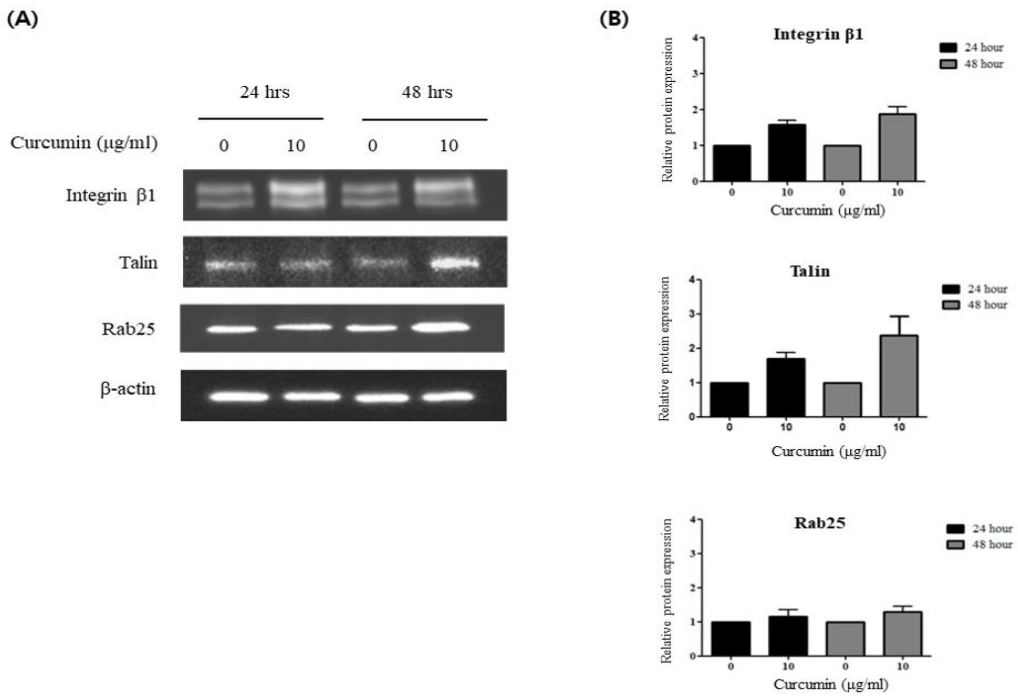
To investigate the involvement of integrin β1 in the anticancer effect of curcumin, we analyzed the expression of integrin β1 after curcumin treatment of HCT 116 cells. Curcumin was added at a concentration of 10 μg/mL, which is the optimal dose between anticancer effect and cytotoxicity, as observed in previous experiments (Figure 1A). As shown in Figure 2, the expression of integrin β1 significantly increased at 24 and 48 h after treatment of HCT 116 cells with curcumin.
To determine whether talin or rab25 is involved in the activation of integrin β1 by curcumin, the expressions of talin and rab25 were assessed in the same manner. HCT 116 cells were treated with 10 μg/mL of curcumin, and the expression of talin and rab25 was assessed using western blotting after 24 and 48 h. The expression of talin increased after curcumin treatment, but the expression of rab25 did not show any significant change (Figure 2). This suggested that the activation of integrin β1 by curcumin occurs in a talin-dependent manner but not in a rab25-dependent manner.

3.3. Curcumin Induces Integrin β1 Activation via a Rab25-Independent Pathway

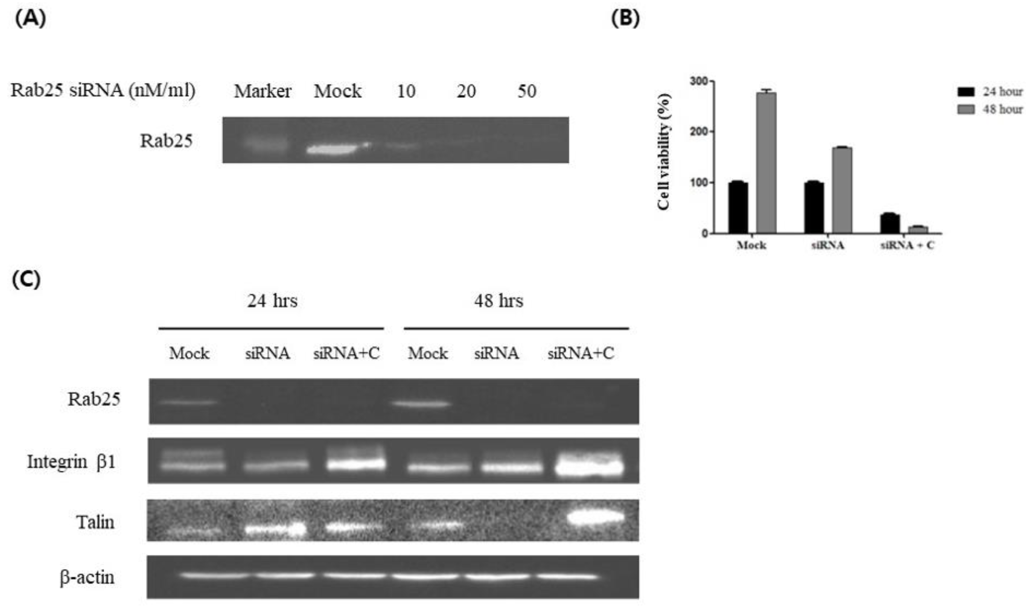
Although trafficking by rab25 is one of the major mechanisms by which integrin β1 is activated, curcumin increased the expression of integrin β1 without changing the expression of rab25 (Figure 2). To determine whether the interaction between curcumin and integrin β1 occurs independently of rab25, we performed further experiments by modulating the expression of rab25. HCT 116 cells were transfected with rab25-specific siRNA, and the efficacy of the knockdown was validated using western blotting, which confirmed a substantial reduction following treatment with 20 nM rab25 siRNA (Figure 3A). After successful rab25 knockdown, a reduction in the cell number was observed, and this phenomenon was further enhanced by curcumin treatment in the rab25 knockdown group (Figure 3B).
The expression of rab25, integrin β1, and talin was evaluated after curcumin treatment in the rab25 knockdown group. The expression of rab25 in the rab25 knockdown group did not increase after curcumin treatment. The expression of integrin β1 was not significantly different in the rab25 knockdown group and increased again after curcumin treatment. Similarly, the expression of talin increased after curcumin treatment (Figure 3C). These results suggested that curcumin exerts its anticancer effect by regulating the expression of integrin β1 through a talin-mediated pathway rather than through rab25.

3.4. Visualization of the Expression of Integrin β1 and Talin in Response to Curcumin

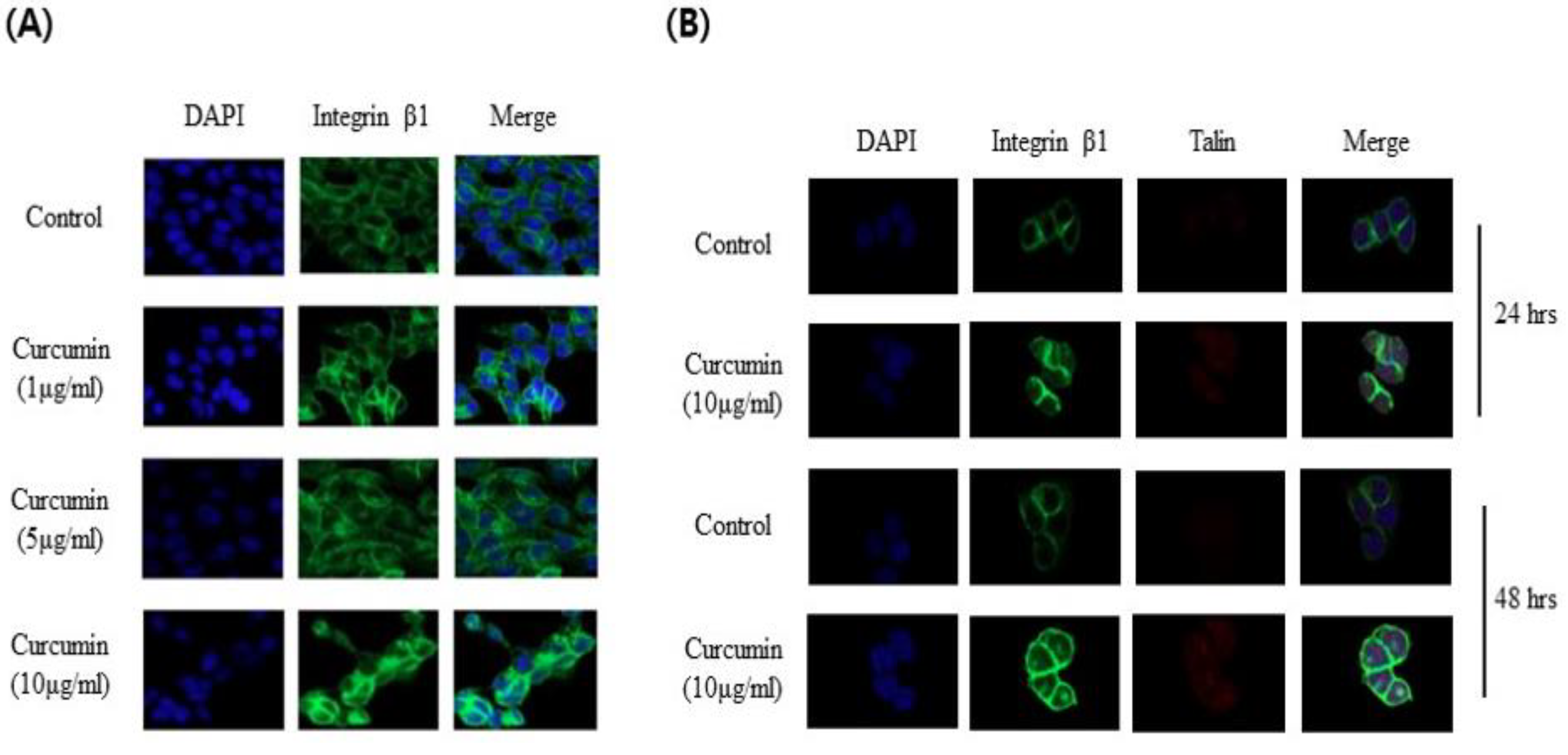
To determine the spatial distribution and quantitative expression levels of integrin β1 and talin in response to curcumin, confocal microscopy was used in HCT 116 cells. Confocal microscopy revealed a significant dose-dependent increase in the expression of integrin β1 within the cytosol after curcumin treatment (Figure 4A). The HCT 116 cells were treated with 10 μg/mL of curcumin and observed via confocal microscopy using integrin β1 and talin monoclonal antibodies after 24 and 48 h. The expression levels of both integrin β1 and talin increased after curcumin treatment compared with those in the control group (Figure 4B).

4. Discussion

In this study, we investigated the role of integrin β1 in the anticancer effect of curcumin in colon cancer. We found that curcumin exerts its anticancer effects by modulating the expression of integrin β1 and that this modulation occurs through a talin-mediated pathway and not through rab25. In particular, the cytotoxic effects of curcumin were observed only in cancer cells and not in normal cells, suggesting that curcumin exhibits cancer-specific cytotoxicity.
Many studies have investigated the anticancer effects of curcumin in various types of cancer. Curcumin inhibits cancer growth by inhibiting MMPs, NF-κB, TNF-α, HER-2, and EGFR [21,22,23]. Curcumin exerts its anticancer effects by regulating the expression of molecules involved in cell adhesion [24,25]. Studies have shown that curcumin inhibits cancer progression by regulating various cellular pathways, including EGFR/PDGFR, AKT/mTOR, and MAPK, which are either signaling partners or downstream molecules of integrin [8,9,10,11,12]. Previous studies on the integrin-mediated anticancer effect of curcumin have shown mixed results, with some studies showing that curcumin upregulates integrin expression and others showing that it downregulates it. Ray et al. demonstrated that curcumin exhibits antimetastatic properties by increasing integrin α5β1 and αvβ3 expression in lung cancer [26]. Coleman et al. showed that curcumin inhibits integrin β4 in breast cancer cells [13]. In contrast, a study reported that curcumin exerts anti-apoptotic and anti-catabolic effects by increasing integrin levels [27]. Mani et al. reported conflicting results, with curcumin upregulating integrin β1 and β4 and downregulating integrin β2 in bladder cancer cells [28]. In our study, we showed that curcumin inhibited cell proliferation by increasing the expression of integrin β1 in colon cancer cells. This effect was also mediated by talin, a key molecule that regulates integrin activity.
Integrins, which mediate cell adhesion to extracellular matrix ligands and cellular counter receptors, are a family of 24 heterodimeric receptors composed of α- and β-subunits [17]. Activation of integrins can be induced either by cytoplasmic events (‘‘inside-out’’ activation) or by extracellular stimuli (‘‘outside-in’’ activation) [18]. Talin is a large, multi-domain protein containing FERM domains. Talin, a key integrin regulator, binds to the cytoplasmic tail of the integrin β subunit to induce “inside-out” activation of integrin [19]. Rab proteins are members of the Ras superfamily of GTPases and are involved in recycling proteins from the endosome to the plasma membrane. Rab25 promotes the “outside-in” activation of integrins through a recycling process [20]. The present study showed that curcumin acts as an anticancer agent without modulating integrin recycling. Our findings shed light on the pivotal role of the integrin-talin interplay in mediating the tumor-suppressive effects of curcumin in colon cancer.
Curcumin, which accounts for 2–8% of the chemicals in turmeric, is thought to be the primary source of the plant’s yellow-gold color and is also responsible for numerous other properties [3,4]. Owing to its diverse capabilities and low intrinsic toxicity, curcumin has had a significant impact on a wide range of pharmacological discoveries, including anticancer, anti-inflammatory, antibacterial, and antioxidant drugs [5,6,7]. The low toxicity of curcumin is one of its most important properties, making it a suitable therapeutic agent. Low doses of curcumin have no adverse effects. High doses of curcumin have been shown to inhibit cancer cell proliferation without affecting normal cells [29,30,31]. These properties have led to its widespread use in cancer treatments [32]. In this study, we found that the integrin-mediated anticancer properties of curcumin in colon cancer cells concurrently attenuated its harmful effects on normal fibroblasts. In addition to the anticancer effects of curcumin, some studies have shown that curcumin inhibits proteins associated with drug resistance and enhances the efficacy of anticancer drugs [33].
This study underscores the clinical relevance of curcumin as a selective anticancer agent, particularly for colon cancer. Our findings suggest that medicinal herbs, such as curcumin, which possesses nontoxic chemopreventive properties, may be promising candidates in this regard. Although we did not delineate a specific pathway, our research highlights the integral role of the integrin-mediated pathway, independent of integrin trafficking modulation, in the anticancer properties of curcumin. Our study provides valuable insights into the diverse functions of common molecular entities, such as integrins, in tumorigenesis, along with the location of the tumor, which would be useful in selecting specific anticancer therapies appropriate for each person.

5. Conclusions

In conclusion, curcumin exerts cancer-specific cytotoxicity by modulating the expression of integrin β1. This modulation occurs through a talin-mediated pathway and not through rab25. Further investigation of the complex interplay between curcumin and integrin-mediated signaling molecules holds promise for the development of targeted anticancer therapeutics for colon cancer.

Author Contributions

Conceptualization: Hyeon Kyeong Kim, Bo Young Oh, Ryung-Ah Lee; Methodology: Hyeon Kyeong Kim, Kyung Sook Hong, Gyoung Tae Noh; Formal analysis and Investigation: Hyeon Kyeong Kim, Kyung Sook Hong; Writing - original draft preparation: Hyeon Kyeong Kim; Writing - review and editing: Ryung-Ah Lee, Bo Young Oh; Funding acquisition: Kyung Sook Hong, Gyoung Tae Noh; Resources: Ryung-Ah Lee, Kyung Sook Hong, Gyoung Tae Noh; Supervision: Ryung-Ah Lee, Bo Young Oh.

Funding

none.

Institutional Review Board Statement

Not applicable.

Informed Consent Statement

Not applicable.

Data Availability Statement

The datasets generated and/or analyzed during the current study are available from the corresponding author on reasonable request.

Acknowledgments

We express our gratitude to Jin Soo Kim for his invaluable contributions to this project. His expertise and guidance have been crucial in navigating the challenges encountered along the way.

Conflicts of Interest

The authors declare no conflicts of interest.

References

  1. Mansouri, K.; Rasoulpoor, S.; Daneshkhah, A.; Abolfathi, S.; Salari, N.; Mohammadi, M.; Rasoulpoor, S.; Shabani, S. Clinical effects of curcumin in enhancing cancer therapy: A systematic review. BMC Cancer 2020, 20, 791. [Google Scholar] [CrossRef] [PubMed]
  2. Ahmadi, F.; Valadbeigi, S.; Sajjadi, S.E.; Shokoohinia, Y.; Azizian, H.; Taheripak, G. Grandivittin as a natural minor groove binder extracted from Ferulago macrocarpa to ct-DNA, experimental and in silico analysis. Chem Biol Interact 2016, 258, 89–101. [Google Scholar] [CrossRef] [PubMed]
  3. Koroth, J.; Nirgude, S.; Tiwari, S.; Gopalakrishnan, V.; Mahadeva, R.; Kumar, S.; Karki, S.S.; Choudhary, B. Investigation of anti-cancer and migrastatic properties of novel curcumin derivatives on breast and ovarian cancer cell lines. BMC Complement Altern Med 2019, 19, 273. [Google Scholar] [CrossRef] [PubMed]
  4. Chattopadhyay, I.; Biswas, K.; Bandyopadhyay, U.; Banerjee, R.K. Turmeric and curcumin: Biological actions and medicinal applications. Current Science 2004, 87, 44–53. [Google Scholar]
  5. Maheshwari, R.K.; Singh, A.K.; Gaddipati, J.; Srimal, R.C. Multiple biological activities of curcumin: a short review. Life Sci 2006, 78, 2081-7. [Google Scholar] [CrossRef] [PubMed]
  6. Ono, K.; Hasegawa, K.; Naiki, H.; Yamada, M. Curcumin has potent anti-amyloidogenic effects for Alzheimer's beta-amyloid fibrils in vitro. J Neurosci Res 2004, 75, 742–50. [Google Scholar] [CrossRef] [PubMed]
  7. Sun, C.; Zhang, S.; Liu, C.; Liu, X. Curcumin Promoted miR-34a Expression and Suppressed Proliferation of Gastric Cancer Cells. Cancer Biother Radiopharm 2019, 34, 634–641. [Google Scholar] [CrossRef] [PubMed]
  8. Zhou, Y.; Zheng, S.; Lin, J.; Zhang, Q.J.; Chen, A. The interruption of the PDGF and EGF signaling pathways by curcumin stimulates gene expression of PPARgamma in rat activated hepatic stellate cell in vitro. Lab Invest 2007, 87, 488–98. [Google Scholar] [CrossRef] [PubMed]
  9. Chen, A.; Xu, J.; Johnson, A.C. Curcumin inhibits human colon cancer cell growth by suppressing gene expression of epidermal growth factor receptor through reducing the activity of the transcription factor Egr-1. Oncogene 2006, 25, 278–87. [Google Scholar] [CrossRef]
  10. Yu, S.; Shen, G.; Khor, T.O.; Kim, J.H.; Kong, A.N. Curcumin inhibits Akt/mammalian target of rapamycin signaling through protein phosphatase-dependent mechanism. Mol Cancer Ther 2008, 7, 2609–20. [Google Scholar] [CrossRef]
  11. Chaudhary, L.R.; Hruska, K.A. Inhibition of cell survival signal protein kinase B/Akt by curcumin in human prostate cancer cells. J Cell Biochem 2003, 89, 1–5. [Google Scholar] [CrossRef] [PubMed]
  12. Squires, M.S.; Hudson, E.A.; Howells, L.; Sale, S.; Houghton, C.E.; Jones, J.L.; Fox, L.H.; Dickens, M.; Prigent, S.A.; Manson, M.M. Relevance of mitogen activated protein kinase (MAPK) and phosphotidylinositol-3-kinase/protein kinase B (PI3K/PKB) pathways to induction of apoptosis by curcumin in breast cells. Biochem Pharmacol 2003, 65, 361–76. [Google Scholar] [CrossRef] [PubMed]
  13. Coleman, D.T.; Soung, Y.H.; Surh, Y.J.; Cardelli, J.A.; Chung, J. Curcumin Prevents Palmitoylation of Integrin β4 in Breast Cancer Cells. PLoS One 2015, 10, e0125399. [Google Scholar] [CrossRef] [PubMed]
  14. Soung, Y.H.; Chung, J. Curcumin inhibition of the functional interaction between integrin α6β4 and the epidermal growth factor receptor. Mol Cancer Ther 2011, 10, 883–91. [Google Scholar] [CrossRef] [PubMed]
  15. Javadi, S.; Zhiani, M.; Mousavi, M.A.; Fathi, M. Crosstalk between Epidermal Growth Factor Receptors (EGFR) and integrins in resistance to EGFR tyrosine kinase inhibitors (TKIs) in solid tumors. Eur J Cell Biol 2020, 99, 151083. [Google Scholar] [CrossRef] [PubMed]
  16. Bachmeier, B.E.; Mohrenz, I.V.; Mirisola, V.; Schleicher, E.; Romeo, F.; Höhneke, C.; Jochum, M.; Nerlich, A.G.; Pfeffer, U. Curcumin downregulates the inflammatory cytokines CXCL1 and -2 in breast cancer cells via NFkappaB. Carcinogenesis 2008, 29, 779–89. [Google Scholar] [CrossRef] [PubMed]
  17. Sun, Z.; Costell, M.; Fässler, R. Integrin activation by talin, kindlin and mechanical forces. Nat Cell Biol 2019, 21, 25–31. [Google Scholar] [CrossRef]
  18. Margadant, C.; Kreft, M.; de Groot, D.J.; Norman, J.C.; Sonnenberg, A. Distinct roles of talin and kindlin in regulating integrin α5β1 function and trafficking. Curr Biol 2012, 22, 1554–63. [Google Scholar] [CrossRef]
  19. Pernier, J.; Cardoso Dos Santos, M.; Souissi, M.; Joly, A.; Narassimprakash, H.; Rossier, O.; Giannone, G.; Helfer, E.; Sengupta, K.; Le Clainche, C. Talin and kindlin cooperate to control the density of integrin clusters. J Cell Sci 2023, 136. [Google Scholar] [CrossRef]
  20. Goldenring, J.R.; Nam, K.T. Rab25 as a tumour suppressor in colon carcinogenesis. Br J Cancer 2011, 104, 33–6. [Google Scholar] [CrossRef]
  21. Duvoix, A.; Blasius, R.; Delhalle, S.; Schnekenburger, M.; Morceau, F.; Henry, E.; Dicato, M.; Diederich, M. Chemopreventive and therapeutic effects of curcumin. Cancer Lett 2005, 223, 181–90. [Google Scholar] [CrossRef] [PubMed]
  22. Qadir, M.I.; Naqvi, S.T.; Muhammad, S.A. Curcumin: a Polyphenol with Molecular Targets for Cancer Control. Asian Pac J Cancer Prev 2016, 17, 2735–9. [Google Scholar]
  23. Niedzwiecki, A.; Roomi, M.W.; Kalinovsky, T.; Rath, M. Anticancer Efficacy of Polyphenols and Their Combinations. Nutrients 2016, 8. [Google Scholar] [CrossRef]
  24. Sullivan, D.E.; Ferris, M.; Nguyen, H.; Abboud, E.; Brody, A.R. TNF-alpha induces TGF-beta1 expression in lung fibroblasts at the transcriptional level via AP-1 activation. J Cell Mol Med 2009, 13, 1866–76. [Google Scholar] [CrossRef] [PubMed]
  25. Sa, G.; Das, T. Anti cancer effects of curcumin: cycle of life and death. Cell Div 2008, 3, 14. [Google Scholar] [CrossRef]
  26. Ray, S.; Chattopadhyay, N.; Mitra, A.; Siddiqi, M.; Chatterjee, A. Curcumin exhibits antimetastatic properties by modulating integrin receptors, collagenase activity, and expression of Nm23 and E-cadherin. J Environ Pathol Toxicol Oncol 2003, 22, 49–58. [Google Scholar] [PubMed]
  27. Shakibaei, M.; Schulze-Tanzil, G.; John, T.; Mobasheri, A. Curcumin protects human chondrocytes from IL-l1beta-induced inhibition of collagen type II and beta1-integrin expression and activation of caspase-3: an immunomorphological study. Ann Anat 2005, 187, 487–97. [Google Scholar] [CrossRef]
  28. Mani, J.; Fleger, J.; Rutz, J.; Maxeiner, S.; Bernd, A.; Kippenberger, S.; Zöller, N.; Chun, F.K.; Relja, B.; Juengel, E.; et al. Curcumin combined with exposure to visible light blocks bladder cancer cell adhesion and migration by an integrin dependent mechanism. Eur Rev Med Pharmacol Sci 2019, 23, 10564–10574. [Google Scholar] [CrossRef] [PubMed]
  29. Anand, P.; Sundaram, C.; Jhurani, S.; Kunnumakkara, A.B.; Aggarwal, B.B. Curcumin and cancer: an "old-age" disease with an "age-old" solution. Cancer Lett 2008, 267, 133–64. [Google Scholar] [CrossRef]
  30. Boon, H.; Wong, J. Botanical medicine and cancer: a review of the safety and efficacy. Expert Opin Pharmacother 2004, 5, 2485–501. [Google Scholar] [CrossRef]
  31. Wargovich, M.J. Nutrition and cancer: the herbal revolution. Curr Opin Gastroenterol 1999, 15, 177–80. [Google Scholar] [CrossRef] [PubMed]
  32. Banik, K.; Ranaware, A.M.; Harsha, C.; Nitesh, T.; Girisa, S.; Deshpande, V.; Fan, L.; Nalawade, S.P.; Sethi, G.; Kunnumakkara, A.B. Piceatannol: A natural stilbene for the prevention and treatment of cancer. Pharmacol Res 2020, 153, 104635. [Google Scholar] [CrossRef] [PubMed]
  33. Keyvani-Ghamsari, S.; Khorsandi, K.; Gul, A. Curcumin effect on cancer cells' multidrug resistance: An update. Phytother Res 2020, 34, 2534–2556. [Google Scholar] [CrossRef] [PubMed]
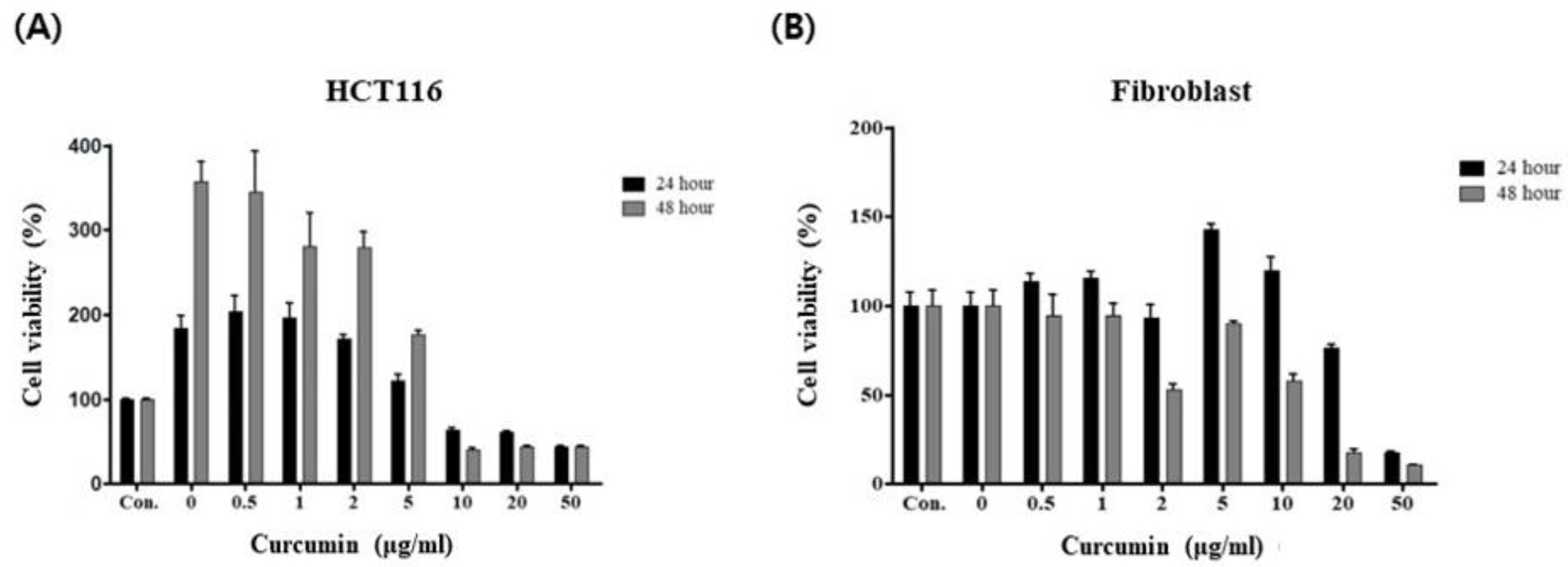
Figure 1. MTT assay after treatment of HCT 116 and fibroblast cells with curcumin. (A) After treatment of HCT 116 cells with curcumin, cellular proliferation is inhibited in a dose-dependent and time-dependent manner; (B) After treatment of fibroblast cells with curcumin, no significant change in cellular proliferation is observed.
Preprints 107988 g001
Figure 2. After treatment of HCT 116 cells with curcumin, an increase in integrin β1 and talin expression is observed, but no significant changes in rab25 expression are observed. (A) Expression of integrin β1, talin, and rab25, depending on curcumin treatment, is analyzed using western blotting; (B) Transcriptional levels of integrin β1, talin, and rab25 are measured using real-time polymerase chain reaction. Columns = mean ± SD (n = 3).
Preprints 107988 g002
Figure 3. Treatment of HCT 116 cells with rab25 siRNA decreased the expression of rab25 but not integrin β1 or talin. Subsequent treatment with curcumin increased the expression levels of integrin β1 and talin. (A) Knockdown of rab25 expression after siRNA transfection of HCT 116 cells; (B) MTT assay after sequentially treating HCT 116 cells with rab25 siRNA (20 nM) and curcumin (10 μg/mL); (C) Western blot analysis after sequentially treating HCT 116 cells with rab25 siRNA (20 nM) and curcumin (10 μg/mL). C = curcumin.
Preprints 107988 g003
Figure 4. Confocal microscopy after treatment of HCT 116 cells with curcumin. (A) Expression of cytosolic integrin β1 increased with curcumin dose. Original magnification, 63×; (B) Expression of integrin β1 and talin increased after curcumin treatment. Original magnification, 40×.
Preprints 107988 g004
Disclaimer/Publisher’s Note: The statements, opinions and data contained in all publications are solely those of the individual author(s) and contributor(s) and not of MDPI and/or the editor(s). MDPI and/or the editor(s) disclaim responsibility for any injury to people or property resulting from any ideas, methods, instructions or products referred to in the content.
Copyright: This open access article is published under a Creative Commons CC BY 4.0 license, which permit the free download, distribution, and reuse, provided that the author and preprint are cited in any reuse.
Prerpints.org logo

Preprints.org is a free preprint server supported by MDPI in Basel, Switzerland.

Subscribe

© 2024 MDPI (Basel, Switzerland) unless otherwise stated