Submitted:
19 August 2024
Posted:
20 August 2024
Read the latest preprint version here
Abstract
Keywords:
1. Introduction
2. Clinical Trial Results and 2023-2024 Campaign
2.1. Clinical Trial Results
2.1.1. Phase 1 and 2a
2.1.2. Results of Phase 2b and 3 Trials
2.1.3. Deaths in Trials
2.2. Phamacovigilance Data and Results of the 2023-2024 Season Immunization Campaign
2.2.1. USA Results of Immunization Campaign
2.2.2. Luxembourg Results of Immunization Campaign
2.2.3. France Results of Immunization Campaign
2.2.4. Spain Results of Immunization Campaign
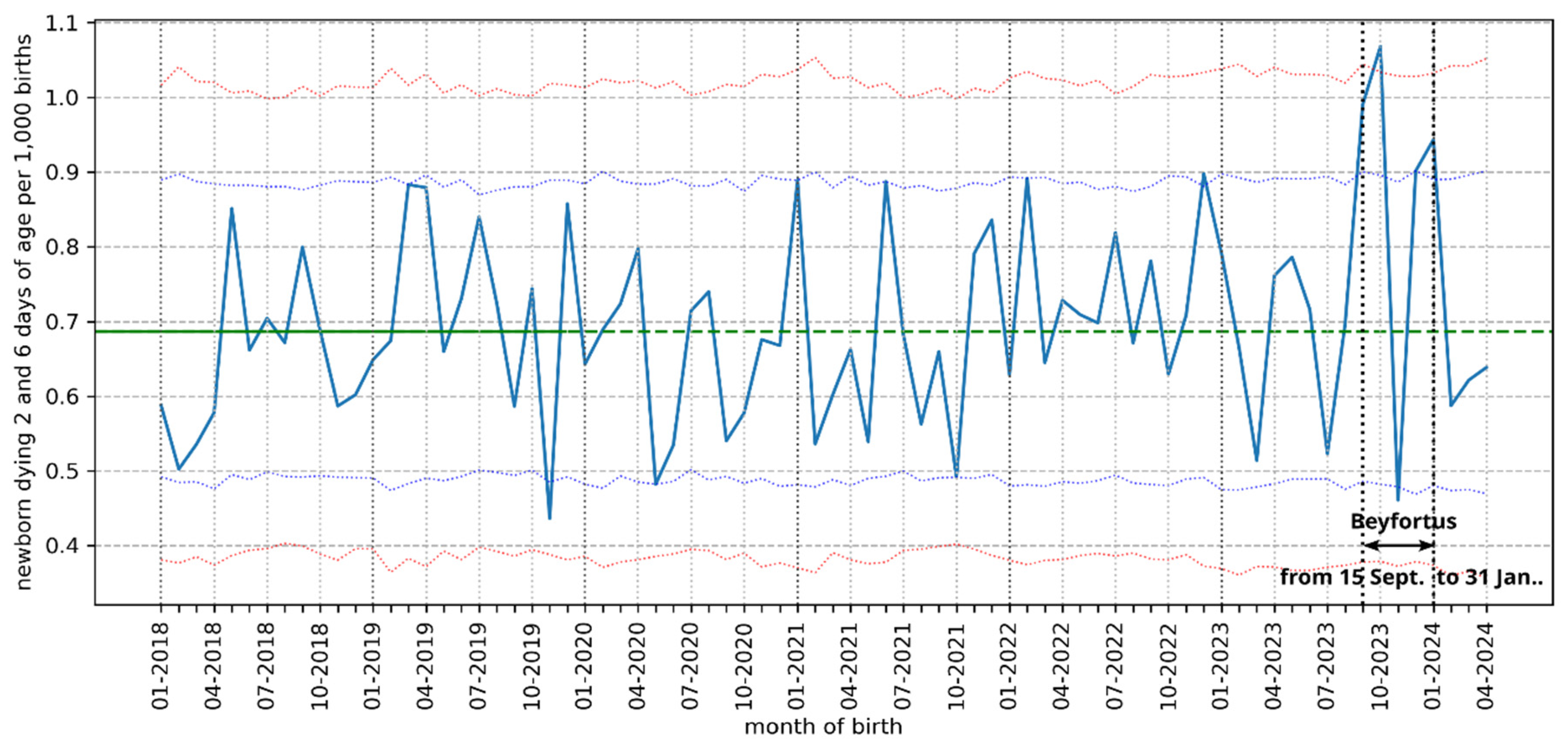
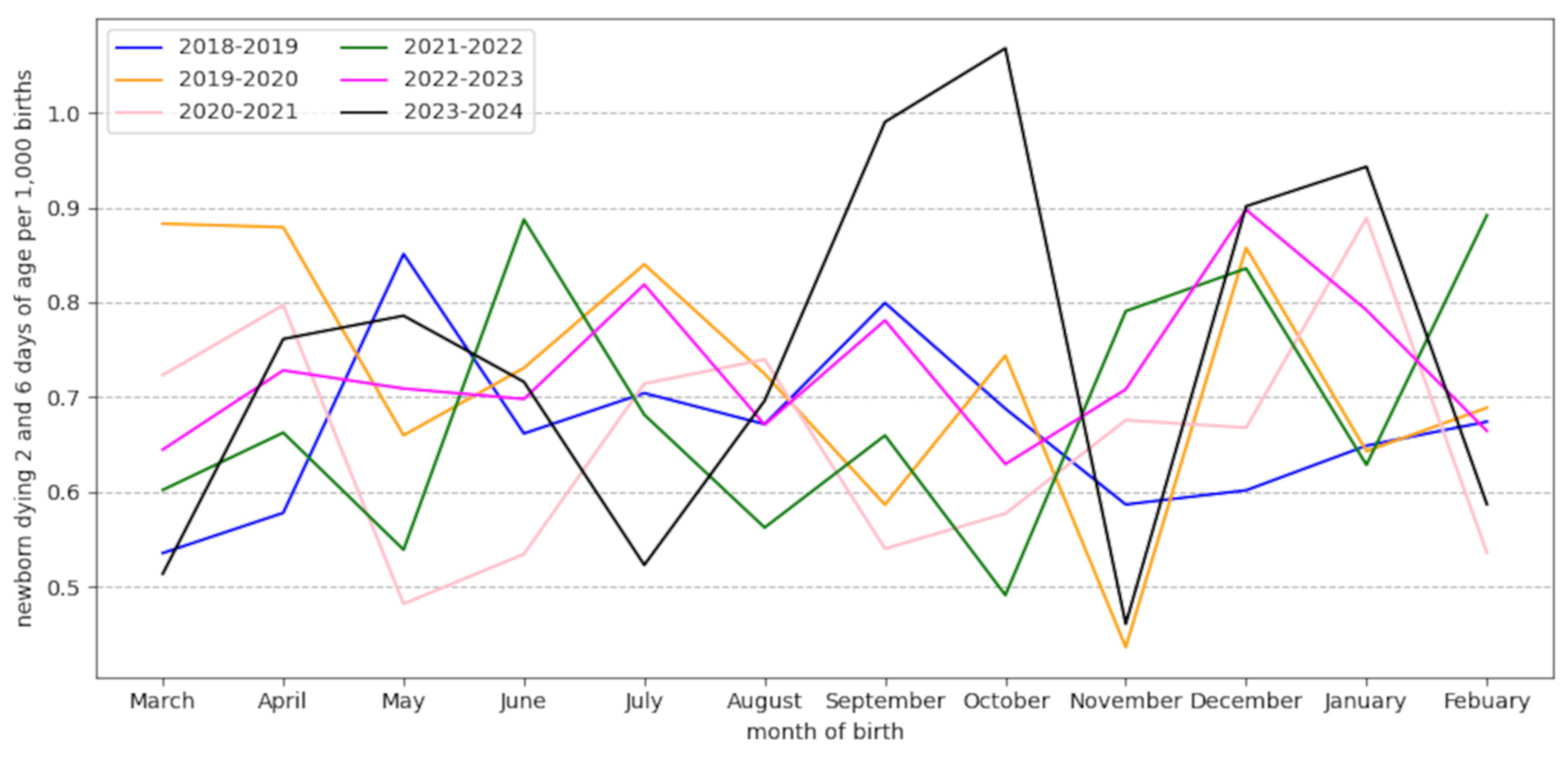
2.3. Neonatal Deaths in France


3. Design of Nirsevimab
4. Definition, Role and Localization of FcRn: Based on Our Knowledge of the Role of FcRn, What Might Be the Consequences of a mAb’s Increased Affinity for This Receptor?
4.1. Transport of Free and Antigen-Bound IgG
4.2. Intracellular Transport of Viruses by FcRn
5. What Are the Mechanisms of ADE in Viral Infections and Following Antiviral Vaccinations, and How Might an RSV F-Protein mAb with Increased Affinity to FcRn Be Involved? Could mAb Binding to Other FcγRs Be Involved?
5.1. Mechanisms of ADE of Viral Infections by Specific Anti-Viral Antibodies
5.2. ADE during Infections and Vaccinations with RSV or Other Viruses
5.3. Role of Complement in ADE (RSV and Other Viruses)
5.4. Role of IgG Binding to FcγR in ADE during RSV Infection
5.5. Involvement of the Monocytic Lineage in ADE (in Case of Viral Infection and after RSV Vaccination)
5.6. Immune System Disruption Caused by IgG binding to FcRn
5.7. Known ADE with Mabs against RSV and Other Viruses
5.8. Importance of Antibody Levels and Quality: ADE Can Occur in the Presence of Low Levels of Strongly Neutralizing RSV Antibodies
6. How Were the Factors Likely to Cause ADE with Nirsevimab Assessed?
6.1. Pharmacokinetics
6.2. Study of Nirsevimab Binding to FcγR In Vitro and Ex-Vivo and of Possible ADE in Animals by Manufacturers
6.2.1. In Vitro and Ex Vivo Studies
6.2.2. Animal Challenge Studies
6.3. Clinical Trials
7. Economic Benefits of Nirsevimab: Price and All-Cause Hospitalization Rates
8. Conclusion
Funding
Informed Consent Statement
Acknowledgments
Conflicts of Interest
Abbreviations
| ADE | antibody dependent enhancement (of the infection) |
| ADCC | antibody- dependent cell-mediated cytotoxicity |
| ADCD | antibody-dependent complement deposition |
| ADCP | antibody- dependent cellular phagocytosis |
| ADNKA | antibody-dependent NK cell activation |
| ADNP | antibody-dependent neutrophil phagocytosis |
| APC | antigen presenting cell |
| ARR | absolute reduction risk |
| ASL | airway surface liquid |
| ARI | acute respiratory infection |
| CDC or CDCC | complement-dependent cytotoxicity |
| CDCP | complement-dependent cell-mediated phagocytosis |
| CR | complement receptors |
| EMA | European Medicines Agency |
| EPAR | European Public Assessment Report |
| ERD | enhanced respiratory disease |
| F | RSV membrane fusion protein |
| Fc | IgG crystallizable fragment |
| FcγR | IgG Fc receptor |
| FcRn | Fc neonatal receptor |
| hRSV | human respiratory syncytial virus |
| IC | immune-complex |
| LRTI | low tract respiratory infection |
| mAb | monoclonal antibody |
| PICU | pediatric intensive care unit |
| RSV | respiratory syncytial virus |
| URTI | upper respiratory tract infection |
| VAED | Vaccine-associated enhanced disease, a form of ADE, antibody-dependent enhancement |
| VAERD | Vaccine-associated enhanced respiratory disease |
References
- Li, Y.; Wang, X.; Blau, D.M.; Caballero, M.T.; Feikin, D.R.; Gill, C.J.; Madhi, S.A.; Omer, S.B.; Simões, E.A.F.; Campbell, H.; et al. Global, regional, and national disease burden estimates of acute lower respiratory infections due to respiratory syncytial virus in children younger than 5 years in 2019: a systematic analysis. Lancet 2022, 399, 2047–2064. [Google Scholar] [CrossRef] [PubMed] [PubMed Central]
- Rocca, A.; Biagi, C.; Scarpini, S.; Dondi, A.; Vandini, S.; Pierantoni, L.; Lanari, M. Passive Immunoprophylaxis against Respiratory Syncytial Virus in Children: Where Are We Now? Int J Mol Sci 2021, 22, 3703. [Google Scholar] [CrossRef] [PubMed] [PubMed Central]
- Osiowy, C.; Horne, D.; Anderson, R. Antibody-dependent enhancement of respiratory syncytial virus infection by sera from young infants. Clin Diagn Lab Immunol 1994, 1, 670–677. [Google Scholar] [CrossRef] [PubMed] [PubMed Central]
- Zhu, Q.; McLellan, J.S.; Kallewaard, N.L.; Ulbrandt, N.D.; Palaszynski, S.; Zhang, J.; Moldt, B.; Khan, A.; Svabek, C.; McAuliffe, J.M.; et al. A highly potent extended half-life antibody as a potential RSV vaccine surrogate for all infants. Sci Transl Med 2017, 9, eaaj1928. [Google Scholar] [CrossRef] [PubMed]
- Acevedo, O.A.; Díaz, F.E.; Beals, T.E.; Benavente, F.M.; Soto, J.A.; Escobar-Vera, J.; González, P.A.; Kalergis, A.M. Contribution of Fcγ Receptor-Mediated Immunity to the Pathogenesis Caused by the Human Respiratory Syncytial Virus. Front Cell Infect Microbiol 2019, 9, 75. [Google Scholar] [CrossRef] [PubMed] [PubMed Central]
- Diethelm-Varela, B.; Soto, J.A.; Riedel, C.A.; Bueno, S.M.; Kalergis, A.M. New Developments and Challenges in Antibody-Based Therapies for the Respiratory Syncytial Virus. Infect Drug Resist 2023, 16, 2061–2074. [Google Scholar] [CrossRef] [PubMed] [PubMed Central]
- Rigter, A.; Widjaja, I.; Versantvoort, H.; Coenjaerts, F.E.; van Roosmalen, M.; Leenhouts, K.; Rottier, P.J.; Haijema, B.J.; de Haan, C.A. A protective and safe intranasal RSV vaccine based on a recombinant prefusion-like form of the F protein bound to bacterium-like particles. PLoS One 2013, 8, e71072. [Google Scholar] [CrossRef] [PubMed] [PubMed Central]
- Gong, X.; Luo, E.; Fan, L.; Zhang, W.; Yang, Y.; Du, Y.; Yang, X.; Xing, S. Clinical research on RSV prevention in children and pregnant women: progress and perspectives. Front Immunol 2024, 14, 1329426. [Google Scholar] [CrossRef] [PubMed] [PubMed Central]
- Kampmann, B.; Madhi, S.A.; Munjal, I.; Simões, E.A.F.; Pahud, B.A.; Llapur, C.; Baker, J.; Pérez Marc, G.; Radley, D.; Shittu, E.; et al. Bivalent Prefusion F Vaccine in Pregnancy to Prevent RSV Illness in Infants. N Engl J Med 2023, 388, 1451–1464. [Google Scholar] [CrossRef] [PubMed]
- European Union Risk Management Plan (EU RMP) for Beyfortus® (Nirsevimab) 3 May 2021. Available online: https://www.ema.europa.eu/en/documents/rmp-summary/beyfortus-epar-risk-management-plan_en.pdf (accessed on 25 April 2024).
- Article 14(9) EC-726/2004 of the European Parliament and of the Council of 31 March 2004 laying down Community procedures for the authorisation and supervision of medicinal products for human and veterinary use and establishing a European Medicines Agency. Available online: http://data.europa.eu/eli/reg/2004/726/oj.
- Suh, M.; Movva, N.; Jiang, X.; Bylsma, L.C.; Reichert, H.; Fryzek, J.P.; Nelson, C.B. Respiratory Syncytial Virus Is the Leading Cause of United States Infant Hospitalizations, 2009-2019: A Study of the National (Nationwide) Inpatient Sample. J Infect Dis 2022, 226 (Suppl 2), S154–S163. [Google Scholar] [CrossRef] [PubMed] [PubMed Central]
- Nair, H.; Simões, E.A.; Rudan, I.; Gessner, B.D.; Azziz-Baumgartner, E.; Zhang, J.S.F.; Feikin, D.R.; Mackenzie, G.A.; Moiïsi, J.C.; Roca, A.; et al. Global and regional burden of hospital admissions for severe acute lower respiratory infections in young children in 2010: a systematic analysis. Lancet 2013, 381, 1380–1390. [Google Scholar] [CrossRef] [PubMed] [PubMed Central]
- Del Riccio, M.; Spreeuwenberg, P.; Osei-Yeboah, R.; Johannesen, C.K.; Fernandez, L.V.; Teirlinck, A.C.; Wang, X.; Heikkinen, T.; Bangert, M.; Caini, S.; et al. Burden of Respiratory Syncytial Virus in the European Union: estimation of RSV-associated hospitalizations in children under 5 years. J Infect Dis 2023, 228, 1528–1538. [Google Scholar] [CrossRef] [PubMed] [PubMed Central]
- Bulletin Infections Respiratoires Aiguës, publié le 17 avril 2024. Available online: https://www.santepubliquefrance.fr/maladies-et-traumatismes/maladies-et-infections-respiratoires/grippe/documents/bulletin-national/infections-respiratoires-aigues-grippe-bronchiolite-covid-19-.-bilan-de-la-saison-2023-2024 (accessed on 22 April 2024).
- Suss, R.J.; Simões, E.A.F. Respiratory Syncytial Virus Hospital-Based Burden of Disease in Children Younger Than 5 Years, 2015-2022. JAMA Netw Open 2024, 7, e247125. [Google Scholar] [CrossRef] [PubMed]
- Domachowske, J.B.; Khan, A.A.; Esser, M.T.; Jensen, K.; Takas, T.; Villafana, T.; Dubovsky, F.; Griffin, M.P. Safety, Tolerability and Pharmacokinetics of MEDI8897, an Extended Half-life Single-dose Respiratory Syncytial Virus Prefusion F-targeting Monoclonal Antibody Administered as a Single Dose to Healthy Preterm Infants. Pediatr Infect Dis J 2018, 37, 886–892. [Google Scholar] [CrossRef] [PubMed] [PubMed Central]
- Griffin, M.P.; Khan, A.A.; Esser, M.T.; Jensen, K.; Takas, T.; Kankam, M.K.; Villafana, T.; Dubovsky, F. Safety, Tolerability, and Pharmacokinetics of MEDI8897, the Respiratory Syncytial Virus Prefusion F-Targeting Monoclonal Antibody with an Extended Half-Life, in Healthy Adults. Antimicrob Agents Chemother 2017, 61, e01714-16. [Google Scholar] [CrossRef] [PubMed]
- Hammitt, L.L.; Dagan, R.; Yuan, Y.; Baca Cots, M.; Bosheva, M.; Madhi, S.A.; Muller, W.J.; Zar, H.J.; Brooks, D.; Grenham, A.; et al. Nirsevimab for Prevention of RSV in Healthy Late-Preterm and Term Infants. N Engl J Med 2022, 386, 837–846. [Google Scholar] [CrossRef] [PubMed]
- Domachowske, J.; Madhi, S.A.; Simões, E.A.F.; Atanasova, V.; Cabañas, F.; Furuno, K.; Garcia-Garcia, M.L.; Grantina, I.; Nguyen, K.A.; Brooks, D.; et al. Safety of Nirsevimab for RSV in Infants with Heart or Lung Disease or Prematurity. N Engl J Med 2022, 386, 892–894. [Google Scholar] [CrossRef] [PubMed]
- Simões, E.A.F.; Madhi, S.A.; Muller, W.J.; Atanasova, V.; Bosheva, M.; Cabañas, F.; Baca Cots, M.; Domachowske, J.B.; Garcia-Garcia, M.L.; Grantina, I.; et al. Efficacy of nirsevimab against respiratory syncytial virus lower respiratory tract infections in preterm and term infants, and pharmacokinetic extrapolation to infants with congenital heart disease and chronic lung disease: a pooled analysis of randomised controlled trials. Lancet Child Adolesc Health 2023, 7, 180–189. [Google Scholar] [CrossRef] [PubMed] [PubMed Central]
- Drysdale, S.B.; Cathie, K.; Flamein, F.; Knuf, M.; Collins, A.M.; Hill, H.C.; Kaiser, F.; Cohen, R.; Pinquier, D.; Felter, C.T.; et al. Nirsevimab for Prevention of Hospitalizations Due to RSV in Infants. N Engl J Med 2023, 389, 2425–2435. [Google Scholar] [CrossRef] [PubMed]
- Griffin, M.P.; Yuan, Y.; Takas, T.; Domachowske, J.B.; Madhi, S.A.; Manzoni, P.; Simões, E.A.F.; Esser, M.T.; Khan, A.A.; Dubovsky, F.; et al. Single-Dose Nirsevimab for Prevention of RSV in Preterm Infants. N Engl J Med 2020, 383, 415–425. [Google Scholar] [CrossRef] [PubMed]
- EMA-EPAR Beyfortus-Nirsevimab. Available online: https://www.ema.europa.eu/en/medicines/human/EPAR/beyfortus#assessment-history (accessed on 15 April 2024).
- HAS nirsevimab Transparency Committee, 19 July 2023. Available online: https://www.has-sante.fr/plugins/ModuleXitiKLEE/types/FileDocument/doXiti.jsp?id=p_3476336 (accessed on 22 April 2024). https://www.has-sante.fr/upload/docs/application/pdf/2023-11/beyfortus_19072023_summary_ct20356_en.pdf.
- Sullivan, K.; Sullivan, B. Does nirsevimab prevent lower respiratory infections caused by respiratory syncytial virus? J Perinatol 2024, 44, 767–769. [Google Scholar] [CrossRef] [PubMed] [PubMed Central]
- FDA Biologics License Application (BLA) 761328 Nirsevimab Antimicrobial Drugs Advisory Committee Meeting June 8, 2023 Division of Antivirals, Office of Infectious Diseases Center for Drug Evaluation and Research. Available online: https://www.fda.gov/media/169322/download (accessed on 13 April 2024)https://web.archive.org/web/20230613110427/.
- EudraVigilance European database of suspected adverse drug reaction reports. Available online: https://www.adrreports.eu/fr/eudravigilance.html (accessed on 15 April 2024).
- Pharmacovigilance d’Île de France, campagne d’immunisation contre le VRS-Beyfortus, 31 janvier 2024. Available online: https://www.pharmacovigilance-iledefrance.fr/d%C3%A9tails-dune-br%C3%A8ve/campagne-dimmunisation-contre-le-vrs-beyfortus-nirs%C3%A9vimab (accessed on 22 April 2024). https://www.pharmacovigilance-iledefrance.fr/détails-dune-brève/campagne-dimmunisation-contre-le-vrs-beyfortus-nirsévimab.
- US-FDA FDA Approves New Drug to Prevent RSV in Babies and Toddlers. Available online: https://www.fda.gov/news-events/press-announcements/fda-approves-new-drug-prevent-rsv-babies-and-toddlers (accessed on 15 April 2024).
- Jones, J.M.; Fleming-Dutra, K.E.; Prill, M.M.; et al. Use of Nirsevimab for the Prevention of Respiratory Syncytial Virus Disease Among Infants and Young Children: Recommendations of the Advisory Committee on Immunization Practices — United States, 2023. MMWR Morb Mortal Wkly Rep 2023, 72, 920–925. [Google Scholar] [CrossRef]
- Sánchez Luna, M.; Fernández Colomer, B.; Couce Pico, M.L.; en representación de la Junta Directiva de la Sociedad española de Neonatología SENEO Comisión de Infecciones SENEO y Comisión de Estándares de SENEO. Recommendations of the Spanish Society of Neonatology for the prevention of severe respiratory syncytial virus infections with nirsevimab, for the 2023-2024 season. An Pediatr (Engl Ed) 2023, 99, 264–265. [Google Scholar] [CrossRef] [PubMed]
- New immunisation to protect newborns and young children against bronchiolitis, The Luxembourg Governement press release, September 22, 2023. Available online: https://gouvernement.lu/en/actualites/toutes_actualites/communiques/2023/09-septembre/22-immunisation-bronchiolite-nourrissons.html (accessed on 17 May 2024).
- CDC, Nirsevimab Coverage, Children 0 to 19 months, United States, Data are current through February 29, 2024. Available online: https://www.cdc.gov/vaccines/imz-managers/coverage/rsvvaxview/nirsevimab-coverage-children-0-19months.html (accessed on 15 April 2024).
- RSV-NET Interactive Dashboard, CDC. Available online: https://www.cdc.gov/rsv/research/rsv-net/dashboard.html (accessed on 15 April 2024).
- Moline, H.L.; et al. Early Estimate of Nirsevimab Effectiveness for Prevention of Respiratory Syncytial Virus–Associated Hospitalization Among Infants Entering Their First Respiratory Syncytial Virus Season — New Vaccine Surveillance Network, October 2023–February 2024. https://www.cdc.gov/mmwr/volumes/73/wr/mm7309a4.htm?s_cid=mm7309a4_w.
- Setia, M.S. Methodology Series Module 2: Case-control Studies. Indian J Dermatol 2016, 61, 146–151. [Google Scholar] [CrossRef] [PubMed] [PubMed Central]
- Ernst, C.; Bejko, D.; Gaasch, L.; Hannelas, E.; Kahn, I.; Pierron, C.; Del Lero, N.; Schalbar, C.; Do Carmo, E.; Kohnen, M.; et al. Impact of nirsevimab prophylaxis on paediatric respiratory syncytial virus (RSV)-related hospitalisations during the initial 2023/24 season in Luxembourg. Euro Surveill 2024, 29, 2400033. [Google Scholar] [CrossRef] [PubMed] [PubMed Central]
- Assad, Z.; Romain, A.S.; Aupiais, C.; Shum, M.; Schrimpf, C.; Lorrot, M.; Corvol, H.; Prevost, B.; Ferrandiz, C.; Giolito, A.; et al. Nirsevimab and Hospitalization for RSV Bronchiolitis. N Engl J Med 2024, 391, 144–154. [Google Scholar] [CrossRef] [PubMed]
- EPI-PHARE Utilisation du Nirsévimab (Beyfortus®) en ville en France lors de la première campagne de prévention (saison 2023/2024) April 8, 2024. Available online: https://www.epi-phare.fr/rapports-detudes-et-publications/utilisation-beyfortus/ (accessed on 3 June 2024).
- Bronchiolite: bilan de la surveillance hivernale 2022-2023, Santé Publique France, July 19, 2023. Available online: https://www.santepubliquefrance.fr/les-actualites/2023/bronchiolite-bilan-de-la-surveillance-hivernale-2022-2023 (accessed on 1 May 2024).
- Paireau, J.; Durand, C.; Raimbault, S.; Cazaubon, J.; Mortamet, G.; et al. Nirsevimab effectiveness against cases of respiratory syncytial virus bronchiolitis hospitalised in pediatric intensive care units in France, September 2023–January 2024. 2024. pasteur-04501286. Available online: https://hal.science/pasteur-04501286 (accessed on 12 March 2024).
- Vaux, S.; Viriot, D.; Forgeot, C.; Pontais, I.; Savitch, Y.; Barondeau-Leuret, A.; Smadja, S.; Valette, M.; Enouf, V.; Parent du Chatelet, I. Bronchiolitis epidemics in France during the SARS-CoV-2 pandemic: The 2020-2021 and 2021-2022 seasons. Infect Dis Now 2022, 52, 374–378. [Google Scholar] [CrossRef] [PubMed] [PubMed Central]
- Delestrain, C.; Danis, K.; Hau, I.; Behillil, S.; Billard, M.N.; Krajten, L.; Cohen, R.; Bont, L.; Epaud, R. Impact of COVID-19 social distancing on viral infection in France: A delayed outbreak of RSV. Pediatr Pulmonol 2021, 56, 3669–3673. [Google Scholar] [CrossRef] [PubMed] [PubMed Central]
- International Classification of Diseases, Tenth Revision, International Statistical Classification of Diseases and Related Health Problems 10th Revision latest version 2019, WHO. Available online: https://icd.who.int/browse10/2019/en (accessed on 12 May 2024).
- La sanidad pública paga 209 euros por cada vacuna infantil contra la bronquiolitis, Civio, October 30, 2023. Available online: https://civio.es/medicamentalia/2023/10/30/nirsevimab-beyfortus-precio-virus-respiratorio-sincitial/.
- Servicio Andaluz de Salud, El Distrito Serranía recibe un reconocimiento por la cobertura de la vacuna combinada frente a difteria, tétanos, tosferina y poliomielitis 30 January 2024. Available online: https://www.sspa.juntadeandalucia.es/servicioandaluzdesalud/todas-noticia/el-distrito-serrania-recibe-un-reconocimiento-por-la-cobertura-de-la-vacuna-combinada-frente (accessed on 10 February 2024).
- Administradas 452.000 vacunas por virus respiratorios, Ondacero.es; April 8, 2024. Available online: https://www.ondacero.es/emisoras/asturias/noticias/administradas-452000-vacunas-virus-respiratorios_202404086613a4ec099903000111cfb3.html (accessed on 23 April 2024).
- La Comunidad de Madrid reduce un 90% los ingresos hospitalarios de menores de un año tras incorporar la vacuna contra la bronquiolitis, Communiqué de presse de la Comunidad de Madrid, April 16,2024. Available online: https://www.comunidad.madrid/notas-prensa/2024/04/16/comunidad-madrid-reduce-90-ingresos-hospitalarios-menores-ano-incorporar-vacuna-bronquiolitis (accessed on 23 April 2024).
- Ministerio de ciencia, innovacion y universidades, Gobierno de Espana, Informes semanales de vigilancia centinela de IRAs y de IRAG: Gripe, Covid-19 y otros virus respiratorios. Available online: https://www.isciii.es/QueHacemos/Servicios/VigilanciaSaludPublicaRENAVE/EnfermedadesTransmisibles/Paginas/VIGILANCIA-CENTINELA-DE-INFECCION-RESPIRATORIA-AGUDA.aspxhttps://web.archive.org/web/20240122052000 (accessed on 23 April 2024). https://www.isciii.es/QueHacemos/Servicios/VigilanciaSaludPublicaRENAVE/EnfermedadesTransmisibles/Documents/GRIPE/Informes%20semanales/Temporada_2023-24/Informe%20semanal_SiVIRA_022024.pdfhttps://www.isciii.es/QueHacemos/Servicios/VigilanciaSaludPublicaRENAVE/EnfermedadesTransmisibles/Documents/GRIPE/Informes%20semanales/Temporada_2022-23/Informe%20semanal_SiVIRA_102023.pdf.
- FOLLOW-UP REPORT ON IMMUNIZATION WITH NIRSEVIMAB IN GALICIA Dirección Xeral de Saúde Pública Data up to week 13, 2024 (31-03-2024). Available online: https://www.nirsegal.es/en (accessed on 23 April 2024). https://assets-global.website-files.com/65774b0d3a50ee58b24dba82/660e95b8142542e4cae669d2_Report_RSV_week13.pdf.
- Ares-Gómez, S.; Mallah, N.; Santiago-Pérez, M.I.; Pardo-Seco, J.; Pérez-Martínez, O.; Otero-Barrós, M.T.; Suárez-Gaiche, N.; Kramer, R.; Jin, J.; Platero-Alonso, L.; et al. Effectiveness and impact of universal prophylaxis with nirsevimab in infants against hospitalisation for respiratory syncytial virus in Galicia, Spain: initial results of a population-based longitudinal study. Lancet Infect Dis 2024. [Google Scholar] [CrossRef] [PubMed]
- Evaluation of the Effectiveness and Impact of Nirsevimab Administered as Routine Immunization (NIRSE-GAL). Available online: https://classic.clinicaltrials.gov/ct2/show/NCT06180993 (accessed on 10 May 2024).
- Ezpeleta, G.; Navascués, A.; Viguria, N.; Herranz-Aguirre, M.; Juan Belloc, S.E.; Gimeno Ballester, J.; Muruzábal, J.C.; García-Cenoz, M.; Trobajo-Sanmartín, C.; Echeverria, A.; et al. Effectiveness of Nirsevimab Immunoprophylaxis Administered at Birth to Prevent Infant Hospitalisation for Respiratory Syncytial Virus Infection: A Population-Based Cohort Study. Vaccines 2024, 12, 383. [Google Scholar] [CrossRef]
- Boletin de Salud Publica de Navarra, n°126, September 2023. Available online: http://www.navarra.es/home_es/Gobierno+de+Navarra/Organigrama/Los+departamentos/Salud/Organigrama/Estructura+Organica/Instituto+Navarro+de+Salud+Publica/Publicaciones/Publicaciones+profesionales/Epidemiologia/Boletin+ISP.htm (accessed on 1 May 2024). http://www.navarra.es/NR/rdonlyres/AECCD760-AB2A-4841-818A-FA53478FD6DC/488194/BOL126INT1.pdf.
- DGS-URGENT 9, 08/24/2023, DGS-URGENT N°2023_14, PREVENTION MEDICAMENTEUSE DES BRONCHIOLITES A VRS A PARTIR DE SEPTEMBRE. Available online: https://sante.gouv.fr/IMG/pdf/dgs-urgent_2023-14_-_traitement_preventif_vrs.pdf (accessed on 30 August 2023).
- DREES, La Naissance: caractéristiques des accouchements. Available online: https://drees.solidarites-sante.gouv.fr/sites/default/files/2021-07/Fiche%2024%20-%20La%20naissance%20-%20caract%C3%A9ristiques%20des%20accouchements.pdf (accessed on 23 April 2024). https://drees.solidarites-sante.gouv.fr/sites/default/files/2021-07/Fiche%2024%20-%20La%20naissance%20-%20caractéristiques%20des%20accouchements.pdf.
- INSEE Décès et Mortalité. Available online: https://www.insee.fr/fr/statistiques/7767420?sommaire=7764286 (accessed on 30 April 2024). https://www.insee.fr/fr/statistiques/6959517?sommaire=4487854.
- Nombre mensuel de naissances (de janvier 2015 à octobre 2023). Available online: https://www.insee.fr/fr/statistiques/7758827?sommaire=5348638 (accessed on 30 April 2024). https://www.insee.fr/fr/statistiques/8064935?sommaire=7944361.
- Journal Officiel, France, 12/231/2023. Available online: https://www.senat.fr/questions/jopdf/2023/2023-12-21_seq_20230050_0001_p000.pdf (accessed on 3 June 2024).
- DGS Beyfortus, fin de la campagne de distribution, 12/26/2023, reference MARS n° 2023-23, limited diffusion. Available online: https://sante.gouv.fr/professionnels/article/dgs-urgent (accessed on 12 January 2024).
- COUR DES COMPTES 06.05.2024 La politique de périnatalité, p.37. Available online: https://www.ccomptes.fr/fr/publications/la-politique-de-perinatalite (accessed on 6 August 2024).
- Sanders, S.L.; Agwan, S.; Hassan, M.; van Driel, M.L.; Del Mar, C.B. Immunoglobulin treatment for hospitalised infants and young children with respiratory syncytial virus infection. Cochrane Database Syst Rev 2019. [Google Scholar] [CrossRef]
- Resch, B. Product review on the monoclonal antibody palivizumab for prevention of respiratory syncytial virus infection. Hum Vaccin Immunother 2017, 13, 2138–2149. [Google Scholar] [CrossRef] [PubMed] [PubMed Central]
- Roopenian, D.C.; Akilesh, S. FcRn: the neonatal Fc receptor comes of age. Nat Rev Immunol 2007, 7, 715–725. [Google Scholar] [CrossRef] [PubMed]
- Patel, D.D.; Bussel, J.B. Neonatal Fc receptor in human immunity: Function and role in therapeutic intervention. J Allergy Clin Immunol 2020, 146, 467–478. [Google Scholar] [CrossRef] [PubMed]
- Dall’Acqua, W.F.; Kiener, P.A.; Wu, H. Properties of human IgG1s engineered for enhanced binding to the neonatal Fc receptor (FcRn). J Biol Chem 2006, 281, 23514–23524. [Google Scholar] [CrossRef] [PubMed]
- Qi, T.; Cao, Y. In Translation: FcRn across the Therapeutic Spectrum. Int J Mol Sci 2021, 22, 3048. [Google Scholar] [CrossRef] [PubMed] [PubMed Central]
- Spiekermann, G.M.; Finn, P.W.; Ward, E.S.; Dumont, J.; Dickinson, B.L.; Blumberg, R.S.; Lencer, W.I. Receptor-mediated immunoglobulin G transport across mucosal barriers in adult life: functional expression of FcRn in the mammalian lung. J Exp Med 2002, 196, 303–310, Erratum in J Exp Med 2003, 197, 1601. [Google Scholar] [CrossRef] [PubMed]
- Pyzik, M.; Sand, K.M.K.; Hubbard, J.J.; Andersen, J.T.; Sandlie, I.; Blumberg, R.S. The Neonatal Fc Receptor (FcRn): A Misnomer? Front Immunol 2019, 10, 1540. [Google Scholar] [CrossRef] [PubMed] [PubMed Central]
- Schlachetzki, F.; Zhu, C.; Pardridge, W.M. Expression of the neonatal Fc receptor (FcRn) at the blood-brain barrier. J Neurochem 2002, 81, 203–206. [Google Scholar] [CrossRef] [PubMed]
- Kim, K.J.; Fandy, T.E.; Lee, V.H.; Ann, D.K.; Borok, Z.; Crandall, E.D. Net absorption of IgG via FcRn-mediated transcytosis across rat alveolar epithelial cell monolayers. Am J Physiol Lung Cell Mol Physiol 2004, 287, L616–L622. [Google Scholar] [CrossRef] [PubMed]
- Pyzik, M.; Kozicky, L.K.; Gandhi, A.K.; Blumberg, R.S. The therapeutic age of the neonatal Fc receptor. Nat Rev Immunol 2023, 23, 415–432. [Google Scholar] [CrossRef] [PubMed] [PubMed Central]
- Pyzik, M.; Rath, T.; Lencer, W.I.; Baker, K.; Blumberg, R.S. FcRn: The Architect Behind the Immune and Nonimmune Functions of IgG and Albumin. J Immunol 2015, 194, 4595–4603. [Google Scholar] [CrossRef] [PubMed] [PubMed Central]
- Fischer, H. Function of Proton Channels in Lung Epithelia. Wiley Interdiscip Rev Membr Transp Signal 2012, 1, 247–258. [Google Scholar] [CrossRef] [PubMed] [PubMed Central]
- Garland, A.L.; Walton, W.G.; Coakley, R.D.; Tan, C.D.; Gilmore, R.C.; Hobbs, C.A.; Tripathy, A.; Clunes, L.A.; Bencharit, S.; Stutts, M.J.; et al. Molecular basis for pH-dependent mucosal dehydration in cystic fibrosis airways. Proc Natl Acad Sci U S A 2013, 110, 15973–15978. [Google Scholar] [CrossRef] [PubMed] [PubMed Central]
- Abou Alaiwa, M.H.; Beer, A.M.; Pezzulo, A.A.; Launspach, J.L.; Horan, R.A.; Stoltz, D.A.; Starner, T.D.; Welsh, M.J.; Zabner, J. Neonates with cystic fibrosis have a reduced nasal liquid pH; a small pilot study. J Cyst Fibros 2014, 13, 373–377. [Google Scholar] [CrossRef] [PubMed] [PubMed Central]
- Schultz, A.; Puvvadi, R.; Borisov, S.M.; Shaw, N.C.; Klimant, I.; Berry, L.J.; Montgomery, S.T.; Nguyen, T.; Kreda, S.M.; Kicic, A.; et al. Airway surface liquid pH is not acidic in children with cystic fibrosis. Nat Commun 2017, 8, 1409. [Google Scholar] [CrossRef] [PubMed] [PubMed Central]
- Challa, D.K.; Wang, X.; Montoyo, H.P.; Velmurugan, R.; Ober, R.J.; Ward, E.S. Neonatal Fc receptor expression in macrophages is indispensable for IgG homeostasis. MAbs 2019, 11, 848–860. [Google Scholar] [CrossRef] [PubMed] [PubMed Central]
- Ye, L.; Zeng, R.; Bai, Y.; Roopenian, D.C.; Zhu, X. Efficient mucosal vaccination mediated by the neonatal Fc receptor. Nat Biotechnol 2011, 29, 158–163. [Google Scholar] [CrossRef] [PubMed] [PubMed Central]
- Zhao, X.; Zhang, G.; Liu, S.; Chen, X.; Peng, R.; Dai, L.; Qu, X.; Li, S.; Song, H.; Gao, Z.; et al. Human Neonatal Fc Receptor Is the Cellular Uncoating Receptor for Enterovirus B. Cell 2019, 177, 1553–1565.e16. [Google Scholar] [CrossRef] [PubMed] [PubMed Central]
- Morosky, S.; Wells, A.I.; Lemon, K.; Evans, A.S.; Schamus, S.; Bakkenist, C.J.; Coyne, C.B. The neonatal Fc receptor is a pan-echovirus receptor. Proc Natl Acad Sci U S A 2019, 116, 3758–3763. [Google Scholar] [CrossRef] [PubMed] [PubMed Central]
- Burstin, S.J.; Brandriss, M.W.; Schlesinger, J.J. Infection of a macrophage-like cell line, P388D1 with reovirus; effects of immune ascitic fluids and monoclonal antibodies on neutralization and on enhancement of viral growth. J Immunol 1983, 130, 2915–2919 PMID: 6304193. [Google Scholar] [PubMed]
- Maidji, E.; McDonagh, S.; Genbacev, O.; Tabata, T.; Pereira, L. Maternal antibodies enhance or prevent cytomegalovirus infection in the placenta by neonatal Fc receptor-mediated transcytosis. Am J Pathol 2006, 168, 1210–1226. [Google Scholar] [CrossRef] [PubMed] [PubMed Central]
- Gupta, S.; Gach, J.S.; Becerra, J.C.; Phan, T.B.; Pudney, J.; Moldoveanu, Z.; Joseph, S.B.; Landucci, G.; Supnet, M.J.; Ping, L.H.; et al. The Neonatal Fc receptor (FcRn) enhances human immunodeficiency virus type 1 (HIV-1) transcytosis across epithelial cells. PLoS Pathog 2013, 9, e1003776, Erratum in PLoS Pathog 2013, 9. [Google Scholar] [CrossRef] [PubMed]
- Gupta, S.; Pegu, P.; Venzon, D.J.; Gach, J.S.; Ma, Z.M.; Landucci, G.; Miller, C.J.; Franchini, G.; Forthal, D.N. Enhanced in vitro transcytosis of simian immunodeficiency virus mediated by vaccine-induced antibody predicts transmitted/founder strain number after rectal challenge. J Infect Dis 2015, 211, 45–52. [Google Scholar] [CrossRef] [PubMed] [PubMed Central]
- Taylor, A.; Foo, S.S.; Bruzzone, R.; Dinh, L.V.; King, N.J.; Mahalingam, S. Fc receptors in antibody-dependent enhancement of viral infections. Immunol Rev 2015, 268, 340–364. [Google Scholar] [CrossRef] [PubMed] [PubMed Central]
- Banoun, H. Measles and Antibody-Dependent Enhancement (ADE): History and Mechanisms. Explor Res Hypothesis Med 2022, 7, 246–252. [Google Scholar] [CrossRef]
- Tirado, S.M.; Yoon, K.J. Antibody-dependent enhancement of virus infection and disease. Viral Immunol 2003, 16, 69–86. [Google Scholar] [CrossRef] [PubMed]
- van Mechelen, L.; Luytjes, W.; de Haan, C.A.; Wicht, O. RSV neutralization by palivizumab, but not by monoclonal antibodies targeting other epitopes, is augmented by Fc gamma receptors. Antiviral Res 2016, 132, 1–5. [Google Scholar] [CrossRef] [PubMed]
- Jang, M.J.; Kim, Y.J.; Hong, S.; Na, J.; Hwang, J.H.; Shin, S.M.; Ahn, Y.M. Positive association of breastfeeding on respiratory syncytial virus infection in hospitalized infants: a multicenter retrospective study. Clin Exp Pediatr 2020, 63, 135–140. [Google Scholar] [CrossRef] [PubMed] [PubMed Central]
- Pasittungkul, S.; Thongpan, I.; Vichaiwattana, P.; Thongmee, T.; Klinfueng, S.; Suntronwong, N.; Wanlapakorn, N.; Vongpunsawad, S.; Poovorawan, Y. High seroprevalence of antibodies against human respiratory syncytial virus and evidence of respiratory syncytial virus reinfection in young children in Thailand. Int J Infect Dis 2022, 125, 177–183. [Google Scholar] [CrossRef] [PubMed]
- Barr, F.E.; Pedigo, H.; Johnson, T.R.; Shepherd, V.L. Surfactant protein-A enhances uptake of respiratory syncytial virus by monocytes and U937 macrophages. Am J Respir Cell Mol Biol 2000, 23, 586–592. [Google Scholar] [CrossRef] [PubMed]
- Polack, F.P.; Alvarez-Paggi, D.; Libster, R.; Caballero, M.T.; Blair, R.V.; Hijano, D.R.; de la Iglesia Niveyro, P.X.; Menendez, D.R.; Gladwell, W.; Avendano, L.M.; et al. Fatal enhanced respiratory syncytial virus disease in toddlers. Sci Transl Med 2021, 13, eabj7843. [Google Scholar] [CrossRef] [PubMed] [PubMed Central]
- Xu, L.; Ma, Z.; Li, Y.; Pang, Z.; Xiao, S. Antibody dependent enhancement: Unavoidable problems in vaccine development. Adv Immunol 2021, 151, 99–133. [Google Scholar] [CrossRef] [PubMed]
- von Kietzell, K.; Pozzuto, T.; Heilbronn, R.; Grössl, T.; Fechner, H.; Weger, S. Antibody-mediated enhancement of parvovirus B19 uptake into endothelial cells mediated by a receptor for complement factor C1q. J Virol 2014, 88, 8102–8115. [Google Scholar] [CrossRef] [PubMed]
- Crowe, J.E., Jr. Human Antibodies for Viral Infections. Annu Rev Immunol 2022, 40, 349–386. [Google Scholar] [CrossRef] [PubMed]
- Polack, F.P.; Teng, M.N.; Collins, P.L.; Prince, G.A.; Exner, M.; Regele, H.; et al. A role for immune complexes in enhanced respiratory syncytial virus disease. J Exp Med 2002, 196, 859–865. [Google Scholar] [CrossRef] [PubMed]
- Golebski, K.; Hoepel, W.; van Egmond, D.; de Groot, E.J.; Amatngalim, G.D.; Beekman, J.M.; Fokkens, W.J.; van Drunen, C.M.; den Dunnen, J. FcγRIII stimulation breaks the tolerance of human nasal epithelial cells to bacteria through cross-talk with TLR4. Mucosal Immunol 2019, 12, 425–433. [Google Scholar] [CrossRef] [PubMed]
- Bournazos, S.; Gupta, A.; Ravetch, J.V. The role of IgG Fc receptors in antibody-dependent enhancement. Nat Rev Immunol 2020, 20, 633–643. [Google Scholar] [CrossRef] [PubMed] [PubMed Central]
- Halstead, S.B.; Venkateshan, C.N.; Gentry, M.K.; Larsen, L.K. Heterogeneity of infection enhancement of dengue 2 strains by monoclonal antibodies. J Immunol 1984, 132, 1529–1532. [Google Scholar] [PubMed]
- Thomas, S.; Smatti, M.K.; Ouhtit, A.; Cyprian, F.S.; Almaslamani, M.A.; Thani, A.A.; Yassine, H.M. Antibody-Dependent Enhancement (ADE) and the role of complement system in disease pathogenesis. Mol Immunol 2022, 152, 172–182. [Google Scholar] [CrossRef] [PubMed] [PubMed Central]
- Cirino, N.M.; Panuska, J.R.; Villani, A.; Taraf, H.; Rebert, N.A.; Merolla, R.; Tsivitse, P.; Gilbert, I.A. Restricted replication of respiratory syncytial virus in human alveolar macrophages. J Gen Virol 1993, 74 Pt 8, 1527–1537. [Google Scholar] [CrossRef] [PubMed]
- Makris, S.; Bajorek, M.; Culley, F.J.; Goritzka, M.; Johansson, C. Alveolar Macrophages Can Control Respiratory Syncytial Virus Infection in the Absence of Type I Interferons. J Innate Immun 2016, 8, 452–463. [Google Scholar] [CrossRef] [PubMed] [PubMed Central]
- Wang, Y.; Zheng, J.; Wang, X.; Yang, P.; Zhao, D. Alveolar macrophages and airway hyperresponsiveness associated with respiratory syncytial virus infection. Front Immunol 2022, 13, 1012048. [Google Scholar] [CrossRef] [PubMed] [PubMed Central]
- Vanderven, H.A.; Kent, S.J. Fc-mediated functions and the treatment of severe respiratory viral infections with passive immunotherapy—A balancing act. Front Immunol 2023, 14, 1307398. [Google Scholar] [CrossRef] [PubMed] [PubMed Central]
- Gimenez, H.B.; Keir, H.M.; Cash, P. In vitro enhancement of respiratory syncytial virus infection of U937 cells by human sera. J Gen Virol 1989, 70 Pt 1, 89–96. [Google Scholar] [CrossRef] [PubMed]
- Krilov, L.R.; Anderson, L.J.; Marcoux, L.; Bonagura, V.R.; Wedgwood, J.F. Antibody-mediated enhancement of respiratory syncytial virus infection in two monocyte/macrophage cell lines. J Infect Dis 1989, 160, 777–782. [Google Scholar] [CrossRef] [PubMed]
- Gómez, R.S.; Ramirez, B.A.; Céspedes, P.F.; Cautivo, K.M.; Riquelme, S.A.; Prado, C.E.; González, P.A.; Kalergis, A.M. Contribution of Fcγ receptors to human respiratory syncytial virus pathogenesis and the impairment of T-cell activation by dendritic cells. Immunology 2016, 147, 55–72. [Google Scholar] [CrossRef] [PubMed] [PubMed Central]
- Vogelzang, A.; Lozza, L.; Reece, S.T.; Perdomo, C.; Zedler, U.; Hahnke, K.; Oberbeck-Mueller, D.; Dorhoi, A.; Kaufmann, S.H. Neonatal Fc Receptor Regulation of Lung Immunoglobulin and CD103+ Dendritic Cells Confers Transient Susceptibility to Tuberculosis. Infect Immun 2016, 84, 2914–2921. [Google Scholar] [CrossRef] [PubMed] [PubMed Central]
- Cines, D.B.; Zaitsev, S.; Rauova, L.; Rux, A.H.; Stepanova, V.; Krishnaswamy, S.; Sarkar, A.; Kowalska, M.A.; Zhao, G.; Mast, A.E.; et al. FcRn augments induction of tissue factor activity by IgG-containing immune complexes. Blood 2020, 135, 2085–2093. [Google Scholar] [CrossRef] [PubMed] [PubMed Central]
- Hubbard, J.J.; Pyzik, M.; Rath, T.; Kozicky, L.K.; Sand, K.M.K.; Gandhi, A.K.; Grevys, A.; Foss, S.; Menzies, S.C.; Glickman, J.N.; et al. FcRn is a CD32a coreceptor that determines susceptibility to IgG immune complex-driven autoimmunity. J Exp Med 2020, 217, e20200359. [Google Scholar] [CrossRef] [PubMed] [PubMed Central]
- Lee, C.H.; Romain, G.; Yan, W.; Watanabe, M.; Charab, W.; Todorova, B.; Lee, J.; Triplett, K.; Donkor, M.; Lungu, O.I.; et al. IgG Fc domains that bind C1q but not effector Fcγ receptors delineate the importance of complement-mediated effector functions. Nat Immunol 2017, 18, 889–898. [Google Scholar] [CrossRef] [PubMed] [PubMed Central]
- Gimenez, H.B.; Chisholm, S.; Dornan, J.; Cash, P. Neutralizing and enhancing activities of human respiratory syncytial virus-specific antibodies. Clin Diagn Lab Immunol 1996, 3, 280–286. [Google Scholar] [CrossRef] [PubMed] [PubMed Central]
- Katzelnick, L.C.; Gresh, L.; Halloran, M.E.; Mercado, J.C.; Kuan, G.; Gordon, A.; Balmaseda, A.; Harris, E. Antibody-dependent enhancement of severe dengue disease in humans. Science 2017, 358, 929–932. [Google Scholar] [CrossRef] [PubMed] [PubMed Central]
- Mokhtary, P.; Pourhashem, Z.; Mehrizi, A.A.; Sala, C.; Rappuoli, R. Recent Progress in the Discovery and Development of Monoclonal Antibodies against Viral Infections. Biomedicines 2022, 10, 1861. [Google Scholar] [CrossRef]
- Brady, T.; Cayatte, C.; Roe, T.L.; Speer, S.D.; Ji, H.; Machiesky, L.; Zhang, T.; Wilkins, D.; Tuffy, K.M.; Kelly, E.J. Fc-mediated functions of nirsevimab complement direct respiratory syncytial virus neutralization but are not required for optimal prophylactic protection. Front Immunol 2023, 14, 1283120. [Google Scholar] [CrossRef] [PubMed] [PubMed Central]
- van Erp, E.A.; Luytjes, W.; Ferwerda, G.; van Kasteren, P.B. Fc-Mediated Antibody Effector Functions During Respiratory Syncytial Virus Infection and Disease. Front Immunol 2019, 10, 548. [Google Scholar] [CrossRef] [PubMed] [PubMed Central]
- Dall’Acqua, W.F.; Woods, R.M.; Ward, E.S.; Palaszynski, S.R.; Patel, N.K.; Brewah, Y.A.; Wu, H.; Kiener, P.A.; Langermann, S. Increasing the affinity of a human IgG1 for the neonatal Fc receptor: biological consequences. J Immunol 2002, 169, 5171–5180. [Google Scholar] [CrossRef] [PubMed]
- Cingoz, O. Motavizumab. MAbs 2010, 1, 439–442. [Google Scholar] [CrossRef] [PubMed] [PubMed Central]
- Liu, L. Antibody glycosylation and its impact on the pharmacokinetics and pharmacodynamics of monoclonal antibodies and Fc-fusion proteins. J Pharm Sci 2015, 104, 1866–1884. [Google Scholar] [CrossRef] [PubMed]
- Dekkers, G.; Treffers, L.; Plomp, R.; Bentlage, A.E.H.; de Boer, M.; Koeleman, C.A.M.; Lissenberg-Thunnissen, S.N.; Visser, R.; Brouwer, M.; Mok, J.Y.; et al. Decoding the Human Immunoglobulin G-Glycan Repertoire Reveals a Spectrum of Fc-Receptor- and Complement-Mediated-Effector Activities. Front Immunol 2017, 8, 877. [Google Scholar] [CrossRef] [PubMed] [PubMed Central]
- Hiatt, A.; Bohorova, N.; Bohorov, O.; Goodman, C.; Kim, D.; Pauly, M.H.; Velasco, J.; Whaley, K.J.; Piedra, P.A.; Gilbert, B.E.; et al. Glycan variants of a respiratory syncytial virus antibody with enhanced effector function and in vivo efficacy. Proc Natl Acad Sci U S A 2014, 111, 5992–5997. [Google Scholar] [CrossRef] [PubMed] [PubMed Central]
- Zheng, K.; Bantog, C.; Bayer, R. The impact of glycosylation on monoclonal antibody conformation and stability. MAbs 2011, 3, 568–576. [Google Scholar] [CrossRef] [PubMed] [PubMed Central]
- Kecse-Nagy, C.; Szittner, Z.; Papp, K.; Hegyi, Z.; Rovero, P.; Migliorini, P.; Lóránd, V.; Homolya, L.; Prechl, J. Characterization of NF-κB Reporter U937 Cells and Their Application for the Detection of Inflammatory Immune-Complexes. PLoS One 2016, 11, e0156328. [Google Scholar] [CrossRef] [PubMed] [PubMed Central]
- Munoz, F.M.; Cramer, J.P.; Dekker, C.L.; Dudley, M.Z.; Graham, B.S.; Gurwith, M.; Law, B.; Perlman, S.; Polack, F.P.; Spergel, J.M.; et al. Brighton Collaboration Vaccine-associated Enhanced Disease Working Group. Vaccine-associated enhanced disease: Case definition and guidelines for data collection, analysis, and presentation of immunization safety data. Vaccine 2021, 39, 3053–3066. [Google Scholar] [CrossRef] [PubMed] [PubMed Central]
- Taylor, G. Animal models of respiratory syncytial virus infection. Vaccine 2017, 35, 469–480. [Google Scholar] [CrossRef] [PubMed] [PubMed Central]
- Shoukat, A.; Abdollahi, E.; Galvani, A.P.; Halperin, S.A.; Langley, J.M.; Moghadas, S.M. Cost-effectiveness analysis of nirsevimab and maternal RSVpreF vaccine strategies for prevention of Respiratory Syncytial Virus disease among infants in Canada: a simulation study. Lancet Reg Health Am 2023, 28, 100629. [Google Scholar] [CrossRef] [PubMed] [PubMed Central]
- An Advisory Committee Statement (ACS) National Advisory Committee on Immunization (NACI) Statement on the prevention of respiratory syncytial virus (RSV) disease in infants: Supplementary systematic review of economic evidence. Available online: https://www.canada.ca/content/dam/phac-aspc/documents/services/publications/vaccines-immunization/national-advisory-committee-immunization-statement-prevention-respiratory-syncytial-virus-disease-infants-supplementary-systematic-review-economic-evidence/naci-appendix-2024-05-17.pdf (accessed on 26 May 2024).
- Neumann, S.; Alverson, B. Nirsevimab: The Hidden Costs. Hosp Pediatr 2024, 14, e2024007739. [Google Scholar] [CrossRef] [PubMed]
- AAP news, Sanofi raising price of RSV immunization nirsevimab April 12, 2024. Available online: https://publications.aap.org/aapnews/news/28657/Sanofi-raising-price-of-RSV-immunization (accessed on 20 April 2024).
- BEYFORTUS 50 mg sol inj ser préremplie, Vidal. Available online: https://www.vidal.fr/medicaments/beyfortus-50-mg-sol-inj-ser-preremplie-242583 (accessed on 26 May 2024).
- Circuit de prescription du Nirsevimab (BEYFORTUS®) ARS de Bretagne. Available online: https://www.bretagne.ars.sante.fr/system/files/2023-09/Presentation%20Dr%20Lefevre.pdf (accessed on 10 October 2023).
- Sanofi senkt Preis für Beyfortus, Apotheke-Adhoc, 16 May 2024. Available online: https://www.apotheke-adhoc.de/nachrichten/detail/pharmazie/sanofi-senkt-preis-fuer-beyfortus/ (accessed on 4 June 2024).
- Gebretekle, G.B.; Yeung, M.W.; Ximenes, R.; Cernat, A.; Simmons, A.E.; Killikelly, A.; Siu, W.; Rafferty, E.; Brousseau, N.; Tunis, M.; Tuite, A.R. Cost-effectiveness of RSVpreF vaccine and nirsevimab for the prevention of respiratory syncytial virus disease in Canadian infants. medRxiv 2024. [Google Scholar] [CrossRef]
| Absolute reduction risk RSV hospitalization | Argument in favor of ADE | Bias | |
|---|---|---|---|
| Domachowske 2018—phase 1—healthy preterm [17] | not evaluated | LRTI in treated group: 5 (5/69) LRTI in placebo group: 0 (0/16) |
6.86% of treated participants are excluded |
| Griffin 2017 adults [18] | not evaluated | URTI in 19% of treated participants URTI in 9% of placebo group | No search of RSV carried out |
| Hammitt phase 2 [19] | 1 % | Treated participants hospitalized longer than placebo | 8,1% of treated are excluded |
| Simöes [21] | 2 % | See [12,16] | 2% of treated are excluded |
| Griffin 2020 preterms [23] | 3,3 % | Within 20-30 days after treatment LRTI all causes are equally frequent in both groups (early ADE not excluded) | 5,8% of treated are excluded |
| Drysdale open label [22] | 1,2 % | More severe infections (any cause) in the treated group than in the placebo group | 0,22% of treated are excluded Investigator is sometimes the treating physician |
| Nationwide immunization coverage | Effectiveness RRR: relative risk reduction ARR: absolute risk reduction |
Argument in favor of ADE | Biases | |
|---|---|---|---|---|
| USA-CDC Moline [36] | 20,0 % | RRR 90% against RSV hospitalization | 77% of RSV hospitalization observed within 7 days of injection | symptoms < 7 days of injection are excluded |
| Luxembourg Ernst [38] | 84 % | not evaluated | Impossible to assess | immune status of participants not studied |
| France Paireau [42] | unknown | RRR 74,4-80,6% against ICU admission due to RSV | occurence of PICU admission <8 days of injection (or unknown): 45% | 52% of cases excluded Case/control study 5/1 |
| France Assad [39] | unknown | RRR against RSV bronchiolitis 80 % | No effectiveness during 7 first days | Case/control study 1/0,5 non-representative population? |
| Spain Ares-Gomez [52] | > 90% | * RRR 82% against RSV hospitalization * ARR of RSV LRTI admission: 1,6% * RRR 66,2% against all causes hospitalizations (comparison between seasons) |
Nosocomial cases excluded (unknown number) | * Non-comparable populations? * No case-control possible * Exclusion of seasons of low epidemic intensity and retention of the exceptional 2022-2023 season |
Disclaimer/Publisher’s Note: The statements, opinions and data contained in all publications are solely those of the individual author(s) and contributor(s) and not of MDPI and/or the editor(s). MDPI and/or the editor(s) disclaim responsibility for any injury to people or property resulting from any ideas, methods, instructions or products referred to in the content. |
© 2024 by the authors. Licensee MDPI, Basel, Switzerland. This article is an open access article distributed under the terms and conditions of the Creative Commons Attribution (CC BY) license (http://creativecommons.org/licenses/by/4.0/).


