Preprint
Article

Arduino-Powered Device for the Study of Talbot-Plateau law: White Perception Beyond the Visual Trichromatic Critical Flicker Fusion Frequency

Altmetrics

Downloads

99

Views

29

Comments

0

A peer-reviewed article of this preprint also exists.

This version is not peer-reviewed

Submitted:

20 June 2024

Posted:

21 June 2024

You are already at the latest version

Alerts
Abstract
Arduino microcontrollers are being used for a wide range of technological and biomedical applications such as image classification, computer vision, brain-computer interaction and vision experiments. Here I present a new cost-effective mini-device based on RGB LED flicker stimulation for the assessment of polychromatic temporal resolution of visual function. The visual device configuration allows for relative luminance thresholding, mono- and polychromatic critical flicker fusion frequency assessment. The results demonstrate the steady white visual perception of a trichromatic flicker stimulus beyond the critical flicker fusion frequency, as predicted by Talbot´s law for monochromatic stimuli. The trichromatic critical flicker fusion frequency (tFCC) depends on the relative luminance threshold at the light-adaptation level. Finally, wavefront measurements demonstrate that high-order aberrations improve the temporal resolution of visual function.
Keywords: 
Subject: Medicine and Pharmacology  -   Ophthalmology

1. Introduction

Visual Acuity is the widespread test to evaluate visual function [1]. However, contrast sensitivity, stereopsis, color perception and temporal vision are also important factors of visual function that are frequently relegated in clinical practice [2]. The temporal aspects of vision are mainly visual latency [3], perisaccadic compensation, persistence [4], temporal integration [5] and resolution [6].
Time perception is also influenced by non-temporal aspects such as contrast and orientation of the visual test [7]. Visual persistence can be understood as prolonged visual perception for a short time after the physical stimuli is removed. From the point of view of retinal physiology, visual persistence is due to light-adaptive gain control mechanisms in the response of ganglion cells [8].
The perception of objects in motion requires the integration of spatiotemporal information [9]. In this sense, the temporal integration window (TIW) deals with the duration of a stimulus to be perceived with a duration of a single instant. Stimuli presented in sequence within a TIW of around 40 milliseconds are integrated into a single visual perception. Larger TIWs allow for temporal resolution and then the subjective perceptual experience of motion [10]. Cortical visual impairment [11] (CVI) affects processing functions in the temporal, parietal and frontal lobes of the brain, one of the CVI characteristics is the slow visual response when a visual target is presented, this delayed response is called visual latency and could require suprathreshold stimuli to achieve a visual response [12].
The common way to measure the temporal resolution of visual performance is the Critical Flicker Frequency Fusion (CFF), which is the measurement of the temporal frequency of a periodically modulated flickering light at which the stimuli cannot be distinguished at any modulation amplitude [13].
Its experimental simplicity has made the CFF the most evaluated representative temporal aspect of the visual function [14]. CFF has been reported as a potential functional measure of temporal vision in multiple sclerosis [15,16,17], retrobulbar neuritis [18], demyelinating optic neuritis [19] or cognitive performance [20,21].
Spatial resolution and color vision are mediated by cone photoreceptors that encode information for blue-yellow (B/Y) and red-green (R/G) channels and luminance through different cortical mechanisms [22,23]. Furthermore, the R/G and B/Y components combine for spatiotemporal modulation of color vision [24]. In this sense, patients with degenerative retinopathy suffer from color vision impairment that can be examined psychophysically with chromatic CFF tests. Gregori et al., [25] found altered CFF values for red light in patients with optics neuritis and impaired CCF for blue stimuli in patients with diabetic retinopathy.
In addition, the trichromatic (red, green and yellow) CFF was proposed as a visual test to discriminate between cataract patients with and without macular affection [26]. The finding revealed a greater sensitivity in yellow-CFF to identify macular diseases.
Prior to this work, an Arduino-based LED stimulation device was reported for cognitive research in rodent models and heterochromatic flicker photometry in humans [27]. Taking into account the relationship between retinal and post-retinal vision disorders and color vision impairment, the objective of this work is to present new a portable and cost-effective Arduino-powered device for the assessment of the chromatic critical flicker frequency fusion. The device allows the subjective evaluation of the CFF of almost any possible RGB combination and has been tested in 30 young adult volunteers. A new experimental psychophysical phenomenon related to the Talbot-Plateau law is observed: at a given flicker frequency in sequential sampling of red, green and blue colors, a continuous white light stimulus is perceived. In addition, ocular wavefront measurements revealed that high-order aberrations improve the temporal resolution of vision.

2. Materials and Methods

2.1. Chromatic Critical Flicker Fusion Frequency Device

The chromatic critical flicker fusion frequency (cCFF) device consists of a flickering visual stimulus driven by an open-source USB programmable 32-bit microprocessor (Arduino UNO, arduino.cc). A RGB LED (L-154A4SURKQBDZGW, Kingbright) with 465/525/630 nm predominant wavelengths is controlled by pulse-width modulation (PWM) signals through the Arduino controller. Figure 1 shows the design of the tCFF device, the red/green/blue cathodes are connected through three resistors (R1-R3, 330 Ω) to the Arduino digital pins with PWM capability (in this case, -9, -10 and -11 for channels red, green and blue, respectively). While the possible combination of the three RGB channels (expressed from 0 to 255 saturation values, i.e., 100 % contrast modulation) allows for the generation of 16.777.216 colors, the experiment focused on the flickering of three monochromatic stimuli for 465 (blue), 525 (green) and 630 (red) nm RGB channels.
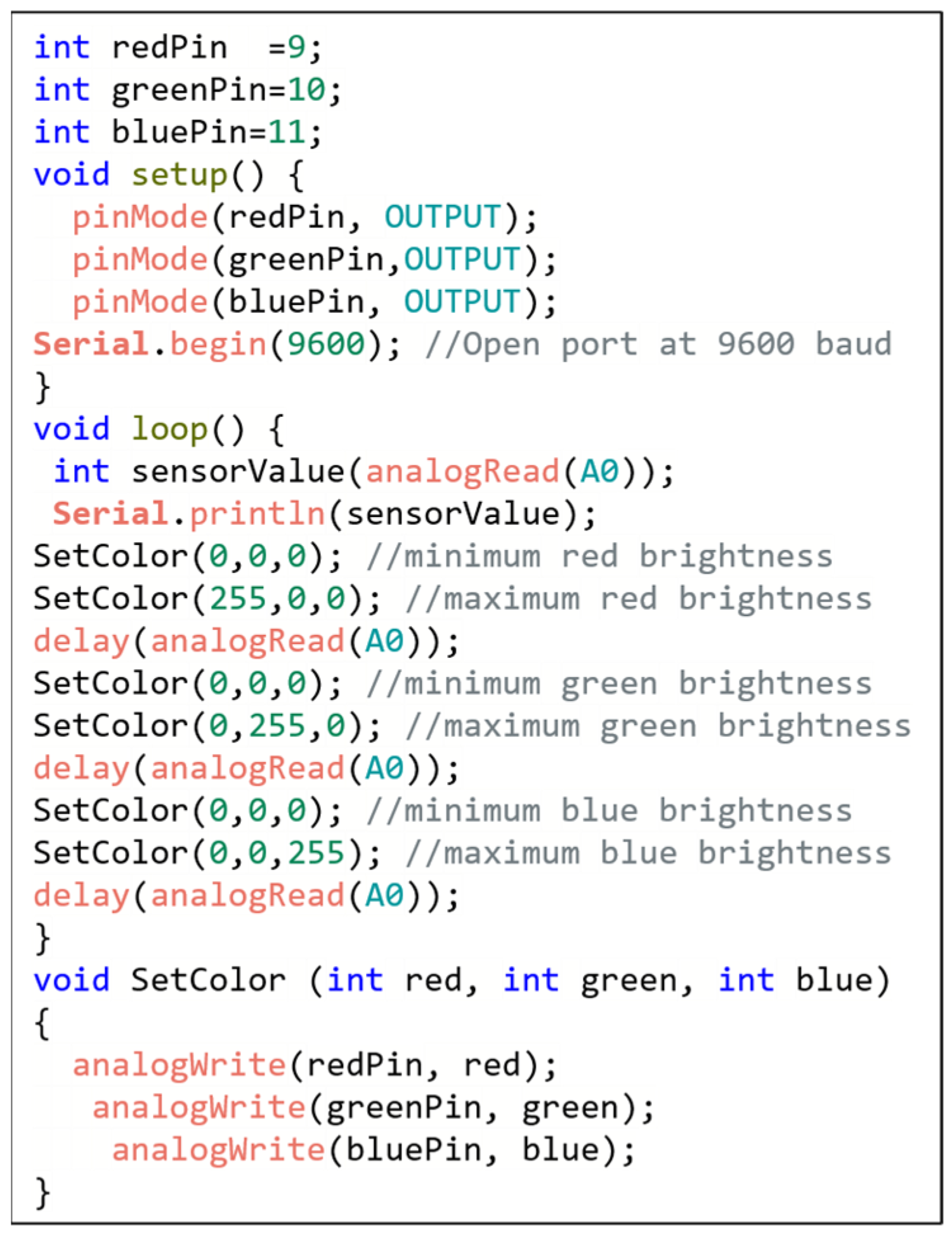
The Arduino code (Sketch) to control the cCFF device is shown in Figure 2. The Integer “sensorValue” reads value from the potentiometer (P) connected to the analog in (A0), then the function “delay ()” controls the pause length of the “sensorValue” value in milliseconds. Finally, the “Serial.printl ()” command prints the flicker rate data (in ms). The luminance modulation of the RGB LED was set between 0 and 255 (maximum brightness) for each color to provide maximum temporal contrast of the stimulus.

2.2. Ocular Wavefront

The Ocular wavefront was measured to characterize the optical quality of each participant’s eye and to explore whether the temporal resolution is affected or related to eye’s aberrations. A Laser Ray Tracing commercial device (iTrace, Tracey Technologies, Texas (USA)) was used for monocular wavefront measurements of the eye. Wavefront measurements were carried-out with the same levels of light-adaptation as the rest of the experiments. The parameters measured were the root mean square low-and high-order aberrations (LOA and HOA RMS, respectively) values and the specific number of coma, spherical aberration and trefoil HOA terms.

2.3. Photostress Recovery Time

The photo-stress recovery time (PRT) test measures the macular function and is a potential biomarker for age-related macular degeneration disease. Therefore, recovery time after macular photobleaching may detect early functional deficits of cone photoreceptors [28]. A previously reported total disability glare vision optical instrument [29] was used to measure the time recovery after retinal photo-stress for a 100 % Michelson Contrast visual target subtending 14º of the visual field.

2.4. Chromatic Critical Flicker Fusion Frequency

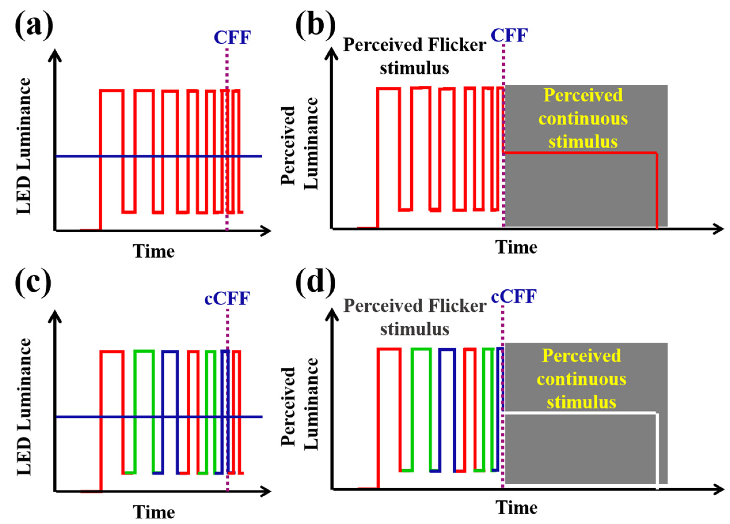
The Talbot-Plateau law establishes a psychophysical aspect of vision related to temporal integration and visual persistence [30]. The T-P law states that when a flickering visual stimulus reaches the CFF, it will be perceived as a fused (continuous) stimulus with the same brightness as a steady stimulus with the same physical luminance [31]. In that sense, the concept of chromatic critical fusion frequency (cCFF) can be illustrated as shown in Figure 3. Figure 3a shows a periodically modulated luminance of a monochromatic light source with an increasing flickering rate. Figure 3b shows the luminance that an observer would perceive before reaching the CFF limit. Once the CFF threshold is exceeded, the visual perception is that of a continuous stimulus having the same luminance. Now let us consider the case of an RGB tristimulus pulse train (Figure 3c), the perceived luminance should be a steady stimulus of the average luminance of the sampled flicker. Then, if the red, green and blue channels merge, the visual perception of a trichromatic flicker stimulus beyond the cCFF limit should be a steady white visual perception (Figure 3d).
The experimental measurements of CFFs depend on physical factors such as brightness of the stimuli, wavelength, contrast, size, eccentricity [32] or individual patient characteristics [33]. Therefore, to establish reasonable comparisons they must be under the same experimental conditions. The experimental conditions in which the experiments were carried-out in this study are summarized in Table 1.

2.5. Participants and experimental protocol

Thirty healthy young volunteers (19 ± 1 years old) participated in the study. None of them presented brain or cognitive disorders that could alter the assessment of temporal vision. Each subject, after 10-minutes of light adaptation to the ambient illumination (200 Lux), was subjected to three sequential measurements: 1) ocular wavefront; 2) chromatic critical flicker fusion frequency and 3) Photo-stress recovery time. All measurements were performed monocularly in the right eye.
The cCFF experiments were carried-out at a viewing distance of 600 mm from the RGB LED. For red, green, blue and achromatic stimuli CFF measurements, the volunteers were asked to indicate when the flickering light became steady. Regarding the chromatic CFF, participants were also asked to indicate when the tristimulus disappeared and continuous white light was perceived.

2.6. Statistical Analysis

The statistical analysis consisted of Pearson Product Moment Correlation and paired t-test to establish possible relationships and find significant differences between the studied variables, respectively. Statistical analysis and graphical representation of data were carried-out using Sigmaplot 12.0 scientific Software. The Arduino circuit designs were obtained using a JavaScript implementation available in [34].

3. Results

3.1. Chromatic Critical Flicker Fusion Frequency

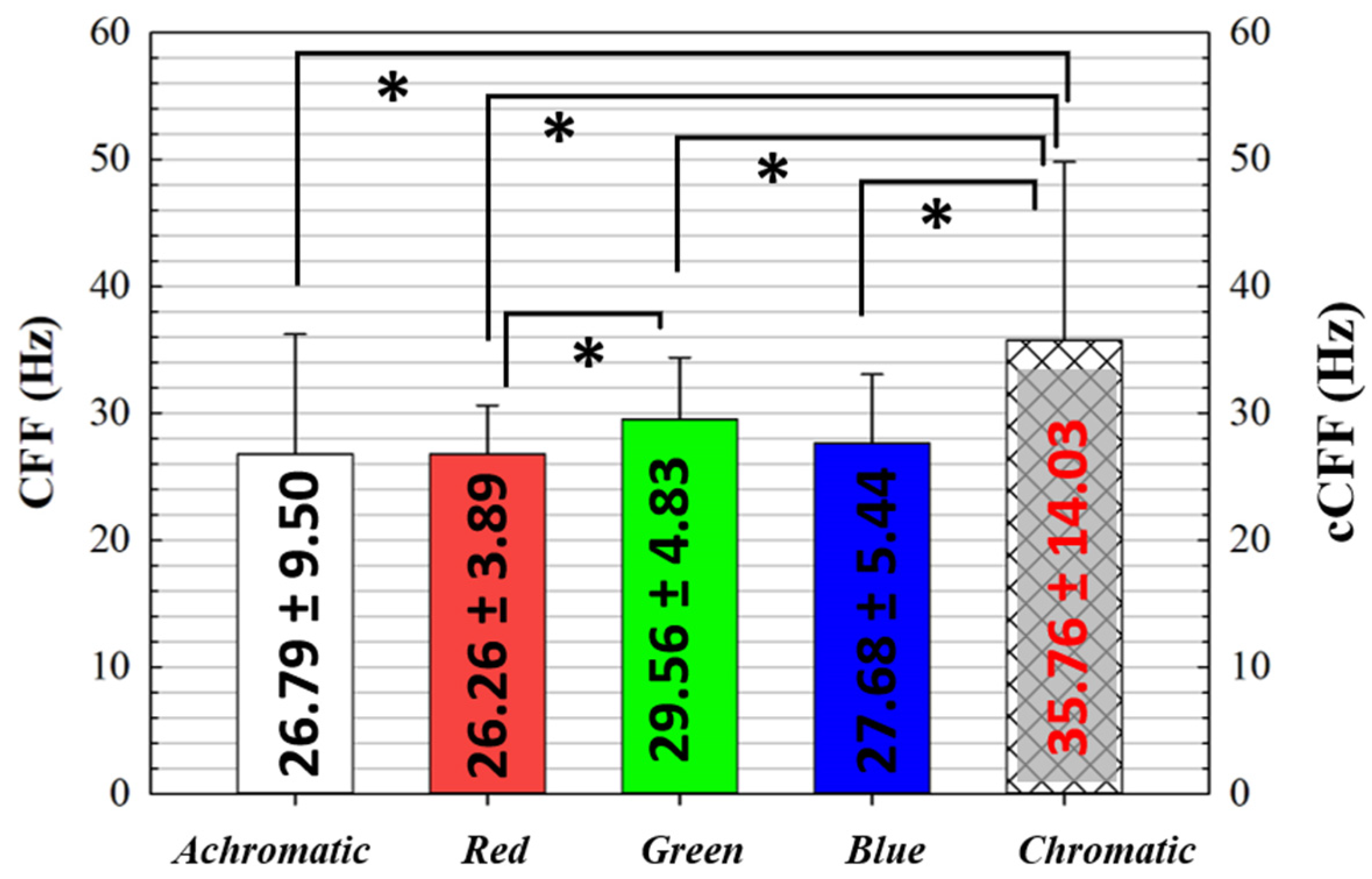
The RGB LED color can be easily modulated by modifying the Arduino Sketch, providing single color flickering for monochromatic CFF evaluation. Figure 4 compares the averaged CFF values for white, red, green and blue colors. The maximum CCF value was found for green light (CFF=29.56 ± 4.83 Hz), which was found to be statistically higher (p=0.006) than CFF for red light (CFF= 26.26 ± 3.89 Hz).
Furthermore, Figure 4 compares the cCFF value with those CFF values obtained for white, red, green and blue colors. For a flicker frequency higher than 35.76 ± 14.03 Hz, the trichromatic flickering is perceived as steady white perception. A statistical analysis revealed that the critical frequency for the perception of fused white is significantly higher than the CFF values for white, red, green and blue separated colors. In addition, the Pearson correlation coefficient did not reveal any correlation between cCFF and the CFF for achromatic and red, green and blue colors.

3.2. cCFF and Photostress Recovery Time

The above results showed that cCFF is statistically longer than CFF for red, green and blue stimuli and achromatic light, but cCFF and the mentioned CFFs did not show any statistical relationship. Furthermore, the CFF for green light was found to be statistically greater than for red stimulus. This section explores whether cCFF is related to the photo-stress recovery time (PRT). Additionally, the potential of differential color flicker to detect color vision impairment is considered [25]. The red/green modulation contrast is defined as:
R / G   C o n t r a s t   M o d u l a t i o n = C F F G C F F R C F F G + C F F R
Where CFFG and CFFR are the CFF values for green and red stimuli, respectively.
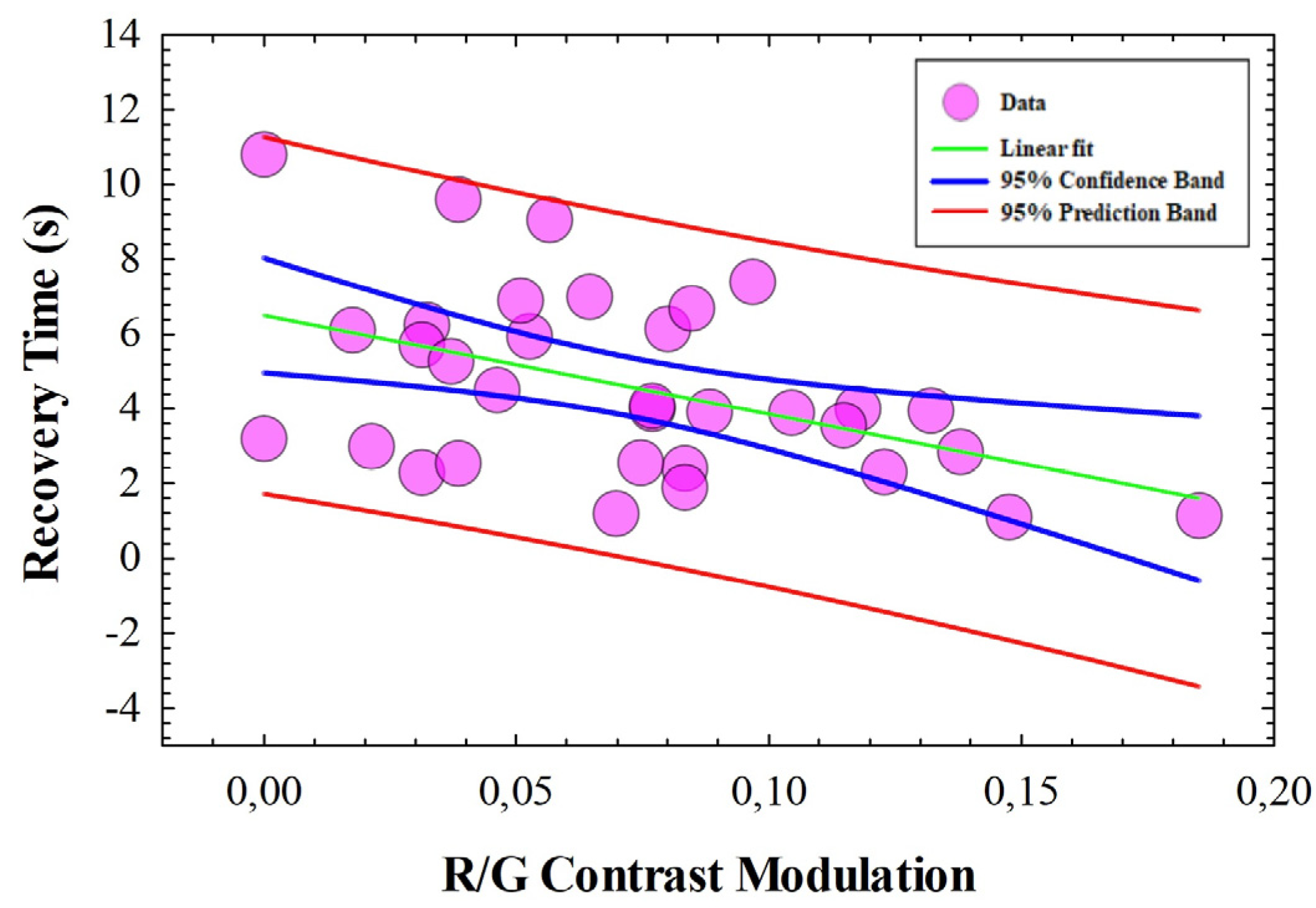
A Pearson Product Moment Correlation was performed to establish any correlation between PRT and the cCFFs shown in Figure 4. Table 2 shows the results of the statistical analysis that revealed no correlation (correlation coefficient p-value) between PRT and any CFF. However, a negative correlation was found (R2 = -0.45, p=0.008) between the PRT and the R/G contrast modulation parameter as depicted in Figure 5.

3.3. Influence of the optical quality of the eye on cCFF

Finally, this section analyzes whether spatial factors of the optical pathway of the visual processing have any implications in the temporal integration of trichromatic visual stimuli. Table 3 shows the averaged aberrometric data from wavefront measurements for all subjects involved in the study.
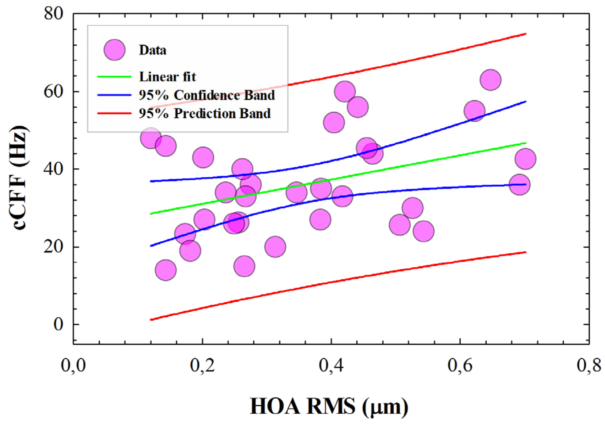
A statistical analysis revealed that cCFF is not affected by LOA (Pearson Product Moment Correlation, p=0.426). However, a positive correlation was found between HOA and cCFF (R2=0.503, p=0.004) as shown in Figure 6. These results show that high-order aberrations improve trichromatic temporal resolution of vision. Specifically, the HOA coma and trefoil terms show the greatest contribution to this effect. No relationships were found between HOA and CFF for red, green, blue, achromatic stimuli and R/G contrast modulation.

4. Discussion and Conclusions

This study presents a new cost-effective portable mini-device for trichromatic temporal resolution assessment of the human vision powered by Arduino technology. The use of pulse-width modulation (PWM) signals to control the brightness and flicker frequency of LEDs controlled by Arduino devices, may raise questions about possible limitations with respect to the use of other technologies based on data acquisition cards (DAQs). However, previous works have reported accurate irradiance output [27] and timing precision of the LEDs based on PWM signals [35]. Those precision tests demonstrated the reliability of using Arduino boards for psychophysical experiments. In contrast, cost-effective LED driving Arduino-based systems can be controlled using open-source programming environments. Additionally, Arduino devices are based on open hardware processors that can be modified by the user to extend the main capabilities of Arduino or to communicate multiple boards wirelessly.
Teiraki et al. [27] reported a LED-based visual stimulator driven by an open-source Arduino microcontroller. Among other interesting applications, they demonstrated an application to measure the density of ocular media in humans based on heterochromatic flicker photometry.
Here a new setup demonstrates an application for assessing the temporal resolution of visual performance using a flickering trichromatic stimulus. CFF was measured for the individual red, green and blue channels and for white light. The highest CFF threshold for green light was found to be significantly higher than for red light.
Regarding the cCFF, the integration frequency was significantly higher than the rest of the color stimuli. That is, while the visual perception of flickering of a monochromatic stimulus would disappear, a trichromatic stimulus flickering at the same frequency would continue to be visible.
Considering the association between the human visual perception of high-frequency flicker stimuli with cortical mechanisms [36], exploring the human visual perception of trichromatic flicker at higher temporal frequencies could help better understand the outcomes of psychophysical tasks and isolate those contributions from the magnocellular pathway.
Section 3.2 explored the relationships between the chromatic temporal resolution of the visual system and macular function measured by photo-stress recovery time (PRT).
No relationships were found between CFFs for red, green, blue light, cCFF and PRT. However, a statistical negative correlation was found between the red/green modulation contrast and PRT. Theoretically, the presence of macular pigment is to enhance the visual performance in glare vision conditions [37]. In that sense, a higher macular pigment density will results in shorter PRTs. The results reported here showed that PRT and cCFF are independent factors of the visual function. Therefore, the contrast modulation parameter defined for red and green CFF measurements appears to be related to the macular pigment density rather than temporal processing of color vision.
Finally, Section 3.3 studied the influence of ocular aberrations on the chromatic CFF. No relationships were found between cCFF and low-order aberrations (i.e., refractive errors), however a positive linear correlation was found between HOA and cCFF. The results demonstrate that high-order ocular aberrations improve the temporal resolution of the visual function.
Spatial contrast sensitivity and HOA are inversely correlated; One of the main mechanisms that degrade visual quality in corneal diseases such as keratoconus is the presence of increased HOA [38]. Furthermore, HOA plays a compensatory mechanism in spatial contrast sensitivity when intraocular scattering effects are also significant [39]. However, the influence of ocular aberrations on temporal aspects of vision is lacking in the literature.
A possible explanation for the relationship between HOA and cCFF can be found in the spatiotemporal mechanisms in human vision processing: spatial and temporal information interact with the magnocellular and parvocellular pathways, respectively. More specifically, there is an inhibitory parvo-magnocellular interaction: improving temporal resolution deteriorates spatial resolution [40]. Therefore, it can be concluded that while on the one hand HOA degrades the optical quality of the eye (in terms of spatial resolution), on the other hand it improves the temporal resolution.
Recently, it has been reported that the human visual cortex is sensitive to flicker stimuli that induce changes in neural activity [41]. In neurodegenerative diseases such as Alzheimer’s, the brain undergoes from electrophysiological changes that are susceptible to reacting to neurostimulation therapies based on chromatic flickering at 40 Hz [42]. One of the drawbacks of studying brain activity with perceived flickering is a high level of patient discomfort [43]. The results reported here showed a mean value for cCFF of 37.76 ± 14.03 Hz, then a trichromatic flicker stimulus at 40 Hz falls inside the steady perceived stimuli regime according to the Talbot´s law, allowing brain activity to be studied at that critical frequency while the patient looks at a continuous white visual stimulus.
To conclude, a new cost-effective mini-device for the study of polychromatic temporal resolution of visual function based on Arduino technology is presented. The cCFF device allowed us the study of the critical flicker fusion of a chromatic stimulus, providing a steady white visual perception beyond the critical frequency limit. The contrast modulation of CFF values for red and green stimuli was found to be related to photo-stress recovery time and therefore more related to macular pigment density than to temporal resolution of color vision. Furthermore, increased ocular HOA improves the temporal resolution of the visual function.
Future work will include characterization of the temporal sensitivity of color vision using the chromatic CFF device in patients with retinal visual impairment.

Funding

This research received no external funding.

Institutional Review Board Statement

The study was conducted in accordance with the Declaration of Helsinki, and approved by the Ethics Committee of the Health Sciences Institute of Aragon, Spain. (protocol code: C.P.-C.I. PI20/377, date of approval: 14 July 2020).

Informed Consent Statement

Informed consent was obtained from all subjects involved in the study.

Data Availability Statement

Dataset is available upon reasonable request.

Conflicts of Interest

The authors declare no conflicts of interest.

References

  1. Z. E. Snow. Visual Acuity as a Measurement of Visual Function. Methods Mol. Biol. 2023, 2560, 141–144. [Google Scholar]
  2. Bennett, C.R.; Bex, P.J.; Bauer, C.M.; Merabet, L.B. The Assessment of Visual Function and Functional Vision. Semin. Pediatr. Neurol. 2019, 31, 30–40. [Google Scholar] [CrossRef] [PubMed]
  3. P. Lennie. “The physiological basis of variations in visual latency. Vis. Res. 1981, 21, 815–824. [Google Scholar] [CrossRef]
  4. M. Coltheart. “The persistences of vision”. Philos. Trans. R. Soc. Lond. B. Biol. Sci. 1980, 290, 57–69. [Google Scholar] [CrossRef] [PubMed]
  5. Wutz, A.; Muschter, E.; van Koningsbruggen, M.G.; Weisz, N.; Melcher, D. Temporal Integration Windows in Neural Processing and Perception Aligned to Saccadic Eye Movements. Curr. Biol. 2016, 26, 1659–1668. [Google Scholar] [CrossRef] [PubMed]
  6. Borghuis, B.G.; Tadin, D.; Lankheet, M.J.; Lappin, J.S.; van de Grind, W.A. Temporal Limits of Visual Motion Processing: Psychophysics and Neurophysiology. Vision 2019, 3, 5. [Google Scholar] [CrossRef] [PubMed]
  7. A. Bowling, W. Lovegrove. Two components to visible persistence: Effects of orientation and contrast Vis. Res. 1981, 21, 1241–1251.
  8. Yeonan-Kim, J.; Francis, G. Retinal spatiotemporal dynamics on emergence of visual persistence and afterimages. Psychol. Rev. 2019, 126, 374–394. [Google Scholar] [CrossRef] [PubMed]
  9. Gibbon, J.; Malapani, C.; Dale, C.L.; Gallistel, C. Toward a neurobiology of temporal cognition: advances and challenges. Curr. Opin. Neurobiol. 1997, 7, 170–184. [Google Scholar] [CrossRef] [PubMed]
  10. Melcher, D.; Wutz, A.; Drewes, J.; Fairhall, S. The Role of Temporal Integration Windows in Visual Perception. Procedia - Soc. Behav. Sci. 2014, 126, 92–93. [Google Scholar] [CrossRef]
  11. Chokron, S.; Kovarski, K.; Dutton, G.N. Cortical Visual Impairments and Learning Disabilities. Front. Hum. Neurosci. 2021, 15. [Google Scholar] [CrossRef] [PubMed]
  12. T. Muth, J.D. Schipke, A.K. Brebeck, S. Dreyer. Assessing Critical Flicker Fusion Frequency: Which Confounders? A Narrative Review. Medicina (Kaunas), 2023, 59, 800.
  13. R. S. Brenton, H.S. R. S. Brenton, H.S. Thompson, C. Maxner. Critical flicker frequency: a new look at an old test [internet]. In: New Methods of Sensory Visual Testing. New York: Springer; 1989, 29–52.
  14. R.S. Brenton, H.S. R.S. Brenton, H.S. Thompson, C. Maxner. Critical flicker frequency: a new look at an old test. In: New Methods of Sensory Visual Testing. New York: Springer;1989, 29–52.
  15. Titcombe, A.F.; Willison, R.G. FLICKER FUSION IN MULTIPLE SCLEROSIS. J. Neurol. Neurosurg. Psychiatry 1961, 24, 260–265. [Google Scholar] [CrossRef] [PubMed]
  16. M. L.Daley, R.L.Swank, C.M. Ellison. Flicker fusion thresholds in multiple sclerosis: a functional measure of neurological damage. Arch Neurol. 1979, 36, 292–295. [Google Scholar] [CrossRef] [PubMed]
  17. Brandt, A.U.; Martinez-Lapiscina, E.H.; Nolan, R.; Saidha, S. Monitoring the Course of MS With Optical Coherence Tomography. Curr. Treat. Options Neurol. 2017, 19, 15. [Google Scholar] [CrossRef] [PubMed]
  18. Galvin, R.J.; Regan, D.; Heron, J.R. IMPAIRED TEMPORAL RESOLUTION OF VISION AFTER ACUTE RETROBULBAR NEURITIS. Brain 1976, 99, 255–268. [Google Scholar] [CrossRef] [PubMed]
  19. J. Fu, Y. Wang, S. Tan, et al. The clinical application of critical flicker fusion frequency in demyelinating optic neuritis. Adv. Opthalamol. Prac. Res. 2021, 1, 100011. [Google Scholar]
  20. K. Prabu, A. Omprakash, M. Kuppusamy, et al. How does cognitive function measured by the reaction time and critical flicker fusion frequency correlate with the academic performance of students? BMC Med Educ. 2020, 20, 507. [Google Scholar]
  21. W. W. Piispanen, R.V. Lundell, L.J. Tuominen,et al. Assessment of Alertness and Cognitive Performance of Closed Circuit Rebreather Divers With the Critical Flicker Fusion Frequency Test in Arctic Diving Conditions. Front Physiol. 2021, 12, 722915. [Google Scholar] [CrossRef]
  22. Gegenfurtner, K.R. Cortical mechanisms of colour vision. Nat. Rev. Neurosci. 2003, 4, 563–572. [Google Scholar] [CrossRef]
  23. Lee, B.B. Paths to colour in the retina. Clin. Exp. Optom. 2004, 87, 239–248. [Google Scholar] [CrossRef]
  24. McKeefry, D.; Murray, I.; Kulikowski, J. Red–green and blue–yellow mechanisms are matched in sensitivity for temporal and spatial modulation. Vis. Res. 2001, 41, 245–255. [Google Scholar] [CrossRef] [PubMed]
  25. Gregori, B.; Papazachariadis, O.; Farruggia, A.; Accornero, N. A differential color flicker test for detecting acquired color vision impairment in multiple sclerosis and diabetic retinopathy. J. Neurol. Sci. 2011, 300, 130–134. [Google Scholar] [CrossRef] [PubMed]
  26. Xu, G.; Luo, Y.; Qi, H.; Liu, S.; Fu, J.; Ye, Z.; Li, Z. Trichromatic critical flicker frequency as potential visual test in cataract and macula disease patients. Graefe's Arch. Clin. Exp. Ophthalmol. 9. [CrossRef]
  27. P. Teiraki, R.P. Najjar, H. Malkki, et al. “An inexpensive Arduino-based LED stimulator system for vision research” J Neuroscience Methods. 2012, 211, 227–236.
  28. Rodriguez, J.D.; Lane, K.; Hollander, D.; Shapiro, A.; Saigal, S.; Hertsenberg, A.J.; Wallstrom, G.; Narayanan, D.; Angjeli, E.; Abelson, M.B.; et al. Cone photoreceptor macular function and recovery after photostress in early non-exudative age-related macular degeneration. Clin. Ophthalmol. 2018, ume 12, 1325–1335. [Google Scholar] [CrossRef]
  29. Ávila, F.J.; Casado, P. Optical instrument for the study of time recovery from total disability glare vision. Appl. Opt. 2022, 61, 2438–2443. [Google Scholar] [CrossRef]
  30. Nelson, T.M.; Bartley, S.H. The Talbot-plateau law and the brightness of restricted numbers of photic repetitions at CFF. Vis. Res. 1964, 4, 403–411. [Google Scholar] [CrossRef] [PubMed]
  31. Greene, E.; Morrison, J. Evaluating the Talbot-Plateau law. Front. Neurosci. 2023, 17. [Google Scholar] [CrossRef] [PubMed]
  32. Eisen-Enosh, A.; Farah, N.; Burgansky-Eliash, Z.; Polat, U.; Mandel, Y. Evaluation of Critical Flicker-Fusion Frequency Measurement Methods for the Investigation of Visual Temporal Resolution. Sci. Rep. 2017, 7, 1–9. [Google Scholar] [CrossRef]
  33. M. Liu, Y. Yan, Q. Xue, L. Gong. “The research and alaysis factors affecting critical flicker frequency” Procedia Manuf. 2015, 3, 4279–4286.
  34. https://github.com/wokwi/avr8js.
  35. D’ausilio, A. Arduino: A low-cost multipurpose lab equipment. Behav. Res. Methods 2011, 44, 305–313. [Google Scholar] [CrossRef] [PubMed]
  36. Skottun, B.C. On using very high temporal frequencies to isolate magnocellular contributions to psychophysical tasks. Neuropsychologia 2013, 51, 1556–1560. [Google Scholar] [CrossRef]
  37. Stringham, J.M.; Garcia, P.V.; Smith, P.A.; Hiers, P.L.; McLin, L.N.; Kuyk, T.K.; Foutch, B.K. Macular Pigment and Visual Performance in Low-Light Conditions. 56, 2459; 68. [Google Scholar] [CrossRef]
  38. Shneor, E.; Piñero, D.P.; Doron, R. Contrast sensitivity and higher-order aberrations in Keratoconus subjects. Sci. Rep. 2021, 11, 1–9. [Google Scholar] [CrossRef] [PubMed]
  39. Zhao, J.; Xiao, F.; Zhao, H.; Dai, Y.; Zhang, Y. Effect of higher-order aberrations and intraocular scatter on contrast sensitivity measured with a single instrument. Biomed. Opt. Express 2017, 8, 2138–2147. [Google Scholar] [CrossRef] [PubMed]
  40. Peñaloza, B.; Ogmen, H. Effects of spatial attention on spatial and temporal acuity: A computational account. Attention, Perception, Psychophys. 2022, 84, 1886–1900. [Google Scholar] [CrossRef] [PubMed]
  41. Mankowska, N.D.; Marcinkowska, A.B.; Waskow, M.; Sharma, R.I.; Kot, J.; Winklewski, P.J. Critical Flicker Fusion Frequency: A Narrative Review. Medicina 2021, 57, 1096. [Google Scholar] [CrossRef] [PubMed]
  42. S. De. Focused arousal and the cognitive 40-hz event-related potentials: Differential diagnosis of Alzheimer’s disease. Progress Clin. Biol. Res. 1989, 317, 79–94. [Google Scholar]
  43. M. P. Agger, E.R. Danielsen, M.S. Carstensen, et al. Safety, Feasibility, and Potential Clinical Efficacy of 40 Hz Invisible Spectral Flicker versus Placebo in Patients with Mild-to-Moderate Alzheimer’s Disease: A Randomized, Placebo-Controlled, Double-Blinded, Pilot Study. J Alzheimers Dis. 2023, 92, 653–665. [Google Scholar]
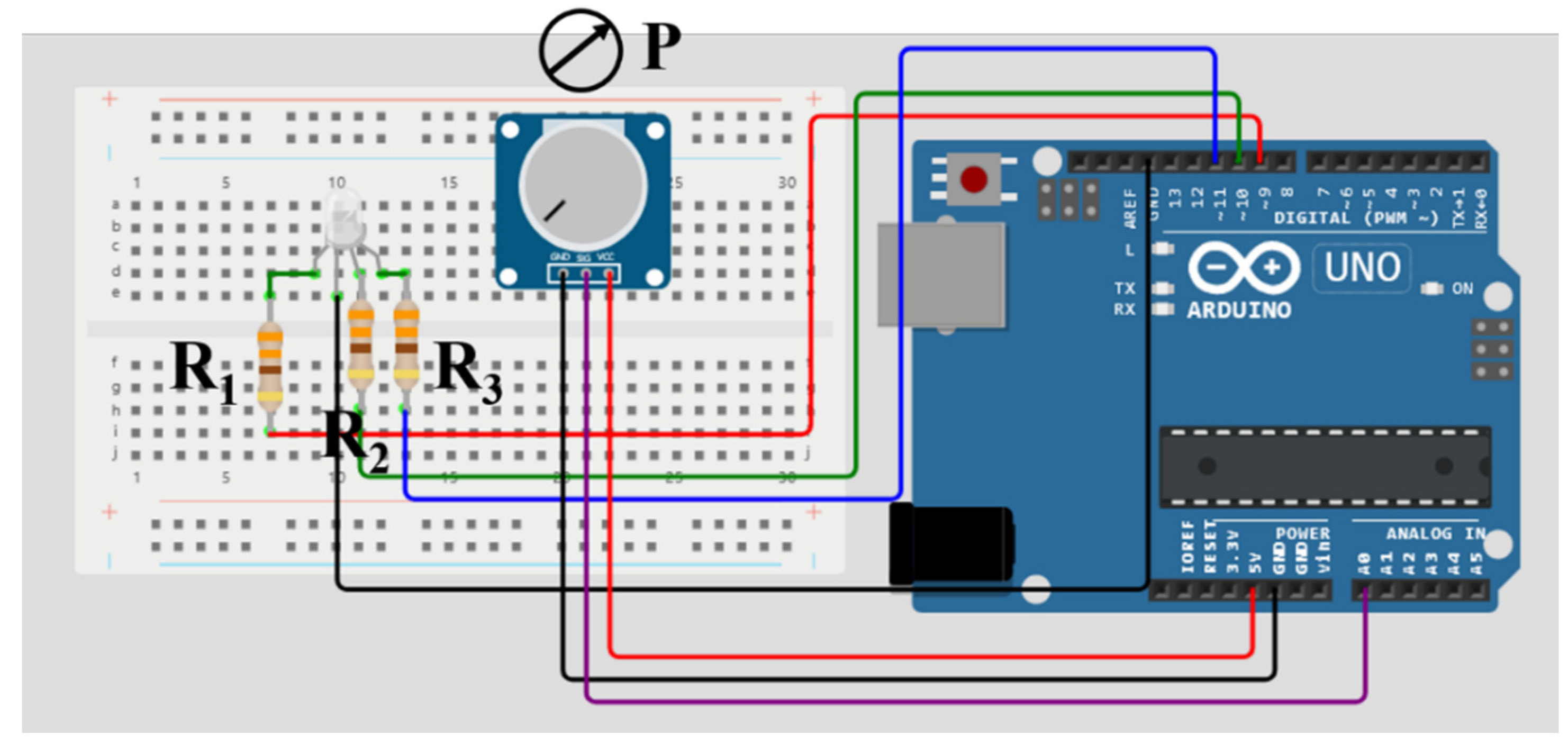
Figure 1. Circuit design for the trichromatic critical flicker frequency fusion device. R1, R2, and R3 are 330 Ω resistors and P a tunable potentiometer.
Preprints 109927 g001
Figure 2. Arduino Sketch to control the cCFF device.
Preprints 109927 g002
Figure 3. Illustration of the Talbot´s law for monochromatic (a, b) and for trichromatic visual stimuli (c, d).
Preprints 109927 g003
Figure 4. mean CFF values for achromatic, red, green and blue colors and chromatic stimuli (lattice bar). The asterisks indicate those groups for which significant differences were found with a significance level of p <0.05.
Preprints 109927 g004
Figure 5. Recovery time as a function of the R/G contrast modulation. The green line, blue and red bands, correspond to the best linear fit, confident and prediction bands, respectively.
Preprints 109927 g005
Figure 6. cCCF as a function of HOA RMS.
Preprints 109927 g006
Table 1. Experimental conditions for the CFF measurements.
Parameter Measure
Luminous intensity [red] 2600 mcd
Luminous intensity [green] 2000 mcd
Luminous intensity [blue] 1800 mcd
Distance of observation 600 mm
Ambient illumination 200 Lux
Eccentricity Foveal vision
CFF red CFF for red stimulus (Hz)
CFF green CFF for green stimulus (Hz)
CFF blue CFF for blue stimulus (Hz)
CFF achromatic CFF for white light stimulus (Hz)
Chromatic CFF (cCFF) CFF for mixed red, green and blue lights.
Table 2. Correlation results (p value) of the PRT versus achromatic, red, green, blue, trichromatic (RGB) stimuli, and R/G contrast modulation.
Achromatic RED GREEN BLUE RGB R/G
PRT p=0.836 p=0.812 p=0.506 p=0.772 p=0.731 p=0.008
Table 3. Root mean square for low- (LOA RMS) and high-order aberrations (HOA RMS), and individual contribution of the coma, spherical aberration (SA) and trefoil high-order terms.
LOA RMS HOA RMS Coma SA Trefoil
2.12 ± 2.52 μm 0.36±0.17 μm 0.21±0.14 μm 0.10±0.07 μm 0.18±0.11 μm
Disclaimer/Publisher’s Note: The statements, opinions and data contained in all publications are solely those of the individual author(s) and contributor(s) and not of MDPI and/or the editor(s). MDPI and/or the editor(s) disclaim responsibility for any injury to people or property resulting from any ideas, methods, instructions or products referred to in the content.
Copyright: This open access article is published under a Creative Commons CC BY 4.0 license, which permit the free download, distribution, and reuse, provided that the author and preprint are cited in any reuse.
Prerpints.org logo

Preprints.org is a free preprint server supported by MDPI in Basel, Switzerland.

Subscribe

© 2024 MDPI (Basel, Switzerland) unless otherwise stated