Preprint
Article

Dogs as Sentinels for Emergent Pathogens: Serological Evidence in a Preserved Area in Brazil

Altmetrics

Downloads

179

Views

158

Comments

0

Submitted:

27 June 2024

Posted:

28 June 2024

You are already at the latest version

Alerts
Abstract
For many tick-borne organisms, dogs may be utilized as sentinel hosts to estimate the risk of human infection. The detection of antibodies in these animals indicates the circulation of pathogens in each location for a set period of time. The standard protocol for the surveillance of tick-borne diseases established by the Brazilian National Reference Laboratory for Rickettsiosis at the Ezequiel Dias Foundation (FUNED) includes testing the seropositivity of dogs for Rickettsia spp. and Ehrlichia canis. Dog serum samples were collected during FUNED’s surveillance protocol in a preserved area in Brazil. Despite Anaplasma phagocytophilum being endemic in Brazil, this agent is not included in FUNED’s standard protocol. To determine if A. phagocytophilum should be included in the standard testing protocol, a collaboration with Fuller Laboratories was established and the seropositivity for A. phagocytophilum was tested. The estimated prevalence for singular infections: 25% Rickettsia spp.; 52.3% E. canis; and 70.5% A. phagocytophilum. The estimated prevalence for co-infections: 17% Rickettsia spp. and E. canis; 18.2% Rickettsia spp. and A. phagocytophilum; 39.8% E. canis and A. phagocytophilum; and 13.6% Rickettsia spp., E. canis, and A. phagocytophilum. These results confirmed a significant presence of A. phagocytophilum and establishes a potential threat upon the public health.
Keywords: 
Subject: Biology and Life Sciences  -   Animal Science, Veterinary Science and Zoology

1. Introduction

The (re)emergence of zoonoses has been linked to significant ecological changes driven by factors such as globalization, habitat modification, urbanization, and climate change, among others [1,2]. Climate change generates both direct and indirect impacts on human health. Indirectly, it causes imbalances in ecosystems, biodiversity, and hydrological/biogeochemical cycles, thereby influencing the morbidity and mortality profiles of diseases, such as those transmitted by vectors. This raises significant concerns in the global health sector, posing a substantial threat to public health as well as to animal health [3,4,5].
Following insects, ticks are the second most capable vectors for transmitting pathogens to humans. They can carry and transmit a diverse range of etiological agents, including bacteria, viruses, protozoa, and helminths, during their blood meals on vertebrate hosts [6,7,8]. This remarkable ability to disseminate pathogens, combined with their indirect impacts, has led to an increase in Tick-Borne Diseases (TBD), which are now recognized as the second leading cause of health issues in both human and veterinary health [9]. Additionally, these diseases have been spreading globally, even reaching regions previously free of such pathogens. This growing prevalence underscores the global relevance of TBD within the framework of One Health, emphasizing the interconnectedness of human, animal, plant, and ecosystem health [10,11,12,13,14,15].
In Brazil, the main zoonosis transmitted by ticks is the Spotted Fever (SF), which can range from mild forms, when associated with Rickettsia parkeri Mata Atlântica strain, to severe forms with a high lethality rate when the agent is the bacterium Rickettsia rickettsii, a fact related to epidemiological scenarios that are quite variable [16,17]. And due to the increase in the number of human cases, SF surveillance in Brazil was markedly influenced by four health actions: I) Brazilian Ministry of Health (BMH) started to consider it as a notifiable disease; II) SF became part of the Notifiable Diseases Information System (SINAN), an agency that aims to collect, gather and disseminate data among the health surveillance network; III) formation of the National Network for Environmental Surveillance for Spotted Fever and other rickettsiosis, initiating training in environmental surveillance of rickettsiosis and IV) SF and other rickettsiosis became part of the list of diseases of immediate compulsory notification, and must be notified within 24 hours [16].
Due to the above, SF surveillance in Brazil encompasses both epidemiological and environmental monitoring, with the following objectives: A) early detection and treatment of suspected cases to reduce lethality; B) investigate and control of outbreaks through the implementation of control measures; C) understand the distribution of the disease by location, time, and affected individuals; D) identify and investigate probable sites of infection (IPLs); and E) recommend the adoption of control and prevention strategies [16].
Additionally, the BMH evaluates the occurrence of another TBD: ehrlichiosis. This disease is caused by bacteria of the genus Ehrlichia sp., which has already been recognized in cats, wild animals, humans and dogs, and is widely detected throughout the country [18,19]. The main species of Ehrlichia in dogs is Ehrlichia canis. E. canis is highly endemic in many regions of Brazil [20] due to the geographic distribution of tick vectors, which contributes directly to the higher prevalence of ehrlichiosis that has been observed [21].
Despite the reports of people affected by E. canis [22,23], the main species of human importance is Ehrlichia chaffeensis, which causes human monocytic ehrlichiosis (HME), a pathogen already identified in animals in Brazil, especially cervids [24]. Due to its zoonotic character, surveillance of circulation in animals becomes relevant in the context of single health.
Another TBD caused by bacteria circulating in the country belongs to the genus Anaplasma sp.. Granulocytic Anaplasmosis is a disease that affects dogs, horses, cats, ruminants and humans, which can be infected by the emerging intracellular bioagent Anaplasma phagocytophilum [25,26,27,28]. The occurrence of anaplasmosis in dogs has been geographically associated with Human Granulocytic Anaplasmosis (HGA) [29], as well as the occurrence of the presence of ixodid ticks in the circulation of A. phagocytophilum in Brazil, despite the lack of identification of the vector responsible for transmission in the tropical region [30]. Although A. phagocytophilum has already been detected in several studies in Brazil [26,27,28,29,30,31], this bioagent is not currently included in the standard surveillance protocol in Brazil.
As the tick Rhipicephalus sanguineus sensu lato is involved in the transmission of Ehrlichia sp. and some species of Anaplasma sp. [32,33], co-infection of ehrlichiosis and anaplasmosis in routine small animal clinical testing is not uncommon, and this co-infection is reported with a prevalence of 9.1% in symptomatic dogs through rapid routine testing [34].
Given the persistent and growing challenges in epidemiology, alongside the knowledge of the interaction between hosts, pathogens and vectors, which is at the heart of the study of TBD, a surveillance method is needed. Within this paradigm, seroepidemiological surveys in sentinel hosts – which have been used as an effective tool and play a crucial role, since sentinels do not pose a threat of direct transmission of diseases to humans – offer valuable insights, which are aligned with the objectives of epidemiological and environmental surveillance in the country [35,36,37,38].
The use of dogs as sentinel hosts for TBD is a well-established surveillance strategy in the scientific literature due to the fact that dogs are susceptible to TBD infections; survive these infections; exhibit detectable and measurable immune responses; and most importantly they closely interact with humans [35]. This strategy has been employed in numerous serological surveys worldwide [39,40,41]. In Brazil, dogs are chosen as sentinel species due to their involvement in infection scenarios, both directly and indirectly, within epidemic and enzootic cycles [36,42,43,44]. Consequently, they serve as bioindicators of the epidemiological status; the detection of antibodies in these animals indicates the circulation of pathogens in specific localities and time periods, aiding in the tracking of TBD risks and monitoring infection trends [45,46,47].
As previously noted, although A. phagocytophilum has been detected in several studies in Brazil [26,27,30,31], this bioagent is not currently included in the standard surveillance protocol of the National Reference Laboratory for Spotted Fever and other Rickettsiosis of the Ezequiel Dias Foundation (FUNED). This study, a collaboration between FUNED and Fuller Laboratories in California, aimed to provide scientifically based information to support the implementation of strategic public health actions. As such, blood samples were obtained from dogs in Serra do Cipó National Park (SCNP) in Minas Gerais, Brazil, and were analyzed serologically for Rickettsia spp., E. canis, and A. phagocytophilum.

2. Materials and Methods

2.1. Fields of Study

This cross-sectional study took place in SCNP, within the state of Minas Gerais, about 100 km from Belo Horizonte (the capital of Minas Gerais) [48], and in the surrounding Retiro and Açude communities, with access to the Areias and Alto Palácio entrances (Figure 1). The SCNP is a Brazilian conservation unit and known as “Jardim do Brasil”, a title given by the landscape architect Burle Marx. With a total area of 33,800 hectares, the unit currently protects several endangered species of Brazilian fauna and flora in the Cerrado biome. The rugged topography, with altitudes ranging from 700 to 1,670 meters in altitude, is located in the southern portion of the Serra do Espinhaço, an important divider between two major Brazilian hydrographic basins: the São Francisco and the Doce River [48]. The SCNP makes it possible to carry out scientific research, recreation and ecological tourism; and has seen 342,476 visitors within the last 5 years [48]. In addition to the purpose of environmental conservation, many traditional communities that have direct contact with preserved environmental aspects are found in the SCNP region, an important factor considering the public health measures expanded to the reality of possible pathogens circulating in the region.

2.2. Obtaining and Processing Samples

Six routine field collection campaigns were carried out by professionals from the municipality between March 2021 and February 2022, resulting in 88 blood samples from dogs (domiciled, semi-domiciled, and stray). These samples were sent to the National Reference Laboratory for Spotted Fever and other Rickettsiosis of FUNED, for antibody research utilizing the Indirect Immunofluorescence Assay (IFA), which is the gold standard methodology according to BMH [16]. IFA analysis for Rickettsia spp., and E. canis followed FUNED’s routine protocol. IFA analysis for A. phagocytophilum followed the instructions of the kit manufacturer: Fuller Laboratories, California, United States of America [49].
The samples were received, sorted, catalogued and stored in a freezer at -80°C until the beginning of the analysis procedures. The samples were thawed, homogenized and diluted in phosphate-buffered saline solution (PBS) pH 7.4, in individualized microtubes with the respective dilutions for Rickettsia spp. 1:64, E. canis 1:40 and A. phagocytophilum 1:80. Then, 25 μl of diluted serum was added to the wells of the respective slides for Rickettsia spp., E. canis and A. phagocytophilum. The slides were incubated at 37ºC for 30 minutes, washed twice in PBS for 5 minutes, and dried at room temperature. Then, 25 μl of anti-IgG antibody specific for each animal species, labelled with fluorescein, prepared at a 1:100 dilution, was added to each well; incubating again for 30 minutes at 37ºC. Next, the slides were washed twice for 5 minutes with PBS and mounted with buffered glycerin and coverslips; then examined under a UV microscope at 400X magnification with fluorescein filters. Antibody titer of 1:64 or higher was considered reactive for Rickettsia spp., 1:40 or higher was considered reactive for E. canis, and 1:80 or higher was considered reactive for A. phagocytophilum.

2.3. Statistical Analysis

It was calculated based on the report of the occurrence of the frequency of serological findings (prevalence with a 95% confidence interval), as well as from the analysis by means of specific tests from the statistical program R, Core Team (2024). R: A language and environment for statistical computing. R Foundation for Statistical Computing, Vienna, Austria. URL https://www.R-project.org/ (https://www.R-project.org/) [50]. A bubble chart generated to illustrate the distribution of the prevalence of simple infections and co-infections is seen in Figure 4. A chi-square test of independence was performed to examine the relation between co-infections of A. phagocytophilum and E. canis, or between co-infections of E. canis and Rickettsia spp., or between co-infections of A. phagocytophilum and Rickettsia spp.

3. Results

Analysis by IFA demonstrated that there was a substantial percentage of sero-reactivity toward the bioagents that was observed in the sentinel hosts within the SCNP (Figure 1).
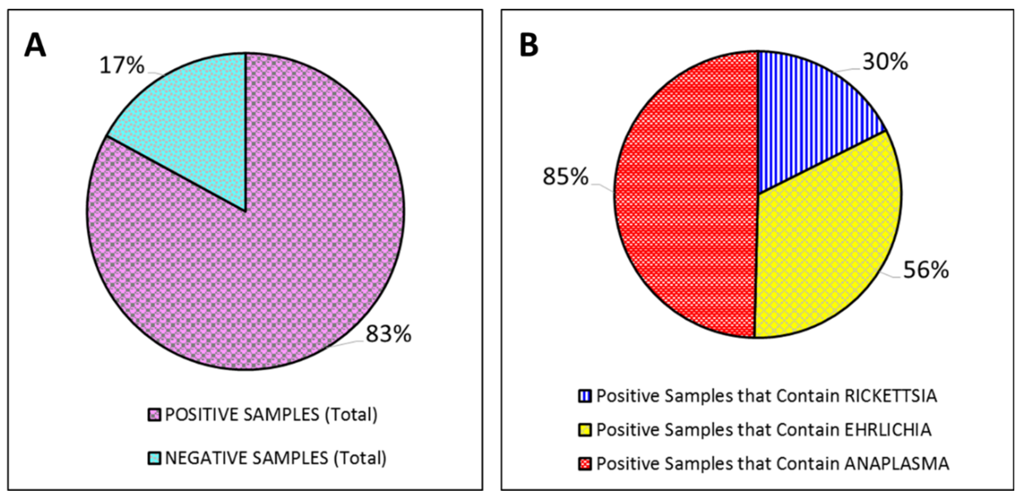
Of the 88 dogs tested, 83% (73/88 samples) tested positive for infections with one or more of the three infectious agents: Rickettsia spp., E. canis, and/or A. phagocytophilum, and 17% (15/88 samples) tested negative for infections with any of the three infectious agents (Figure 2A). Within the subset of the positive samples seen in Figure 2A, 30% (22/73 samples) tested positive for having Rickettsia spp. present, 56% (41/73 samples) tested positive for having E. canis present, and 85% (62/73 samples) tested positive for having A. phagocytophilum present (Figure 2B).
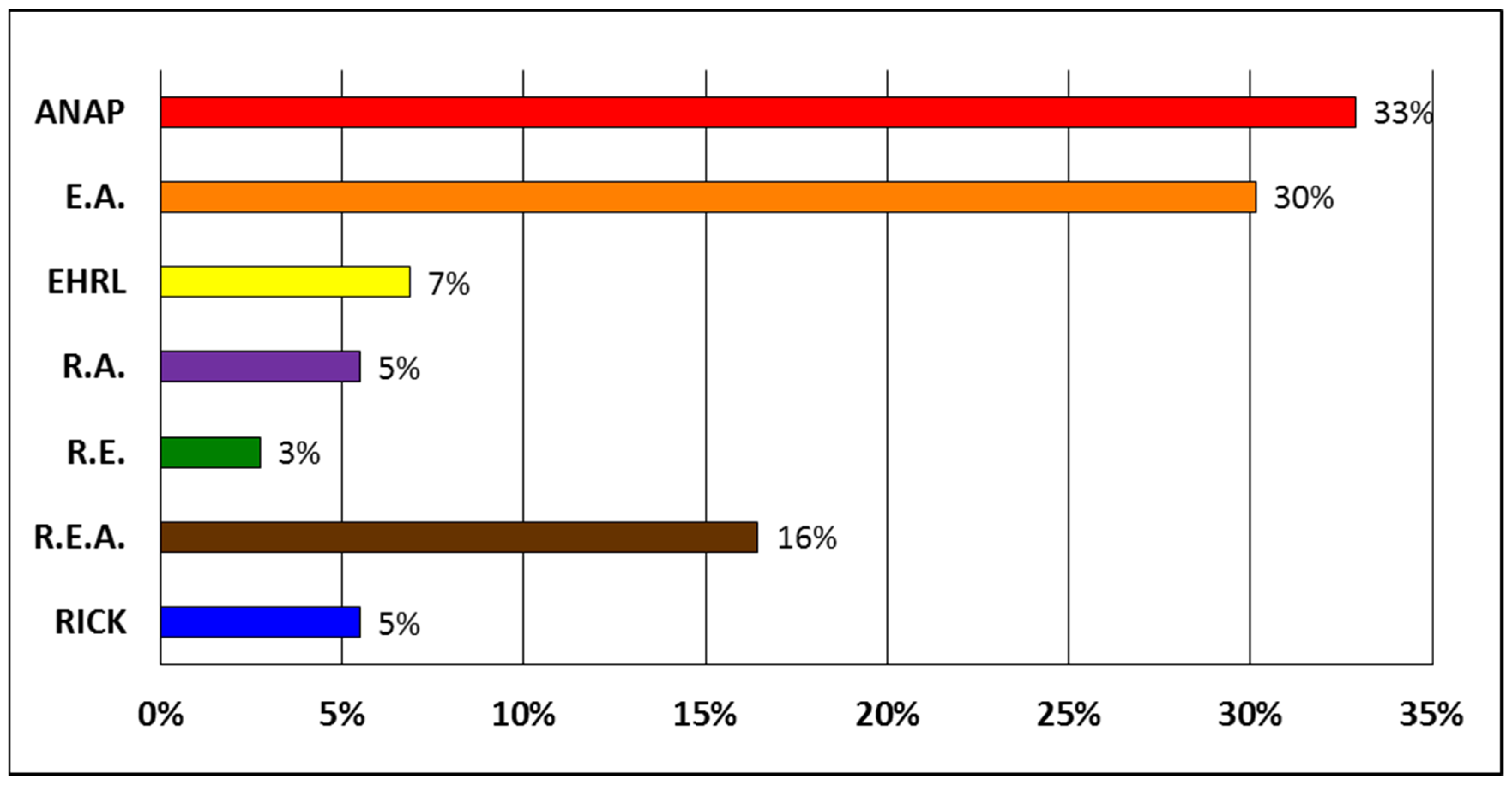
The percentage of positive samples with either single infections or co-infections of Rickettsia spp., E. canis, and/or A. phagocytophilum is demonstrated in Figure 3. Within the subset of the positive samples seen in Figure 2A, 33% (24/73 samples) tested positive for infections with A. phagocytophilum only, 30% (22/73 samples) tested positive for co-infection with E. canis, and A. phagocytophilum, 7% (5/73 samples) tested positive for an infection with E. canis only, 5% (4/73 samples) tested positive for co-infection with Rickettsia spp., and A. phagocytophilum, 3% (2/73 samples) tested positive for co-infection with Rickettsia spp., and E. canis, 16% (12/73 samples) tested positive for co-infection with Rickettsia spp., E. canis, and A. phagocytophilum, 5% (4/73 samples) tested positive for co-infection with Rickettsia spp. only.
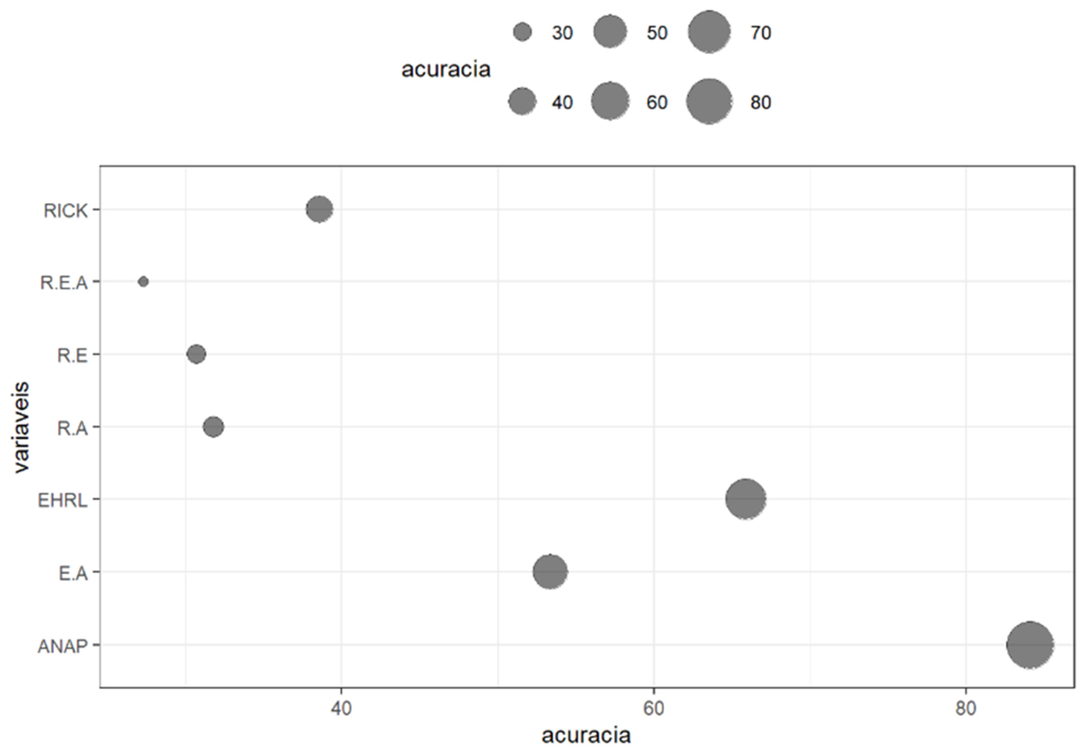
The different instant prevalence for singular infections as well as co-infections are illustrated in the bubble chart seen in Figure 4. The estimated prevalence for singular infections was calculated with respective 95% confidence intervals. For infections that only contained Rickettsia spp., the estimated prevalence was equal to 25% (16.6% - 35.6%). For infections that only contained the E. canis the estimated prevalence was equal to 52.3% (41.5% - 62.9%). For infections that only contained the A. phagocytophilum the estimated prevalence was equal to 70.5% (59.6% - 79.5 %). The estimated prevalence for co-infections was calculated with respective 95% confidence intervals. For co-infections that contained Rickettsia spp. and E. canis the estimated prevalence was equal to 17% (10.2% - 26.9%). For co-infections that contained Rickettsia spp. and A. phagocytophilum the estimated prevalence was equal to 18.2% (11.1% - 28.1%). For co-infections that contained E. canis and A. phagocytophilum the estimated prevalence was equal to 39.8% (29.7% - 50.8%). For co-infections that contained all three: Rickettsia spp., E. canis, and A. phagocytophilum the estimated prevalence was equal to 13.6% (7.5% - 23.0%).
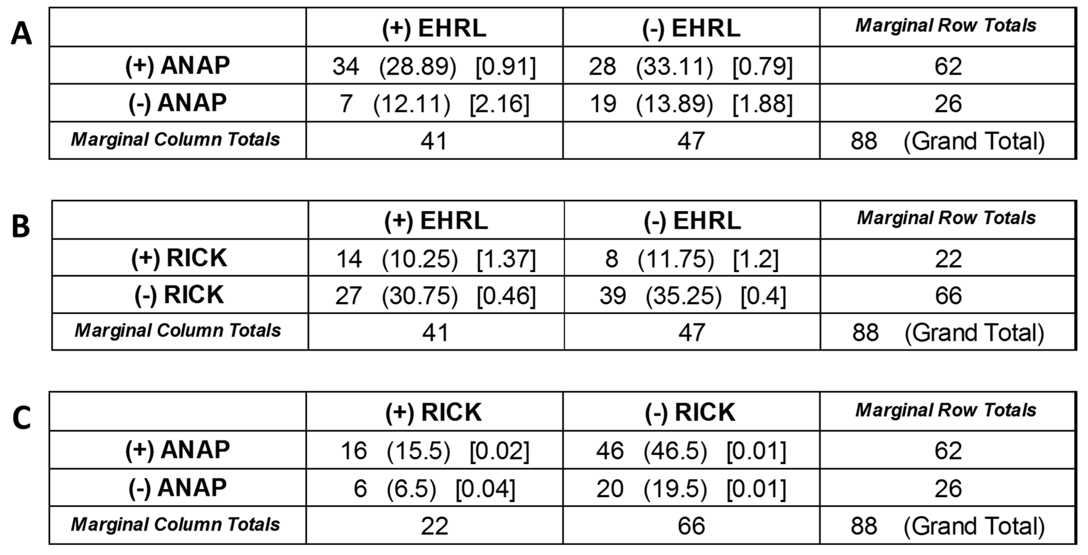
A chi-square test of independence was performed to examine the relation between co-infections of A. phagocytophilum and E. canis, or between co-infections of E. canis and Rickettsia spp., or between co-infections of A. phagocytophilum and Rickettsia spp. (Figure 5). The chi-square statistic between A. phagocytophilum and E. canis co-infections is 5.7367. The p-value is 0.016614. The relation between A. phagocytophilum and E. canis co-infections was significant at p < 0.10. The chi-square statistic with Yates correction is 4.6697. The p-value is 0.030699. The relation between A. phagocytophilum and E. canis co-infections was significant at p < 0.10 with the Yates correction (Figure 5A). The chi-square statistic between E. canis and Rickettsia spp. co-infections is 3.425. The p-value is 0.064216. The relation between E. canis and Rickettsia spp. co-infections was significant at p < 0.10. The chi-square statistic with Yates correction is 2.5726. The p-value is 0.108731. The relation between E. canis and Rickettsia spp. co-infections was not significant at p < 0.10 with the Yates correction (Figure 5B). The chi-square statistic between A. phagocytophilum and Rickettsia spp. co-infections is 0.0728. The p-value is 0.787321. The relation between A. phagocytophilum and Rickettsia spp. co-infections was not significant at p < 0.10. The chi-square statistic with Yates correction is 0. The p-value is 1. Not significant at p < 0.10. The relation between A. phagocytophilum and Rickettsia spp. co-infections was not significant at p < 0.10 with the Yates correction (Figure 5C).

4. Discussion

Between 2007 and 2023, the state of Minas Gerais, confirmed 451 cases of SF (the third highest number of confirmed cases in the country) [51], ranking second in Brazil in number of deaths from SF, with 145 deaths [52]. For many tick-borne organisms, dogs may be utilized as sentinel hosts in order to estimate the risk of human infection [35,47,53]. Previous studies have demonstrated a direct correlation between an increase of seropositivity in these animals and the resulting threat upon the public health of humans [54,55]. In this context, the serological survey in sentinels is essential, adequately reflecting the circulation of TBD in a given location, primarily indicating the presence of the bioagent in these animals.
For the purpose of screening for TBD, IFA serology, a methodology considered the gold standard in Brazil [16], is also frequently used in other countries, mainly because it is more practical and less costly. However, it is important to note that several studies have demonstrated cross-reactions between different members of the bioagents investigated here [25,56,57].
According to the BMH [16], titers that are greater than or equal to 1:64 for Rickettsia spp., 1:40 for E. canis, and 1:80 for A. phagocytophilum in a single sample confirm the diagnosis for the TBD studied. The results seen in Figure 2, demonstrate that positive titers (per the standards of the BMH) were detected in 83% of the collected samples (Figure 2A); and within those positive samples, 30% tested positive with Rickettsia spp. present, 56% tested positive with E. canis present, and 85% tested positive with A. phagocytophilum present (Figure 2B). These results demonstrate the circulation of these agents in the SCNP, and highlight the potential risk of infection, for both the human and animal population, within this location and the surrounding areas.
Previous research indicated that co-infection of multiple TBD pathogens in dogs appears more frequent in endemic areas [58,59]. The results in Figure 3 demonstrate the percentage of the samples where more than one infectious agent was present. Specifically, it was determined that 30% of the samples were co-infected with E. canis, and A. phagocytophilum; 16% of the samples were co-infected with Rickettsia spp., E. canis, and A. phagocytophilum; 5% of the samples were co-infected with Rickettsia spp., and A. phagocytophilum; and 3% of the samples were co-infected with Rickettsia spp., and E. canis.
Furthermore, a previous study found E. canis and Anaplasma spp. co-infecting the tick host R. sanguineus, thus explaining the common occurrence of these two infectious agents co-infecting dogs [60]. The chi-square test of independence seen in Figure 5 demonstrated a significant relationship between A. phagocytophilum and E. canis co-infections in the dogs sampled. The results in Figure 5A demonstrate that the chi-square statistic between A. phagocytophilum and E. canis co-infections was significant at p < 0.10; and was significant at p < 0.10 with the Yates correction. However, the co-infections between E. canis and Rickettsia spp. (Figure 5B) and between A. phagocytophilum and Rickettsia spp. (Figure 5C) were demonstrated to not be statistically related. Thus, our results not only support the previously findings [60], it also demonstrated that this co-infection also occurs in the new (sentinel) host.
Since surveillance in sentinel hosts has direct implications for the prevention, treatment, and control of zoonotic diseases [37], increasing the standard surveillance protocol to include even more endemic pathogens could be beneficial to public health. As such, the question was raised as to whether A. phagocytophilum should be included in FUNED’s standard protocol for the surveillance of tick-borne diseases. The results in Figure 3 also demonstrated the percentage of positive samples with a single infection of either Rickettsia spp., E. canis, or A. phagocytophilum. Specifically, 33% of the samples were found to be infected with A. phagocytophilum only, 7% of the samples were infected with E. canis only, and 5% of the samples were infected with Rickettsia spp. only. The results in Figure 4 demonstrate that the most prevalent infection in this group of samples was for the single infection of A. phagocytophilum. Taken together these results confirmed a significant presence of A. phagocytophilum in the sentinel hosts monitored by FUNED, and establishes a potential threat upon the public health of humans. These results also imply that 33% of the sentinel hosts that were tested would have gone unnoticed and untreated, further permitting the potential spread of this disease.

5. Conclusions

The results of the present study: I) Confirmed the presence of A. phagocytophilum in the sentinel hosts, in addition to the pathogens that are currently included in the standard surveillance protocol performed by FUNED; II) Support the inference of the risk of TBD occurrence in humans and also in other vertebrate hosts in the SCNP; III) Could prove helpful in changing public policies to include testing for Anaplasmosis as a part of the standard surveillance protocol; and IV) Could prove helpful in monitoring, preventing, controlling, and treating another potential threat upon the public health of humans; as well as, providing education, health vigilance, and vector control measures in the respective areas.
Ideally, future studies should include: I) An increase in the serological surveillance methodologies to include research on ticks and etiological agents; II) Identifying and monitoring more land areas in order to determine the potential risk toward the public health of humans; and III) Monitoring the seropositive sentinel animals after treatment in order to quantify the success of the standard surveillance protocol upon the prevention, control and treatment for TBD.

Author Contributions

Conceptualization, L.D.; L.O and A.D.; Methodology, L.D; M.T. and A.D; Software, M.T..; Validation, L.D; M.T.; L.O.; A.D. and L.F.; Formal Analysis, L.D; M.T. and L.O.; Investigation, L.D.; A.D. and L.O.; Resources, L.D.; A.D.; G.F; V.R.; G.P.; D.R. and L.F.; Data Curation, L.D.; M.T.; L.O. and L.F.; Writing—original draft preparation, L.D.; L.O. and A.D.; writing—review and editing, L.D.; L.O. and A.D.; visualization, L.D.; L.O. and A.D.; supervision, L.D.; Project Administration, L.D.; A.D., L.O. and L.F.. All authors have read and agreed to the published version of the manuscript.

Acknowledgments

This work was carried out by the Cipó Project, with support from the Chico Mendes Institute for Biodiversity Conservation, Ezequiel Dias Foundation, University of São Paulo, management of Parna Serra do Cipó, local community leaders, Log Nature and Fuller Laboratories, aiming to contribute to the conservation and collective health of the site, combining research and communication actions.

Conflicts of Interest

The authors declare no conflicts of interest. The funders had no role in the design of the study; in the collection, analyses, or interpretation of data; in the writing of the manuscript; or in the decision to publish the results.

References

  1. Jones, B.A.; Grace, D.; Kock, R.; Alonso, S.; Rushton, J.; Said, M.Y.; McKeever, D.; Mutua, F.; Young, J.; McDermott, J.; Pfeiffer, D.U. Zoonosis emergence linked to agricultural intensification and environmental change. Proc Natl Acad Sci U S A 2012, 110, 8399–8404. [Google Scholar] [CrossRef]
  2. Karesh, W.B.; Dobson, A.; Lloyd-Smith, J.O.; Lubroth, J.; Dixon, M.A.; Bennett, M.; Aldrich, S.; Harrington, T.; Formenty, P.; Loh, E.H.; Machalaba, C.C.; Thomas, M.J.; Heymann, D.L. Ecology of zoonoses: natural and unnatural histories. Lancet 2012, 380, 1936–1945. [Google Scholar] [CrossRef] [PubMed]
  3. Barcellos, C.D.C.; Monteiro, A.M.V.; Corvalán, C.; Gurgel, H.C.; Carvalho, M.S.; Artaxo, P.; Hacon, S.; Ragoni, V. Mudanças climáticas e ambientais e as doenças infecciosas: cenários e incertezas para o Brasil. Epidemiol. Serv. Saúde, 2009; 18, 285–304. [Google Scholar] [CrossRef]
  4. Intergovernmental Panel on Climate Change. Climate change 2014: mitigation of climate change. Available online: https://archive.ipcc.ch/report/ar5/syr/ (accessed on 21 March 2024).
  5. World Health Organization. World Malaria Report 2019. Available online: https://www.who.int/publications/i/item/9789241565721 (accessed on 21 March 2024).
  6. Domingos, A.; Antunes, S.; Borges, L.; Rosario, V.E.D. Approaches towards tick and tick-borne diseases control. Rev. Soc. Bras. Med. 2013, 46, 265–269. [Google Scholar] [CrossRef]
  7. Jongejan, F.; Uilenberg, G. The global importance of ticks. Parasitology 2004, 129. [Google Scholar] [CrossRef] [PubMed]
  8. De la Fuente, J.; Antunes, S.; Bonnet, S.; Cabezas-Cruz, A.; Domingos, A. G.; Estrada-Peña, A.; Johnson, N.; Kocan, K.M.; Mansfield, K.L.; Nijhof, A.M.; Papa, A.; Rudenko, N.; Villar, M.; Alberdi, P.; Torina, A.; Ayllón, N.; Vancova, M.; Golovchenko, M.; Grubhoffer, L.; Caracappa, S.; Fooks, A.R.; Gortazar, C.; Rego, R.O. Tick-pathogen interactions and vector competence: identification of molecular drivers for tick-borne diseases. Front. cell. infect. microbiol. 2017, 7. [Google Scholar] [CrossRef] [PubMed]
  9. Anderson, J.F.; Magnarelli, L.A. Biology of ticks. Infect Dis Clin North Am 2008, 22, 195–215. [Google Scholar] [CrossRef]
  10. Parola, P.; Paddock, C.D.; Socolovschi, C.; Labruna, M.B.; Mediannikov, O.; Kernif, T.; Abdad, M.Y.; Stenos, J.; Bitam, I.; Fournier, P.-E.; Raoult, D. Update on tick-borne rickettsioses around the world: a geographic approach. Clin Microbiol Rev 2013, 26, 657–702. [Google Scholar] [CrossRef]
  11. Polo, G.; Labruna, M.B.; Ferreira, F. Satellite hyperspectral imagery to support tick-borne infectious diseases surveillance. PLoS One 2015, 10. [Google Scholar] [CrossRef]
  12. Oliveira, S.V.D.; Guimarães, J.N.; Reckziegel, G.C.; Neves, B.M.D.C.; Araújo-Vilges, K.M.D.; Fonseca, L.X.; Pinna, F.V.; Pereira, S.V.C.; Caldas, E.P.; Gazeta, G.S.; Gurgel-Gonçalves, R. An update on the epidemiological situation of spotted fever in Brazil. J. Venom. Anim. Toxins Incl. Trop. Dis 2016, 22. [Google Scholar] [CrossRef]
  13. Satjanadumrong, J.; Robinson, M.T.; Hughes, T.; Blacksell, S.D. Distribution and ecological drivers of spotted fever group Rickettsia in Asia. Ecohealth 2019, 16, 611–626. [Google Scholar] [CrossRef]
  14. Piotrowski, M.; Rymaszewska, A. Expansion of tick-borne rickettsioses in the world. Microorganisms 2020, 8. [Google Scholar] [CrossRef] [PubMed]
  15. Springer, A.; Glass, A.; Probst, J.; Strube, C. Tick-borne zoonoses and commonly used diagnostic methods in human and veterinary medicine. Parasitol. Res. 2021, 120, 4075–4090. [Google Scholar] [CrossRef]
  16. Ministerio da Saúde do Brasil. Guia de vigilância em saúde, 5th ed. 2022. Available online: https://bvsms.saude.gov.br/bvs/publicacoes/guia_vigilancia_saude_5ed_rev_atual.pdf (accessed on 21 March 2024).
  17. Durães, L.S.; Bitencourth, K.; Ramalho, F.R.; Nogueira, M.C.; Nunes, E.D.C.; Gazêta, G.S. Biodiversity of potential vectors of rickettsiae and epidemiological mosaic of spotted fever in the State of Paraná, Brazil. Front. Public Health 2021, 9. [Google Scholar] [CrossRef]
  18. Vieira, R.F.D.C.; Biondo, A.W.; Guimarães, A.M.S.; Santos, A.P.D.; Santos, R.P.D.; Dutra, L.H.; Diniz, P.P.V.P.; Morais, H.A.; Messick, J.B.; Labruna, M.B.; Vidotto, O. Ehrlichiosis in Brazil. Rev. Bras. Parasitol. Vet. 2011, 20, 01–12. [Google Scholar] [CrossRef] [PubMed]
  19. Vieira, R.F.D.C.; Vieira, T.S.W.J.; Nascimento, D.D.A.G.; Martins, T.F.; Krawczak, F.S.; Labruna, M.B.; Chandrashekar, R.; Marcondes, M.; Biondo, A.W.; Vidotto, O. Serological survey of Ehrlichia species in dogs, horses and humans: zoonotic scenery in a rural settlement from southern Brazil. Rev Inst Med Trop Sao Paulo 2013, 55, 335–340. [Google Scholar] [CrossRef] [PubMed]
  20. Guedes, P.E.B.; Oliveira, T.N.D.A.; Carvalho, F.S.; Carlos, R.S.A.; Albuquerque, G.R.; Munhoz, A.D.; Wenceslau, A.A.; Silva, F.L. (2015). Canine ehrlichiosis: prevalence and epidemiology in northeast Brazil. Rev. Bras. Parasitol. Vet. 2015, 24, 115–121. [Google Scholar] [CrossRef]
  21. Aziz, M.U.; Hussain, S.; Song, B.; Ghauri, H.N.; Zeb, J.; Sparagano, O.A. Ehrlichiosis in Dogs: A Comprehensive Review about the Pathogen and Its Vectors with Emphasis on South and East Asian Countries. Vet. Sci. 2023, 10. [Google Scholar] [CrossRef]
  22. Perez, M.; Bodor, M.; Zhang, C.; Xiong, Q.; Rikihisa, Y. Human Infection with Ehrlichia canis accompanied by Clinical Signs in Venezuela. Ann. N. Y. Acad. Sci. 2006, 1078, 110–117. [Google Scholar] [CrossRef]
  23. Silva, A.B.; Canseco, S.P.; De la Torre, M.D.P.G.; Silva, A.M.; Mayoral, M.Á.; Mayoral, L.P.C.; Martínez, J.L.; Pérez-Campos, E. Asymptomatic human infection from contact with dogs: a case of human ehrlichiosis. Gaceta Médica de México 2014, 150, 171–174. [Google Scholar]
  24. Machado, R.Z.; Duarte, J.M.B.; Dagnone, A.S.; Szabó, M.P.J. Detection of Ehrlichia chaffeensis in Brazilian marsh deer (Blastocerus dichotomus). Vet. Parasitol. 2006, 139, 262–266. [Google Scholar] [CrossRef]
  25. Carrade, D.D.; Foley, J.E.; Borjesson, D.L.; Sykes, J.E. Canine granulocytic anaplasmosis: a review. J. Vet. Intern. Med. 2009, 23, 1129–1141. [Google Scholar] [CrossRef] [PubMed]
  26. Santos, H.A.; Thomé, S.M.; Baldani, C.D.; Silva, C.B.; Peixoto, M.P.; Pires, M.S.; Vitari, G.L.V.; Costa, R.L.; Santos, T.M.; Angelo, I.C.; Santos, L.A.; Faccini, J.L.H.; Massard, C.L. Molecular epidemiology of the emerging zoonosis agent Anaplasma phagocytophilum (Foggie, 1949) in dogs and ixodid ticks in Brazil. Parasit. Vectors. 2013, 6, 1–10. [Google Scholar] [CrossRef] [PubMed]
  27. Dos Santos, T.M.; Roier, E.C.R.; Pires, M.S.; Santos, H.A.; Vilela, J.A.R.; Peckle, M.; Paulino, P.G.; Baldani, C.D.; Massard, C.L. Molecular evidence of Anaplasma phagocytophilum and Theileria equi coinfection in horses from Rio de Janeiro, Brazil. VAS 2019, 7. [Google Scholar] [CrossRef] [PubMed]
  28. Dumler, J.S.; Choi, K.S.; Garcia-Garcia, J.C.; Barat, N.S.; Scorpio, D.G.; Garyu, J.W.; Grab, D.J.; Bakken, J.S. Human granulocytic anaplasmosis and Anaplasma phagocytophilum. Emerg. Infect. Dis. 2005, 11, 1828–1834. [Google Scholar] [CrossRef] [PubMed]
  29. Atif, F.A.; Mehnaz, S.; Qamar, M.F.; Roheen, T.; Sajid, M.S.; Ehtisham-ul-Haque, S.; Kashif, M.; Said, M.B. Epidemiology, Diagnosis, and Control of Canine Infectious Cyclic Thrombocytopenia and Granulocytic Anaplasmosis: Emerging Diseases of Veterinary and Public Health Significance. Vet. Sci. 2021, 8, 2–20. [Google Scholar] [CrossRef] [PubMed]
  30. Silveira, J.A.G.; Reis, I.A.; Estevam, L.G.T.M.; Pinto, M.C.C.; Zweygarth, E.; Passos, L.M.F.; Paz, G.F. 2017. Important frequency of Anaplasma phagocytophilum infection in a population of domiciled dogs in an urbanized area in south-eastern Brazil. Pesq. Vet. 2017, 37, 958–962. [Google Scholar] [CrossRef]
  31. Dantas-Torres, F.; Figueredo, L.A.; Sales, K.G.D.S.; Miranda, D.E.D.O.; Alexandre, J.L.D.A.; da Silva, Y.Y.; da Silva, L.G.; Valle, G.R.; Ribeiro, V.M.; Otranto, D.; Deuster, K.; Pollmeier, M.; Altreuther, G. Prevalence and incidence of vector-borne pathogens in unprotected dogs in two Brazilian regions. Parasit Vectors 2020, 13, 1–7. [Google Scholar] [CrossRef] [PubMed]
  32. Sousa, V.R.F.; Avaliação clínica, morfológica, hematológica, bioquímica e biomolecular de cães naturalmente infectados por Ehrlichia canis e Anaplasma platys. Universidade Federal Rural do Rio de Janeiro 2006. Available online: https://rima.ufrrj.br/jspui/handle/20.500.14407/9664 (accessed on 21 March 2024).
  33. Šlapeta, J.; Halliday, B.; Chandra, S.; Alanazi, A. D.; Abdel-Shafy, S. Rhipicephalus linnaei (Audouin, 1826) recognised as the “tropical lineage” of the brown dog tick Rhipicephalus sanguineus sensu lato: Neotype designation, redescription, and establishment of morphological and molecular reference. Ticks Tick Borne Dis 2022, 13. [Google Scholar] [CrossRef]
  34. Peixoto, C.S.; Alterações oculares e hematológicas em cães acometidos por Ehrlichia canis e co-infecções. Universidade de Brasília 2019. Available online: http://www.realp.unb.br/jspui/handle/10482/37471 (accessed on 21 March 2024).
  35. Schmidt, P.L. Companion animals as sentinels for public health. Vet Clin North Am Small Anim Pract 2009, 39, 241–250. [Google Scholar] [CrossRef]
  36. Silva, M.E.; Ribeiro, R.R.; Costa, J.O.; Moraes-Filho, J.; Pacheco, R.C.; Labruna, M.B. Prevalência de anticorpos anti-Rickettsia spp. em cães da cidade de Belo Horizonte, MG. Arq. Bras. Med. Vet. Zootec. 2010, 62, 1007–1010. [Google Scholar] [CrossRef]
  37. Clow, K.M.; Leighton, P.A.; Pearl, D.L.; Jardine, C.M. A framework for adaptive surveillance of emerging tick-borne zoonoses. One Health 2019, 7. [Google Scholar] [CrossRef] [PubMed]
  38. Durães, L.S.; Bitencourth, K.; de Oliveira, S.V.; Gazêta, G.S. Fiebre maculosa en Brasil: contexto histórico y actual. In Enfermedades rickettsiales en Latinoamérica, 1st ed.; Arias, S.P., Jaramillo, A.C., Buriticá, S.M., Eds.; Fondo Editorial Biogénesis: Medellín, Colombia, 2020; pp. 240–266. ISBN 978-958-5596-67-2. [Google Scholar]
  39. Campos, S.D. , Cunha, N.C.D., Machado, C.S., de Souza, T.V., Fonseca, A.B.M., Pinter, A.; Fonseca, A.H.; Almosny, N.R.P. Circulação de Rickettsias do Grupo da Febre Maculosa em cães no entorno de Unidades de Conservação Federais do estado do Rio de Janeiro: evidência sorológica e fatores associados. Pesq. Vet. 2017, 37, 1307–1312. [Google Scholar] [CrossRef]
  40. Springer, A.; Glass, A.; Topp, A.K.; Strube, C. Zoonotic tick-borne pathogens in temperate and cold regions of Europe—A review on the prevalence in domestic animals. Front. vet. sci. 2020, 7. [Google Scholar] [CrossRef] [PubMed]
  41. Davitt, C.; Traub, R.; Batsukh, B.; Battur, B.; Pfeffer, M.; Wiethoelter, A.K. Knowledge of Mongolian veterinarians towards canine vector-borne diseases. One Health 2022, 15. [Google Scholar] [CrossRef] [PubMed]
  42. Gehrke, F.S.; Gazeta, G.S.; Souza, E.R.; Ribeiro, A.; Marrelli, M.T.; Schumaker, T.T.S. Rickettsia rickettsii, Rickettsia felis and, Rickettsia sp TwKM03 infecting Rhipicephalus sanguineus and Ctenocephalides felis collected from dogs in a Brazilian Spotted Fever focus in the state of Rio de Janeiro/Brazil. Clin Microbiol Infect 2009, 15, 267–268. [Google Scholar] [CrossRef] [PubMed]
  43. Moraes-Filho, J.; Pinter, A.; Pacheco, R.C.; Gutmann, T.B.; Barbosa, S.O.; Gonzáles, M.A.R. M.; Muraro, M.A.; Cecílio, S.R.M.; Labruna, M.B. New Epidemiological Data on Brazilian Spotted Fever in an Endemic Area of the State of São Paulo, Brazil. Vector Borne Zoonotic Dis 2009, 9, 73–78. [Google Scholar] [CrossRef] [PubMed]
  44. Rozental, T.; Ferreira, M.S.; Gomes, R.; Costa, C.M.; Barbosa, P.R.A.; Bezerra, I.O.; Garcia, M.H.O.; Oliveira E Cruz, D.M.; Galliez, R.; Oliveira, S.; Brasil, P.; Rezende, T.; De Lemos, E.R.S. A cluster of Rickettsia rickettsii infection at an animal shelter in an urban area of Brazil. Epidemiol Infect 2015, 143, 2446–2450. [Google Scholar] [CrossRef] [PubMed]
  45. Lemos, E.R.S.; Machado, R.D.; Coura, J.R.; Guimarães, M.A.A.; Freire, N.M.S. Infestation by ticks and detection of antibodies to spotted fever group Rickettsiae in wild animals captured in the State of São Paulo, Brazil. A preliminary report. Mem Inst Oswaldo Cruz 1996, 91, 701–702. [Google Scholar] [CrossRef] [PubMed]
  46. Souza, C.E.; Camargo, L.B.; Pinter, A.; Donalisio, M.R. High seroprevalence for Rickettsia rickettsii in equines suggests risk of human infection in silent areas for the Brazilian spotted fever. PLoS ONE 2016, 11, 1–9. [Google Scholar] [CrossRef]
  47. Mahachi, K.; Kontowicz, E.; Anderson, B.; Toepp, A. J.; Lima, A. L.; Larson, M.; Wilson, G.; Grinnage-Pulley, T.; Bennett, C.; Ozanne, M.; Anderson, M.; Fowler, H.; Parrish, M.; Saucier, J.; Tyrrell, P.H.; Palmer, Z.; Buch, J.; Chandrashekar, R.; Scorza, B.; Brown, G.; Oleson, J.J.; Petersen, C.A. Predominant risk factors for tick-borne co-infections in hunting dogs from the USA. Parasit Vectors 2020, 13, 1–12. [Google Scholar] [CrossRef]
  48. Instituto Chico Mendes de Conservação da Biodiversidade. Parque Nacional da Serra do Cipó. Available online: https://www.icmbio.gov.br/parnaserradocipo/guia-do-visitante.html. (accessed on 20 May 2024).
  49. Fuller Laboratories. Anaplasma phagocytophilum (EED-120) Instructions for Use, Original 7/95, Current Version D (1/04). Fullerton, California, United States of America. Available online: https://fullerlaboratories.com/wp-content/uploads/2023/05/EED-120-English.pdf. (accessed on 15 May 2024).
  50. Pagano, M.; Gauvreau, K. Princípios de Bioestatística, 2nd ed.; Pioneira Thomson Learning: São Paulo, SP, Brazil, 2004. [Google Scholar]
  51. Ministério da Saúde do Brasil. Casos confirmados de Febre Maculosa. Brasil, Grandes Regiões e Unidades Federadas (Infecção) - 2007 a 2024. Available online: https://www.gov.br/saude/pt-br/assuntos/saude-de-a-a-z/f/febre-maculosa/situacao-epidemiologica/casos-confirmados-de-febre-maculosa-brasil-grandes-regioes-e-unidades-federadas-infeccao-2007-a-2024/view (accessed on 15 May 2024).
  52. Ministério da Saúde do Brasil. Óbitos confirmados de Febre Maculosa. Brasil, Regiões e Unidades Federadas (Infecção) - 2007 a 2024. Available online: https://www.gov.br/saude/pt-br/assuntos/saude-de-a-a-z/f/febre-maculosa/situacao-epidemiologica/obitos-de-febre-maculosa-brasil-grandes-regioes-e-unidades-federadas-infeccao-2007-a-2024/view (accessed on 21 March 2024).
  53. De Paiva Diniz, P.P.V.; Schwartz, D.S.; De Morais, H.S.A.; Breitschwerdt, E.B. Surveillance for zoonotic vector-borne infections using sick dogs from southeastern Brazil. Vector Borne Zoonotic Dis 2007, 7, 689–698. [Google Scholar] [CrossRef] [PubMed]
  54. Breitschwerdt, E.B.; Papich, M.G.; Hegarty, B.C.; Gilger, B.; Hancock, S.I.; Davidson, M.G. Efficacy of Doxycicline, azithromicin, or trovafloxacin for treatment of experimental rocky mountain spotted fever in dogs. Antimicrob Agents Chemother 1999, 43, 813–821. [Google Scholar] [CrossRef] [PubMed]
  55. Sangioni, L.A.; Horta, M.C.; Vianna, M.C.B.; Gennari, S.M.; Soares, R.M.; Galvão, M.A.M.; Schumaker, T.T.S.; Ferreira, F.; Vidotto, O.; Labruna, M.B. Rickettsial infection in animals and Brazilian spotted fever endemicity. Emerg. Infect. Dis. 2005, 11, 265–270. [Google Scholar] [CrossRef] [PubMed]
  56. Inokuma, H.; Nane, G.; Uechi, T.; Yonahara, Y.; Brouqui, P. , Okuda; M.; Onishi, T. Survey of tick infestation and tick-borne ehrlichial infection of dogs in Ishigaki Island, Japan. J Vet Med Sci 2001, 63, 1225–1227. [Google Scholar] [CrossRef] [PubMed]
  57. Dreher, U.M.; De La Fuente, J.; Hofmann-Lehmann, R.; Meli, M. L.; Pusterla, N.; Kocan, K. M.; Woldehiwet, Z.; Braun, U.; Regula, G.; Staerk, K.D.C.; Lutz, H. Serologic cross-reactivity between Anaplasma marginale and Anaplasma phagocytophilum. Clin Vaccine Immunol 2005, 12, 1177–1183. [Google Scholar] [CrossRef] [PubMed]
  58. Beall, M.J.; Chandrashekar, R.; Eberts, M.D.; Cyr, K.E.; Diniz, P.P.V.P.; Mainville, C. , Hegarty, B.C.; Crawford, J.M.; Breitschwerdt, E.B. Serological and molecular prevalence of Borrelia burgdorferi, Anaplasma phagocytophilum, and Ehrlichia species in dogs from Minnesota. Vector Borne Zoonotic Dis 2008, 8, 455–464. [Google Scholar] [CrossRef]
  59. Bowman, D.; Little, S.E.; Lorentzen, L.; Shields, J.; Sullivan, M.P.; Carlin, E.P. Prevalence and geographic distribution of Dirofilaria immitis, Borrelia burgdorferi, Ehrlichia canis, and Anaplasma phagocytophilum in dogs in the United States: results of a national clinic-based serologic survey. Vet. Parasitol. 2009, 160, 138–148. [Google Scholar] [CrossRef]
  60. Cetinkaya, H.; Matur, E.; Akyazi, I.; Ekiz, E.E.; Aydin, L.; Toparlak, M. Serological and molecular investigation of Ehrlichia spp. and Anaplasma spp. in ticks and blood of dogs, in the Thrace Region of Turkey. Ticks Tick Borne Dis 2016, 7, 706–714. [Google Scholar] [CrossRef]
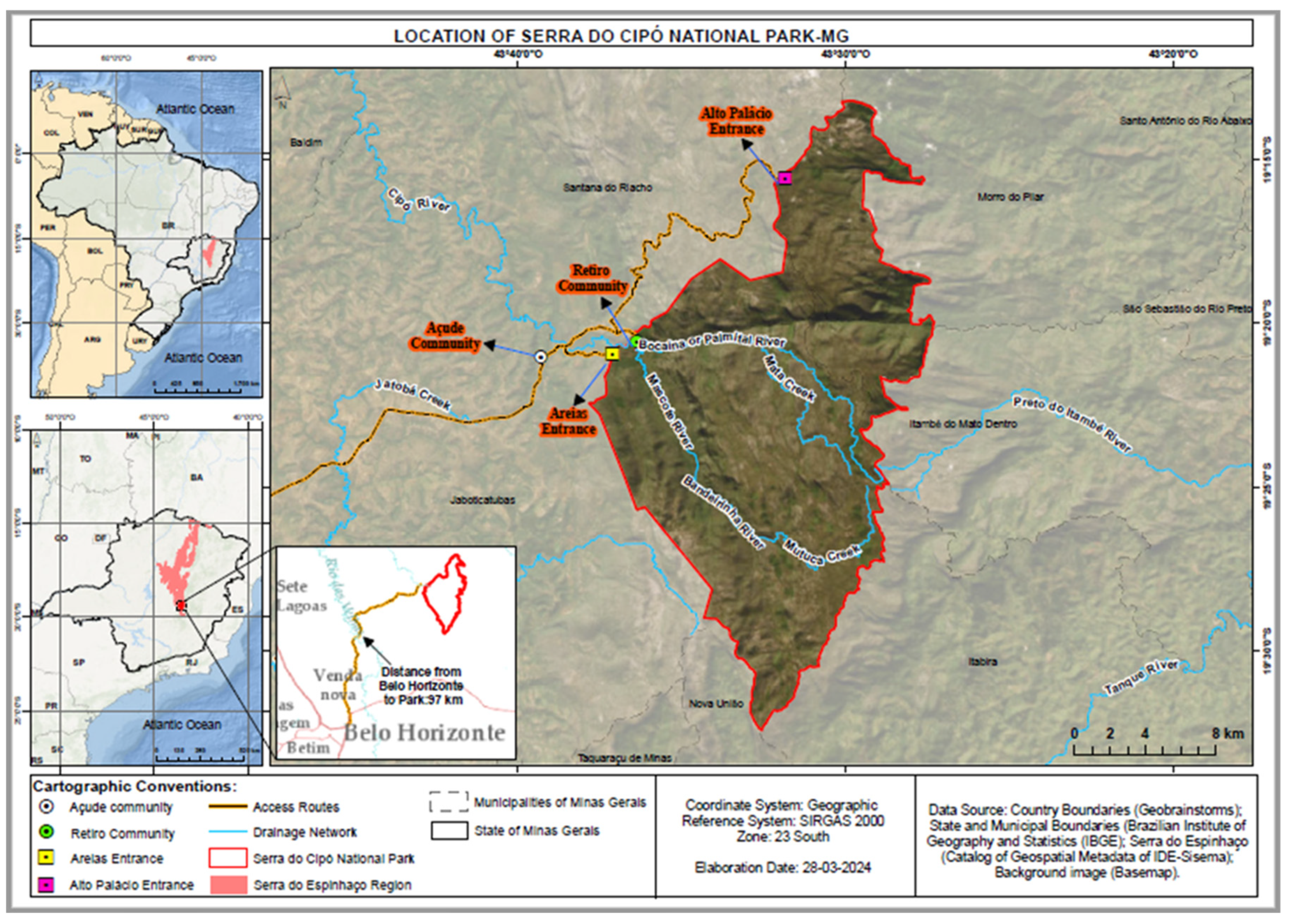
Figure 1. Blood samples from sentinel animals (both domiciled and stray dogs) were collected at the Serra do Cipó National Park (SCNP), which is located about 100 km from the capital of Belo Horizonte in the state of Minas Gerais. Blood samples were also obtained from the surrounding areas of the Retiro and Açude Communities, with access to the Areias and Alto Palácio gates.
Preprints 110614 g001
Figure 2. Six fieldwork campaigns were carried out between March 2021 and February 2022 at the Serra do Cipó National Park (SCNP), resulting in a total of 88 blood samples collected from dogs (domiciled and stray). 2A) The total percentages of samples that tested positive (83% [73/88 samples]) for infections with one or more of the three infectious agents: Rickettsia spp., Ehrlichia canis, and/or Anaplasma phagocytophilum; as well as the total percentage of samples that tested negative (17% [15/88 samples]) for infections with any of three infectious agents. 2B) Within the subset of the positive samples seen in Figure 2A, 30% (22/73 samples) tested positive for having Rickettsia spp. present, 56% (41/73 samples) tested positive for having E. canis present, and 85% (62/73 samples) tested positive for having A. phagocytophilum present.
Preprints 110614 g002
Figure 3. The percentage of positive samples with either single infections or co-infections of Rickettsia spp., Ehrlichia canis, and/or Anaplasma phagocytophilum (within the subset of the positive samples seen in Figure 2A). Abbreviations: RICK (Rickettsia spp. only), R.E.A. (Rickettsia spp., E. canis, and A. phagocytophilum), R.E. (Rickettsia spp., and E. canis), R.A. (Rickettsia spp., and A. phagocytophilum), EHRL (E. canis only), E.A. (E. canis, and A. phagocytophilum), ANAP (A. phagocytophilum only).
Preprints 110614 g003
Figure 4. The prevalence of Rickettsia spp., Ehrlichia canis, and Anaplasma phagocytophilum infections and co-infections. Each bubble represents a respective infection or co-infection type, and the size indicates the prevalence of the respective infection or co-infection within the PNSC geographic area. Abbreviations: RICK (Rickettsia spp. only), R.E.A. (Rickettsia spp., E. canis, and A. phagocytophilum), R.E. (Rickettsia spp., and E. canis), R.A. (Rickettsia spp., and A. phagocytophilum), EHRL (E. canis only), E.A. (E. canis, and A. phagocytophilum), ANAP (A. phagocytophilum only).
Preprints 110614 g004
Figure 5. A chi-square test of independence was performed to examine the relation between co-infections of Anaplasma phagocytophilum and Ehrlichia canis, or between co-infections of E. canis and Rickettsia spp., or between co-infections of A. phagocytophilum and Rickettsia spp. 5A) The chi-square statistic between A. phagocytophilum and E. canis co-infections was significant at p < 0.10; and was significant at p < 0.10 with the Yates correction. 5B) The chi-square statistic between E. canis and Rickettsia spp. co-infections was significant at p < 0.10; but was not significant at p < 0.10 with the Yates correction. 5C) The chi-square statistic between A. phagocytophilum and Rickettsia spp. co-infections was not significant at p < 0.10; and was not significant at p < 0.10 with the Yates correction. Abbreviations: (+) RICK (samples that contain Rickettsia spp.), (-) RICK (samples that do not contain Rickettsia spp.), (+) EHRL (samples that contain E. canis), (-) EHRL (samples that do not contain E. canis), (+) ANAP (samples that contain A. phagocytophilum), (-) ANAP (samples that do not contain A. phagocytophilum).
Preprints 110614 g005
Disclaimer/Publisher’s Note: The statements, opinions and data contained in all publications are solely those of the individual author(s) and contributor(s) and not of MDPI and/or the editor(s). MDPI and/or the editor(s) disclaim responsibility for any injury to people or property resulting from any ideas, methods, instructions or products referred to in the content.
Copyright: This open access article is published under a Creative Commons CC BY 4.0 license, which permit the free download, distribution, and reuse, provided that the author and preprint are cited in any reuse.
Prerpints.org logo

Preprints.org is a free preprint server supported by MDPI in Basel, Switzerland.

Subscribe

© 2024 MDPI (Basel, Switzerland) unless otherwise stated