Preprint
Article

Design Modulated Light on Physiological and Molecular Processes in Phenolic Compounds Production of Ocimum basilicum L.

Altmetrics

Downloads

112

Views

74

Comments

0

Submitted:

28 June 2024

Posted:

28 June 2024

You are already at the latest version

Alerts
Abstract
Microgreens (young, edible plantlets) are a remarkable opportunity for both consumers, to benefit from health promoting compounds and for applied research, as such organisms have a very high plasticity related to environmental cues, allowing biotechnology development with low costs. Ocimum basilicum species naturally synthesize valuable, phenolic compounds, among which rosmarinic acid is most prominent. Basil plantlets were grown for 10 days under either full spectrum light (white light) or modulated blue:red:far-red:UV spectrum with an additional factorization, by applying fertilisation. Biomass accumulation reached up to 0.8 g/20 plantlets, while chlorophyll fluorescence was in the 0.75 – 0.78 range and remained uniform across treatments, indicating that no significant stress was exerted under modified light treatment. However, total phenolic contents and, in particular, rosmarinic acid contents were markedly enhanced (up to 7.5 mg/g in the red cultivar) under modulated light treatment and fertilization, compared to full spectrum light. Moreover, gene expression was enhanced, 1.3-6.3 fold for genes coding for enzymes involved in phenylpropanoid synthesis patwhays, such as phenylalanine ammonia lyase (PAL), Catechol-O-methyltransferase (COMT) and renin-angiotensin system (RAS). Overall, light modulation coupled with fertilization led to the production of basil microgreens with up to 10% more total phenolics and up to 25% more rosmarinic acid.
Keywords: 
Subject: Biology and Life Sciences  -   Horticulture

1. Introduction

Phenolic compounds are secondary metabolites found in high concentration in medicinal and aromatic plants, as well as in micro plantlets that are used in various cuisines. Epidemiologically, a diet rich in polyphenols protects against diseases such as cancer, diabetes, osteoporosis, cardiovascular and neurological diseases [1], underpinned by mechanisms such as inflammation, oxidative stress, and cell ageing. For instance, inflammation caused by pathogens or toxic compounds [2],such as free radicals may exceed the antioxidant defence and lead to cell ageing,, progressive loss of tissue and organ function [3], associated with the progression of diseases such as diabetes [4], Alzheimer’s and Parkinson’s diseases [5,6]. In this sense, dietary bioactive molecules can positively influence tissue metabolism and alleviate oxidative and inflammatory effects at cellular level [7], while inadequate nutrient consumption may lead to imbalance between antioxidant defense and pro-oxidant load that induces oxidative stress [8]. Relevant examples are polyphenols, which are good electron or hydrogen atom donors may neutralize free radicals and reactive oxygen species (ROS). Moreover, polyphenols act at different cellular sites, leading to antioxidant, antimicrobial, anti-inflammatory or other biological functions, through several mechanisms [9,10]. Moreover, polyphenols alleviate the structural and functional damages caused by oxidative damage in mitochondria by regulating the expression of some antioxidant enzymes, such as superoxid dismutase (SOD), glutathione S-transferase (GST) and glutathion peroxidase (GSH-Px) [9].
Also, phenolic compounds exert anti-inflammatory through effects on gene expression, positively correlating with the levels of immune cell populations or cytokines production, or may inhibit the activity of enzymes such as cyclooxygenase (COX-2), lypooxygenase (LOX), inducible nitric oxide synthase (iNOS) [11],nuclear factor-kB (NF-kB), nuclear factor-erythroid factor 2-related factor 2 (Nrf-2) [1] and activate enzymes such as phase-II antioxidant detoxifying enzymes, mitogen activated protein kinase (MAPK), protein kinase-C.
Furthermore, polyphenols act on different sites of bacterial cells, altering the structure or metabolic pathways or may inhibit the gene expression related to virulence factors produced by bacterial pathogens, exhibiting also antibacterial properties [12].
While mean dietary phenolic intake range from around 255 mg/day in US citizens [13] and up to 1756 mg/day in European citizens [14], major benefits from consumption were described, such as decreased body fat, body mass index(BMI), waist and hip circumference [15] or lower serum pro/anti-inflammatory biomarkers’ ratio such as interleukin-10 (IL-10), T helper 1/T helper 2 balance (Th1:Th2), interleukin-1 (IL-1), interleukin-2 (IL-2) and interferon-gamma (IFN-γ) [16]. However, there is a great degree of variability in phenolic consumption, depending on the source, from olive-based foods in Mediterranean countries, to coffee in European Non-Mediterranean countries [17], to tea in Asian countries [13]. Moreover, age could be a factor influencing dietary phenolic intake, as European adolescents have a mean of only 326 mg/day [18]. However, phenolic substances are truly relevant for human health only by repeated intake, as most are degraded after 1-2 hours, with phenolic acids having a longer retention time, however [19] and also considering that the effects of dietary phenolic on human physiology are also influenced by conditioning or cooking [20]. Thus, it appears logical that constant consumption of dietary phenolic may be a real solution to the occurrence of some chronic diseases, such as malnutrition, cardiovascular diseases, obesity, diabetes, cancer, neurodegenerative disorders [21].
Dietary phenolic in microgreens stand out, as they share a set of very desirable traits related to healthy nutrition: young plants have a high moisture content and, hence, palatability, are easy to grow and have quick harvest turnaround times, are rich in enzymes, chlorophyll pigments etc. Also, microgreens have high nutritional quality, low environmental impacts, and broad consumer acceptance [22]. In the same time, microgreens are susceptible to influence of many environmental cues that offer the opportunity of modulating specific pathways, and, hence, increase bioactive production. The abundance of phenolic is high in basil microgreens, the most representative compounds being chicory acid, rosmarinic acid and caffeic acid, with values up to 17.58 mg/g dry matter [13]. The synthesis of this kind of phenolic acids was already proven that can be increased by modulating the spectrum of light delivered to microgreens, including in basil ones [23]. Various light regimes showed that bioactive compounds production can be stimulated, for instance, total phenolic and anthocyanin compounds under different blue:red proportions [24]. Other reports established that certain wavelengths increase nutraceutical and mineral contents [25] or specific bioactive such as rosmarinic acid [26] in various basil cultivars. While an undoubtable effect of light on the synthesis of specific compounds is agreed upon, only some works, such as [27,28] report the effect (usually beneficial) of the applied light treatments on the physiology of plants, as such effects are fundamental to designing proper technological setups for mass cultivation. Much less are the reports on the effects such treatments have on molecular mechanisms, such as gene expression, in order to truly characterize the process. While red, blue and UV appear to be main wavelengths affecting phenolic synthesis in microgreens [29], this aspect is addressed in papers focused on Lamiaceae species such as Mentha piperita [30] or Salvia verticillata [31], but not in Ocimum, as far as a reasonable literature screening.
Building on previous results, the present paper, based on selected blue:red:UV spectrum, further explores the effects and mechanisms involved in primary and secondary plant metabolism modulation by light, by quantifying key metabolites and also looking at normal physiological processes and specific genes’ expression.

2. Results

Plants exposed to light regimes exhibited various grades of effects in phenotypical traits but in biochemical and physiological processes as well. Regarding biomass accumulation, plants under either colored or light treatment recorded little differences, with respect to the fresh mass/20 plantlets and also to water content (Figure 1). The green cultivar consistently recorded higher biomass accumulation not from water, rather from organic matter (Figure 1), compared to the red cultivar. The different light spectra induced some significant variation, between the water and fertilizer treated plantlets, in the green cultivar
With respect to physiological processes such as the efficiency of the second photosystem, the applied factors (light and fertilization) did not exert influence. Overall, ΦPSII values ranged between 0.75-0.79 in the red cultivar and between 0.76-0.78 in the green cultivar (Table 1). The former recorded lower values of both light-adapted steady state fluorescence (Fs) and maximal fluorescence (Fm’), which is attributable to the intrinsically lower chlorophyll contents in this cultivar.
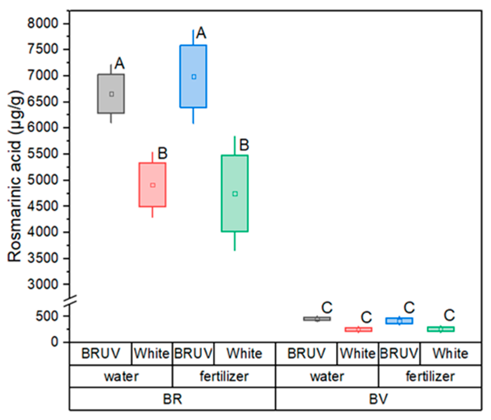
As the efficiency of the second photo system was comparable among treatments, no indication of stress development in plants could be observed. In a similar pattern, chlorophyll contents were higher in the green cultivar and the lowest value of chlorophyll contents were recorded in the red cultivar under fertilization. Total phenolic contents recorded marked differences among treatments with the highest values in plantlets under modified spectrum illumination in the red basil cultivar (Table 2). Among quantified specific phenolic acids, rosmarinic acid was most abundant with values up to 8 micrograms per gram under modulated light treatment in the red basil cultivar. Overall, the red cultivar had the highest phenolic acid contents with significant increases under blue red and UV illumination (Figure 2).
Gene expression of selected phenolic pathways were reduced in plantlets under blue red and UV treatment come on with similar values under water or fertilization (Figure 3).

3. Discussion

With an astounding structural diversity, natural phenolic substances are known for their valuable, health-promoting properties and offer the opportunity to be used as part of a regular diet or may serve as starting points for further enhancement of structure and function [12]. Phenolic compounds are a class of secondary metabolites that play pivotal roles in plant physiology and adaptation to the environment. They are involved in a wide range of processes, including defense against pathogens, protection from UV radiation, attraction of pollinators, and modulation of plant-microbe interactions [32]. The biosynthesis of phenolic compounds in plants is a complex process, involving various enzymes and associated genes such as phenylalanine ammonia lyase (PAL), caffeic acid O-methyltransferase (COMT), rosmarinic acid synthase (RAS), and tyrosine aminotransferase (TAT). Phenylalanine ammonia lyase is a key enzyme in the phenolic synthesis pathway, catalyzing the deamination of phenylalanine to form cinnamic acid, the entry point for phenolic biosynthesis in plants [33]. Meanwhile, caffeic acid O-methyltransferase is involved in the methylation of hydroxycinnamic acids, converting them into their corresponding methyl esters. This enzyme contributes to lignin biosynthesis and regulates the accumulation of various phenolic compounds [34]. Rosmarinic acid synthase, responsible for the synthesis of rosmarinic acid, catalyzes the condensation of caffeic acid with 3,4-dihydroxyphenyllactic acid. Tyrosine aminotransferase is involved in the conversion of tyrosine to p-coumaric acid, a precursor of various phenolic compounds and is crucial for the biosynthesis of flavonoids and other phenolics [35,36].
Several studies have highlighted that the spectral quality of light can significantly influence the production of phenolic compounds. For instance, an experiment with spring barley acclimated to different spectral qualities—white, blue, green, and red—at various irradiances found a complex interaction between photosynthetically active radiation (PAR) irradiance and spectral components in the accumulation of phenolic compounds [37,38]. The impact of light quantity (intensity and photoperiod) and quality (spectral composition) extends to plant growth and physiology, interacting with other environmental parameters and cultivation factors. This complexity influences plant behavior and metabolism, including the synthesis of phenolic compounds, as it was seen when comparing the effects of blue, red, and a combination of blue and red lights on metabolism of young wheat plants, which is related to stress responses and secondary metabolite production [39].
The spectral composition of light plays a significant role in photosynthesis and the overall functioning of photosystems in plants. The diversity of effects observed due to variations in light spectrum is attributed to three main factors: the activation of different photoreceptors, the variable efficiency of different spectral components in driving photosynthesis, and the depth of penetration of these spectral components into the leaf [40]. Different wavelengths of light are known to trigger various photoreceptors in plants, such as phytochromes for red light and cryptochromes for blue light, which subsequently influence plant growth and metabolism. The influence of blue light (maximum 450 nm) and red light (maximum 660 nm) on plant growth and metabolism is recognized, but not fully understood, however the spectral composition of light modifies the expression of light-dependent genes and impacts the growth, photosynthesis, and physiological responses in plants, as observed in seedlings [41,42].
Blue and red lights are known to have significant effects on PSII and PSI [43]. Blue light, particularly in the range of 400-500 nm, has been shown to enhance the rate of photosynthesis and stomatal opening, leading to increased CO2 assimilation [44,45]. Also, the use of red and blue LED spectra has been shown to increase the accumulation of polyphenols, flavonoids, and other phytochemicals, although not necessarily enhancing antioxidant activity [46], possibly due to premature plant allocation of metabolites to alternative pathways (such as curcumin synthesis). The mechanisms appears to be related to the stress, induced by high light intensity or specific light spectral compositions, which activates plant response mechanisms that include the production of phenolic compounds through hormonal pathways [47].
Red light, predominantly absorbed by chlorophyll, increases the efficiency of PSII [48], while far-red wavelengths, such as those used in our research, lead to higher yield also by enhancing PSII efficiency through reduced the heat dissipation of PSII, increased light energy available for photosynthesis and decrease in NPQ through faster reoxidation of plastoquinone and reopening of the PSII reaction center [49]. UV light, particularly UV-B (280-315 nm), has been proved to increase phenolic acids synthesis through the UVR8 photoreceptor, which interacts with the COP1/HYH/HY5 signaling pathway and leads to increases in mRNA levels and activities of phenylalanine ammonia lyase (PAL), cinnamic acid 4-hydroxylase (C4H), 4-coumarate coenzyme A ligase (4CL), p-coumaric acid 3-hdroxylase (C3H), caffeic acid O-methyltransferase (COMT) [50]. UV also increases phenolic contents by increasing PAL activity as a response to induced energy excess in mitochondrial electron transport chain and reactive oxygen species (ROS) generation and by enhancing vitamin C production and, thus, protection of phenolic substances from degradation [51].
Rosmarinic acid production, in particular, was shown to increase by a few folds in Lamiaceae plants by exposure to blue:red:far-red treatments, following increases in PAL, TAT, and hydroxyphenyl pyruvate reductase (HPPR) enzymes, but with minimal effects on chlorophyll contents, as shown in our study [52]. The mechanisms appears to be a modified balance of transcript levels of downstream genes (C4H, chalcone synthase (CHS), chalcone isomerase (CHI), and RAS) and upstream genes (PAL, TAT, and HPPR) [53].
As such, our results point to the fact that phenolic acids production was increased, as a result of light modulation and due to the fact that the quality of light can be perceived by plants as a cue for the need to protect from excess energy and that phenylpropanoid pathways are involved in such protection [54]. In the arsenal of plant defense mechanisms against harsh light environments and the overproduction of reactive oxygen species (ROS), phenolic compounds stand out for their significant antioxidative capability. While plants employ various strategies to avert ROS accumulation—including UV-protective epidermal layers, dissipation of surplus light energy as heat, optimizing the architecture of leaves, moving chloroplasts, and transitioning photosystem states—phenolics play a pivotal role in the detoxification process. These compounds are part of the plant’s sophisticated antioxidant system that springs into action under stress to neutralize ROS. Alongside enzymatic antioxidants like (SOD), catalase (CAT), and ascorbate peroxidase (APX), phenolics are integral low-molecular-weight antioxidants. They work in concert with other antioxidants such as tocopherols, ascorbate, glutathione, and carotenoids, ensuring the equilibrium between ROS production and scavenging is maintained in non-stressful conditions [39].

4. Materials and Methods

4.1. Plant Material and Growth Conditions

Two basil cultivars were used for microgreens production, “Sweet Genovese”, a green, acyanic cultivar, and “Red Rubin”, a red pigmented, cyanic cultivar, seeds being provided by VS (author) from the research and educational seed stock of the Life Sciences University in Iasi. For each treatment, approximately 150 seeds were sowed in plastic boxes (5 boxes per treatment, 3 used for biochemical analyses and 2 for phenotypic measurements), 10 × 10 × 12 cm, using a mixture of general-purpose soil and peat moss 2:1. Boxes were irrigated daily for 1 minute using automated drip systems, with either tap water or fertilizer. The fertilizer was prepared according to the recipe presented in Khater et al ) [55] and had the final concentrations: N:P:K 210:31:234 ppm. The minerals were introduced as the following salts: NH4NO3, P2O5 and C2K2O4xH2O. Each light treatment was provided by a Phytofy RL LED unit (OSRAM, Golden Dragon, Munich, Germany), from a distance of 30 cm from the top of the boxes. The two light treatments applied were a control variant, using a white LED program (0:0:0:0:0:1, UV:blue:green:red:far-red:white, in µmoles) and a colored program (1:9:0:9:3:0, UV:blue:green:red:far-red:white), respectively. After seeding, the boxes were kept in the dark for 3 days and afterwards, total PPFD (Photonic Flux Density) for the two treatments were 160 and, respectively, 161 µmoles/m2/s. The emission spectra of LED lights (according to OSRAM software) used are given in Figure 4. Plants were collected for biochemical and gene expression analyses 10 days after germination.

4.2. Analyses

Chlorophyll pigments were analyzed non-destructively, using a MC-100 Chlorophyll Concentration Meter (Apogee Instruments), by measuring 24 leaves/treatment/basil cultivar. Chlorophyll fluorescence related parameters - Fs – steady state fluorescence, Fm’ – maximal light adapted fluorescence and ΦPSII – quantum efficiency of the photosystem II, were measured using an FMS2 fluorimeter (HansaTech, Norfolk, UK) for 12 cotyledons/treatment, during light treatment period. Chlorophyll related analyses were performed at the end of the experiment before harvest.
Total phenolic content and antioxidant activity were determined in microtiter plates according to the methods described by Herald [56]. Briefly, total phenolic contents were assayed using Folin-Ciocalteu reagent, expressing results as gallic acid equivalents (GAE)/mg, while antioxidant activity was measured as % inhibition of DPPH free radical in ethanolic extracts. The reads were performed with BioTek Epoch 2 microplate spectrophotometer (Agilent, United States). The extracts were prepared from the dry plant and 70 % (w/w) ethanol in a ratio of 1:9, by maceration at 50˚ for 60 minutes. For High Performance liquid chromatography (HPLC) determination the extracts were filtered through a polyethersulfone (PES) membrane with 0.22 µm diameter pores.
Identification and quantification of the phenolic compounds from samples were performed on a Waters 2695e Alliance HPLC system coupled with a 2998 PDA Detector. The resulting chromatograms were processed using Empower software. Separation was achieved on a Waters XBridge column C18 column (50 x 4.6 mm, 3.5 µm), maintained at 30°C. The mobile phase A consisted in a solution of 0.1 % trifluoroacetic acid (TFA) in water, while for mobile phase B a solution of 0.1 % TFA in acetonitrile was used. The gradient program was: 0–4 min 100% (A), 5–20 min 98% (A), 27–30 min 96% (A), 32–35 min 90% (A), 40–45 min 82% (A), 50–53 min 0% (A), 55–60 min 100% (A). The flow rate was set up at 0.7 mL/min and the injection volume was 20 µL. HPLC/DAD analyses were performed monitoring the 280 nm wavelength. The identification of phenolic compounds was realized by comparing retention time with the available standards. The phenolic compounds quantification was performed using standard curves of external standards, obtained by plotting HPLC peak areas against the concentrations (µg/mL) (r2 > 0.99).
Gene expression analysis was carried out using qRT-PCR commercial assays on a Applied Biosystems QuantStudio5 real time PCR equipment. Total RNA extraction was performed using RNeasy Plant Mini Kit (Qiagen Inc.), from liquid nitrogen frozen cotyledons. RNA extracts were assessed for nucleic acid purity and amount using Qubit fluorometer, then samples were prepared according to manufacturer specifications for amplification and detection, using GoTaq® 1-Step RT-qPCR System (Promega Corp., Madison, USA). ΔΔCt calculations were performed, relative to GADPH reference and expressed logarithmically.

5. Conclusions

The spectral composition of light is a critical factor that influences the functioning of photosystems in plants, affecting a wide range of physiological and developmental processes. Our results showed that a modulation of light quality and quantity, alongside basic fertilization of Ocimum basilicum plants can lead to significant increases in the production of the valuable phenolic substances, in particular rosmarinic acid, with up to 25%. Moreover, basil microgreens retain their biomass production, thus the light treatment does not impede economic reasons and no significant stress is recorded in plants. The breadth of the influence of light spectrum variation on plant life underscores the importance of this area of study, particularly in the context of artificial lighting in agriculture and the potential for targeted manipulation of light spectra to enhance plant growth and productivity.

Author Contributions

“Conceptualization, A.L. and N.-E.P.; methodology, A.L. and G.M.; software, G.C.T. and A.L.; validation, V.S. and M.B..; formal analysis, A.L. and N.-E.P; investigation, N.-E.P.; resources, G.M.; data curation, G.M.; writing—original draft preparation, G.C.T. and N.-E.P; writing—review and editing, A.L.; supervision, V.S.; project administration, A.L.; funding acquisition, A.L. All authors have read and agreed to the published version of the manuscript.

Funding

This research was supported by a grant from the Ministry of Research, Innovation and Digitization, CCCDI-UEFISCDI, project number PN-III-P2-2.1-PED-2021-4380, within PNCDI III.

Data Availability Statement

Data are contained within the article.

Conflicts of Interest

The authors declare no conflict of interest.

References

  1. Rahman, Md. M.; Rahaman, Md. S.; Islam, Md. R.; Rahman, F.; Mithi, F. M.; Alqahtani, T.; Almikhlafi, M. A.; Alghamdi, S. Q.; Alruwaili, A. S.; Hossain, Md. S.; et al. Role of Phenolic Compounds in Human Disease: Current Knowledge and Future Prospects. Molecules 2021, 27, 233. [Google Scholar] [CrossRef] [PubMed]
  2. Ambriz-Perez, D. L.; Leyva-Lopez, N.; Gutierrez-Grijalva, E. P.; Heredia, J. B. Phenolic Compounds: Natural Alternative in Inflammation Treatment. A Review. Cogent Food Agric. 2016, 2. [Google Scholar] [CrossRef]
  3. Liguori, I.; Russo, G.; Curcio, F.; Bulli, G.; Aran, L.; Della-Morte, D.; Gargiulo, G.; Testa, G.; Cacciatore, F.; Bonaduce, D.; et al. Oxidative Stress, Aging, and Diseases. Clin. Interv. Aging 2018, Volume 13, 757–772. [Google Scholar] [CrossRef]
  4. Yaribeygi, H.; Sathyapalan, T.; Atkin, S. L.; Sahebkar, A. Molecular Mechanisms Linking Oxidative Stress and Diabetes Mellitus. Oxid. Med. Cell. Longev. 2020, 2020, 1–13. [Google Scholar] [CrossRef]
  5. Blesa, J.; Trigo-Damas, I.; Quiroga-Varela, A.; Jackson-Lewis, V. R. Oxidative Stress and Parkinson’s Disease. Front. Neuroanat. 2015, 9. [Google Scholar] [CrossRef] [PubMed]
  6. Huang, W.-J.; Zhang, X.; Chen, W.-W. Role of Oxidative Stress in Alzheimer’s Disease. Biomed. Rep. 2016, 4, 519–522. [Google Scholar] [CrossRef] [PubMed]
  7. Cianciosi, D.; Forbes-Hernández, T.; Afrin, S.; Gasparrini, M.; Reboredo-Rodriguez, P.; Manna, P.; Zhang, J.; Bravo Lamas, L.; Martínez Flórez, S.; Agudo Toyos, P.; et al. Phenolic Compounds in Honey and Their Associated Health Benefits: A Review. Molecules 2018, 23, 2322. [Google Scholar] [CrossRef] [PubMed]
  8. Saha, S. K.; Lee, S. B.; Won, J.; Choi, H. Y.; Kim, K.; Yang, G.-M.; Dayem, A. A.; Cho, S. Correlation between Oxidative Stress, Nutrition, and Cancer Initiation. Int. J. Mol. Sci. 2017, 18, 1544. [Google Scholar] [CrossRef] [PubMed]
  9. Yan, Z.; Zhong, Y.; Duan, Y.; Chen, Q.; Li, F. Antioxidant Mechanism of Tea Polyphenols and Its Impact on Health Benefits. Anim. Nutr. 2020, 6, 115–123. [Google Scholar] [CrossRef]
  10. Zeb, A. Concept, Mechanism, and Applications of Phenolic Antioxidants in Foods. J. Food Biochem. 2020, 44. [Google Scholar] [CrossRef]
  11. Hussain, T.; Tan, B.; Yin, Y.; Blachier, F.; Tossou, M. C. B.; Rahu, N. Oxidative Stress and Inflammation: What Polyphenols Can Do for Us? Oxid. Med. Cell. Longev. 2016, 2016, 1–9. [Google Scholar] [CrossRef]
  12. Lobiuc, A.; Pavăl, N.-E.; Mangalagiu, I. I.; Gheorghiță, R.; Teliban, G.-C.; Amăriucăi-Mantu, D.; Stoleru, V. Future Antimicrobials: Natural and Functionalized Phenolics. Molecules 2023, 28, 1114. [Google Scholar] [CrossRef] [PubMed]
  13. Lee, B. H.; Nam, T. G.; Park, N. Y.; Chun, O. K.; Koo, S. I.; Kim, D.-O. Estimated Daily Intake of Phenolics and Antioxidants from Green Tea Consumption in the Korean Diet. Int. J. Food Sci. Nutr. 2016, 67, 344–352. [Google Scholar] [CrossRef] [PubMed]
  14. Grosso, G.; Stepaniak, U.; Topor-Mądry, R.; Szafraniec, K.; Pająk, A. Estimated Dietary Intake and Major Food Sources of Polyphenols in the Polish Arm of the HAPIEE Study. Nutrition 2014, 30, (11–12). [Google Scholar] [CrossRef] [PubMed]
  15. Kapolou, A.; Karantonis, H. C.; Rigopoulos, N.; Koutelidakis, A. E. Association of Mean Daily Polyphenols Intake with Mediterranean Diet Adherence and Anthropometric Indices in Healthy Greek Adults: A Retrospective Study. Appl. Sci. 2021, 11, 4664. [Google Scholar] [CrossRef]
  16. Wisnuwardani, R. W.; De Henauw, S.; Ferrari, M.; Forsner, M.; Gottrand, F.; Huybrechts, I.; Kafatos, A. G.; Kersting, M.; Knaze, V.; Manios, Y.; et al. Total Polyphenol Intake Is Inversely Associated with a Pro/Anti-Inflammatory Biomarker Ratio in European Adolescents of the HELENA Study. J. Nutr. 2020, 150, 1610–1618. [Google Scholar] [CrossRef] [PubMed]
  17. Zamora-Ros, R.; Knaze, V.; Rothwell, J.A.; Hémon, B.; Moskal, A.; Overvad, K.; Tjønneland, A.; Kyrø, C.; Fagherazzi, G.; Boutron-Ruault, M.-C.; et al. Dietary Polyphenol Intake in Europe: The European Prospective Investigation into Cancer and Nutrition (EPIC) Study. Eur. J. Nutr. 2016, 55, 1359–1375. [Google Scholar] [CrossRef] [PubMed]
  18. Wisnuwardani, R. W.; De Henauw, S.; Androutsos, O.; Forsner, M.; Gottrand, F.; Huybrechts, I.; Knaze, V.; Kersting, M.; Le Donne, C.; Marcos, A.; et al. Estimated Dietary Intake of Polyphenols in European Adolescents: The HELENA Study. Eur. J. Nutr. 2019, 58, 2345–2363. [Google Scholar] [CrossRef] [PubMed]
  19. Scalbert, A.; Williamson, G. Dietary Intake and Bioavailability of Polyphenols. J. Nutr. 2000, 130, 2073S–2085S. [Google Scholar] [CrossRef]
  20. Knaze, V.; Rothwell, J.A.; Zamora-Ros, R.; Moskal, A.; Kyrø, C.; Jakszyn, P.; Skeie, G.; Weiderpass, E.; Santucci de Magistris, M.; Agnoli, C.; et al. A New Food-Composition Database for 437 Polyphenols in 19,899 Raw and Prepared Foods Used to Estimate Polyphenol Intakes in Adults from 10 European Countries. Am. J. Clin. Nutr. 2018, 108, 517–524. [Google Scholar] [CrossRef]
  21. Bhaswant, M.; Shanmugam, D. K.; Miyazawa, T.; Abe, C.; Miyazawa, T. Microgreens—A Comprehensive Review of Bioactive Molecules and Health Benefits. Molecules 2023, 28, 867. [Google Scholar] [CrossRef] [PubMed]
  22. Michell, K.A.; Isweiri, H.; Newman, S.E.; Bunning, M.; Bellows, L.L.; Dinges, M.M.; Grabos, L.E.; Rao, S.; Foster, M.T.; Heuberger, A.L.; et al. Microgreens: Consumer Sensory Perception and Acceptance of an Emerging Functional Food Crop. J. Food Sci. 2020, 85, 926–935. [Google Scholar] [CrossRef]
  23. Lobiuc, A.; Vasilache, V.; Oroian, M.; Stoleru, T.; Burducea, M.; Pintilie, O.; Zamfirache, M.-M. Blue and Red LED Illumination Improves Growth and Bioactive Compounds Contents in Acyanic and Cyanic Ocimum Basilicum L. Microgreens. Molecules 2017, 22, 2111. [Google Scholar] [CrossRef]
  24. Hosseini, A.; Zare Mehrjerdi, M.; Aliniaeifard, S. Alteration of Bioactive Compounds in Two Varieties of Basil ( Ocimum Basilicum ) Grown Under Different Light Spectra. J. Essent. Oil Bear. Plants 2018, 21, 913–923. [Google Scholar] [CrossRef]
  25. Viršilė, A.; Laužikė, K.; Sutulienė, R.; Brazaitytė, A.; Kudirka, G.; Samuolienė, G. Distinct Impacts of UV-A Light Wavelengths on Nutraceutical and Mineral Contents in Green and Purple Basil Cultivated in a Controlled Environment. Horticulturae 2023, 9, 1168. [Google Scholar] [CrossRef]
  26. Shiga, T.; Shoji, K.; Shimada, H.; Hashida, S.; Goto, F.; Yoshihara, T. Effect of Light Quality on Rosmarinic Acid Content and Antioxidant Activity of Sweet Basil, Ocimum Basilicum L. Plant Biotechnol. 2009, 26, 255–259. [Google Scholar] [CrossRef]
  27. Vodnik, D.; Vogrin, Ž.; Šircelj, H.; Grohar, M. C.; Medič, A.; Carović-Stanko, K.; Safner, T.; Lazarević, B. Phenotyping of Basil (Ocimum Basilicum L.) Illuminated with UV-A Light of Different Wavelengths and Intensities. Sci. Hortic. 2023, 309, 111638. [Google Scholar] [CrossRef]
  28. Chutimanukul, P.; Wanichananan, P.; Janta, S.; Toojinda, T.; Darwell, C. T.; Mosaleeyanon, K. The Influence of Different Light Spectra on Physiological Responses, Antioxidant Capacity and Chemical Compositions in Two Holy Basil Cultivars. Sci. Rep. 2022, 12, 588. [Google Scholar] [CrossRef]
  29. Zhang, S.; Zhang, L.; Zou, H.; Qiu, L.; Zheng, Y.; Yang, D.; Wang, Y. Effects of Light on Secondary Metabolite Biosynthesis in Medicinal Plants. Front. Plant Sci. 2021, 12, 781236. [Google Scholar] [CrossRef]
  30. Gholamnia, A.; Mosleh Arani, A.; Sodaeizadeh, H.; Tarkesh Esfahani, S.; Ghasemi, S. Expression Profiling of Rosmarinic Acid Biosynthetic Genes and Some Physiological Responses from Mentha Piperita L. under Salinity and Heat Stress. Physiol. Mol. Biol. Plants Int. J. Funct. Plant Biol. 2022, 28, 545–557. [Google Scholar] [CrossRef]
  31. Rizi, M. R.; Azizi, A.; Sayyari, M.; Mirzaie-Asl, A.; Conti, L. Increased Phenylpropanoids Production in UV-B Irradiated Salvia Verticillata as a Consequence of Altered Genes Expression in Young Leaves. Plant Physiol. Biochem. 2021, 167, 174–184. [Google Scholar] [CrossRef] [PubMed]
  32. Dixon, R. A.; Paiva, N. L. Stress-Induced Phenylpropanoid Metabolism. Plant Cell 1995, 1085–1097. [Google Scholar] [CrossRef]
  33. Koshiba, T.; Saito, E.; Ono, N.; Yamamoto, N.; Sato, M. Purification and Properties of Flavin- and Molybdenum-Containing Aldehyde Oxidase from Coleoptiles of Maize. Plant Physiol. 1996, 110, 781–789. [Google Scholar] [CrossRef] [PubMed]
  34. Boudet, A. M.; Kajita, S.; Grima-Pettenati, J.; Goffner, D. Lignins and Lignocellulosics: A Better Control of Synthesis for New and Improved Uses. Trends Plant Sci. 2003, 8, 576–581. [Google Scholar] [CrossRef] [PubMed]
  35. Petersen, M.; Husler, E.; Karwatzki, B.; Meinhard, J. Proposed Biosynthetic Pathway for Rosmarinic Acid in Cell Cultures of Coleus Blumei Benth. Planta 1993, 189. [Google Scholar] [CrossRef]
  36. Petersen, M.; Abdullah, Y.; Benner, J.; Eberle, D.; Gehlen, K.; Hücherig, S.; Janiak, V.; Kim, K. H.; Sander, M.; Weitzel, C.; et al. Evolution of Rosmarinic Acid Biosynthesis. Phytochemistry 2009, 70, 1663–1679. [Google Scholar] [CrossRef] [PubMed]
  37. Kivimäenpä, M.; Mofikoya, A.; Abd El-Raheem, A. M.; Riikonen, J.; Julkunen-Tiitto, R.; Holopainen, J. K. Alteration in Light Spectra Causes Opposite Responses in Volatile Phenylpropanoids and Terpenoids Compared with Phenolic Acids in Sweet Basil ( Ocimum Basilicum ) Leaves. J. Agric. Food Chem. 2022, 70, 12287–12296. [Google Scholar] [CrossRef] [PubMed]
  38. Taulavuori, K.; Hyöky, V.; Oksanen, J.; Taulavuori, E.; Julkunen-Tiitto, R. Species-Specific Differences in Synthesis of Flavonoids and Phenolic Acids under Increasing Periods of Enhanced Blue Light. Environ. Exp. Bot. 2016, 121, 145–150. [Google Scholar] [CrossRef]
  39. Pech, R.; Volná, A.; Hunt, L.; Bartas, M.; Červeň, J.; Pečinka, P.; Špunda, V.; Nezval, J. Regulation of Phenolic Compound Production by Light Varying in Spectral Quality and Total Irradiance. Int. J. Mol. Sci. 2022, 23, 6533. [Google Scholar] [CrossRef]
  40. Ptushenko, O. S.; Ptushenko, V. V.; Solovchenko, A. E. Spectrum of Light as a Determinant of Plant Functioning: A Historical Perspective. Life 2020, 10, 25. [Google Scholar] [CrossRef]
  41. Pashkovskiy, P.; Ivanov, Y.; Ivanova, A.; Kreslavski, V. D.; Vereshchagin, M.; Tatarkina, P.; Kuznetsov, V. V.; Allakhverdiev, S. I. Influence of Light of Different Spectral Compositions on Growth Parameters, Photosynthetic Pigment Contents and Gene Expression in Scots Pine Plantlets. Int. J. Mol. Sci. 2023, 24, 2063. [Google Scholar] [CrossRef]
  42. Tarakanov, I. G.; Tovstyko, D. A.; Lomakin, M. P.; Shmakov, A. S.; Sleptsov, N. N.; Shmarev, A. N.; Litvinskiy, V. A.; Ivlev, A. A. Effects of Light Spectral Quality on Photosynthetic Activity, Biomass Production, and Carbon Isotope Fractionation in Lettuce, Lactuca Sativa L., Plants. Plants 2022, 11, 441. [Google Scholar] [CrossRef]
  43. Paradiso, R.; Proietti, S. Light-Quality Manipulation to Control Plant Growth and Photomorphogenesis in Greenhouse Horticulture: The State of the Art and the Opportunities of Modern LED Systems. J. Plant Growth Regul. 2022, 41, 742–780. [Google Scholar] [CrossRef]
  44. Lazzarin, M.; Meisenburg, M.; Meijer, D.; Van Ieperen, W.; Marcelis, L. F. M.; Kappers, I. F.; Van Der Krol, A. R.; Van Loon, J. J. A.; Dicke, M. LEDs Make It Resilient: Effects on Plant Growth and Defense. Trends Plant Sci. 2021, 26, 496–508. [Google Scholar] [CrossRef]
  45. Pál, M.; Hamow, K. Á.; Rahman, A.; Majláth, I.; Tajti, J.; Gondor, O. K.; Ahres, M.; Gholizadeh, F.; Szalai, G.; Janda, T. Light Spectral Composition Modifies Polyamine Metabolism in Young Wheat Plants. Int. J. Mol. Sci. 2022, 23, 8394. [Google Scholar] [CrossRef]
  46. Marchant, M. J.; Molina, P.; Montecinos, M.; Guzmán, L.; Balada, C.; Castro, M. Effects of LED Light Spectra on the Development, Phytochemical Profile, and Antioxidant Activity of Curcuma Longa from Easter Island. Plants 2022, 11, 2701. [Google Scholar] [CrossRef]
  47. Makowski, W.; Tokarz, B.; Banasiuk, R.; Królicka, A.; Dziurka, M.; Wojciechowska, R.; Tokarz, K. M. Is a Blue–Red Light a Good Elicitor of Phenolic Compounds in the Family Droseraceae? A Comparative Study. J. Photochem. Photobiol. B 2019, 201, 111679. [Google Scholar] [CrossRef]
  48. Zhen, S.; Haidekker, M.; Van Iersel, M. W. Far--red Light Enhances Photochemical Efficiency in a Wavelength--dependent Manner. Physiol. Plant. 2019, 167, 21–33. [Google Scholar] [CrossRef]
  49. Tan, T.; Li, S.; Fan, Y.; Wang, Z.; Ali Raza, M.; Shafiq, I.; Wang, B.; Wu, X.; Yong, T.; Wang, X.; et al. Far-Red Light: A Regulator of Plant Morphology and Photosynthetic Capacity. Crop J. 2022, 10, 300–309. [Google Scholar] [CrossRef]
  50. Wang, M.; Leng, C.; Zhu, Y.; Wang, P.; Gu, Z.; Yang, R. UV-B Treatment Enhances Phenolic Acids Accumulation and Antioxidant Capacity of Barley Seedlings. LWT 2022, 153, 112445. [Google Scholar] [CrossRef]
  51. Rabelo, M. C.; Bang, W. Y.; Nair, V.; Alves, R. E.; Jacobo-Velázquez, D. A.; Sreedharan, S.; De Miranda, M. R. A.; Cisneros-Zevallos, L. UVC Light Modulates Vitamin C and Phenolic Biosynthesis in Acerola Fruit: Role of Increased Mitochondria Activity and ROS Production. Sci. Rep. 2020, 10, 21972. [Google Scholar] [CrossRef]
  52. Chai, W. Y.; Goh, J. K.; Kalavally, V.; Rahman, S.; Lim, Y. Y.; Choo, W. S. Enhancing Rosmarinic Acid Production and Regulating Enzyme Activity in Melissa Officinalis L. Using Spectrally Tunable Light-Emitting Diodes. Ind. Crops Prod. 2023, 204, 117332. [Google Scholar] [CrossRef]
  53. Park, W. T.; Yeo, S. K.; Sathasivam, R.; Park, J. S.; Kim, J. K.; Park, S. U. Influence of Light-Emitting Diodes on Phenylpropanoid Biosynthetic Gene Expression and Phenylpropanoid Accumulation in Agastache Rugosa. Appl. Biol. Chem. 2020, 63, 25. [Google Scholar] [CrossRef]
  54. Marchica, A.; Cotrozzi, L.; Detti, R.; Lorenzini, G.; Pellegrini, E.; Petersen, M.; Nali, C. The Biosynthesis of Phenolic Compounds Is an Integrated Defence Mechanism to Prevent Ozone Injury in Salvia Officinalis. Antioxidants 2020, 9, 1274. [Google Scholar] [CrossRef] [PubMed]
  55. Khater, E.-S.; Bahnasawy, A.; Abass, W.; Morsy, O.; El-Ghobashy, H.; Shaban, Y.; Egela, M. Production of Basil (Ocimum Basilicum L.) under Different Soilless Cultures. Sci. Rep. 2021, 11, 12754. [Google Scholar] [CrossRef]
  56. Herald, T. J.; Gadgil, P.; Tilley, M. High--throughput Micro Plate Assays for Screening Flavonoid Content and DPPH--scavenging Activity in Sorghum Bran and Flour. J. Sci. Food Agric. 2012, 92, 2326–2331. [Google Scholar] [CrossRef]
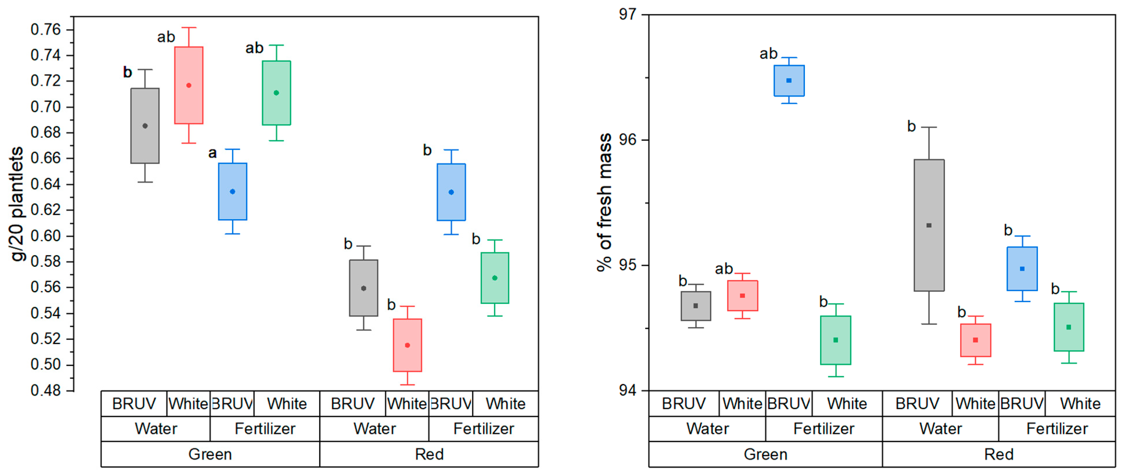
Figure 1. Biomass accumulation (left) and water contents (%of fresh mass)(right) in Ocimum basilicum L. (BRUV – blue:red:UV light treatment, White – full spectrum light treatment).
Preprints 110630 g001
Figure 2. Selected phenolic acids (left) and rosmarinic acid (right) contents in Ocimum basilicum L. (different letters indicate statistical significance, p<0.05) (BRUV – blue:red:UV light treatment, White – full spectrum light treatment; BR – Red basil cultivar, BV – Green basil cultivar).
Preprints 110630 g002
Figure 3. Specific gene expression changes in Ocimum basilicum L., red cultivar (left) and green cultivar (right).
Preprints 110630 g003
Figure 4. LED emission spectra (left) BRUV Treatment; (right) White Treatment).
Preprints 110630 g004
Table 1. Chlorophyll contents and chlorophyll fluorescence markers in Ocimum basilicum L. (BRUV – blue:red:UV light treatment, White – full spectrum light treatment).
Cultivar Fertilizer Treatment Fs Fm’ ΦPSII Chlorophyll (AU)
Green Water BRUV 474.63a±17.32 2106.89ab±72.83 0.78ab±0.01 6.33a±0.28
White 539.99a±21.1 2245.66a±80.99 0.77b±0.01 5.16a±1.91
Fertilizer BRUV 561.67a±89.45 2288.25ab±343.99 0.76ab±0.02 -
White 568.34a±25.77 2378.59a±99.4 0.77ab±0.01 -
Red Water BRUV 287.67b±16.19 1288.27c±64.63 0.79a±0.01 5.97a±0.37
White 321.5b±16.79 1388.1c±54.73 0.78ab±0.01 3.22b±0.37
Fertilizer BRUV 415.59ab±20.99 1652.67bc±90.88 0.75b±0.01 -
White 505.34a±27.53 2083a±133.11 0.76ab±0.01 -
Table 2. Total phenolic contents and antioxidant activity of Ocimum basilicum L. extracts (BRUV – blue:red:UV light treatment, White – full spectrum light treatment).
Cultivar Fertilizer Treatment Total phenolic content (µg GAE/g dry plant mass) Antioxidant activity (% inhibition)
Green Water BRUV 3739.1b±202.11 88.08±1.71a
White 3846.48b±200.94 88.19±4.83a
Fertilizer BRUV 5739.56b±313.12 85.28±1.54a
White 4858.96b±264.71 81.4±3.85a
Red Water BRUV 8194.1ab±476.49 86.96±3.97a
White 7219.1ab±425.58 92.73±1.8a
Fertilizer BRUV 14063.79a±755.69 87.84±1.26a
White 13145.41a±768.87 92.33±1.46a
Disclaimer/Publisher’s Note: The statements, opinions and data contained in all publications are solely those of the individual author(s) and contributor(s) and not of MDPI and/or the editor(s). MDPI and/or the editor(s) disclaim responsibility for any injury to people or property resulting from any ideas, methods, instructions or products referred to in the content.
Copyright: This open access article is published under a Creative Commons CC BY 4.0 license, which permit the free download, distribution, and reuse, provided that the author and preprint are cited in any reuse.
Prerpints.org logo

Preprints.org is a free preprint server supported by MDPI in Basel, Switzerland.

Subscribe

© 2024 MDPI (Basel, Switzerland) unless otherwise stated