Preprint
Article

Evaluation of Gastric Digestion Behavior of Protein Sports Supplements by In Vitro Dynamic Gastrointestinal Digestion Model

Altmetrics

Downloads

138

Views

64

Comments

0

This version is not peer-reviewed

Submitted:

05 July 2024

Posted:

09 July 2024

You are already at the latest version

Alerts
Abstract
Commercial protein supplements are the main sources for the sports nutrition. Nowadays, those protein supplements mainly differ in the ingredients and formulations, possibly affecting the digestion behavior and protein absorption during gastrointestinal digestion. Herein, this work aimed to investigate the gastric digestion behavior and protein hydrolysis of commercial protein supplements by using an in vitro dynamic digestion model. Five commercial protein supplements were selected, including three protein powders and two liquid protein beverages. The results indicated that liquid protein beverages had smaller particle size than protein powder dispersions, and one of the protein powders existed as the microcapsules. During gastric digestion, all samples tended to form aggregates as the pH decreased, and the degree of aggregation was more pronounced in liquid protein beverages than in protein powders, where the intact structure of the microcapsules was gradually disrupted. Protein hydrolysis degree in liquid protein beverages was higher than that in protein powders. One of the liquid products (M20) had the highest protein hydrolysis degree of around 35.74% after 120 min gastric digestion and a half-empty time of 37.82 min. These findings suggest that the formulation and ingredients of commercial protein supplements will affect their digestion behavior, resulting in varying degree of protein hydrolysis. Hence, sports supplements should be carefully designed to provide precise nutrition for the athletes and exercisers.
Keywords: 
Subject: Biology and Life Sciences  -   Food Science and Technology

1. Introduction

With the growing concern on health, exercise has gone from specialization to publicization. During exercise, the body inevitably suffers from fatigue and loss of nutrients, which ultimately causes damage to the body [1]. Therefore, nutritional supplements that can rapidly improve body functions in a short period of time are gradually gaining attention. Nutritional supplements commonly include sugars, proteins, amino acids, vitamins, creatine, and taurine and so on [2,3]. Among them, sports nutrition supplements with high protein content are often used to meet the needs of athletes and sportspersons for daily exercise and high-intensity training, which have a positive impact on the body's ability to balance the breakdown and synthesis of muscle proteins, slimming down, improving body composition, restoring normal functions after exercise, and enhancing immunity.
Currently, most commercially available sports protein supplements in the market mainly differ in the ingredients (mainly in proteins and other nutrients) and formulations (powder or liquid). Proteins are the principal factor to meet the requirements of the body after exercise, due to their composition and digestion behavior. Whey proteins, milk proteins and soy proteins are the main protein ingredients in the commercial protein supplements. After oral administration, proteins will be hydrolyzed by digestive enzymes (pepsin and trypsin) to provide peptide and amino acids for the specific functions [4], such as increasing muscle mass, shortening recovery time, accelerating muscle repair, reducing post-workout muscle loss and increasing metabolic rate and so on [5]. Hence, the hydrolysis of proteins might affect the quality of those products. However, digestion behavior and absorption of proteins is not only related to the physiological properties of the gastrointestinal tract, but also depends on a variety of other factors, including protein types, protein sources, formulations, processing methods, other components (minerals, polysaccharides, polyphenols and others) and intake temperature and so on [6,7,8,9,10].
Whey protein mainly contains α-lactalbumin and β-lactoglobulin, and casein mainly exists as four types of αs1-, αs2-, β-, and κ-casein. During digestion, whey proteins are resistant to the action of pepsin and gastric acid because they are globular and dense proteins that hardly coagulate due to changes in pH, whereas casein proteins are easily broken down into polypeptides by the action of pepsin because of their loose protein structure. The micellar structure of casein is also found to be readily broken down during gastric digestion, followed by coagulation and the production of a mixture of peptides and free amino acids [11]. This results in a slower transport rate of milk casein to the intestine. In contrast to animal proteins, plant proteins are deficient in the essential amino acid composition and contain anti-nutritional factors resulting in lower digestibility [12]. Processing procedures such as heat treatment, thermo-mechanical, ultrasonic, microwave and high pressure homogenization are often required to produce high quality or long shelf-life protein products [13], and these procedures can also lead to changes in the digestive behavior of proteins. The treatment of pasteurization, autoclave and microwave was known to affect the microstructure of proteins in stomach, resulting in different empty rate and digestibility [14]. Raw and autoclaved milk formed a more compact curd structure in the stomach, and hence autoclaved milk empties from the stomach at the lowest rate, and pasteurized milk had the highest protein digestibility. Therefore, it is critical that protein sports supplements should be rationally designed and processed.
Although there have been lots of protein sports supplements, few studies are carried out to evaluate the hydrolysis of proteins in those products. Herein, five popular commercially available sports protein supplements, including three protein powders and two liquid protein beverages, were selected for investigation. The general properties of those products were first characterized. Then, their gastric digestion and emptying behaviors were evaluated by using in vitro dynamic human stomach system. The results may provide supports for the development and rational selection of sports protein nutrition supplements.

2. Materials and Methods

2.1. Materials

All the protein sports supplements in the present study were donated by Shanghai M-Action Health Technology Co., LTD (Shanghai, China). Liquid protein beverage M20 (protein content 20 g/300 mL) mainly contains milk protein concentrate and collagen peptides. Liquid protein beverage M25 (protein content 25 g/300 mL) mainly contains milk protein concentrate, whey protein powder, collagen peptides and calcium caseinate. The other compositions in M25 include 14 vitamins and minerals, while that in M20 is only vitamin D. Protein powder KBT (protein content 88 g/100 g) contains whey protein concentrate and wheat protein. Protein powder SB (protein content 75 g/100 g) contains whey protein concentrate, whey protein isolate, hydrolyzed whey protein, α-Lactalbumin and colostrum. Protein powder TC (protein content 80 g/100 g) contains soy protein isolate, whey protein concentrates and soybean lecithin. Pepsin from porcine gastric mucosa (P7000, EC No.: 232–629-3, ≥250 units/mg solid) was purchased from Sigma (Sigma-Aldrich, USA). All other chemicals and reagents (analytical grade or HPLC grade) were purchased from either Sinopharm Chemical Reagent Co., Ltd (Shanghai, China) or Sigma-Aldrich Company.

2.2. Sample Treatments

The actual total protein content in commercial protein supplements and protein powders was first determined by the Kjeldahl method. Then, the protein content in all samples was set at 50 mg/mL for the digestion experiments. Liquid samples were directly diluted by water. Powder samples were collected and dispersed in water. In detailed, samples of M20, M25, KBT, SB, TC were weighted by 167.07g, 134.68 g, 15.92 g, 16.59 g, 13.66 g, respectively, and then correspondingly added into 62.93g, 95.32 g, 214.08 g, 213.41 g, 216.34 g of water. The mixture was stirred for 30 min before in vitro dynamic digestion experiments.

2.3. In Vitro Dynamic Gastric Digestion

In vitro dynamic human stomach system (DIVHS-Ⅰ, Xiao Dong Pro-health Instrumentation Co., Ltd, Suzhou, China) was used to investigate the digestive behavior and gastric emptying of protein sports supplements. Simulated gastric fluid (SGF) containing pepsin was prepared according to the INFOGEST standard digestion method [15]. The pH value of SGF was adjusted to 1.6 ± 0.1 using hydrochloric acid (HCl). Then SGF and HCl solution (1 mol/L) were held in a water bath at 37°C for 30 min prior to being added to the syringe.
The in vitro dynamic gastric digestion experiments were performed in triplicate following procedure as previously described with minor modifications [14]. Briefly, the oral digestion experiment was not implemented due to the short residence time in the oral cavity. The stomach vessel was pre-injected with 30 mL of SGF to simulate the fasting state of the human body as realistically as possible. Afterwards, 230 mL of sample was introduced into the stomach vessel through the funnel on top of the apparatus within 6 min to achieve a feeding rate of 38 mL/min. During this time, three contractions per min were generated in the stomach vessel by a pneumatically driven roller device. The gastric digestion experiments continued for 2 h and the temperature in the digestion system was maintained at 37°C.
The secretion of SGF and HCl in the stomach were simulated according to the parameters in Table 1. HCl was secreted within the initial 30 min at the rate of 1.0 mL/min. SGF was secreted at different rates during 2 h. The ratio of sample to SGF at the beginning of stomach digestion was 1:1, consistent with in vitro static digestion experiments. The tilting angle was remained at + 2° (0 – 5min), in the next - 0.6° (5 – 30 min), and in the last - 0.4° min (30 – 120min).
Emptied digesta were collected every 15 min during gastric digestion. The visual image, weight and pH value were taken or determined immediately. Emptied digesta of 5 mL was mixed with an equal volume of trichloroacetic acid solution (24%). The mixture was centrifuged and the supernatant was collected to determine the hydrolysis degree of protein. Meanwhile, 1 mL of the digesta sample was taken and the pepsin inhibitor (pepstatin A,10 μL, 0.135mg/mL) was added to inhibit enzyme activity for SDS-PAGE patterns analysis and microstructure observation.

2.4. Microstructure Characterization

The microstructure of samples was observed using confocal laser scanning microscope (CLSM, FV3000, OLYMPUS Corporation, Japan). Proteins were labeled with fast green dye (FCF, 1 wt% dissolved in Ethanol absolute, excitation/emission wavelength = 494/518 nm). 10 μL of the sample was placed on a slide and covered by coverslip, and then the microstructure was observed under a 100× oil immersion lens (raw sample) or 20× (digested sample) lens after the slides were covered.

2.5. Determination of Gastric Emptying Rate

Retention rate of samples at a given time was calculated as the weight of gastric digesta remaining in the stomach vessel divided by the total weight of gastric contents (including samples, SGF and HCl) entering the stomach at the same time. To better quantify gastric emptying behavior, the gastric retention data were fitted to a modified Elashoff power index model [16,17].
y t = 1 ( 1 e k t ) β  
where y(t) is the fraction of digesta remaining in the stomach vessel at time t (min), κ is the gastric emptying rate per minute (1/min), and β is the parameter that measures the degree of concavity of the curve.
The half time (t1/2) of gastric emptying was calculated when y(t) = 0.5 according to the following equation.
t 1 / 2 = 1 k × ln ( 1 0.5 1 β )  
The parameters k, β, t1/2 of gastric emptying model were obtained by nonlinear least squares fitting in GraphPad prism 9.5.

2.6. Determination of Protein Hydrolysis Degree

The hydrolysis degree of protein was determined by Kjeldahl method[18]. Digesta collected at different times of 5 mL was mixed well with 5 mL of trichloroacetic acid solution (24%). Then the mixture was centrifuged at 10,000 × g for 10 min at 4°C and the supernatant was taken. The supernatant (accurate to 0.001 g) was weighed into a digestion tube and processed process at high temperature. The distillation process was achieved using a semi-automatic Kjeldahl apparatus and the distillate was titrated using 0.05053 mol/L HCl. A protein coefficient of 6.38 was used to convert the nitrogen content of the samples to soluble protein content.

2.7. SDS-PAGE Analysis

Protein composition of sports nutrition supplements during gastric digestion was assessed by SDS-PAGE under reducing conditions. After the treatment of pepsin inhibitor, the digesta samples were centrifuged at 8000 × g for 10 min and supernatant was collected. The protein concentration was first determined by using a BCA protein quantification kit (Elabscience Biotechnology Co., Ltd.). Then the protein concentration in each sample was adjusted to the same before the SDS-PAGE analysis. The diluted samples were mixed with loading buffer (5×) and heated for 10 min to denature the proteins sufficiently, and then centrifuged. Then, 20 μL of mixed samples were loaded per lane in 12.5% separating gel, and 5 μL of protein marker (molecular weight range 10-180 kDa, Thermo Fisher Scientific, Waltham, MA, USA) was used to detect the protein band position. The electrophoresis voltage was set at 120 V. After electrophoresis, the gel was stained with Coomassie Brilliant Blue (R-250), then decolorized (approximately 2 hours) and finally imaged using gel documentation system (*GelDoc XR+, Bio-Rad Laboratories, Inc, USA).

2.8. Statistical Analysis

All experimental data were repeated for at least three times and the results were presented as mean ± standard deviation of three replicates. Statistical significances between the samples were analyzed by one-way analysis of variance (ANOVA) followed by Duncan multiple comparisons test using SPSS 25.0 software. P <0.05 was considered as statistically significant.

3. Results and Discussion

3.1. Physical Statement and Particle Size of Commercial Protein Sports Supplements

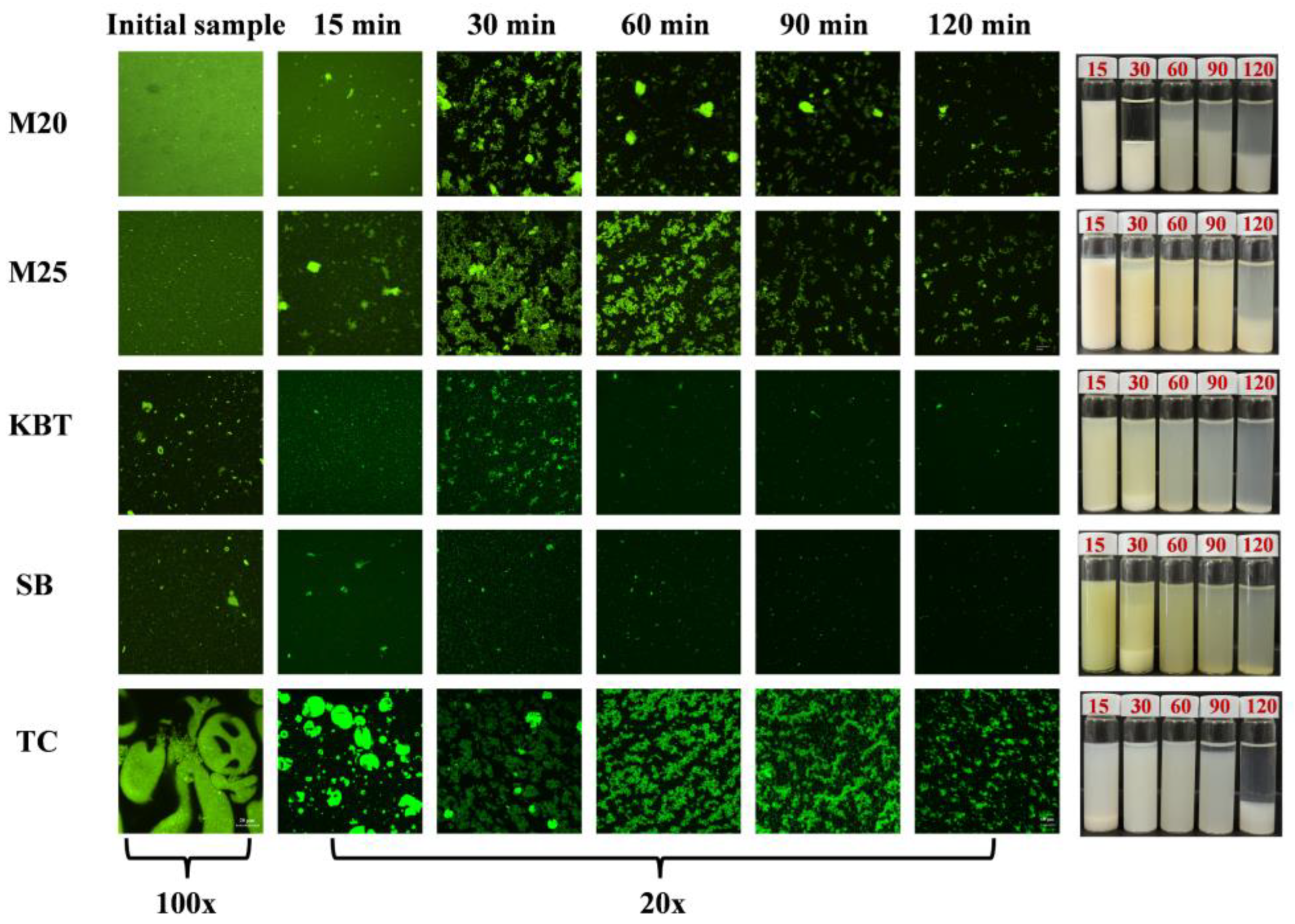
Protein powder samples were first dispersed or diluted in water before the characterization. Compared with liquid protein beverages, protein powder dispersions were prone to precipitation, in which TC sample had the thickest precipitation layer (Figure 1A). The average particle size (d32) of liquid protein beverages M20 and M25 was similar, around 140 nm, which was significantly smaller than that of protein powder dispersions (Figure 1B). Particle sizes of KBT, SB and TC samples were about 500 nm, 850 nm and 45 μm, respectively. Due to the relatively large particle size, TC sample tended to precipitate quickly. Liquid protein beverages displayed mono-modal peak in particle size distribution with the primary peak at approximately 140 nm (Figure 1C). However, KBT and SB samples displayed multiple peaks, ranging from nanometer to micrometer. The primary peak of the TC sample was distributed in the region of larger particle size (approximately 85 μm). The change of particle size d43 also indicated the presence of large particles in KBT and SB, while d43 of M20 and M25 was much smaller (Figure 1B), contributing to the higher uniformity and stability of liquid protein beverages [19,20]. The microstructure further confirmed the above results that TC sample presented as microcapsule structure, while the distribution of proteins in the other samples was more homogenous (Figure 3A). Relatively more large protein aggregates were observed in KBT and SB samples than liquid protein beverages (M20, M25). The results demonstrated that the commercial protein sports supplements had different physical properties, depending on the specific processing procedures.

3.2. Variation of pH Values during Gastric Digestion

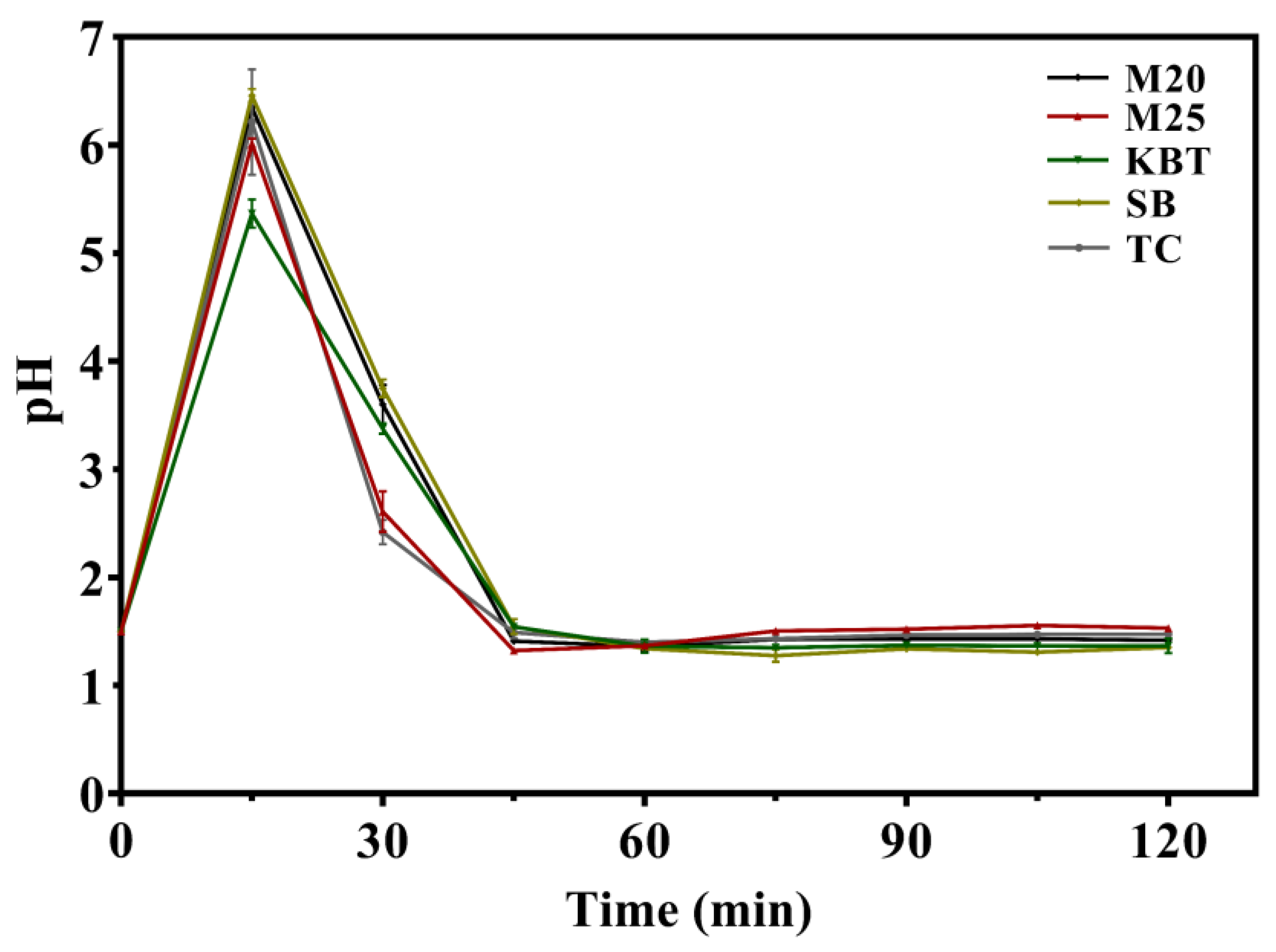
In vitro dynamic digestion system can mimic the relatively real digestion process, such as regulating the secretion rate of digestive fluid and HCl, simulating gastric peristalsis, squeezing, and screening [21]. Hence, the change of pH values in stomach digesta was recorded in Figure 2. Overall, pH changing profiles of all samples followed a similar trend during the gastric digestion. Due to the pre-injection of 30 mL SGF into the stomach, the initial pH of digesta was around 1.6. Subsequently, the intake of samples diluted the SGF and secreted HCl, causing the increase of pH value within 20 min. Due to the continuous secretion of SGF and HCl, pH value gradually decreased again with longer digestion time. After 45 min, pH value of digesta recovered as the initial level and kept constant. Similar pH changing profiles were observed in previous publications, which evaluated in vitro dynamic digestion behavior of curcumin yogurt and infant formula [22,23]. However, the buffering capacity of the chyme also affects the change of intragastric pH value at the same content secrete of HCl. The results in Figure 2 indicated that different protein samples had discrepancy in pH-responsive during gastric digestion. SB showed a higher buffering capacity than other samples within 30 min (~ pH 3.75), followed by M20 (~ pH 3.60) > KBT (~ pH 3.38) > M25 (~ pH 2.61) > TC (~ pH 2.42). After 45 min, the pH values of all samples turned into equilibrium, close to the initial pH value of gastric fluid. Among human milk, bovine milk, and camel milk, pH value of bovine milk during gastric digestion varied more significantly, whereas the digesta of human milk showed stronger buffering capacity against the acidic environment in the stomach [24]. Different compositions of milk lipids and proteins, and the substrate preferences of digestive enzymes used for in vitro digestion were responsible for the variation in pH value. Moreover, thermal processing and homogenization were found to delay the pH decline of bovine milk digesta during gastric digestion [25]. For example, pH value of raw milk decreased significantly in the stomach while that of treated milk tended to be stable after 120 min to 300 min, it is related to the fragmented curd structure formed by homogenization and thermal processing on casein, a quantity of fragmented structures were dispersed in the stomach to act as buffering components against the decline of pH value [26].

3.3. Microstructure Change of Protein Digesta during Gastric Digestion

Due to the pH alteration and enzyme effect, proteins are apt to form aggregation in the gastric digestion [27,28] . After passing through in vitro dynamic digestion system, the digesta was first emptied out at 15 min. The collected samples were unstable and some precipitates were found in the bottom of the bottle (Figure 3). The thickness of precipitation layer altered, due to the aggregation of proteins during gastric digestion. M20 and M25 had higher thickness of precipitation layer, contributing to the formation of large and dense aggregations (Figure 3), which was induced by pepsin to collapse the hairy layer of κ-casein and a decrease in pH [29]. In contrast, protein aggregation was not significant in KBT and SB samples due to the high percentage of whey proteins, which were more soluble in gastric fluid. TC sample retained an integrated microcapsule structure after 15 min of digestion, and the microcapsule structure gradually ruptured and disappeared as digestion progressed. Due to the hydrolysis of proteins by pepsin, the turbidity of digesta decreased with longer digestion time, indicating the breakdown of protein aggregations (Figure 3). It has been reported that protein fractions, thermal processing and homogenization treatments, and fat content could tailor curd production, protein structure and properties during digestion [26,30]. Compared to protein powder, liquid proteins contained a great amount of casein, which tended to aggregate in the presence of gastric fluids, forming the open, porous and flexible structure [31]. Pre-treatment of decalcification increased the degradation of casein in the stomach, due to the formation of looser curd structure [6]. The casein to whey protein ratio was also a factor influencing digestive behavior, especially heat-treated protein samples. Casein content above 50% resulted in the formation of curds and generated the delay in gastric emptying [32]. The casein-rich skimmed goat milk ( 80%) showed a high rate of protein hydrolysis and the formation of a looser clot after heat treatment at 85℃ [33]. Moreover, proteins from human breast milk and a variety of mammalian milk differed in gastric digestion behavior and hydrolysis degrees [7].

3.4. Gastric Emptying Curves

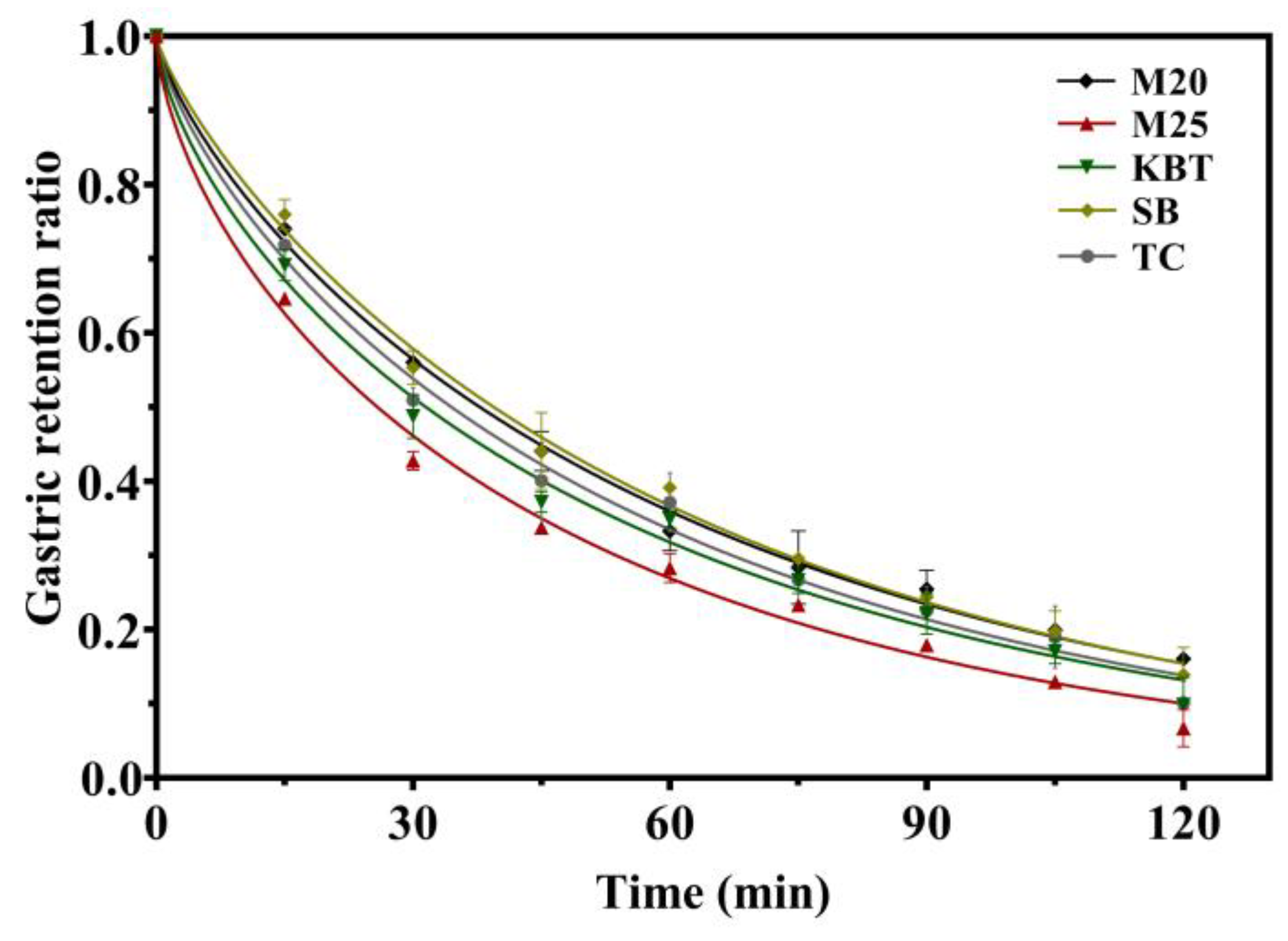
It is known that the digesta in the stomach will be discharged from stomach into the intestines by the action of gastrointestinal peristalsis, which is called as the gastric emptying process. This gastric emptying refers to a complex and critical process to ensure the absorption of nutrients in the intestine [34]. In vitro dynamic digestion system in the present paper can control the tilt angle of stomach vessel to mimic this process by rotating the turntable [35,36,37,38,39]. Figure 4 displayed the gastric emptying curves of different protein samples within 2 h gastric digestion, as represented by the retention rate of the protein digest inside the stomach over time. Similar gastric emptying trends were observed for all the samples which had a decreasing retention ratio with longer digestion time. M25 sample maintained a lower gastric retention rate throughout gastric emptying than other ones (approximately from 0.6462 to 0.0667). Gastric retention ratio of M20 and SB samples were relatively higher, but began to decrease after 30 min digestion, which kept similar trends as the other samples. The casein-rich M20 sample was prone to form solid curd structure that was persisted throughout gastric digestion, ultimately leading to a delay in gastric emptying [32]. In contrast, casein-rich M25 sample also contain a certain amount of whey protein, enabling the formation of soluble curd structure in the stomach through the action of gastric acid and enzyme and facilitating gastric emptying [40]. This may be due to the fact that the denatured whey proteins by the high temperature were joined to the casein, resulting in the structure looser compared to the curd only consisting of caseins [33,41]. Hence, protein-induced coagulation and digestion processes could be controlled through the proper selection of protein components and the ratio of casein to whey protein, thus satisfying diverse nutritional requirements. [23] In order to quantitatively describe the gastric emptying behavior, the retention rate profiles of samples were fitted using the modified Elashoff model (equation 1) [16], which allows a visual and quantitative comparison of their emptying rates [14,38,39]. The coefficient of determination r2 after equation fitting varied between 0 to 1, with r2 approaching 1, indicating that the equation model is suitable for quantifying the gastric emptying behavior of these products in the DIVHS system. After fitting, there were three parameters (k, β and t1/2) to quantifying the empty process. k, β and t1/2 represent the gastric emptying rate per minute, the intercept measuring the concavity of the curve and the half time (t1/2) of gastric emptying, respectively. Table 1 presented that the β values of all samples were less than 1, indicating that the gastric emptying behavior of samples possesses the characteristics of initial rapid emptying without significant lag period. The t1/2 was defined as the time to empty 50% of the meal from stomach into the intestine. Compared to other samples, M25 showed the lowest t1/2 (25.83 ± 0.12 min) and SB sample had the highest t1/2 (39.43 ± 0.98 min) (P<0.05). This implied that the M25 sample could be emptied within a relatively short gastric residence time, facilitating further digestion and absorption of protein.
Table 2. Gastric emptying model parameters for the sports protein supplements.
Sample k × 10-2 (1/min) β t1/2 (min) r2
M20 1.35 ± 0.07b 0.754 ± 0.014b 37.82 ± 1.21a 0.9963
M25 1.55 ± 0.02ab 0.626 ± 0.009c 25.83 ± 0.12d 0.9938
KBT 1.38 ± 0.06b 0.663 ± 0.002c 31.52 ± 1.50c 0.9927
SB 1.40 ± 0.09b 0.806 ± 0.026a 39.43± 0.98a 0.9948
TC 1.41 ± 0.15b 0.726 ± 0.028b 34.58 ± 1.97b 0.9923
Values are expressed as mean ± deviation of the three replications, and values in the same column with different letters differ significantly at the level of p < 0.05.

3.5. Protein Hydrolysis Degree of Commercial Protein Supplements

Due to the presence of protein enzyme in the stomach, proteins can be hydrolyzed into peptides and amino acids. As shown in Table 3, the hydrolysis degree of proteins for all the samples gradually increased during gastric digestion, ranging from 16.16% to 35.74% after 120 min, respectively. Liquid protein beverages (M20 and M25) kept higher protein hydrolysis degree than protein powder dispersions (KBT, SB, TC). After 120-min digestion, the protein hydrolysis degree of M20 and M25 was 35.74 ± 0.33% and 30.44 ± 3.45%, which was 16.16 ± 1.54%, 16.66 ± 0.08%, 17.90 ± 0.58% for KBT, SB, TC, respectively. The results demonstrated that proteins in liquid protein beverages were easier to be hydrolyzed than those in protein powders under the action of pepsin enzyme. Numerous factors have been reported to affect the digestion behavior and hydrolysis of proteins, including protein types [42], processing methods [43], formulations [44], other co-existed components [45]. In the present work, the main protein in liquid protein beverages was casein, which aggregated during digestion to form a curd with a loose structure and large surface area, and then pepsin could diffuse into the curd fast, thus accelerating protein hydrolysis [41,46]. Protein powders mainly contained whey proteins, which might have higher resistance to pepsin during digestion [47]. However, it was difficult to exactly explain why liquid protein beverages showed higher protein hydrolysis degree, since all the protein supplements experienced different processing methods and contained various co-compositions.

3.6. Protein Composition after Gastric Digestion by SDS-PAGE Analysis

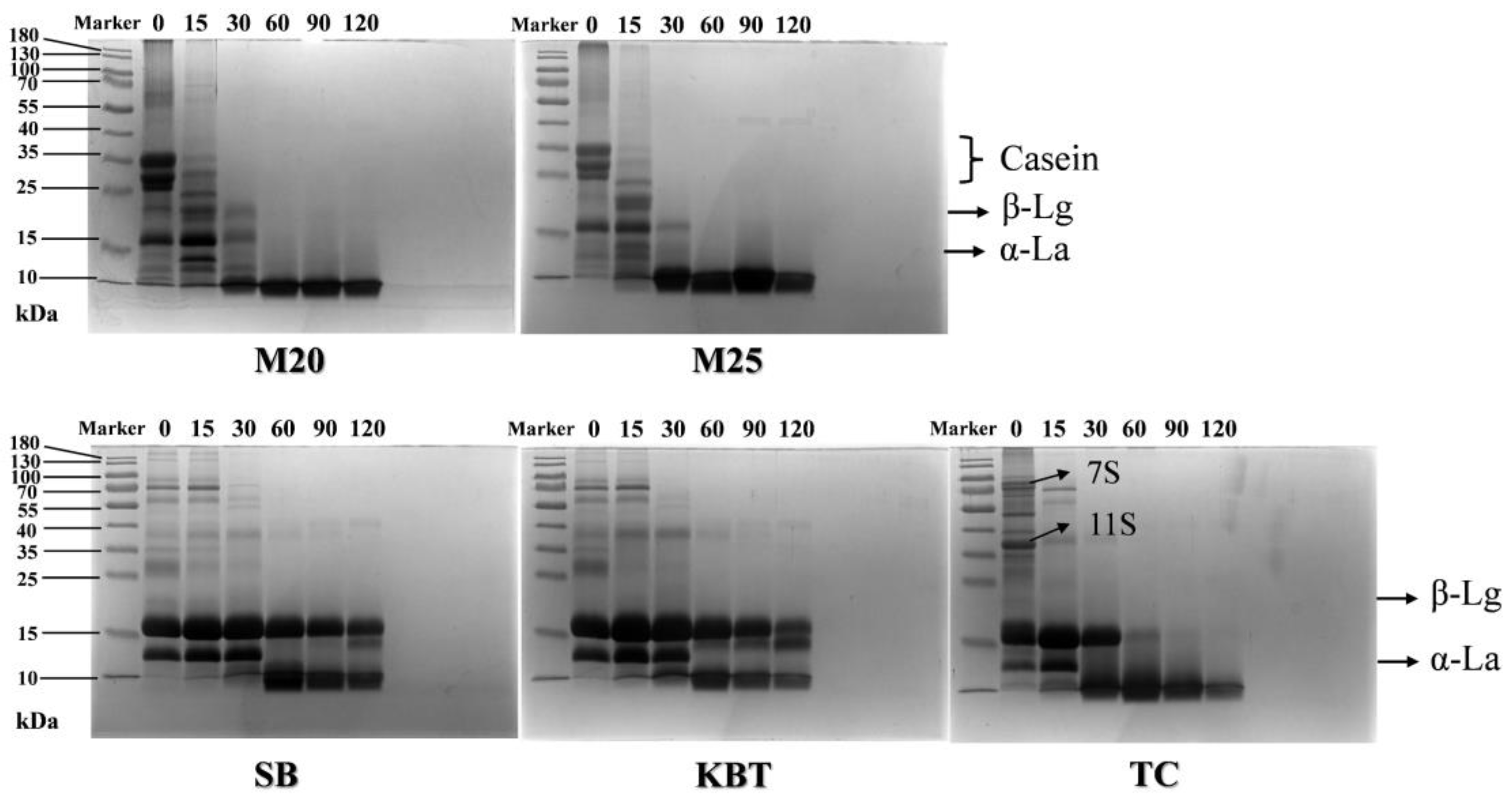
Hydrolysis of proteins causes the produce of peptides and amino acids with lower molecular weight. Before digestion, the main bands in M20 and M25 located at 23 - 35 kDa and 13-19 kDa, corresponding to the presence of casein and whey proteins from milk protein concentrate (Figure 5). The strong bands at 13-19 kDa were observed in all protein powder samples (KBT, SB and TC), corresponding to the presence of β-lactoglobulin (β-Lg) and α-lactalbumin (α-La) from whey protein. TC sample also had the relative strong bands at 67-70 kDa and 35 - 47 kDa, indicating the presence of 7S and 11S globulin protein from soybean protein. For M20 and M25, more bands at low molecular weight were observed after 15 min digestion. The casein bands in M25 were hardly seen after 30 min, and the bands above 10 kDa in M20 were more than those in M25. In addition, the intensity of β-Lg bands gradually became weaker and stronger bands appeared at molecular weight of 10 kDa, especially in M20, indicating the faster hydrolysis of proteins in M20. Liquid protein beverages were possibly subjected to thermal processing and denaturation at high temperatures, facilitating the hydrolysis of casein and β-Lg by pepsin[48]. For protein powder samples (KBT and SB), the intensity of β-Lg and α-La bands kept the same within 30 min digestion. Subsequently, α-La band almost disappeared after 30 min digestion, while strong β-Lg band was still observed after 120 min. It was reported that whey proteins without any treatments, especially β-Lg, were resistant to hydrolysis by pepsin due to their compact spherical structure. [41,49,50,51]. The β-Lg band in TC sample decreased gradually with the increase of the digestion time, and the α -La band disappeared after 30 min. More bands at low molecular weight in TC during digestion were observed than those in KBT and SB, indicating that the combination of soybean protein might enhance the hydrolysis process of whey protein. The results in SDS-PAGE analysis were consistent with those in Table 1, which milk protein concentrate-rich liquid protein beverages presented higher protein hydrolysis degree than whey protein-rich protein powder samples. Taking into account the composition, formulation and processing of all the commercial protein supplements, more factors might be considered to favor the protein hydrolysis and absorption during the design of protein supplements, including thermal, ultrasound, or pulsed electric fields processing [13,52]

4. Conclusions

The protein hydrolysis process of five commercial protein sports supplements including liquid protein beverages and protein powders was evaluated by using in vitro dynamic gastric digestion system. Liquid protein beverages had smaller particle size and more uniform particle size distribution then protein powder dispersions. All the samples showed different microstructure before and after gastric digestion. Liquid protein beverages were apt to form large aggregates than protein powders. Protein hydrolysis degree of liquid protein beverages was much higher than that of protein powder dispersions. After 120 min digestion, M20 had the highest protein hydrolysis degree of 35.74%, which was 2.2, 2.1 and 2.0 times higher than protein powder KBT, SB and TC, respectively. Hence, the findings might suggest that the application of liquid formulation and milk protein concentrate is potentially more favorable for protein absorption than protein powders. It is worthy to note that both the composition of protein supplement and processing procedures will affect digestion behavior of proteins, which should be considered for the precise design and development of high-protein sports supplements.

Author Contributions

Conceptualization, Y.L. and W.D.; methodology, W.L. and Y.L.; data curation, Y.L; formal analysis, W.L. and Y.L.; project administration, Y.L., W.D., C.T., and Y.Z.; visualization, W.L.; validation, W.D., C.T., and Y.Z.; writing—original draft preparation, W.L.; writing—review & editing, supervision, and funding acquisition, Y.L. All authors have read and agreed to the published version of the manuscript.

Acknowledgments

This work was supported by National Natural Science Foundation of China (No. 32372325).

Data Availability Statement

The data that support the findings of this study are available on request from the corresponding author.

Conflicts of Interest

The authors declare no conflicts of interest.

References

  1. Cui, P.; Li, M.; Yu, M.; Liu, Y.; Ding, Y.; Liu, W.; Liu, J. Advances in sports food: Sports nutrition, food manufacture, opportunities and challenges. Food Res. Int. 2022, 157, 111258. [CrossRef]
  2. Alonso, M.R.; Fernández-García, B. Evolution of the use of sports supplements. PharmaNutrition 2020, 14, 100239. [CrossRef]
  3. López-Martínez, M.I.; Miguel, M.; Garcés-Rimón, M. Protein and Sport: Alternative Sources and Strategies for Bioactive and Sustainable Sports Nutrition. Front. Nutr. 2022, 9, 926043. [CrossRef]
  4. Maeda, N.; Dulko, D.; Macierzanka, A.; Jungnickel, C. Analysis of the Factors Affecting Static In Vitro Pepsinolysis of Food Proteins. Molecules 2022, 27, 1260. [CrossRef]
  5. Khatkar, S.K.; Khatkar, A.B.; Mehta, N.; Kaur, G.; Dhull, S.B.; Prakash, S. Effective strategies for elevating the techno-functional properties of milk protein concentrate. Trends Food Sci. Technol. 2023, 140. [CrossRef]
  6. Wang, K.; Liu, D.; Tao, X.; Zhang, J.; Huppertz, T.; Regenstein, J.M.; Liu, X.; Zhou, P. Decalcification strongly affects in vitro gastrointestinal digestion of bovine casein micelles under infant, adult and elderly conditions. Food Hydrocoll. 2023, 139. [CrossRef]
  7. Xiao, T.; Zeng, J.; Zhao, C.; Hou, Y.; Wu, T.; Deng, Z.; Zheng, L. Comparative Analysis of Protein Digestion Characteristics in Human, Cow, Goat, Sheep, Mare, and Camel Milk under Simulated Infant Condition. J. Agric. Food Chem. 2023, 71, 15035–15047. [CrossRef]
  8. Ren, Q.; Keijzer, P.; Wichers, H.J.; Hettinga, K.A. Glycation of goat milk with different casein-to-whey protein ratios and its effects on simulated infant digestion. Food Chem. 2024, 450, 139346. [CrossRef]
  9. Lavoisier, A.; Morzel, M.; Chevalier, S.; Henry, G.; Jardin, J.; Harel-Oger, M.; Garric, G.; Dupont, D. In vitro digestion of two protein-rich dairy products in the ageing gastrointestinal tract. Food Funct. 2023, 14, 9377–9390. [CrossRef]
  10. Nguyen, H.T.; Afsar, S.; Day, L. Differences in the microstructure and rheological properties of low-fat yoghurts from goat, sheep and cow milk. Food Res. Int. 2018, 108, 423–429. [CrossRef]
  11. Miralles, B.; Sanchón, J.; Sánchez-Rivera, L.; Martínez-Maqueda, D.; Le Gouar, Y.; Dupont, D.; Amigo, L.; Recio, I. Digestion of micellar casein in duodenum cannulated pigs. Correlation between in vitro simulated gastric digestion and in vivo data. Food Chem. 2021, 343, 128424. [CrossRef]
  12. Ashkar, F.; Wu, J. Effects of Food Factors and Processing on Protein Digestibility and Gut Microbiota. J. Agric. Food Chem. 2023, 71, 8685–8698. [CrossRef]
  13. Kaur, L.; Mao, B.; Beniwal, A.S.; Abhilasha; Kaur, R.; Chian, F.M.; Singh, J. Alternative proteins vs animal proteins: The influence of structure and processing on their gastro-small intestinal digestion. Trends Food Sci. Technol. 2022, 122, 275–286. [CrossRef]
  14. Liu, Z.; Suolang, Q.; Wang, J.; Li, L.; Luo, Z.; Shang, P.; Chen, X.D.; Wu, P. Formation of structured clots, gastric emptying and hydrolysis kinetics of yak milk during in vitro dynamic gastrointestinal digestion: Impact of different heat treatments. Food Res. Int. 2022, 162, 111958. [CrossRef]
  15. Minekus, M.; Alminger, M.; Alvito, P.; Ballance, S.; Bohn, T.; Bourlieu, C.; Carrière, F.; Boutrou, R.; Corredig, M.; Dupont, D. A standardised static in vitro digestion method suitable for food–an international consensus. Food Funct. 2014, 5, 1113-1124.
  16. Elashoff, J.D.; Reedy, T.J.; Meyer, J.H. Analysis of Gastric Emptying Data. Gastroenterology 1982, 83, 1306–1312. [CrossRef]
  17. Urbain, J.-L.C.; Siegel, J.A.; Charkes, N.D.; Maurer, A.H.; Malmud, L.S.; Fisher, R.S. The two-component stomach: effects of meal particle size on fundal and antral emptying. Eur. J. Nucl. Med. 1989, 15, 254–259. [CrossRef]
  18. Barbano, D.M.; Lynch, J.M.; Fleming, J.R. Direct and Indirect Determination of True Protein Content of Milk by Kjeldahl Analysis: Collaborative Study. J. AOAC Int. 1991, 74, 281–288. [CrossRef]
  19. Wang, Y.; Li, M.; Wen, X.; Tao, H.; Wang, K.; Fu, R.; Tao, H.; Wang, F.; Chen, N.; Ni, Y. Conformational changes and the formation of new bonds achieving robust nanoemulsions by electrostatic interactions between whey protein isolate and chondroitin sulfate. Food Hydrocoll. 2023, 136. [CrossRef]
  20. Yu, C.; Zheng, L.; Cai, Y.; Zhao, Q.; Zhao, M. Desirable characteristics of casein peptides with simultaneously enhanced emulsion forming ability and antioxidative capacity in O/W emulsion. Food Hydrocoll. 2022, 131. [CrossRef]
  21. Chen, L.; Xu, Y.; Fan, T.; Liao, Z.; Wu, P.; Wu, X.; Chen, X.D. Gastric emptying and morphology of a ‘near real’in vitro human stomach model (RD-IV-HSM). J. Food Eng. 2016, 183, 1-8.
  22. Ye, Q.; Ge, F.; Wang, Y.; Wu, P.; Chen, X.D.; Selomulya, C. Digestion of curcumin-fortified yogurt in short/long gastric residence times using a near-real dynamic in vitro human stomach. Food Chem. 2022, 372, 131327. [CrossRef]
  23. Lin, Q.; Ouyang, C.; Luo, N.; Ye, A. Coagulation of model infant formulae: Impact on their in vitro dynamic gastric digestion. Food Hydrocoll. 2023, 141. [CrossRef]
  24. Zou, Z.; Duley, J.A.; Cowley, D.M.; Reed, S.; Arachchige, B.J.; Koorts, P.; Shaw, P.N.; Bansal, N. Digestibility of proteins in camel milk in comparison to bovine and human milk using an in vitro infant gastrointestinal digestion system. Food Chem. 2022, 374, 131704. [CrossRef]
  25. Li, S.; Ye, A.; Pan, Z.; Cui, J.; Dave, A.; Singh, H. Dynamic in vitro gastric digestion behavior of goat milk: Effects of homogenization and heat treatments. J. Dairy Sci. 2022, 105, 965–980. [CrossRef]
  26. Ahlborn, N.G.; Montoya, C.A.; Hodgkinson, S.M.; Dave, A.; Ye, A.; Samuelsson, L.M.; Roy, N.C.; McNabb, W.C. Heat treatment and homogenization of bovine milk loosened gastric curd structure and increased gastric emptying in growing pigs. Food Hydrocoll. 2023, 137. [CrossRef]
  27. Mayar, M.; de Vries, M.; Smeets, P.; van Duynhoven, J.; Terenzi, C. MRI assessment of pH and coagulation during semi-dynamic in vitro gastric digestion of milk proteins. Food Hydrocoll. 2024, 152, 109866.
  28. Wu, X.; Gao, T.; Xu, Z.; Liu, C.; Teng, F.; Li, Y. Effect of combined enzyme and ultrasound treatment on the structure and gel properties of soy protein isolate: A comparative study of alkaline protease and pepsin. Colloids Surfaces A: Physicochem. Eng. Asp. 2024, 687. [CrossRef]
  29. Huppertz, T.; Chia, L.W. Milk protein coagulation under gastric conditions: A review. Int. Dairy J. 2021, 113, 104882. [CrossRef]
  30. Liang, L.; Qi, C.; Wang, X.; Jin, Q.; McClements, D.J. Influence of Homogenization and Thermal Processing on the Gastrointestinal Fate of Bovine Milk Fat: In Vitro Digestion Study. J. Agric. Food Chem. 2017, 65, 11109–11117. [CrossRef]
  31. Wang, X.; Ye, A.; Lin, Q.; Han, J.; Singh, H. Gastric digestion of milk protein ingredients: Study using an in vitro dynamic model. J. Dairy Sci. 2018, 101, 6842–6852. [CrossRef]
  32. Mulet-Cabero, A.-I.; Torcello-Gómez, A.; Saha, S.; Mackie, A.R.; Wilde, P.J.; Brodkorb, A. Impact of caseins and whey proteins ratio and lipid content on in vitro digestion and ex vivo absorption. Food Chem. 2020, 319, 126514. [CrossRef]
  33. Ren, Q.; Boiani, M.; He, T.; Wichers, H.J.; Hettinga, K.A. Heating affects protein digestion of skimmed goat milk proteins with different casein:whey ratios under simulated infant conditions. Food Hydrocoll. 2024, 148. [CrossRef]
  34. Goyal, R.K.; Guo, Y.; Mashimo, H. Advances in the physiology of gastric emptying. Neurogastroenterol. Motil. 2019, 31, e13546. [CrossRef]
  35. Yang, S.; Hu, Z.; Wu, P.; Kirk, T.; Chen, X.D. In vitro release and bioaccessibility of oral solid preparations in a dynamic gastrointestinal system simulating fasted and fed states: A case study of metformin hydrochloride tablets. Int. J. Pharm. 2024, 652, 123869. [CrossRef]
  36. Iqbal, S.; Zhang, P.; Wu, P.; Yin, Q.; Hidayat, K.; Chen, X.D. Modulation of viscosity, microstructure and lipolysis of W/O emulsions by cellulose ethers during in vitro digestion in the dynamic and semi-dynamic gastrointestinal models. Food Hydrocoll. 2022, 128, 107584. [CrossRef]
  37. Peng, Z.; Wu, P.; Wang, J.; Dupont, D.; Menard, O.; Jeantet, R.; Chen, X.D. Achieving realistic gastric emptying curve in an advanced dynamic in vitro human digestion system: experiences with cheese—a difficult to empty material. Food Funct. 2021, 12, 3965–3977. [CrossRef]
  38. Wang, J.; Wu, P.; Wang, J.; Wang, J.; Gu, B.; Ge, F.; Chen, X.D. In vitro gastric digestion and emptying of cooked white and brown rice using a dynamic human stomach system. Food Struct. 2022, 31, 100245.
  39. Zhang, P.; Iqbal, S.; Deng, R.; Duan, X.; Han, H.; Chen, X.D.; Wu, P. Impact of elderly gastrointestinal alterations on gastric emptying and enzymatic hydrolysis of skim milk: An in vitro study using a dynamic stomach system. Food Chem. 2023, 402, 134365. [CrossRef]
  40. Ye, A.; Cui, J.; Carpenter, E.; Prosser, C.; Singh, H. Dynamic in vitro gastric digestion of infant formulae made with goat milk and cow milk: Influence of protein composition. Int. Dairy J. 2019, 97, 76–85. [CrossRef]
  41. Mulet-Cabero, A.-I.; Mackie, A.R.; Wilde, P.J.; Fenelon, M.A.; Brodkorb, A. Structural mechanism and kinetics of in vitro gastric digestion are affected by process-induced changes in bovine milk. Food Hydrocoll. 2019, 86, 172–183. [CrossRef]
  42. Hodgkinson, A.J.; Wallace, O.A.; Boggs, I.; Broadhurst, M.; Prosser, C.G. Gastric digestion of cow and goat milk: Impact of infant and young child in vitro digestion conditions. Food Chem. 2018, 245, 275–281. [CrossRef]
  43. Loveday, S.M. Protein digestion and absorption: the influence of food processing. Nutr. Res. Rev. 2023, 36, 544–559. [CrossRef]
  44. Halabi, A.; Croguennec, T.; Ménard, O.; Briard-Bion, V.; Jardin, J.; Le Gouar, Y.; Hennetier, M.; Bouhallab, S.; Dupont, D.; Deglaire, A. Protein structure in model infant milk formulas impacts their kinetics of hydrolysis under in vitro dynamic digestion. Food Hydrocoll. 2022, 126. [CrossRef]
  45. Nasrabadi, M.N.; Doost, A.S.; Mezzenga, R. Modification approaches of plant-based proteins to improve their techno-functionality and use in food products. Food Hydrocoll. 2021, 118, 106789.
  46. Han, T.; Wang, M.; Wang, Y.; Tang, L. Effects of high-pressure homogenization and ultrasonic treatment on the structure and characteristics of casein. LWT Food Sci. Technol. 2020, 130, 109560. [CrossRef]
  47. Li, J.; Hartinger, C.; Zhu, F. Physicochemical properties of infant formula model emulsions stabilised by different whey protein hydrolysates and characteristics of interfacial peptides. Food Hydrocoll. 2023, 145. [CrossRef]
  48. Tunick, M.H.; Ren, D.X.; Van Hekken, D.L.; Bonnaillie, L.; Paul, M.; Kwoczak, R.; Tomasula, P.M. Effect of heat and homogenization on in vitro digestion of milk. J. Dairy Sci. 2016, 99, 4124–4139. [CrossRef]
  49. Pan, Z.; Ye, A.; Li, S.; Dave, A.; Fraser, K.; Singh, H. Dynamic In Vitro Gastric Digestion of Sheep Milk: Influence of Homogenization and Heat Treatment. Foods 2021, 10, 1938. [CrossRef]
  50. Hu, Z.; Wu, P.; Wang, L.; Wu, Z.; Chen, X.D. Exploring in vitro release and digestion of commercial DHA microcapsules from algae oil and tuna oil with whey protein and casein as wall materials. Food Funct. 2022, 13, 978-989.
  51. Zhao, P.; Yang, X.; Li, D.; Zhang, X.; Wei, W.; Jin, Q.; Wang, X. Development of in vitro digestion simulation of gastrointestinal tract to evaluate lipolysis and proteolysis: Comparison of infant model digestion of breast milk and adult model digestion of cow milk. Food Hydrocoll. 2023, 142. [CrossRef]
  52. Zhong, M.; Sun, Y.; Sun, Y.; Fang, L.; Qi, B.; Xie, F.; Li, Y. Dynamic gastric stability and in vitro lipid digestion of soybean protein isolate and three storage protein-stabilized emulsions: Effects of ultrasonic treatment. Food Res. Int. 2021, 149, 110666. [CrossRef]
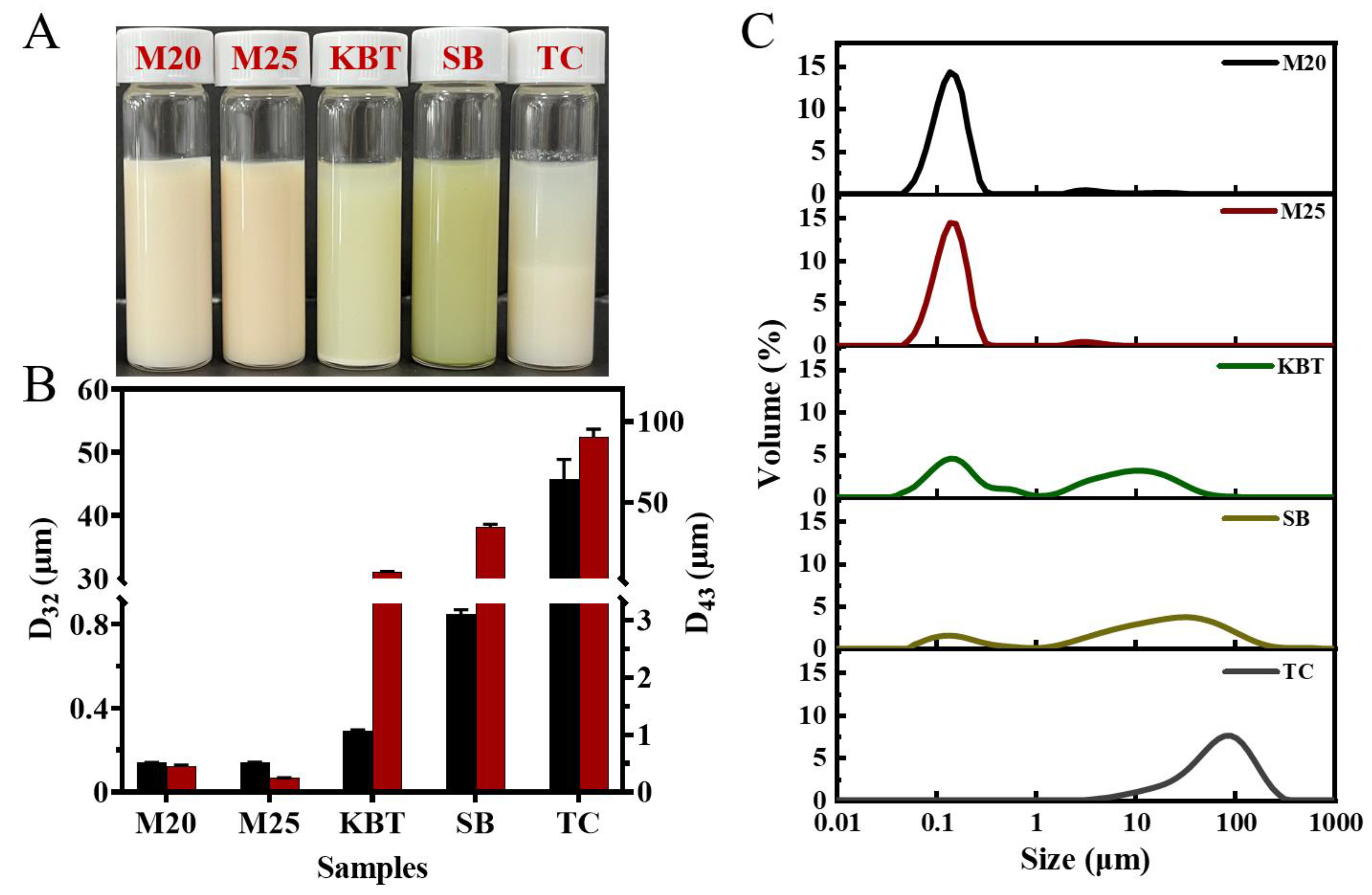
Figure 1. Physical stability of commercial protein sports supplements in liquid statement, (A) visual appearance, (B) particle size and (C) particle size distribution.
Preprints 111305 g001
Figure 2. pH change profiles of protein digesta during gastric digestion.
Preprints 111305 g002
Figure 3. Microstructures of the emptied digesta of sports protein supplements at different time during gastric digestion in the DIVHS system. 15,30,60,90 and 120 represent samples subjected to 15,30,60,90 and 120 min of gastric digestion, respectively. Scale bars for raw and digested samples were 20 µm and 60 µm, respectively.
Preprints 111305 g003
Figure 4. Gastric emptying curves of the sports protein supplements in vitro digestion in the DIVHS system.
Preprints 111305 g004
Figure 5. SDS-PAGE patterns under reducing conditions of sports protein supplements during gastric digestion in the DIVHS system. 0,15, 30, 60, 90 and 120 represent simulated gastric digestion, initial sample, 15, 30, 60, 90 and 120min samples respectively. β-Lg (β-lactoglobulin), α-La (α-lactalbumin), 7S, (β-conglycinin, 7S globulin,) and 11S, (glycinin ,11S globulin).
Preprints 111305 g005
Table 1. Parameters for in vitro dynamic gastric digestion system.
Key parameters Settings
Sample volume 230 mL
pH value of SGF 1.6 ± 0.1
Pre-volume of SGF 30 mL
Pepsin activity 4000 U/ mL
Secretion rate of SGF 0-10 min, 1.2mL/min;
10-20 min, 1.6mL/min;
20-30 min, 2.0mL/min;
30-40 min, 2.5mL/min;
40-50 min, 2.1mL/min;
50-60 min, 1.6 mL/min;
60-120 min, 1.2 mL/min;
Secretion rate of HCl 0-30 min, 1.0 mL/min;
Tilt angle of stomach vessel 0-5min, 2°/min; 5-30min, -0.6°/min; 30-120min, -0.4°/min;
Table 3. Protein hydrolysis degree of the sports protein supplements during gastric digestion.
Time
Sample
15 min 30 min 60 min 90 min 120 min
M20 8.12 ± 0.29Ea 16.95 ± 0.20Da 27.84 ± 1.16Ca 31.86 ± 0.41Ba 35.74 ± 0.33Aa
M25 8.67 ± 0.54Da 18.61 ± 1.45Ca 22.03 ± 1.61BCb 25.68 ± 2.27Bb 30.44 ± 3.45Ab
KBT 4.02 ± 0.81Db 8.38 ± 1.55Cb 10.24 ± 1.44Cc 13.25 ± 1.36Bc 16.16 ± 1.54Ac
SB 3.12 ± 0.60Ebc 7.89 ± 1.62Db 10.84 ± 0.40Cc 13.82 ± 0.10Bc 16.66 ± 0.08Ac
TC 2.34 ± 0.50Ec 7.23 ± 1.10Db 9.99 ± 1.18Cc 13.07 ± 1.55Bc 17.90 ± 0.58Ac
Different capital letters indicate significant differences at different times for the same sample, and different lowercase letters indicate significant differences at the same time for different samples (p < 0.05).
Disclaimer/Publisher’s Note: The statements, opinions and data contained in all publications are solely those of the individual author(s) and contributor(s) and not of MDPI and/or the editor(s). MDPI and/or the editor(s) disclaim responsibility for any injury to people or property resulting from any ideas, methods, instructions or products referred to in the content.
Copyright: This open access article is published under a Creative Commons CC BY 4.0 license, which permit the free download, distribution, and reuse, provided that the author and preprint are cited in any reuse.
Prerpints.org logo

Preprints.org is a free preprint server supported by MDPI in Basel, Switzerland.

Subscribe

© 2024 MDPI (Basel, Switzerland) unless otherwise stated