Preprint
Review

Recent Advances in Phthalocyanine-Based Hybrid Composites for Electrochemical Biosensors

Altmetrics

Downloads

109

Views

68

Comments

0

A peer-reviewed article of this preprint also exists.

Submitted:

10 July 2024

Posted:

10 July 2024

You are already at the latest version

Alerts
Abstract
Biosensors are smart devices that convert biochemical responses to electrical signals. Designing biosensor devices with high sensitivity and selectivity is of great interest because of their wide range of functional operations. However, the major obstacles in the practical application of biosensors are their binding affinity toward biomolecules and the conversion and amplification of the interaction to various signals such as electrical, optical, gravimetric, and electrochemical signals. Additionally, enhancement of sensitivity, limit of detection, time of response, reproducibility, and stability are considerable challenges when designing an efficient biosensor. In this regard, hybrid composites have high sensitivity, selectivity, thermal stability, and tunable electrical conductivities. The integration of phthalocyanines (Pcs) with conductive materials such as carbon nanomaterials or metal nanoparticles (MNPs) improves the electrochemical response, signal amplification, and stability of biosensors. This review explores recent advancements in hybrid Pcs for biomolecule detection. Herein, we discuss the synthetic strategies, material properties, working mechanisms, and integration methods for designing electrochemical biosensors. Finally, the challenges and future directions of hybrid Pc composites for biosensor applications are discussed.
Keywords: 
Subject: Chemistry and Materials Science  -   Electrochemistry

1. Introduction

The pervasive conveniences of modern life are a testament to the remarkable advancements in science and technology. We consistently rely on various devices such as computers, phones, refrigerators, air conditioners, and smoke detectors for seamless interaction with the physical world [1]. Most of these devices would not operate without sensors, which act as our electronic eyes and ears [2]. Electronic devices detect changes in physical or chemical properties, such as pressure, temperature, humidity, motion, and light, by converting them into electrical signals for processing and analysis [3]. Ideal sensors are imperative components of various measurement systems, possessing specific characteristics such as a wide operational range, minimal drift, ease of calibration, high sensitivity, and selectivity [4]. Sensor technology has gained importance in numerous fields, including environmental and food quality monitoring, medical diagnosis and healthcare, automotive and industrial manufacturing, space exploration, and national security [5]. The development of sensors has led to their use in diverse fields, and one particularly exciting area is biosensors. The term ““biosensor”” was coined by Cammann, and its definition was established by IUPAC [6]. Biosensors detect biological processes, diagnose diseases, identify environmental contaminants, and aid in drug discovery. Biosensors function by detecting biomolecules such as proteins, DNA, RNA, enzymes, and hormones, providing valuable insights into health conditions, disease progression, and environmental security [7]. Promptly and accurately identifying biomarkers plays a crucial role in advancing early disease diagnosis and personalized medicine [8]. Biosensor development is continuously advancing with a focus on designing highly sensitive and selective devices for biomolecular detection. However, biosensors face many challenges. Their accuracy and specificity are hampered by complex biological samples and environmental interferences. Device performance in terms of temperature and humidity requires careful control [9]. Additionally, some biosensors have a limited lifespan owing to degradation and require regular calibration for sustained accuracy. Furthermore, the development and maintenance of biosensors is expensive because specialized materials and expertise are required [9]. Figure 1 shows a schematic diagram of the biosensor device used in this study. The biosensor operates via a series of steps involving the interaction of a target analyte with a bioreceptor, leading to the generation of a measurable signal.
Ongoing research is focused on improving biosensor sensitivity, selectivity, and stability, paving the way for wider application in the future. From this perspective, the fabrication of biosensors has gained significant attention for advancement in device functionality and practical applications [11]. The quest for precise and efficient biosensor design and fabrication technique is paramount for unveiling the potential of “smart” biosensor systems [12]. Various biosensor technologies enable biomolecule detection, each with its own operating principle. Optical biosensors use light interactions with biomolecules to measure them, piezoelectric biosensors track mass changes upon biomolecule binding, surface plasmon resonance (SPR) biosensors detect refractive index changes caused by complex biomolecules, and electrochemical biosensors measure the electrical signals produced during biomolecule binding at the electrodes [13]. Electrochemical biosensors are promising because of their sensitivity, affordability, portability, biocompatibility, simplicity, and fast response [14]. A biochemical receptor captures the response of a biological process and transmits it to a transducer [14]. An ideal biosensor is independent of temperature and pH, recyclable, and specific [15]. Electrochemical biosensors consist of three electrodes: reference electrode (RE), counter electrode (CE), and working electrode (WE) [16]. Electrical signal detection involves electrons that are generated or consumed, and it is categorized as potentiometric, cyclic voltammetric (CV), chronoamperometric (CA), differential pulse voltammetry (DPV), impedimetric, and linear sweep voltammetry (LSV) [17]. The sensing mechanism includes the interaction between the analyte and the pinhole surface of the WE to induce a redox reaction. To decrease the overpotential and fouling effect and improve the response, virgin electrodes have been modified with appropriate redox materials or stimulant [17]. To optimize the interaction, an electrode surface has been fabricated with various conducting nano and rare noble materials (Au, Pt, etc.) to facilitate faster electron movement in biosensors [18]. Different redox-active materials such as NPs, metal oxides, carbonaceous materials, polymers, macrocycles, and organic compounds have been employed on the electrode surface because of their large surface area, enhanced conductivity, optical properties, and biocompatibility [18]. However, long-term stability, reproducibility, and cost-effectiveness of biosensors remain challenging issues [19]. In response, various hybrid materials such as graphene oxide (GO)–NPs hybrids [20], polymer–enzyme hybrids [21], carbon nanotube (CNT)–quantum dot hybrids [22], metal–organic framework hybrids [23], and nanostructured composite hybrids [24] have been extensively designed for the development of novel materials with exceptional properties.
Organic-based macrocycles (phthalocyanine (Pc)/porphyrin) are considered reliable materials for signal amplification in electrochemical sensing owing to the simplicity of their synthesis and the ability to fine-tune their electronic properties through the substitution of axial/peripheral groups [25]. N4 macrocycles Pc are interesting because of their unique electronic structure, substantial surface area, distinctive atomic structure, and properties that can be engineered by substituting various functional groups or metal ions [26]. Pc consists of a tetrapyrrole core similar to that of porphyrins in natural systems. This structural resemblance to the heme group in hemoglobin and chlorophyll in photosynthetic organisms is one of the reasons for their bioinspired appeal [26]. The central metal ion in Pc plays a vital role in tuning the catalytic properties. Transition metals, such as Fe, Co, Mn, and Ni, are commonly used as metal centers in Pc [26]. Furthermore, the rigid and planar structure of Pcs provides stability and promotes efficient electron and ion transport during catalytic processes [27]. Additionally, their notable features include high selectivity and a specific size for miniaturized binding geometries, which reduce interference and ensure more accurate data [28]. Furthermore, it offers improved detection limits, enhanced sensitivity, and signal amplification for targeted molecules at minute concentrations, making it a versatile material for biosensing applications [28]. However, the design and fabrication of hybrid materials ensures high stability in harsh environments, and their smooth integration with smart devices underscores the utilization of macrocyclic complexes in biosensor applications.
In recent years, the design and fabrication of biosensors using hybrid materials containing Pc with metal oxide or carbon composites has increased, owing to the large scope of functional operations [29]. Hybrid materials provide unique exploitation owing to the collaborative attributes of each component. Pcs contribute to tunable biocompatibility, leading to improved sensor response and easier processing during fabrication [30]. Carbon nanomaterials (CNs) substantially enhance the electrical conductivity and surface area of biosensors, allowing improved detection efficiency [31]. Furthermore, the incorporation of metal nanoparticles (MNPs) further enhances the sensing performance through their catalytic activity, signal amplification, and unique optical properties [31]. Hence, the deployment of Pc–metal oxide or polymer–metal–carbon (PMC) hybrids has led to the development of biosensors with superior sensitivity, enabling the detection of analytes at lower concentrations [32]. Additionally, PMC hybrids offer improved selectivity, meaning that they can distinguish between the target molecule and potential interferences. The use of these hybrid materials paves the way for the miniaturization of biosensors, making them smaller, more portable, and potentially more suitable for various biomedical applications. Despite these positive observations, ensuring long-term stability, biocompatibility, reproducibility, minimal potential toxicity of NPs, and developing cost-effective production methods, as well as fabricating PMC-based hybrid biosensors, remain paramount challenges [30,31,32]. In this review, we briefly introduce hybrid materials. The synthesis strategies, properties, and sensing mechanisms of the hybrid materials are also incorporated, and their potential for the detection of analytes such as biomarkers, environmental pollutants, and food contaminants is explained. Furthermore, we explore recent advancements in the development and application of PMC-based hybrid materials for biosensors, particularly the integration of sensor systems, and their translation into practical applications. Finally, the challenges and future directions of PMC-based hybrids for biosensors are discussed.

2. Pc Composite Materials for Biosensors

2.1. Carbonaceous Materials

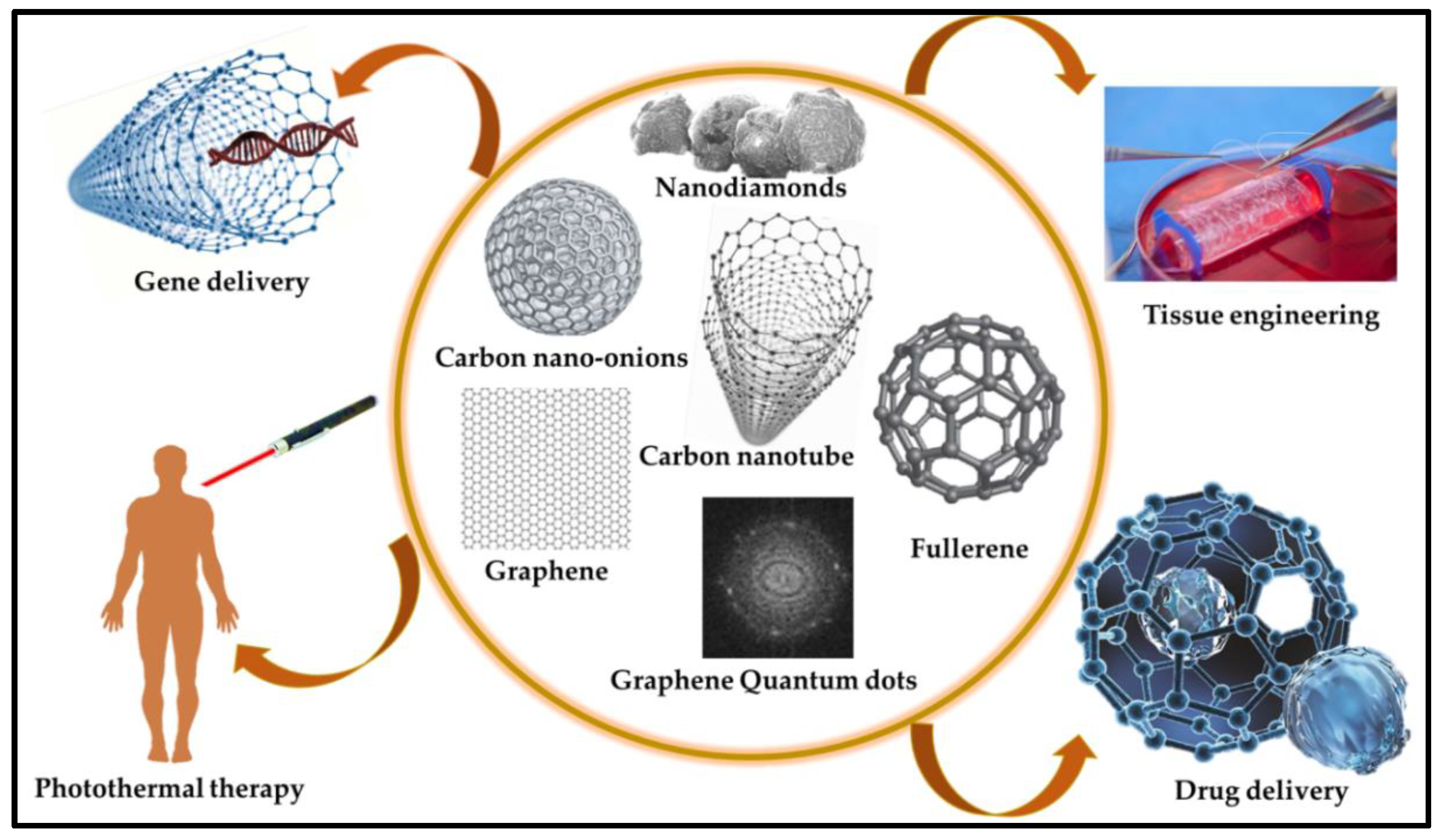
CNs are reliable materials for biosensor applications owing to their affordable electrical properties, robust mechanical strength, large surface area, bioreceptor immobilization, and biocompatibility [33]. CNs are employed in various biosensor devices that manifest binding sites for target biomarkers to facilitate their capture and identification. Moreover, CNs convert the molecular interactions detected on the electrode surface into measurable electrical signals, enabling quantitative analysis. Furthermore, it increases signal amplification and sensitivity [34]. However, efficient signal capture for biomolecular recognition remains a challenge in the development of CNs. The CNs most utilized in biosensors include CNTs, graphene, nanodiamonds, and fullerenes . Some CNs are directly integrated into the bare electrodes, serving a dual role in analyte identification and signal transduction [35]. Recently, Kim et al. [36] developed a single-walled CNT (SWCNT)-based immunosensor for CD4+ T cell detection by addressing the problems of immobilization efficiency, reproducibility, and linear quantification. Through repeated two-step O2 plasma treatment with recovery periods, the team achieved enhanced CNT surface functionalization and enabled linear electrochemical signal generation proportional to the number of bound cells. The SWCNT electrode exhibited a slope of 4.55×10⁻² μA/dec within the target range of 102~106 cells/mL, with a detection limit of 1×10² cells/mL. Reduced CD4+ T cell counts are markers of HIV progression; this immunosensor offers simple, low-cost point-of-care diagnostics, potentially aiding in HIV patient management. Wang et al. [37] studied a novel electrochemical sensor for hyperin detection that was developed using a 3DG-MWCNTs network fabricated directly on a glassy carbon electrode (GCE) using the pulse potential method. In contrast to bare GCE and 3DG-modified electrodes, the 3DG-MWCNTs complex exhibited an increased response toward hyperin redox, which was attributed to the large surface area and excellent conductivity of the 3DG-MWCNTs, facilitating efficient electron transfer. The sensor portrayed a wide linear range of 5.0 × 10⁻9 to 1.5 × 10⁻6 mol L⁻¹ and a low detection limit of 1.0 × 10⁻⁹ mol L⁻¹ with high sensitivity for hyperin detection. Additionally, the sensor displayed remarkable stability, selectivity, and reproducibility in detecting hyperin in various matrices. Similarly, Chaitra et al. [38] designed biomass-based carbon nanospheres using ““touch-me-not”” (Mimosa pudica), which was coated on carbon fiber paper and employed as a host matrix for palladium NPs via electrochemical deposition. The modified electrodes exhibited excellent electrocatalytic activity toward morin oxidation, with a sensitive peak at -0.30 V (vs Saturated calomel electrode) and a low detection limit of 572 fM, within a linear response range of 37.50–130 pM. The sensor successfully analyzed the morin content in mulberry and guava leaves, demonstrating its potential for the sustainable approach of using biomass for sensor fabrication. Similarly, various studies have been conducted on the fabrication of biomaterial–CNT hybrid systems. Through a potent acid treatment, the ensuing development of carboxylic and phenolic groups on protein-associated CNTs along with nucleic acid-functionalized CNTs was observed. This chemical shortening of the SWCNTs’’ nanotube terminations facilitated the covalent immobilization of biomaterials, proteins, and nucleic acids onto the surface of the SWCNTs [39]. An electrochemical biosensor was devised to detect the human carcinogen aflatoxin B1 by employing MWCNTs deposited on indium-tin-oxide (ITO) electrodes [40]. Figure 2 illustrates the various methods used for fabricating carbon-based materials, which are integral to biosensor applications. The versatility, conductivity, and biocompatibility of carbon-based materials render them highly suitable for biosensor development.
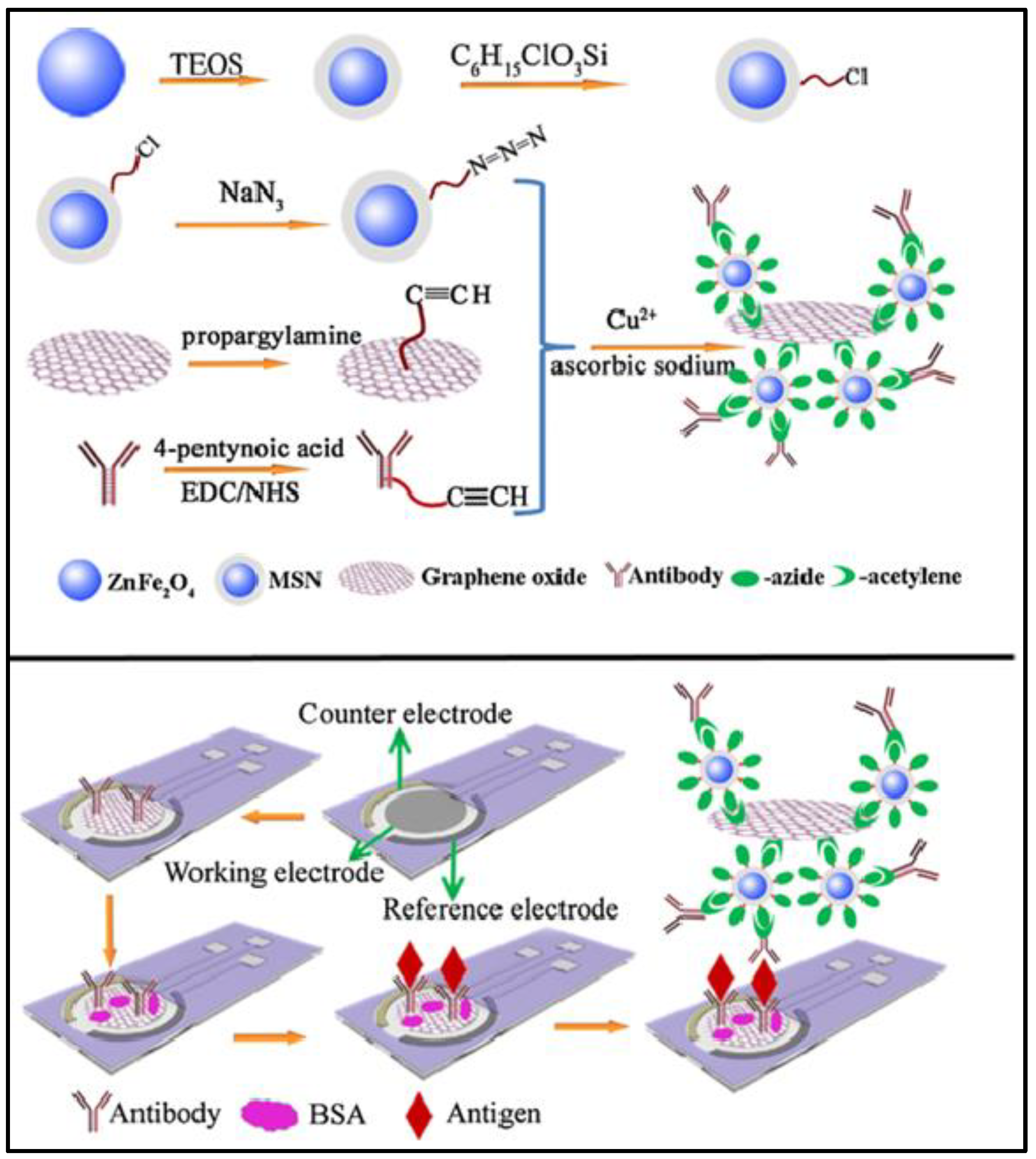
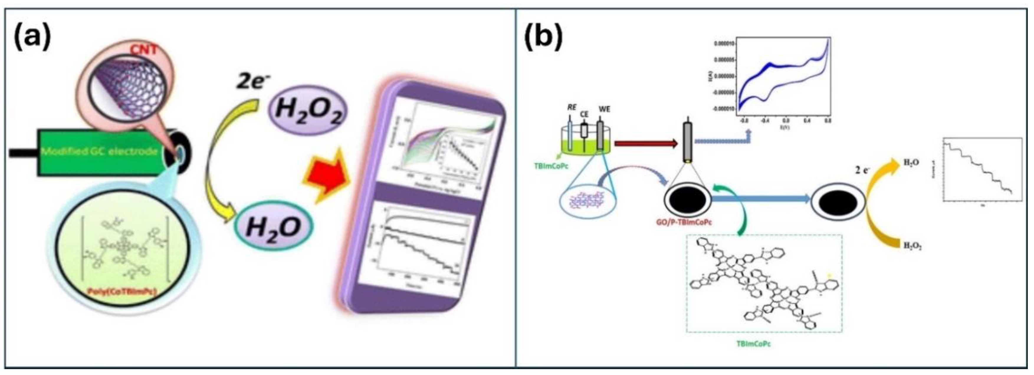
Figure 3 shows a detailed step-by-step plan for making an rGO-coated electrochemical immunosensor specifically designed to detect cancer antigens. The fabrication process involves several key stages, each of which contributes to the creation of a highly selective and specific biosensor. Although CNs-based biosensors are promising, they face practical challenges in biological applications. Notably, biosensor fabrication requires a specific size and helicity of CNs, posing difficulties in size control during synthesis [43]. Moreover, the cost efficiency challenges of large-scale production hampers the widespread use of high-purity nanomaterials, contributing to the prohibitively expensive CNs and limiting their practical commercial applications. In carbon-based biosensors, the immobilization of enzymes on CN-coated electrodes raises concerns about potential damage to biological receptors, affecting biocompatibility, biological activity, and structural stability [44]. Addressing this issue requires the development of reversible, reusable, and enduring systems, as opposed to the prevalent use of irreversible, disposable, and single-use devices, which presents an ongoing challenge in this field. Furthermore, the functionalization and conjugation of CNs with organic compounds or metallic NPs can impart novel properties, including physical, chemical, mechanical, electrical, and optical attributes [45]. This augmentation further enhances their suitability for biosensor applications. Recently, Keshavandaprabhu et al. [46] developed an organic compound-based polymeric cobalt Pc (poly(CoTBImPc)) complex with tetrabenzimidazole (TBIm) substitutions for hydrogen peroxide (H2O2) sensing. Poly(CoTBImPc) showcased distinctive redox properties with peaks corresponding to Co2+/Co1+ and PcII−/PcIII- redox couples. To enhance its conductivity, surface area, and sensitivity, the complex was coated onto a CNT-modified GCE (GCE/CNT/poly(CoTBImPc)). This resulted in a highly sensitive amperometric sensor for H2O2 that exhibited a linear response in the 10–100 nM range and a remarkable limit of detection (LOD) of 2 nM. Additionally, the sensor exhibited exceptional reproducibility, repeatability, and long-term stability without any loss of catalytic activity. Notably, it maintained high selectivity for H2O2 even in the presence of potential interfering species, demonstrating its reliability for practical applications. Imadadulla et al. [47] synthesized and characterized novel CoTBIPc substituted with TBIm. Electrochemical studies revealed the redox behavior of the Co(II)/Co(I) metal center. CoTBIPc underwent electropolymerization on the GCE, forming film (GCE/poly(CoTBIPc) for H2O2 detection. To improve the surface area and sensitivity, CoTBIPc was electropolymerized on GO-coated (GCE/GO/poly(CoTBIPc). GCE/GO/poly(CoTBIPc) displayed enhanced catalytic activity with a linear H2O2 detection range of 2–300 μM compared to 3–160 μM of GCE/poly(CoTBIPc). Amperometric sensors using both the modified electrodes showed linear responses with LODs of 0.8 μM and 0.6 μM for H2O2 detection, respectively. GCE/GO/poly(CoTBIPc) offered superior reproducibility, repeatability, and stability while maintaining high selectivity for H2O2. This study demonstrated the potential of CoTBIPc-based electrodes for H2O2 sensing with improved performance. Recent studies utilized materials based on their mechanical and electrical properties for biosensor applications via the amalgamation of CNPs. The integration of CNPs with biotechnology provides an economical method for the real identification of targets and leverages specific antibody recognition in biosensor development. Figure 4 shows the design and fabrication steps for two types of electrodes, CNT-modified and GO-modified electrodes, for the electrochemical detection of H2O2. Both methods exploit the unique properties of CNs to enhance the sensitivity and performance of the sensors in H2O2 detection [46,47]. Table 1 provides an overview of different CNs, their unique properties, and their suitability for detecting various analytes in biosensor applications [48,49,50,51,52,53,54,55,56,57,58,59,60].

2.2. Metal and Metal Oxide NPs

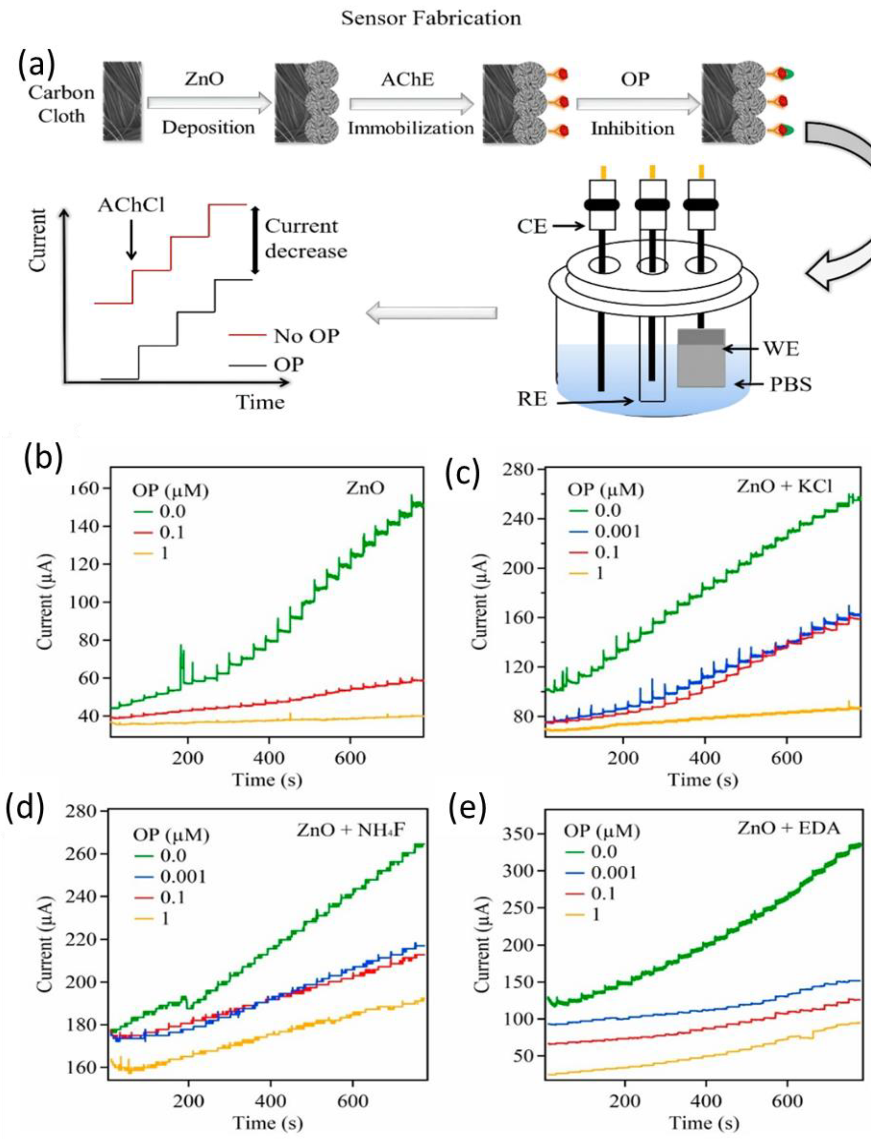
MNPs act as ““electron wires”” to transport the electrons produced in the bio-reaction to sensing electrodes [60]. The integration of MNPs into biosensors enhances the functionality of biosensor devices. MNPs serve as transducers because of their unique physiochemical properties in the 1 to 100 nanoscale range, providing a substantial surface-to-volume ratio and facilitating the sensing and detection of biomolecules [61]. Various MNPs such as Pb, Pd, Cu, and Se NPs, and metal oxide NPs, such as zinc oxide (ZnO), iron oxide (Fe2O3), copper oxide (CuO), titanium dioxide (TiO2), and indium oxide (InO2) NPs, have been used in sensing applications [62,63]. Functionalization of NPs with specific ligands or biomolecules on the surface imparts selectivity for the target analyte, harnessing the unique properties of MNPs such as their high surface area, catalytic activity, and optical properties, making them suitable candidates for sensing [64]. Among the various MNPs, gold, silver, and platinum have been extensively studied because of their well-established chemical inertness on a macroscopic scale. Despite their chemical inertness in larger dimensions, these noble metals exhibit unique physicochemical characteristics when scrutinized at the nanoscale. Guo et al. [65] studied an amperometric glucose biosensor using both Rh and Au NPs. The Rh NP-modified Pt electrode contributed substantially to the surface area, with numerous active sites enhancing the electrocatalytic activity. Meanwhile, Au NPs strategically positioned near the active regions of the enzyme facilitated the oxidation of peroxide molecules, thereby amplifying the sensitivity of the glucose sensor. The functionalization of Rh and Au NPs played a crucial role in optimizing the biosensor performance for efficient glucose detection. Additionally, the functionalization of MNPs is defined by the physical or electrochemical changes that occur after biomolecular analytes bind to the immobilized receptor-target on the surface of the MNP. MNPs exhibit various functions, including immobilizing platforms, accelerating electron transfer, catalyzing chemiluminescent reactions, amplifying changes in mass, and enhancing refractive index modification [66]. Figure 5 provides a comprehensive overview of the fabrication and modification of ZnO NPs and their subsequent amperometric responses when deposited on carbon paper. The comparison of different ZnO samples, such as pristine, potassium chloride (KCl)-doped, ammonium fluoride (NH₄F)-doped, and ethylenediamine (EDA)-modified ZnO, demonstrates the impact of various modifications on the electrochemical performance, with the EDA modification showing the most pronounced enhancement [67].
Zhu et al. [68] investigated the fluorescence quenching capabilities of silver-coated gold triangular nanoplates (Ag-Au TNPs) for carcinoembryonic antigen (CEA) detection. SPR-induced nonradiative energy transfer from CEA to the Ag-Au TNPs significantly quenched CEA fluorescence, demonstrating a higher quenching efficiency than that of bare gold TNPs. This effect intensified with increasing silver coating thickness and CEA concentration, with a lower detection limit of 5 pg/mL for the CEA, which is a testament to the enhanced sensitivity of the Ag-Au TNPs. Further, Lan et al. [69] developed a chemiluminescence (CL) flow-through biosensor for glucose detection by immobilizing Glucose oxidase (GOD) and horseradish peroxidase (HRP) on eggshell membranes using glutaraldehyde. The CL process involved enzymatic glucose oxidation, producing D-gluconic acid and H2O2, with H2O2 oxidizing luminol to emit CL in the presence of HRP. The immobilization conditions (time, GOD/HRP ratio, and glutaraldehyde concentration) were thoroughly studied. The biosensor exhibited stable storage at 4 °C for 5 months, with quick response, high sensitivity, simple operation, and assembly. Successfully applied for human serum glucose determination, this biosensor holds promise for practical applications.
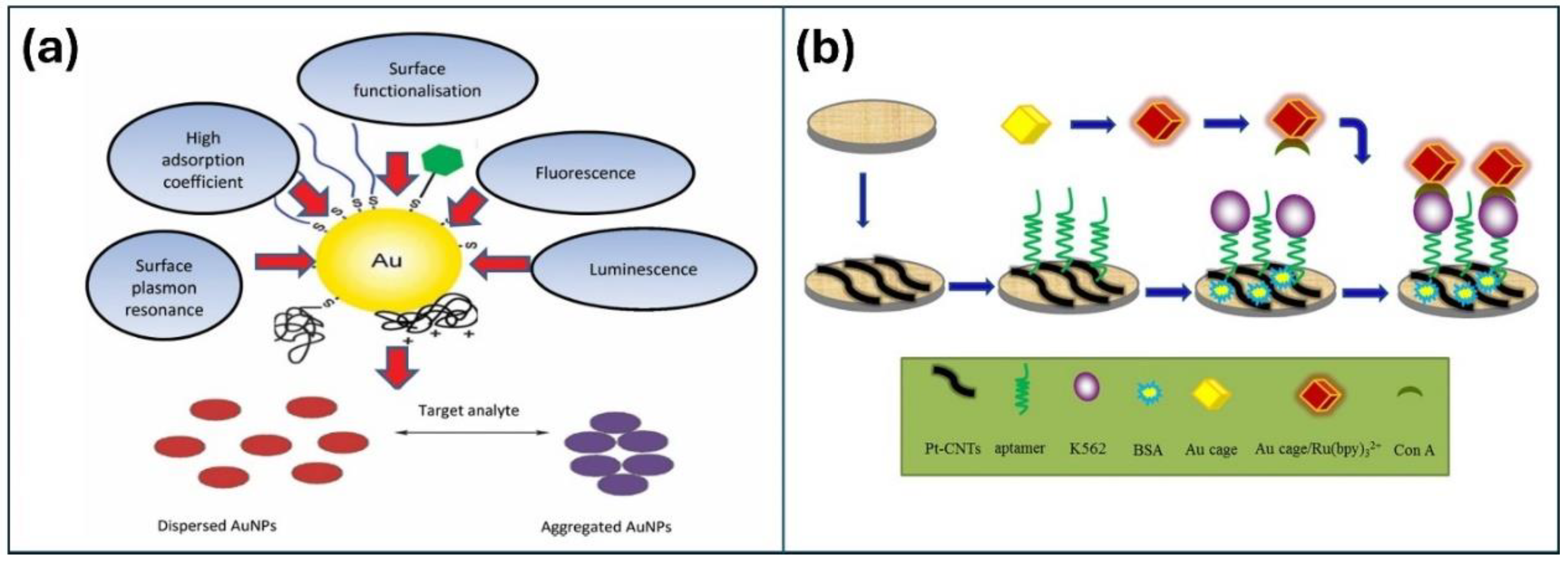
Lei et al. [70] designed an electrochemiluminescence (ECL) cytosensor for highly sensitive and selective cytosensing of K562 cancer cells. The research group immobilized PtNPs on CNTs on the WE, which enhanced both the electronic transmission rate and surface area, as shown in Figure 6. The electrode was further modified with aptamers for precise capture of cancer cells. The researchers also devised a novel class of nanoprobes by integrating concanavalin A (Con A) for specific recognition and signal amplification using Au cage/Ru(bpy)32+ nanostructures. Operating on a sandwich-type cytosensor format, the ECL signals increased proportionally with the quantity of K562 cancer cells, reflecting the augmented presence of Au cage/Ru(bpy)32+ labeled Con A. The cytosensor demonstrated outstanding analytical performance across a broad detection range of 500 to 5.0 × 10⁶ cells mL−1, and a detection limit of 500 cells mL−1. Additionally, the method exhibited high precision, longer stability, and reproducibility. Similarly, Liang et al. [71] presented a highly sensitive self-enhanced ECL cytosensor for monitoring cell apoptosis, employing ruthenium-silica composite nanoparticles (Ru-N-SiNPs) labeled with annexin V as signal probes. The as-designed Ru-N-SiNPs resulted in enhanced ECL intensity and higher emission efficiency, which were attributed to a shorter electron transfer path and reduced energy loss. The developed ECL cytosensor successfully evaluated the efficacy of paclitaxel against MDA-MB-231 breast cancer cells within a concentration range of 1 to 200 nM. The detection limit was determined to be 0.3 nM, and the correlation coefficient was 0.9917, indicating improved accuracy.
Optimization relies mainly on the synthesis of various metal/metal oxide NPs. Gold, silver, platinum, copper, palladium, lead, selenium, and metal oxides have been synthesized using chemical, physical, and biological synthesis methods [72]. Chemical methods provide control over size and shape. Physical methods such as mechanical milling and vapor deposition furnish a controlled surface morphology and crystal structure. Additionally, biological methods such as green synthesis employ plants, algae, fungi, and microorganisms for NPs synthesis, to produce biocompatible NPs with biomolecule encapsulation further enhancing biocompatibility and reducing toxicity. Biological methods are cost-effective and scalable. Furthermore, the cellular uptake of MNPs is influenced by their physicochemical properties, and their biocompatibility involves both qualitative and quantitative analyses [73]. Despite these synthesis methods, it still faces many challenges, such as precisely controlling particle size and morphology, ensuring high purity and reproducibility, and achieving large-scale production without compromising environmental friendliness and cost-effectiveness. Additionally, assessing the impact of physicochemical properties on cellular uptake and developing robust biocompatibility assessments are crucial for addressing potential toxicity concerns. Recently, a synergistic combination of MNPs with polymers or carbonaceous materials has shown promise in biosensor applications. This study was conducted by Liu et al. [74], who explored the synergistic effects by utilizing two distinct types of noble MNPs in an electrochemical biosensor, showing enhanced electron transfer. This study revealed that Pd-Co alloy NPs embedded in carbon nanofibers exhibit superior analytical capabilities, particularly for hydrogen peroxide and nitrite sensing. This synergistic effect was manifested as a reduced overpotential and significantly higher reduction currents for H2O2, as well as an increased oxidation peak and decreased overpotential in the nitrite cyclic voltammograms. This underscores the effectiveness of employing a combination of noble MNPs to improve the performance of electrochemical biosensors for detecting specific analytes. Table 2 provides a detailed overview of the capabilities of various MNPs for the detection of different analytes. Each entry in the table highlights the MNP used, the target analyte, the detection method employed, the sensitivity and detection limits achieved, and the notable features that contribute to their effectiveness. This demonstrates the significant role of MNPs in enhancing the performance of biosensors in various analytical applications [75,76,77,78,79,80,81,82,83,84].

2.3. Polymeric Pc Materials

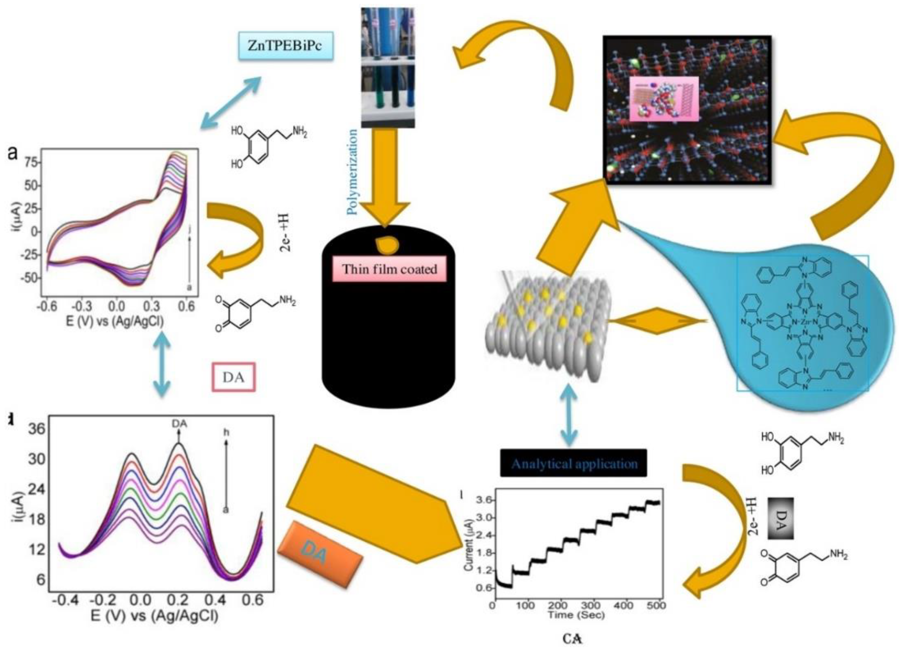
Polymeric Pcs are a group of aromatic macrocyclic polymers known for their unique electronic properties, rich redox behavior, strong absorption capability, tunable biocompatibility, and high thermal and electrochemical stabilities in corrosive media [85]. These outlined electrocatalytic properties contribute to the high efficacy of Pc/porphyrin and its analogs, which are similar to naturally occurring porphyrin macrocycles such as vitamin B-12, hemoglobin, cytochrome-c, and chlorophyll [86]. Several MPcs such as ZnPc, CoPc, RhPc, NiPc, and TiPc have been widely employed in dyes and pigments, photovoltaic devices, electronics, photodynamic therapy, sensors, and biosensors [87]. Pcs are used to enhance the sensitivity and selectivity of biosensors. For instance, Pcs act as mediators in redox reactions, facilitate electron transfer, and improve biosensor efficiency [88]. CoPc is widely used as a redox mediator in enzymatic glucose biosensors. Additionally, the catalytic activity, stability, and electrochemical sensitivity of CoPc contribute to its overall performance, making it the most preferred candidate for biosensors. However, ZnPc faces instability in biosensors owing to its weak electrochemical activity and low electrical conductivity [89]. Therefore, graphene nanosheets and CNTs have been incorporated into MPcs to enhance electron transfer and improve their electrical conductivity [89]. M. Pari et al. (Figure 7) demonstrated the effective detection of DA using a composite of rGO and zinc tetra [4-{2-[(E)-2-phenylethenyl]-1H-benzimidazol-1-yl}] Pc (Zn(II)TPEBiPc). The rGO-Zn(II)TPEBiPc composite-modified electrode showed better performance compared to Zn(II)TPEBiPc film, with a detection limit of 6 nM and sensitivity of 2.8784 μA μM–1 cm–2 for DA detection in the range of 20 nM to 1.0 μM. Furthermore, it exhibited good stability and repeatability and was successfully applied for DA detection in pharmaceutical drugs [90]. Similarly, S. Malali et al. worked on graphene-based biosensors for selective DA detection using hybrid tetra-amino cobalt (II) Pc (TACoPc) and polyaniline (PANI) nanofibers (TACoPc/PANI hybrid). The hybrid synthesized via a one-step process exhibited superior DA detection performance compared to conventional methods. The TACoPc/PANI hybrid-modified electrode displayed a high sensitivity of 1.212 μA μM-1 cm⁻² and low detection limit of 0.064 μM for DA within a 20–200 μM concentration range in a phosphate buffer solution with pH 7. The synergistic effect of PANI and TACoPc eliminated the interference from ascorbic acid (AA), which is a major challenge for DA detection [91]. Sajjan et al. [92] designed the peripheral amine of Pc to form a polymeric film on the electrode surface in DMSO. The polymeric film-modified electrode exhibited excellent voltammetric and amperometric detection of DA, with linear responses of 100–4000 nmol L-1 and 100–1000 nM, respectively. Additionally, the amperometric analysis displayed a high correlation coefficient (R2 = 0.999), low LOD (20 nmol L-1), and good sensitivity (0.024 μA nmol-1 cm-2).
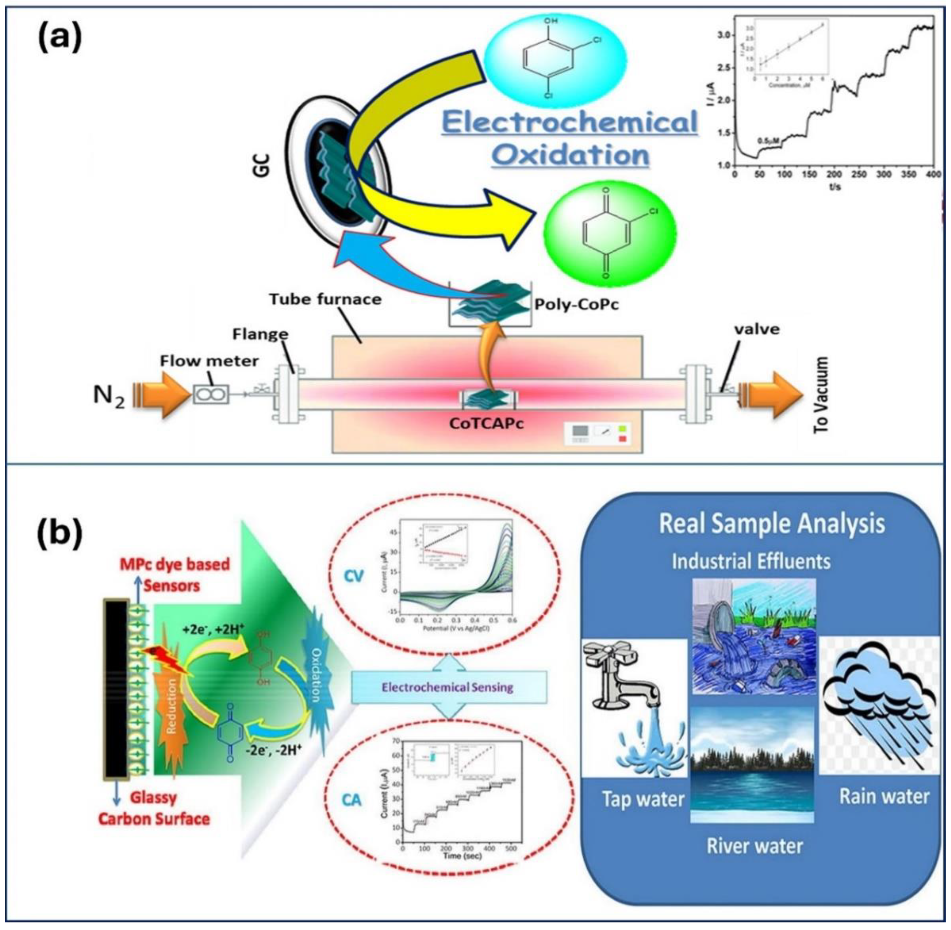
Imadadulla et al. [93] synthesized a cobalt Pc sheet polymer (poly-CoPc) through the thermal condensation of cobalt tetracarboxylic acid Pc (CoTCAPc) and investigated for 2,4-dichlorophenol (DCP) analyte (Figure 8(a)). The polymer exhibited high thermal stability (up to 420 °C) and redox-active behavior and was successfully immobilized on GCE (GC/poly-CoPc). Voltammetric analysis revealed a linear response for DCP in the concentration range of 1–36 µM with an LOD of 0.35 µM and a sensitivity of 0.08 µA µM⁻¹. Amperometric measurements showed a linear response for DCP between 0.5–10 µM, with an even lower LOD of 0.15 µM and a sensitivity of 0.0384 µA µM⁻¹. The sensor displayed excellent stability, repeatability, reproducibility, and high selectivity toward DCP even in the presence of various other alcohols. These results suggest that poly-CoPc holds promise as a sensitive and selective electrochemical sensor for DCP. Nemakal et al. investigated amide bridge cobalt Pc (CoTAMFCAPc) complexes for hydroquinone sensing and confirmed that the furan-containing complex had superior performance [94]. Similarly, Shantaraj et al. [96] designed a novel cobalt (II) Pc polymeric material (poly-CoTPzPyPc) for detection of l-arginine. The designed material exhibited a linear response to l-arginine concentration in the range of 10–100 μM with an LOD of 2.5 μM. The diffusion coefficient for l-arginine was calculated as 1.67 × 10⁻⁶ cm2/s. The rotating disc electrode performance confirmed the 2e- transfer process during l-arginine oxidation. CA studies showed the catalytic response for l-arginine in the range of 2–60 μM with an LOD of 0.6 μM. The poly-CoTPzPyPc film demonstrated excellent selectivity toward l-arginine in the presence of biomolecules. Furthermore, the sensor displayed good stability and satisfactory performance in the analysis of real samples. These findings suggest that poly-CoTPzPyPc has the potential to detect and monitor l-arginine in biological samples. Sajjan et al. [97] further explored the remarkable sensing capabilities of a GCE/CoTTIMPPc electrode toward 4-nitrophenol. Keshavandaprabhu et al. [98] synthesized a dark blue cobalt (II) Pc complex (CoTBrImPc). The complex exhibited promising electrocatalytic activity toward L-cysteine detection. Immobilized on GCE, it demonstrated excellent performance with a low detection limit of 3 nM and a high sensitivity of 2.99 μA nM⁻¹ cm⁻². Notably, the linear response ranged from 10 to 100 nM, indicating the potential application of CoTBrImPc in L-cysteine sensing. These advancements highlight the versatility and potential of polymeric Pc-based materials in developing next-generation sensors for various analytes (Figure 8(b)). Table 3 summarizes the diverse applications and performance characteristics of polymeric Pc materials in the detection of various analytes. Each entry in the table highlights the specific target analyte, the detection method employed, the sensitivity, detection limit, and advantages of each material. This underscores the versatility and effectiveness of polymeric Pcs in analytical sensing applications [90,91,92,93,94,95,96,97,98].

3. Pc-Based Hybrid Composites

In recent years, the integration of an organic hybrid composite of Pc with metal oxides or CNs in biosensors has been promising owing to the unique combination of properties and synergistic framework. Pc provides tunable biocompatibility and enhances sensor responsiveness and processability, whereas CNs (CNTs, graphene, and GO) contribute to electrical conductivity and a large surface area [99]. MNPs exhibit catalytic activity, signal amplification, and unique optical properties that further enhance the sensing competence of hybrid materials [100]. The integration of PMC-based hybrids reinforces the performance of biosensors with improved sensitivity, detection limit, selectivity, multifunctionality, and miniaturization [101]. The improved biosensing performance of hybrid composites is explained below.
a) Combining MPc with carbon materials in a hybrid composite overcomes the MPc aggregation issue and enhances biosensor sensitivity [102].
b) An 18 п-electron system in MPc combined with carbon materials results in the optimization of hybrid composites with high porosity, surface area, and conductivity. This contributes to improved specificity in detecting biomolecules and enhances selective detection [103].
c) The integration of carbon materials into a hybrid composite not only enhances electrical conductivity and electron mobility, but also facilitates efficient electron transfer, ensuring a rapid biosensor response [104,105].
d) Carbon materials in hybrid composites solve the stacking arrangement issue in MPc and improve dispersion, diffusion, and adsorption to overcome issues related to long-term stability [106].
e) The immobilization of the hybrid composite on the electrode surface in a hierarchical structure leads to strong affinity and stability, thus contributing to an efficient biosensing process [107].
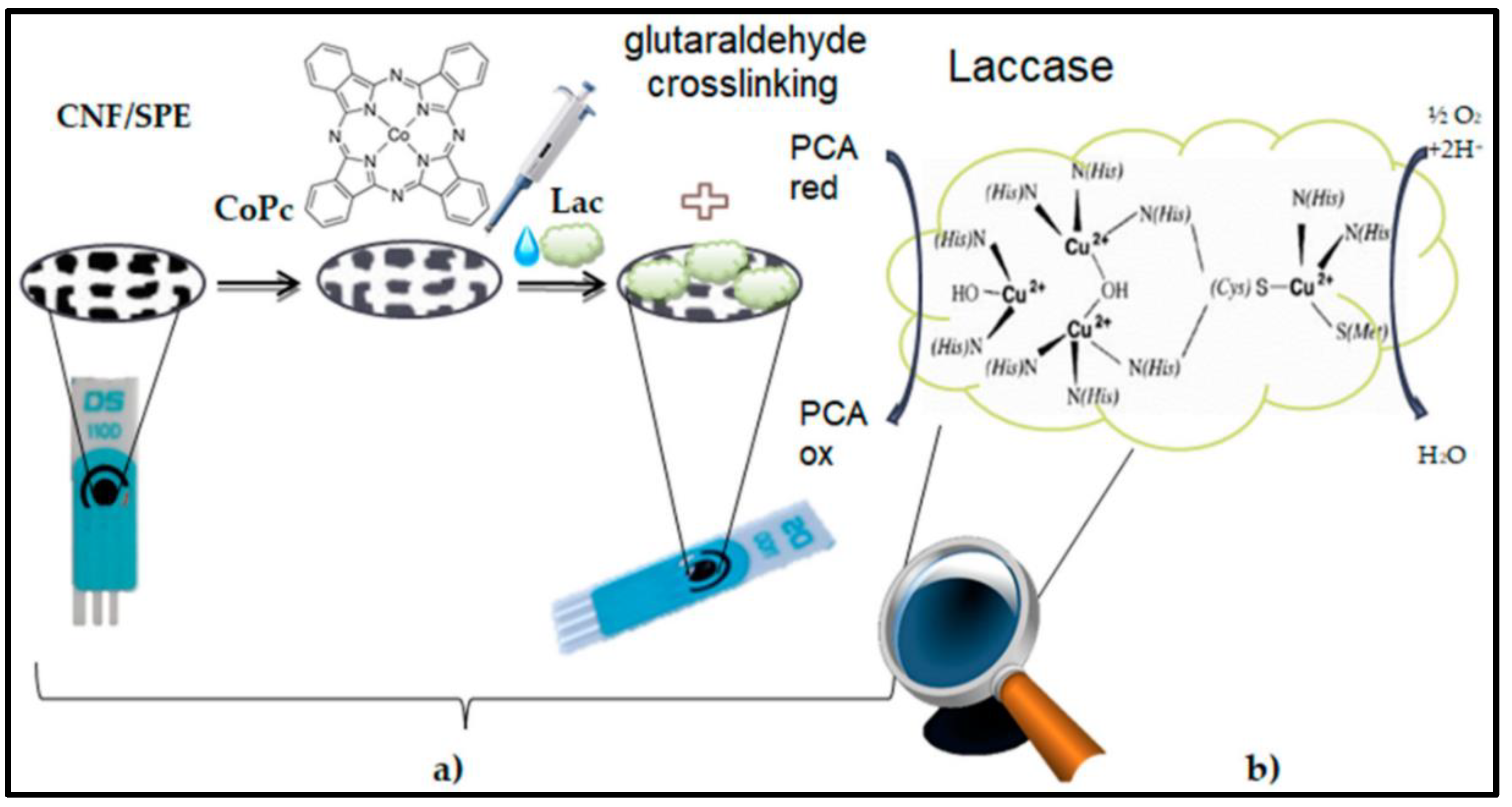
Figure 9 illustrates the step-by-step preparation process of a laccase-based biosensor on a screen-printed electrode (SPE) modified with cobalt Pc-modified carbon nanofibers (CoPc-CNFs). The fabrication procedure involves several key steps. Carbon nanofibers are initially treated to enhance their surface properties and facilitate the binding of the cobalt Pc molecules. Cobalt Pc, known for its catalytic properties and stability, is then immobilized onto the surface of the carbon nanofibers through a suitable deposition method, such as drop-casting or electrodeposition. An SPE is utilized as the substrate for the biosensor owing to its ease of fabrication, portability, and compatibility with different detection techniques. The SPE is fabricated by printing a layer of conductive ink onto a substrate and pasting CoPc-CNFs onto the WE surface area. The laccase enzyme has ability to catalyze the oxidation of phenolic compounds immobilized on the CoPc-CNFs-modified electrode surface. The immobilization process involves incubating the electrode in a laccase-containing solution under controlled conditions, allowing the enzyme to adsorb or bind to the electrode surface. The enzymatic oxidation mechanism of protocatechuic acid (PCA) in the presence of laccases is shown in Figure 9(b). Laccase catalyzes PCA oxidation by transferring electrons from the substrate to molecular O2, resulting in the formation of reactive intermediates that lead to the formation of quinone products. In the oxidation process, electrons are transferred from PCA to the laccase enzyme, which undergoes a redox cycle involving the active sites of copper ions (Cu2+ and Cu3+), facilitating the oxidation reaction. This enzymatic oxidation of PCA leads to the formation of a quinone product via the release of protons and electrons. Figure 9 provides valuable insights into the fabrication and enzymatic oxidation mechanism of a laccase-based biosensor for the detection of phenolic compounds such as PCA. The integration of CoPc-modified CNFs for laccase immobilization enhances catalytic activity and sensitivity in biosensor applications.
Additionally, the incorporation of carbon materials like graphene or nanotubes disrupts π-π stacking in Pcs which hinders the accessibility of active sites for biomolecule interaction. The improved dispersion maximizes the surface area for biomolecule adsorption during sensor fabrication. Furthermore, the 18 π-electron system of MPcs interacts with the carbon network to optimize the electronic structure of the composite. This enhanced π-conjugation facilitates efficient electron transfer and improves interfacial charge transfer between the immobilized biomolecule and the composite, leading to superior detection level of Hydrazine (Hz). Additionally, the excellent conductivity of carbon materials significantly increases the overall conductivity of the composite, resulting in a fast response and increased sensitivity during fabrication. Moreover, the optimized structure during fabrication maximizes the surface area for biomolecule interaction, which promotes strong binding with target biomolecules and facilitates efficient mass transport, thereby boosting the sensitivity and response time of the biosensor [109].
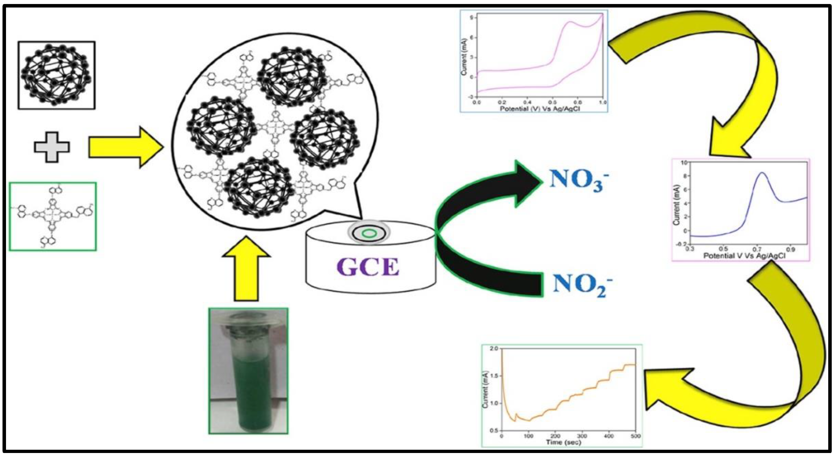
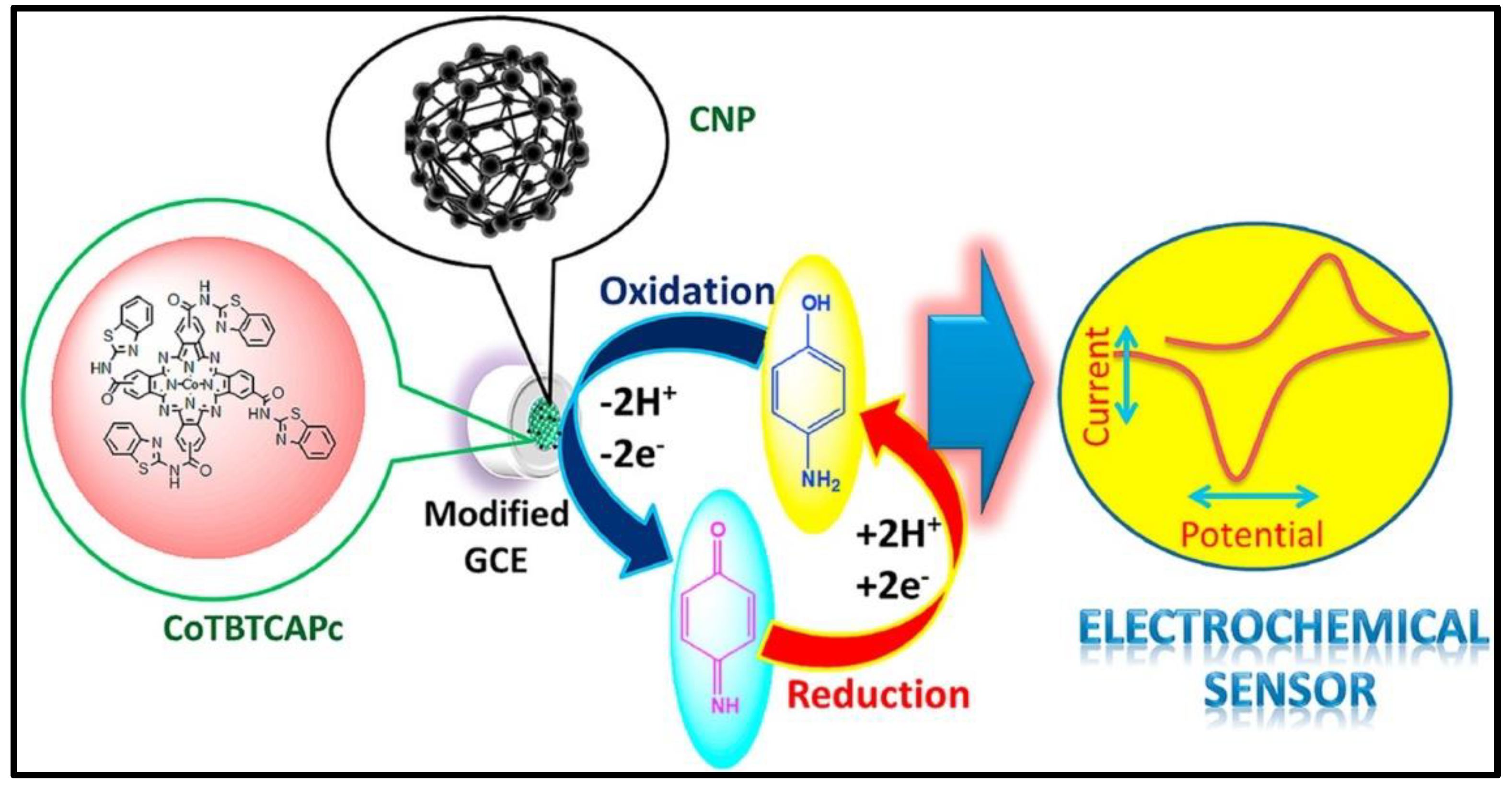
Hybrid composites of rGO have gained attention for biosensing applications because of their unique properties. rGO provides high conductivity and a large surface area, facilitating efficient electron transfer and increasing biomolecule interaction. This translates into potentially faster and more sensitive biosensors, aiding in the development of high-performance biosensors with modified electrode surfaces that exhibit minimal interfacial resistance, exceptional stability, and efficient electron transfer between the electrolyte and electrode. Recently, Jilani et al. [110] explored the potential of carbonaceous and metal phthalocyanine (MPc) hybrid composites for nitrite-sensing applications. As shown in Figure 10, they synthesized a novel composite material, cobalt (II) tetramethylquinoline oxy bridged Pc (CoTM-QOPc) and used it for nitrite sensing. The sensor exhibited a linear detection range of 0.3 to 120 μmol/L, using CV, and 0.2 to 170 μmol/L, using CA. The detection limit achieved was 0.1 μmol/L for CV and 0.06 μmol/L for CA, with good sensitivity of 0.765 μA μM⁻¹ cm⁻² (CV) and 1.204 μA μM⁻¹ cm⁻² (CA). Recognizing the potential for further improvements, they strategically incorporated CNP into the CoTM-QOPc matrix. This carbonaceous and MPc hybrid composite (CoTM-QOPc/CNP) offered an enhanced electrocatalytic response for nitrite oxidation compared to the electrode modified with Pc alone. The linear detection range for nitrite sensing using the composite electrode was even broader, spanning from 0.2 to 200 μmol/L (CV), 0.2 to 225 μmol/L (DPV), and 0.1 to 350 μmol/L (CA). The detection limit also improved remarkably, reaching 0.06 μmol/L for both CV and DPV, and 0.033 μmol/L for CA. The sensitivity of the CoTM-QOPc/CNP composite electrode was also superior, with values of 2.298 μA μM⁻¹ cm⁻² (CV), 1.031 μA μM⁻¹ cm⁻² (DPV), and 1.237 μA μM⁻¹ cm⁻² (CA). This highlights the remarkable utilization of carbonaceous materials in Pc-based sensors. The CoTM-QOPc/CNP composite electrode demonstrated not only enhanced sensitivity and detection limits for nitrite but also impressive selectivity, even in the presence of interfering ions such as AA, carbonate, urea, phosphate, and glucose. This study paves the way for the development of highly selective and sensitive biosensors for real-world applications. Similarly, Shambulinga et al. [111] designed an oxy-bridge cobalt Pc polymer (poly(TazoCoPc)) to enhance the conjugation effect for nitrite detection. Furthermore, poly(TazoCoPc) doped with CNP was utilized for electrochemical voltammetric and amperometric nitrite sensors. The poly(TazoCoPc)/CNP composite demonstrated superior electrocatalytic activity for nitrite oxidation compared to pure poly(TazoCoPc). The amperometric sensor showed excellent performance in detection of nitrite concentrations ranging from 20 nM to 1 μM with a detection limit of 6 nM and a sensitivity of 0.137 mA/μM. The modified electrode exhibited high selectivity with no interference from ions such as Mg2+, SO42−, K+, CO32−, and NO3−. Similarly, Manjunath et al. [112] designed a cobalt (II) tetra-β-[N(2-(1,3-benzothiazole)) carboxamide] Pc (CoTBTCAPc) for detection of 4-aminophenol (AP) (Figure 11). The GCE/CoTBTCAPc electrode displayed poor charge transfer, whereas the composite electrode with CNP (GCE/CNP-CoTBTCAPc) showed improved charge transfer. Both electrodes exhibited reduced overpotential and increased oxidation peak current. For 4-AP detection in phosphate buffer, they exhibited linear responses with detection limits of 13 nM (GCE/CoTBTCAPc) and 11 nM (GCE/CNP-CoTBTCAPc). DPV showed sensitivities of 0.0328 and 0.4179 μA nM−1 cm−2, while amperometry showed sensitivities of 0.4008 and 0.8887 μA nM−1 cm−2, with LODs of 40 and 30 nM. The GCE/CNP-CoTBTCAPc electrode was selective for 4-AP in the presence of interferents, making it suitable for the analysis of real samples, such as 4-AP in paracetamol tablets.
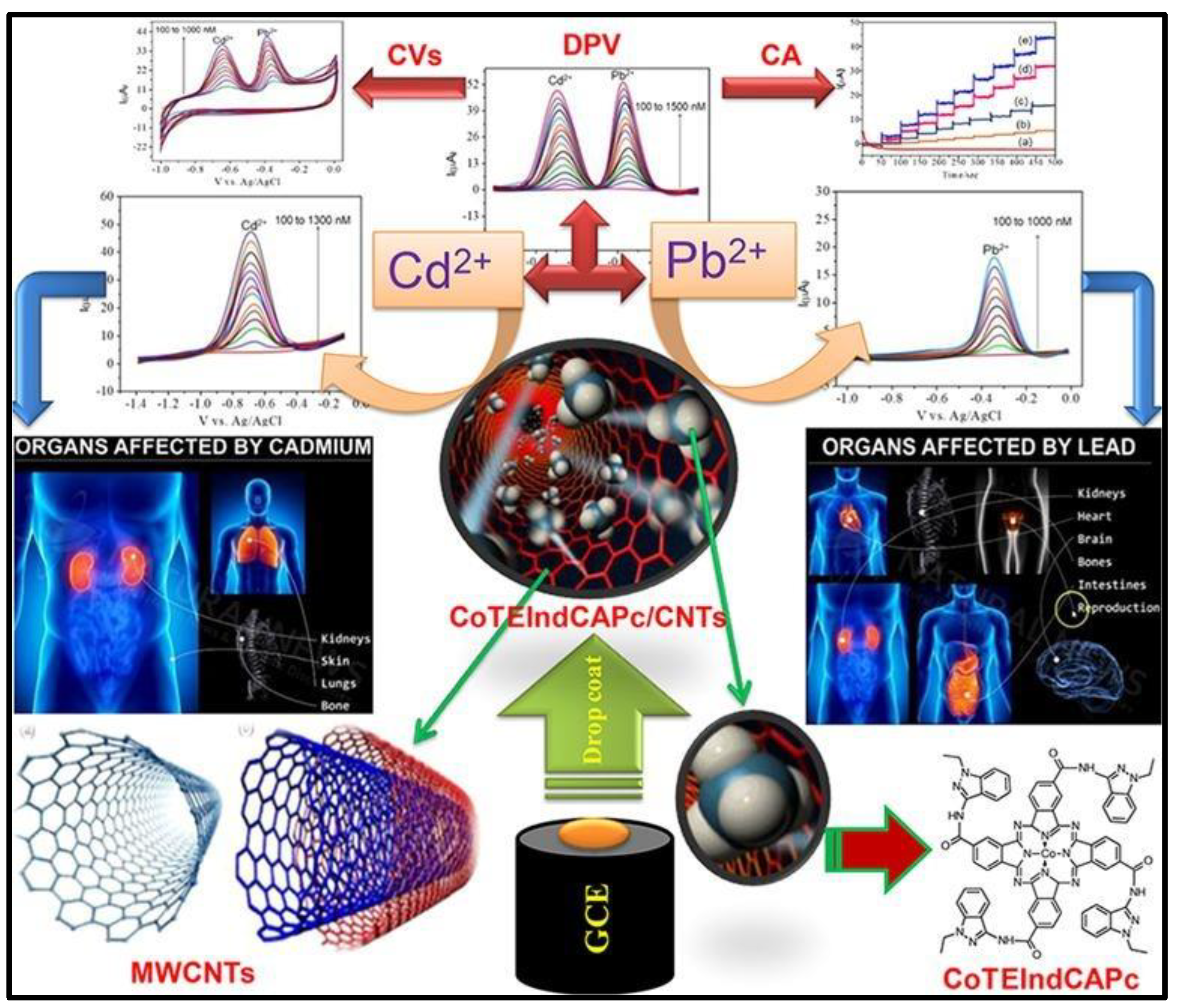
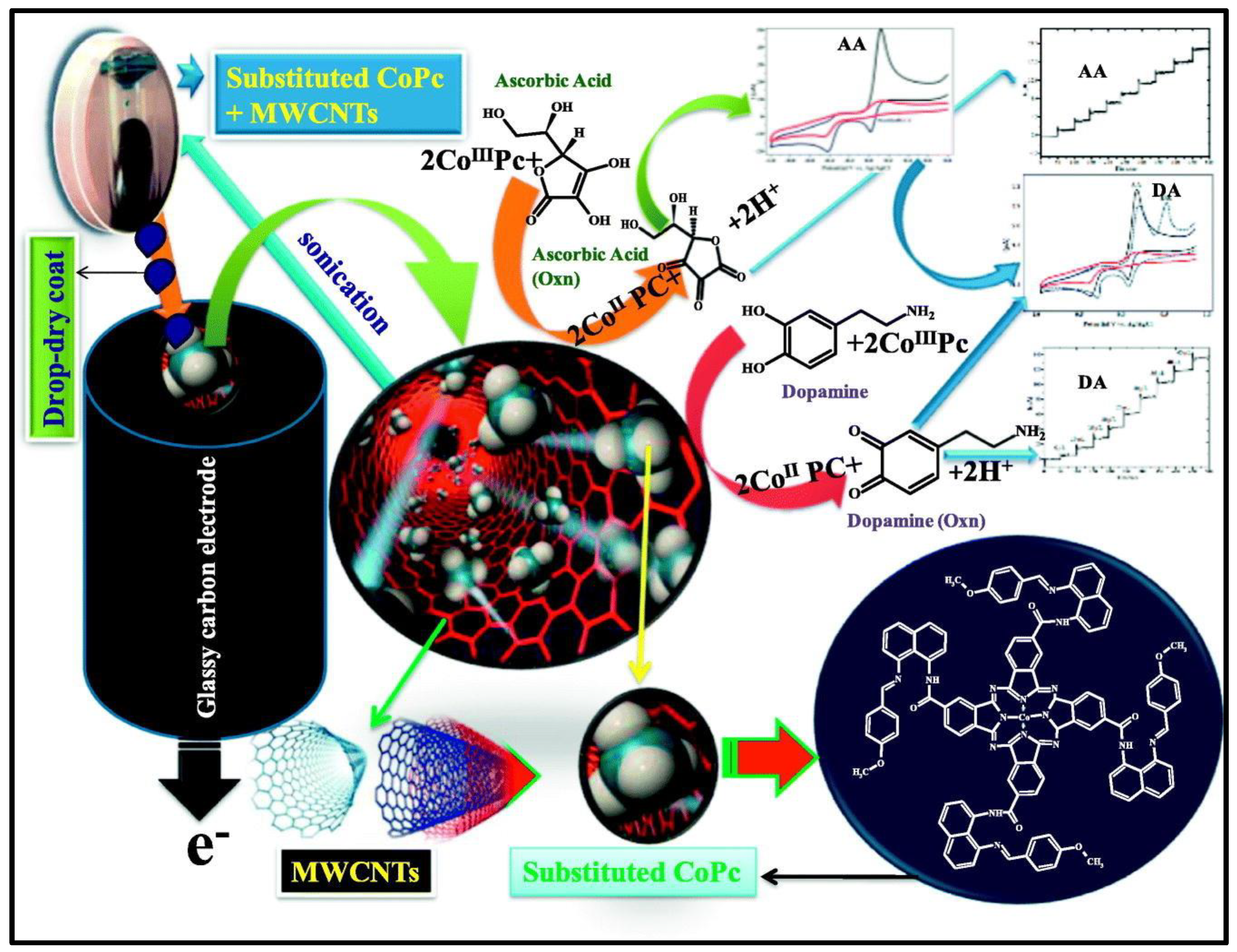
CNs, particularly MWCNTs, have immense potential as next-generation biosensors. In Pc-based composites, MWCNTs improve conductivity and enhance electron transfer with faster response times and potentially higher sensitivity. Additionally, the MWCNTs increase the effective surface area by providing more space for biomolecule interactions and potentially more binding sites. These CNs contribute to the overall stability of the composite by offering mechanical support, leading to a more robust and long-lasting biosensing activity. Hence, the synergistic effects of incorporating MWCNTs into Pc composites provide great possibilities for the development of superior biosensors with enhanced sensitivity, stability, and performance [113,114]. The electrocatalytic performance of a CoTEIndCAPc/MWCNTs/GCE electrode for Cd²⁺ and Pb²⁺ detection is illustrated in Figure 12. The electrode was designed and utilized for CV, DPV, and CA measurements of electrochemical activity. The electrode showed excellent sensitivity with low detection limits of 10 nmol L⁻¹ for Cd²⁺ and 9 nmol L⁻¹ for Pb²⁺, and high reproducibility, highlighting its potential for biological applications [114]. Recently, Mounesh et al. [115] presented an intriguing approach for biosensing applications using carbonaceous and MPc hybrid composites. They synthesized a novel tetra-8-[(E)-(4-methoxybenzylidene) amino] naphthalene-1-amine cobalt (II) Pc (CoTMBANAPc) through an amide bridge linkage, using cobalt (II) tetracarboxylic acid Pc (CoTCAPc) as the initial material. This synthesized macromolecule displayed excellent solubility in aprotic organic solvents, providing valuable insights into the composition and structure of the material. The GCE/MWCNT-CoTMBANAPc electrode exhibited remarkable promise for the simultaneous detection of AA and DA using CV, DPV, and CA techniques. The detection performance within the linear response of the concentration range of 7.5 to 70 nM for both AA and DA. The composite exhibited a lower detection limit of 6.6 μM for AA and 0.33 nM for DA. Furthermore, the GCE/MWCNT-CoTMBANAPc electrode displayed excellent stability, sensitivity, and reproducibility within the micromolar range. However, exploring the selectivity of these sensors in complex biological matrices where additional interfering species are present is necessary. Keshavanand Prabhu et al. [116] developed FeTBImPc, modified it with CNP, and immobilized it on a GCE for the detection of DA. The composite electrodes (GCE/CNP-FeTCAPc and GCE/CNP-polyFeTBImPc) showed excellent electrocatalytic activity toward DA oxidation, with lower detection limits of 14 nM. The GCE/CNP-polyFeTBImPc sensor exhibited superior performance with a high sensitivity of 67.2039 mA nM⁻¹ cm⁻². However, carbonaceous and metal–Pc hybrid composites face many challenges. The aggregation of Pc molecules limits the available surface area for biomolecule interaction and hinders sensor performance. The long-term stability of the composites under real-world conditions requires further investigation. Figure 13 provides a comprehensive overview of the applications of hybrid Pc composites as biosensors. The cobalt (II) Pc-modified GCE exhibited enhanced performance for the detection of AA and DA. Table 4 highlights the versatility of various hybrid Pc composites in terms of their sensitivity and selectivity [109,110,111,112,113,114,115,116,117,118].

4. Fabrication of Hybrid Pc in Three-Electrode System

The fabrication of hybrid Pc materials for electrochemical applications using a three-electrode system involves several advanced techniques aimed at significantly enhancing the conductivity, stability, and catalytic efficiency of the WE. In a typical three-electrode setup, the WE is where the electrochemical reaction of interest occurs, the CE completes the electrical circuit, and the RE maintains a stable potential, allowing for precise measurement of the potential and current of the WE. Thin film deposition techniques, such as chemical vapor deposition (CVD) and sputtering, are essential for achieving precise control over film thickness, consistency, and uniformity. In CVD, volatile precursors decay and react on the substrate, forming a thin, uniform film of Pc, which is crucial for the formation of deformity-free layers and effective electron transfer. Faltering involves the physical injection of material from an objective through high-energy ion bombardment. The material is then deposited onto the WE surface, providing a controlled and uniform coating that is fundamental for steady electrochemical performance. Electrochemical deposition further customizes the WE by integrating different metal ions into the electrode surface, thus fitting the catalytic properties of Pcs. This technique involves immersing the WE in an electrolyte containing metal ions and applying a voltage, which reduces the metal ions and deposits them on the electrode surface. This technique is particularly beneficial for framing nanostructured surfaces that significantly enhance catalytic activity and stability. Furthermore, self-gathering procedures, including the development of self-assembled monolayers (SAMs), are utilized to create coordinated molecular structures on the WE. SAMs involve immersing the WE in a solution containing particles with a functional group that binds strongly to the electrode material. These molecules spontaneously form a monolayer, providing a coordinated design that improves charge energy kinetics and sensor responsiveness by ensuring that the Pc molecules are optimally oriented for electron transfer.
Hybridization with nanomaterials, such as graphene, CNTs, rGO, and MNPs, is achieved through techniques such as solution-phase mixing, in situ development, or layer-by-layer assembly. This integration significantly expands the surface area and conductivity of the WE, leading to improved electron transfer rates and electrochemical performance. The collaboration between Pcs and nanomaterials results in composite materials with superior electron transfer rates and enhanced strengths under electrochemical conditions. Advanced lithographic methods, including photolithography and electron beam lithography, are used to design the WE with high accuracy, facilitating the formation of complex and miniaturized electrode designs. Photolithography uses light-sensitive photoresists to achieve micron-level accuracy, whereas electron beam lithography offers considerably greater resolution for nanoscale fabrication. These techniques ensure the optimization of the cathode surface for high electrochemical activity, making hybrid Pc materials ideal for various applications. Using these fabrication techniques, researchers can optimize the WE interface in a three-electrode system framework, thereby significantly enhancing the performance and reliability of electrochemical biosensors.

5. Conclusions, Challenges, and Future Perspectives

This manuscript highlights the exciting potential of macrocycle–macromolecule hybrids for biomolecule sensor applications. These hybrids offer excellent durability, precision, and sensitivity, overcoming difficulties and challenges such as redox behavior, cost, and synthetic complexity. However, further advancements are expected to achieve unrivalled sensitivity and ultra-low detection limits, particularly in intricate biological matrices. The careful selection of reactants with flexible functional groups during macromolecule synthesis is critical for sensor power against interference and environmental variations. Real-time biomolecule detection in different medical conditions is fundamental for the development of portable and remote biosensors. The resolution of biocompatibility and stability issues, combined with the ability to functionalize these hybrid sensors for multi-analyte identification, holds enormous potential for customized medical healthcare, surpassing traditional diagnostic strategies. Optimizing the analyte–electrode interface and sensor variability is key to addressing biocompatibility, stability, and reproducibility concerns. Investigating novel materials, nanostructures, and coatings offers promising ways toward enhanced accuracy and efficiency. The incorporation of progressive nanostructures appears to be particularly encouraging for advancing biosensor innovation. These nanostructures offer unique benefits, such as increased surface area and improved signal transduction, ultimately resulting in superior sensitivity, extremely low detection limits, and improved noise control in biosensing applications. Standardization initiatives and regulatory cooperation are crucial for effective clinical collaboration. A multidisciplinary approach that coordinates expertise in materials science, biotechnology, and engineering is fundamental for fully understanding the capability and potential of biosensing platforms and improving global health conditions.

Author Contributions

Shivalingayya G and Keshavananada Prabhu C P wrote the manuscript. Hur JH supervised the study. All authors listed have made substantial, direct, and intellectual contributions to the work and have approved it for publication. All authors have read and agreed to the published version of the manuscript.

Acknowledgments

This research was supported by the Basic Science Research Capacity Enhancement Project through the Korea Basic Science Institute (National Research Facilities and Equipment Center) grant funded by the Ministry of Education (2019R1A6C1010016) and the Basic Science Research Program through the National Research Foundation of Korea (NRF) grant funded by the Ministry of Education (NRF-2021R1F1A1050130).

References

  1. Naresh,V.; Lee, N.A. Review on Biosensors and Recent Development of Nanostructured Materials-Enabled Biosensors. Sensors. 2021, 21, 1109. [CrossRef]
  2. Kulkarni, M.B.; Ayachit, N.H.; Aminabhavi, T.M. Biosensors and Microfluidic Biosensors: From Fabrication to Application. Biosensors. 2022, 12, 543. [Google Scholar] [CrossRef] [PubMed]
  3. Abid, H.; Mohd, J.; Ravi, P.S. R, Suman.; S, Rab. Biosensors applications in medical field: A brief review, Sensors International, 2021, 2, 202. [CrossRef]
  4. Mujawar, M.A.; H, Gohel.; Bhardwaj, S.K.; S. Srinivasan.; N, Hickman.; A.Kaushik, Nano-enabled biosensing systems for intelligent healthcare: towards COVID-19 management, Materials Today Chemistry, 2020, 17. [CrossRef]
  5. B, Katey.; Ioana, V.; Anita, N.P.; Alexandrina U. A Review of Biosensors and Their Applications, ASME Open J. Engineering. 2023, 2, 020201. [CrossRef]
  6. Thevenot, D.R.; Toth, K.; Durst, R.A.; Wilson, G.S. Electrochemical biosensors: Recommended definitions and classification. Pure Appl. Chem. 1999, 71, 2333–2348. [Google Scholar] [CrossRef]
  7. Singh,A.; Sharma,A.; Ahmed, A.; Sundramoorthy, A.K.; Furukawa, H.; Arya, S.; Khosla, A. Recent Advances in Electrochemical Biosensors: Applications, Challenges, and Future Scope. Biosensors. 2021, 11, 336. [CrossRef]
  8. Shivalingayya ; Preeti R.K.; Sangappa, K.G.; Shashidhar.; Ashajyoti, C.; Arunkumar, L. Multifunctional Nanoparticles for Biomedical Applications, Journal of Chemical, Biological and Physical Sciences, JCBPS; Section B. 2022, 12, 410-422. [CrossRef]
  9. Costa, P.; Nunespereira, J.; Oliveira, JA.;, et al. High-performance graphene-based carbon nanofiller/polymer composites for piezoresistive sensor applications. Compos Sci Technol. 2017, 153, 241–252. [CrossRef]
  10. Campaña, A.; Florez, S.; Noguera, M.; Fuentes, O.; Ruiz, P.P.; Cruz, J.; Osma, J. Enzyme-based electrochemical biosensors for microfluidic platforms to detect pharmaceutical residues in wastewater. Biosensors. 2019, 9, 41. [Google Scholar] [CrossRef]
  11. Lee, J.; Kim, J.; Kim, S.; Min, D.H. Biosensors based on graphene oxide and its biomedical application. Adv Drug Deliv Rev. 2016, 1, 105, 275–287. [Google Scholar] [CrossRef]
  12. Basova, T.V.; Ray, A.K. Review—Hybrid materials based on phthalocyanines and metal nanoparticles for chemi-resistive and electrochemical sensors: A Mini-Review. ECS Journal of Solid State Science and Technology. 2020, 9, 061001. [Google Scholar] [CrossRef]
  13. Kuntoji, G.; Kousar, N.; Gaddimath, S.; Koodlur Sannegowda, L. Macromolecule–Nanoparticle-Based Hybrid Materials for Biosensor Applications. Biosensors. 2024, 14, 277. [Google Scholar] [CrossRef]
  14. Soto, D.; Orozco, J. Hybrid Nanobioengineered Nanomaterial- Based Electrochemical Biosensors. Molecules. 2022, 27, 3841. [Google Scholar] [CrossRef]
  15. Huai-Song, W. metal–organic frameworks for biosensing and bioimaging applications, Coordination Chemistry Reviews. 2017, 349, 139-155. [CrossRef]
  16. Kuntoji, G.; Kousar, N.; Gaddimath, S.; Koodlur Sannegowda, L. Macromolecule–Nanoparticle-Based Hybrid Materials for Biosensor Applications. Biosensors 2024, 14, 277. [Google Scholar] [CrossRef]
  17. Manjunatha, N.; Lokesh, K.S. Hybrid composites based on phthalocyanine and carbonaceous materials for sensing applications: a review, International Journal of Biosensors & Bioelectronics. 2021, 7(3), 84‒89. [CrossRef]
  18. Jipei, Y.; Nikolai, G.; Alexander, Eychmüller. Application of Polymer Quantum Dot-Enzyme Hybrids in the Biosensor Development and Test Paper Fabrication. Anal. Chem. 2012, 84, 11, 5047–5052. [CrossRef]
  19. Skladal, P.; Mascini, M. Sensitive detection of pesticides using amperometric sensors based on cobalt phthalocyanine-modified composite electrodes and immobilized choline esterases. Biosensors& Bioelectronics. 1992, 7, 335–343. [Google Scholar] [CrossRef]
  20. Lv, N.; Li, Q.; Zhu, H.; Mu, S.; Luo, X.; Ren, X.; Liu, X.; Li, S.; Cheng, C.; Ma, T. Electrocatalytic Porphyrin/Phthalocyanine-Based Organic Frameworks: Building Blocks, Coordination Microenvironments, Structure-Performance Relationships. Adv Sci (Weinh). 2023, 10(7), e2206239. [Google Scholar] [CrossRef] [PubMed]
  21. John, R.A.; Anthony, Guiseppi-Elie. Responsive Polymers in the Fabrication of Enzyme-Based Biosensors, Biomaterials Science. 2020, 1267-1286. [CrossRef]
  22. Mehrab, P.; Erfan, R.; Maryam, R.K.; Amirmasoud, S.; Razieh, B.; Abbas, R.; Luiz, F.R.; Ferreira. Properties and application of carbon quantum dots (CQDs) in biosensors for disease detection: A comprehensive review, Journal of Drug Delivery Science and Technology. , 2023, 80, 104156. [CrossRef]
  23. Zhihong, Zhang.; Yafei, Lou.; Chuanpan, Guo.; Qiaojuan, Jia.; Yingpan, Song.; Jia, Y.T.; Shuai, Z.; Minghua, W.; Linghao, He.; Miao, D. Metal–organic frameworks (MOFs) based chemosensors/biosensors for analysis of food contaminants, Trends in Food Science & Technology. 118, 2021, 569-588. [CrossRef]
  24. Naresh, V. .; Lee, N. A Review on Biosensors and Recent Development of Nanostructured Materials-Enabled Biosensors. Sensors. 2021, 21, 1109. [Google Scholar] [CrossRef]
  25. Sorokin, AB. Phthalocyanine metal complexes in catalysis. Chem Rev. 2013, 9(113), 8152–91. [Google Scholar] [CrossRef]
  26. Zagal, JH.; Bedioui, F.; Dodelet, JP. N4-macrocyclic Complexes. NY. Springer. 2006.
  27. Hong, Q.; Chen, S. Facile one-step fabrication of phthalocyanine– graphene–bacterial–cellulose nanocomposite with superior catalytic performance. Nanomaterials. 2020, 10, 1673. [Google Scholar] [CrossRef]
  28. Browne, MP.; Novotny, F.; Sofer, Z.; et al. 3D printed graphene electrodes’ electrochemical activation. ACS Appl Mater Interfaces. 2018, 10, 40294–40301. [CrossRef]
  29. Gaddimath, S.; Payamalle,S.; Channabasavana HundiPuttaningaiah, K.P.; Hur, J. RecentAdvances in pH and RedoxResponsive Polymer Nanocompositesfor Cancer Therapy. J. Compos. Sci. 024, 8, 28. [CrossRef]
  30. Sun, G.; Wei, X.; Zhang, D.; Huang, L.; Liu, H.; Fang, H. Immobilization of Enzyme Electrochemical Biosensors and Their Application to Food Bioprocess Monitoring. Biosensors. 2023, 13, 886. [Google Scholar] [CrossRef]
  31. Verma, S.; Thakur, D.; Pandey, C.M.; Kumar, D. Recent Prospects of Carbonaceous Nanomaterials-Based Laccase Biosensor for Electrochemical Detection of Phenolic Compounds. Biosensors. 2023, 13, 305. [Google Scholar] [CrossRef]
  32. Kamalasekaran, K.; Magesh, V.; Atchudan, R.; Arya, S.; Sundramoorthy, A.K. Development of Electrochemical Sensor Using Iron (III) Phthalocyanine/Gold Nanoparticle /Graphene Hybrid Film for Highly Selective Determination of Nicotine in HumanSalivary Samples. Biosensors. 2023, 13, 839. [Google Scholar] [CrossRef]
  33. Mahesh, M.S.; G, Manasa.; Ronald, J.M.; K, Mondal.; N.P, Shetti. Fundamentals of bio-electrochemical sensing, Chemical Engineering Journal Advances. 2023, 16, 100516. [CrossRef]
  34. Mikuła, E. Recent Advancements in Electrochemical Biosensors for Alzheimer’‘s’ Disease Biomarkers Detection. Curr Med Chem. 2021, 28(20), 4049-4073.
  35. H.S, Hwang.; Jae, W. J.; Yoong, A.K.; Mincheol, C. Carbon Nanomaterials as Versatile Platforms for Biosensing Applications, Micromachines. 2020, 11, 814. [CrossRef]
  36. Joonhyub, Kim.; Gayoung, P.; Seoho, Lee.; Suk-Won, H.; Namki, M.; Kyung-Mi, Lee. Single Wall Carbon Nanotube Electrode SystemCapable of Quantitative Detection of CD4+ T Cells, Biosensors and Bioelectronic. [CrossRef]
  37. Lijie, Wang.; Wenke, J.; Yanju, Wu.; Fei, Wang.; Lu, Kui. Direct Fabrication of 3D Graphene–Multi Walled Carbon Nanotubes Network and Its Application for Sensitive Electrochemical Determination of Hyperin, Int. J. Electrochem. Sci., 2019, 14, 481 – 493. [CrossRef]
  38. Chaithra, K.P.; Akshaya, K.B.; Maiyalagan, T.; Gurumurthy, H.; Anitha, V.; Louis, G. Unique Host Matrix to Disperse Pd Nanoparticles for Electrochemical Sensing of Morin: Sustainable Engineering Approach. ACS Biomater. Sci. Eng. 2020, 6, 5264–52731. [Google Scholar] [CrossRef]
  39. Diao, P.; Liu, Z.; Wu, B.; Nan, X.; Zhang, J.; Wei, Z. Chemically assembled single-wall carbon nanotubes and their electrochemistry. Chemphyschem. 2002, 3, 898–901 101002/1439. [Google Scholar] [CrossRef] [PubMed]
  40. Singh, C.; Srivastava, S.; Ali, M.A.; Gupta, T.K.; Sumana, G.; Srivastava, A.; Mathur, R.B.; Malhotra, B.D. Carboxylated multiwalled carbon nanotubes based biosensor for aflatoxin detection. Sens. Actuators Chem. 2013, 185, 258–264. [Google Scholar] [CrossRef]
  41. Mahor, A.; Singh, P.P.; Bharadwaj, P.; Sharma, N.; Yadav, S.; Rosenholm, J.M.; Bansal, K.K. Carbon-Based Nanomaterials for Delivery of Biologicals and Therapeutics: A Cutting-Edge Technology. 2021, 7, 19. [CrossRef]
  42. Ge, S.; Sun, M.; Liu, W.; Li, S.; Wang, X.; Chu, C.; Yan, M.; Yu, J. Disposable electrochemical immunosensor based on peroxidase-like magnetic silica–graphene oxide composites for detection of cancer antigen 153. Sens. Actuators B Chem. 2014, 192, 317–326. [Google Scholar] [CrossRef]
  43. Sumit, M.; Joginder, S.; Rohit, G.; Yajvinder, S.; Vivek, C.; Ahmad, Umar.; Ahmed, A.I.; Sheikh, A.; Sadia, Ameen.; Sotirios, B. Nanomaterials-based biosensor and their applications: A review, Heliyon. 9, 9, 2023. [CrossRef]
  44. Pratik, J.; Rupesh, M.; Roger, J.N. Biosensing applications of carbon-based materials, Current Opinion in Biomedical Engineering. 2021, 18, 100274. [CrossRef]
  45. Aoife, C.; Power, B.G.; Shaneel, C.; James, C. Carbon nanomaterials and their application to electrochemical sensors: a review, Nanotechnol Rev. 2018, 7(1), 19–41. [CrossRef]
  46. Keshavananda, C.P., Kenkera, R.N.; Shambhulinga A.; Shivalingayya.; Lokesh, K.S. Novel polymeric cobalt tetrabenzimidazole phthalocyanine for nanomolar detection of hydrogen peroxide, RSC Sustain. 2023,1, 128-138. [CrossRef]
  47. Imadadulla, M.; Manjunatha, N.; Veeresh, A.S.; Dayananda, B.P.; Lokesh, K.S. Electropolymerized film of cobalt tetrabenzimidazolephthalocyanine for the T amperometric detection of H2O2, Journal of Electroanalytical Chemistry. 2018, 826, 96–103. [CrossRef]
  48. Zribi, B.; Roy, E.; Pallandre, A.; Chebil, S.; Koubaa, M.; Mejri, N.; Magdinier Gomez, H.; Sola, C.; Korri-Youssoufi, H.; Haghiri-Gosnet, A.-M. A microfluidic electrochemical biosensor based on multiwall carbon nanotube/ferrocene for genomic DNA detection of Mycobacterium tuberculosis in clinical isolates. Biomicrofluidics. 2016. [CrossRef]
  49. Zong-hua, W.; Qiong-lin, L.; Yi-ming, W.; Guo-an, Luo. Carbon nanotube-intercalated graphite electrodes for simultaneous determination of dopamine and serotonin in the presence of ascorbic acid, Journal of Electroanalytical Chemistry. 540, 2003, 129-134. [CrossRef]
  50. Khan, R.; Pal, M.; Kuzikov, A.V.; Bulko, T.; Suprun, E.V.; Shumyantseva, V.V. Impedimetric immunosensor for detection of cardiovascular disorder risk biomarker. Mater. Sci. Eng. C 2016, 68, 52–58. [Google Scholar] [CrossRef] [PubMed]
  51. Yang, L.; Li, X.; Yan, S.; Wang, M.; Liu, P.; Dong, Y.; Zhang, C. Single-walled carbon nanotubes–carboxyl- functionalized graphene oxide-based electrochemical DNA biosensor for thermolabile hemolysin gene detection. Anal. Methods. 2015, 7, 5303–5310. [Google Scholar] [CrossRef]
  52. Xing, R.M.; Yang, H.T.; Li, S.N.; Yang, J.H.; Zhao, X.Y.; Wang, Q.L.; Liu, S.H.; Liu, X.H. A sensitive and reliable rutin electrochemical sensor based on palladium phthalocyanine-MWCNTs-Nafion nanocomposite. J. Solid State. Electr. 2017, 21, 1219–1228. [Google Scholar] [CrossRef]
  53. Yang, H.T.; Li, B.Y.; Cui, R.J.; Xing, R.M.; Liu, S.H. Electrochemical sensor for rutin detection based on Au nanoparticle-loaded helical carbon nanotubes. J. Nanopart. Res. 2017, 19, 354. [Google Scholar] [CrossRef]
  54. Jing, S.S.; Zheng, H.J.; Zhao, L.; Qu, L.B.; Yu, L.L. A novel electrochemical sensor based on WO3 nanorods-decorated poly(sodium 4-styrenesulfonate) functionalized graphene nanocomposite modified electrode for detecting of puerarin. Talanta. 2017, 174, 477–485. [Google Scholar] [CrossRef] [PubMed]
  55. Li, H.F.; Wang, S.; Cui, F.; Zhuo, B.B.; Zhao, C.R.; Liu, W.L. Sensitive and selective detection of puerarin based on the hybrid of reduced graphene oxide and molecularly imprinted polymer. J. Pharmaceut. Biomed. 2020, 185, 113221. [Google Scholar] [CrossRef] [PubMed]
  56. Wang, L.J.; Jia, W.K.; Wu, Y.J.; Wang, F.; Kui, L. Direct Fabrication of 3D Graphene-Multi Walled Carbon Nanotubes Network and Its Application for Sensitive Electrochemical Determination of Hyperin. Int. J. Electrochem. Sc. 2019, 14, 481–493. [Google Scholar] [CrossRef]
  57. Meng, R.Q.; Li, Q.L.; Zhang, S.J.; Tang, J.K.; Ma, C.L.; Jin, R.Y. GQDs/PEDOT Bilayer Films Modified Electrode as a Novel Electrochemical Sensing Platform for Rutin Detection. Int. J. Electrochem. Sc. 2019, 14, 11000–11011. [Google Scholar] [CrossRef]
  58. Zhao, P.C.; Ni, M.J.; Xu, Y.T.; Wang, C.X.; Chen, C.; Zhang, X.R.; Li, C.Y.; Xie, Y.X.; Fei, J.J. A novel ultrasensitive electrochemical quercetin sensor based on MoS2-carbon nanotube @ graphene oxide nanoribbons / HS-cyclodextrin / graphene quantum dots composite film. Sens. Actuators, 2019, 299, 126997. [Google Scholar] [CrossRef]
  59. Senocak, A.; Khataee, A.; Demirbas, E.; Doustkhah, E. Ultrasensitive detection of rutin antioxidant through a magnetic micro-mesoporous graphitized carbon wrapped Co nanoarchitecture. Sensor Actuat. B-Chem. 2020, 312, 127939. [Google Scholar] [CrossRef]
  60. Malekzad, H.; Zangabad, P.S.; Mirshekari, H.; Karimi, M.; Hamblin, M.R. Noble metal nanoparticles in biosensors: recent studies and applications. Nanotechnol Rev. 2017, 27, 6(3), 301–329. [Google Scholar] [CrossRef]
  61. Keshavananada prabhu C P.; Aralekallu, S.; Sajjan, V. A.; Palanna, M.; Kumar, S.; Sannegowda, L. K. Non-precious cobalt phthalocyanine-embedded iron ore electrocatalysts for hydrogen evolution reactions. Sustainable Energy & Fuels, 2021,5(5), 1448-1457. [CrossRef]
  62. Shivalingayya, G.; K. B, Chandrakala.; Arunkumar, L.; Santhoshkumar, Dani.; C. P, Keshavananda Prabhu.; Giddaerappa.; L.K. Sannegowda. Ilmenite-type NiTiO3 nanoparticles for oxygen evolution reaction, Journal of Applied Electrochemistry. [CrossRef]
  63. N, Tripathy.; Deok-Ho, Kim. Metal oxide modified ZnO nanomaterials for biosensor applications, Nano Convergence. 2018, 5, 1-10. [CrossRef]
  64. L.K, Sannegowda.; K.R, Venugopala Reddy.; K.H, Shivaprasad. Stable nano-size copper and its oxide particles using cobalt tetraamino phthalocyanine as a stabilizer; Application to electrochemical activity, RSC Advances. 2014, 4, 11367-11374. [CrossRef]
  65. Guo, X.; Liang, B.; Jian, J.; Zhang, Y.; Ye, X. Glucose biosensor based on a platinum electrode modified with rhodium nanoparticles and with glucose oxidase immobilized on gold nanoparticles. Microchim Acta. 2014, 181, 519–25. [Google Scholar] [CrossRef]
  66. Han, W.; Yang, J.; Jiang, B.; Wang, X.; Wang, C.; Guo, L.; Sun, Y.; Liu, F.; Sun, P.; Lu, G. Conductometric ppb-Level CO Sensors Based on In2O3 Nanofibers Co-Modified with Au and Pd Species. Nanomaterials. 2022, 12, 3267. [Google Scholar] [CrossRef]
  67. Fallatah, A.; Kuperus, N.; Almomtan, M.; Padalkar, S. Sensitive Biosensor Based on Shape-Controlled ZnONanostructures Grown on Flexible Porous Substrate for Pesticide Detection. Sensors. 2022, 22, 3522. [Google Scholar] [CrossRef]
  68. Zhu, J.; Wang, J-F.; Li, J-J.; Zhao, J-W. Tuning the fluorescence quenching properties of plasmonic Ag-coated-au triangular nanoplates: application in ultrasensitive detection of CEA. Plasmonics. 2015, 11, 565–572. [CrossRef]
  69. Lan, D.; Li, B.; Zhang, Z. Chemiluminescence flow biosensor for glucose based on gold nanoparticle-enhanced activities of glucose oxidase and horseradish peroxidase. Biosens Bioelectron. 2008, 24, 940–944. [Google Scholar] [CrossRef] [PubMed]
  70. Ge, L.; Su, M.; Gao, C.; Tao, X.; Ge, S. Application of Au cage/Ru (bpy) 32+ nanostructures for the electrochemiluminescence detection of K562 cancer cells based on aptamer. Sensor Actuator B. 2015, 214, 144–151. [Google Scholar] [CrossRef]
  71. Liang, W.; Zhuo, Y.; Xiong, C.; Zheng, Y.; Chai, Y.; Yuan, R. Ultrasensitive cytosensor based on self-enhanced electrochemiluminescent ruthenium-silica composite nanoparticles for efficient drug screening with cell apoptosis monitoring. Anal Chem. 2015, 87, 12363–12371 101021/acsanalchem5b03822. [Google Scholar] [CrossRef] [PubMed]
  72. Mohadeseh, Safaei.; Mohammad, M.; Foroughi.; Nasser, E.; Shohreh, J.; Ali, Omidi.; Mehrdad, Khatami. A review on metal-organic frameworks: Synthesis and applications, TrAC Trends in Analytical Chemistry. 118, 2019, 401-425. [CrossRef]
  73. Prasad, G.J.; Namrata, W.G.; Abdul, H.B.; Mohan, G.K. Metal nanoparticles synthesis: An overview on methods of preparation, advantages and disadvantages, and applications, Journal of Drug Delivery Science and Technology. 53, 2019, 101174. [CrossRef]
  74. Liu, D.; Guo, Q.; Zhang, X.; Hou, H.; You, T. PdCo alloy nanoparticle-embedded carbon nanofiber for ultrasensitive nonenzymatic detection of hydrogen peroxide and nitrite. J Colloid Interf Sci. 2015, 450, 168–173. [Google Scholar] [CrossRef]
  75. Bernardo-Boongaling, V.R.R.; Serrano, N.; García-Guzmán,J.J.; Palacios-Santander,J.M.; Díaz-Cruz, J.M. Screen-PrintedElectrodes Modified with Green-Synthesized Gold Nanoparticles for the Electrochemical Determination of Aminothiols. J. Electroanal. Chem. 2019, 847, 113184. [CrossRef]
  76. Ravalli,A.; dosSantos, G.P.; Ferroni, M.; Faglia, G.; Yamanaka, H.; Marrazza,G. New Label Free CA 125 Detection Based on Gold Nanostructured Screen-Printed Electrode. Sens. Actuators B Chem. 2013, 179. [CrossRef]
  77. Baradoke, A.; Jose, B.; Pauliukaite, R.; Forster, R.J. Properties of Anti-CA125 Antibody Layers on Screen-Printed Carbon Electrodes Modified by Gold and Platinum Nanostructures. Electrochim. Acta. 2019, 306, 299–306. [Google Scholar] [CrossRef]
  78. Phetsang, S.; Jakmunee, J.; Mungkornasawakul, P.; Laocharoensuk, R.; Ounnunkad, K. Sensitive Amperometric Biosensors for Detection of Glucose and Cholesterol Using a Platinum/Reduced Graphene Oxide/Poly(3-Aminobenzoic Acid) Film-Modified Screen-Printed Carbon Electrode. Bioelectrochemistry. 2019, 127, 125–135.
  79. Reitz, E.; Jia, W.; Gentile, M.; Wang, Y.; Lei, Y. CuO Nanospheres Based Nonenzymatic Glucose Sensor. Electroanalysis. 2008, 20, 2482–2486. [Google Scholar] [CrossRef]
  80. K.K. Naik.; C.S, Rout. Electrodeposition of ZnCo2O4 nanoparticles for biosensing applications, RSC Adv. 2015, 5, 79397–79404. [CrossRef]
  81. Jianjun, L. Shiwei, L.; Yue, Y.; Kai, Liu.; Wencai, Du. Highly selective and sensitive glucose sensors based on organic electrochemical transistors using TiO2 nanotube arrays-based gate electrodes, Sensors and Actuators B: Chemical. 2015, 208, 457-463. [CrossRef]
  82. Yuan, M.; Liu, A.; Zhao, M.; Dong, W.; Zhao, T.; Wang, J.; Tang, W. Bimetallic PdCu nanoparticle decorated three-dimensional graphene hydrogel for non-enzymatic amperometric glucose sensor. Sensor Actuator B. 2014, 19, 707–714. [Google Scholar] [CrossRef]
  83. Chang, G.; Shu, H.; Ji, K.; Oyama, M.; Liu, X.; He, Y. Gold nanoparticles directly modified glassy carbon electrode for non-enzymatic detection of glucose. Appl Surf Sci. 2014, 288, 524–529. [Google Scholar] [CrossRef]
  84. Yin, G.; Xing, L.; Ma, X-J.; Wan, J. Non-enzymatic hydrogen peroxide sensor based on a nanoporous gold electrode modified with platinum nanoparticles. Chem Pap. 2014, 68, 435–441. [CrossRef]
  85. Shantharaja.; Giddaerappa.; Lokesh, K.S. Phthalocyanine based metal- organic frame work with carbon nanoparticles as hybrid catalyst for oxygen reduction reaction, Electrochimica Acta. 2023, 456, 142405. [CrossRef]
  86. Leznoff, C.C.; Lever, A.B.P. Phthalocyanines: properties and applications vols. 1–4. New York. VCH Publishers. [CrossRef]
  87. Giddaerappa.; Nemakal, M.; Shantharaja.; Mirabbos, H.; Lokesh, K.S. Tetraphenolphthalein Cobalt(II) Phthalocyanine Polymer Modified with Multiwalled Carbon Nanotubes as an Efficient Catalyst for the Oxygen Reduction Reaction, ACS Omega. 2022, 7, 14291−14304. [CrossRef]
  88. K.I, Ozoemena.; T, Nyokong. Novel amperometric glucose biosensor based on an ether-linked cobalt(II) phthalocyanine–cobalt(II) tetraphenylporphyrin pentamer as a redox mediator, Electrochim. Acta. 2006, 51, 5131. [CrossRef]
  89. Liu, D.; Long, Y.T. Superior Catalytic Activity of Electrochemically Reduced Graphene Oxide Supported Iron Phthalocyanines toward Oxygen Reduction Reaction. ACS Appl Mater Interfaces. 2015, 4, 7(43), 24063-8. [CrossRef]
  90. Malathesh, Pari.; K.R.V, Reddy.; Fasiulla.; Chandrakala, K.B. Amperometric determination of dopamine based on an interface platform comprising tetra-substituted Zn2+ phthalocyanine film layer with embedment of reduced graphene oxide, Sensors and Actuators A: Physical. 2020, 316, 112377. [CrossRef]
  91. Sarvajith, M.S.; Harish, M.; Nimbegondi, K.; Mruthyunjayachari, C. D.; Fasiulla, Khan. Synthesis and Electrochemical Investigation of Tetra Amino Cobalt (II) Phthalocyanine Functionalized Polyaniline Nanofiber for the Selective Detection of Dopamine, Electroanalysis. 2020, 32(8), 1807-1817. [CrossRef]
  92. Veeresh, A.S.; Imadadulla, M.; Manjunatha, N.; Shambulinga, A.; Hemantha, K.K.R.; Sreenivasa, Swamy.; Lokesh, K.S. Synthesis and electropolymerization of cobalt tetraaminebenzamidephthalocyanine macrocycle for the amperometric sensing of dopamine, Journal of Electroanalytical Chemistry. 2019, 838, 33–40. [CrossRef]
  93. Imadadulla, M.; Manjunatha, N.; Shambhulinga, A.; Veeresh, A.S.; T.R, Divakara.; Manjunatha, P.; C.P, Keshavanandaprabu.; Lokesh, K.S. Phthalocyanine sheet polymer based amperometric sensor for the selective detection of 2,4-dichlorophenol, Journal of Electroanalytical Chemistry. 2020, 871, 114292. [CrossRef]
  94. Manjunatha, N.; Giddaerappa.; Shantharaja.; Veeresh, A.S.; Lokesh, K.S. Novel amide coupled phthalocyanines: Synthesis and structure-property relationship for electrocatalysis and sensing of hydroquinone, Journal of Electroanalytical Chemistry. 2021, 898, 115657. [CrossRef]
  95. Veeresh, A.S.; Shambhulinga, A.; Manjunatha, N.; Manjunatha, P.; C.P, Keshavananda Prabhu.; Lokesh, K.S. Nanomolar detection of lead using electrochemical methods based on a T novel phthalocyanine, Inorganica Chimica Acta. 2020, 506, 119564. [CrossRef]
  96. Shantharaja.; Manjunatha, N.; Giddaerappa.; Lokesh, K.S. Biocompatible polymeric pyrazolopyrimidinium cobalt(II) phthalocyanine: An efficient electroanalytical platform for the detection of l-arginine, Sensors and Actuators A. 2021, 324, 112690. [CrossRef]
  97. Veeresh, A.S.; Shambhulinga, A.; Manjunatha, N.; Manjunatha, P.; C. P, Keshavananda Prabhu.; Lokesh, K.S. Nanomolar detection of 4-nitrophenol using Schiff-base phthalocyanine, Microchemical Journal. 2021, 164, 105980. [CrossRef]
  98. C.P, Keshavananda Prabhu.; Nemakal, M.; Aralekallu, S.; Mohammed, I.; K.H, Shivaprasad.; M.K, Amshumali.; K.S, Lokesh. Synthesis and characterization of novel imine substituted phthalocyanine for sensing of L-cysteine, Journal of Electroanalytical Chemistry. 2019, 834, 30–137. [CrossRef]
  99. Gonçalo, D.; João, C.; Bruno, V.; Leticia, G.; Carina, A.; Maria, A.; João, R.; Pedro, V.B. Noble Metal Nanoparticles for Biosensing Applications, Sensors. 2012, 12, 1657-1687. [CrossRef]
  100. Zhibing, Liao.; Liu, Yao.; Yan, Liu.; Yaohui, Wu.; Yonghong, Wang.; Ge, Ning. Progress on nanomaterials based-signal amplification strategies for the detection of zearalenone, Biosensors and Bioelectronics. 2021, 9, 100084. [CrossRef]
  101. M.S, Sumitha.; T.S, Xavier. Recent advances in electrochemical biosensors – A brief review, Hybrid Advances. 2023, 2, 100023. [CrossRef]
  102. Duanduan, Yin.; Jian, Liu.; Xiangjie, Bo.; Mian, Li.; Liping, Guo. Porphyrinic metal-organic framework/macroporous carbon composites for electrocatalytic applications, Electrochimica Acta. 2017, 247, 41-49. [CrossRef]
  103. Sami, U.; Aziz, R.; Tayyaba, Najam.; Ismail, H.; Shazia, A.; Rashid, Ali.; Muhammad, U.S.; S.S, Ahmad Shah.; Muhammad, A.N. Advances in metal-organic framework@activated carbon (MOF@AC) composite materials: Synthesis, characteristics and applications, Journal of Industrial and Engineering Chemistry. 2024. [CrossRef]
  104. Basova, T.V.; Ray, A.K. Review—Hybrid materials based on phthalocyanines and metal nanoparticles for chemi-resistive and electrochemical sensors: A Mini-Review. ECS Journal of Solid-State Science and Technology. 2020, 9, 061001. [CrossRef]
  105. Gonçalves.; Josué, M; Bernardo, A.I.; Paulo, R.M.; Lúcio, A. Recent advances in electroanalytical drug detection by porphyrin/phthalocyanine macrocycles: developments and future perspectives. Analyst 146, 2021, 2, 365-381. [CrossRef]
  106. Nobuhle, N.; Sithi, M.; Tebello, N. Electrocatalytic Activity of Cobalt Phthalocyanines Revisited: Effect of the Number of Oxygen Atoms and Conjugation to Carbon Nanomaterials, electrocatalysis, 2021, 12, 499-515. [CrossRef]
  107. Costa, P.; Nunespereira, J.; Oliveira, JA.; et al. High-performance graphene-based carbon nanofiller/polymer composites for piezoresistive sensor applications. Compos Sci Technol. 2017, 153, 241–252. [Google Scholar] [CrossRef]
  108. Bounegru, A.V.; Apetrei, C. Development of a Novel Electrochemical Biosensor Based on Carbon Nanofibers–Cobalt Phthalocyanine–Laccase for the Detection of p-Coumaric Acid in Phytoproducts. Int. J. Mol. Sci. 2021, 22, 9302. [Google Scholar] [CrossRef] [PubMed]
  109. Manjunatha, N.; Shambhulinga, A.; Imadadulla, M.; et al. Electropolymerized octabenzimidazole phthalocyanine as an amperometric sensor for hydrazine. Journal of Electroanalytical Chemistry. 2019, 839, 238–246. [Google Scholar] [CrossRef]
  110. Jilani, B.S.; Mounesh.; Malathesh, P.; et al. Cobalt (II) tetra methyl-quinoline oxy bridged phthalocyanine carbon nano particles modified glassy carbon electrode for sensing nitrite: A voltammetric study. Materials Chemistry and Physics. 2020, 121920. [CrossRef]
  111. Shambhulinga, A.; Imadadulla, M.; Manjunatha, N.; et al. Synthesis of novel azo group substituted polymeric phthalocyanine for amperometric sensing of nitrite. Sensors & Actuators: B Chemical. 2019, 282, 417–425. [Google Scholar] [CrossRef]
  112. Manjunatha, N.; Shambhulinga, A.; Imadadulla, M.; et al. Nanomolar detection of 4-aminophenol using amperometric sensor based on a novel phthalocyanine. Electrochimica Acta. 2019, 318, 342–353. [Google Scholar] [CrossRef]
  113. Manjunatha, N.; Shambhulinga, A.; Imadadulla, M.; et al. Chemisorbed palladium phthalocyanine for simultaneous determination of biomolecules. Microchemical Journal. 2018, 143, 82–91. [Google Scholar] [CrossRef]
  114. Mounesh.; Reddy, K.R.V. Decorated CoPc with appliance of MWCNTs on GCE: Sensitive and reliable electrochemical investigation of heavy metals. Microchemical Journal. 2021, 160, 05610. [CrossRef]
  115. Mounesh.; Bhvimane, S.J.; Malatesh, Pari.; K.R. Venugopala Reddy.; K.S. Lokesh. Simultaneous and sensitive detection of ascorbic acid in presence of dopamine using MWCNTs-decorated cobalt (II) phthalocyanine modified GCE, Microchemical Journal. 2019, 147, 755–763. [CrossRef]
  116. Keshavanandaprabhu, C.P.; Manjunatha, N.; Shambhulinga, A.; Imadadulla, M.; Manjunatha, P.; Veeresh A.S.; Akshitha, D.; Lokesh, K.S. A comparative study of carboxylic acid and benzimidazole phthalocyanines and their surface modification for dopamine sensing, Journal of Electroanalytical Chemistry. 2019, 847, 113262. [CrossRef]
  117. Mania, V.; Huanga, S.T.; Devasenathipathy, R.; et al. Electropolymerization of cobalt tetraamino-phthalocyanine at reduced graphene oxide for electrochemical determination of cysteine and hydrazine. RSC Adv. 2016, 6(44), 38463–38469. [CrossRef]
  118. Tang, C.; Umasankar, Y.; Chen, S.M.s Simultaneous determination of adenine guanine and thymine at multi-walled carbon nanotubes incorporated with poly(new fuchsin) composite film. Anal Chim Acta. 2009, 636, 19–27. [CrossRef]
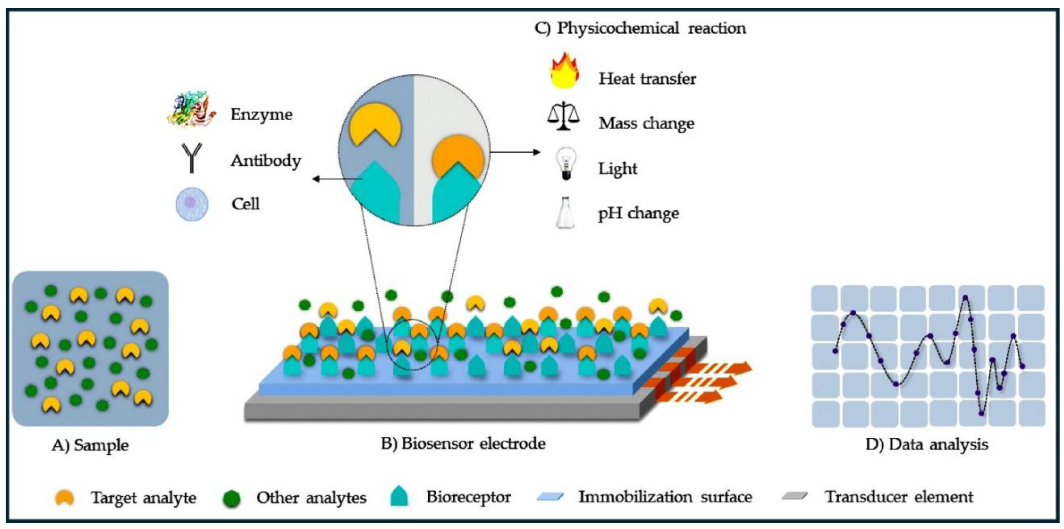
Figure 1. The schematic of a biosensor device, where the target analyte (A) interacts with the bioreceptor (B) immobilized on an electrode surface. This interaction triggers a physicochemical reaction (C) that the transducer element converts into a measurable signal. This signal is then analyzed (D) to detect and quantify the target analyte [10].
Preprints 111733 g001
Figure 2. Various fabrication methods of carbon-based materials in biosensor applications [41].
Preprints 111733 g002
Figure 3. Schematics of the fabrication of magnetic silica-graphene oxide (MSN/GO)-coated electrochemical immunosensor for the detection of cancer antigen 153 (CA 153). Reproduced with permission [42] Copyright 2014 from Elsevier Publications.
Preprints 111733 g003
Figure 4. The fabrication of CNT and GO for detection of H2O2 (a) CNT-modified electrode [46], and (b) GO-modified electrode for detection of H2O2. Reproduced with permission [47] Copyright 2018 from Elsevier Publications.
Preprints 111733 g004
Figure 5. (a) Fabrication of metal oxide NPs and the amperometric response of ZnO samples deposited on carbon paper. The responses for four different ZnO samples are compared: (b) pristine ZnO, (c) ZnO doped with KCl, (d) ZnO doped with NH₄F, and (e) ZnO modified with EDA [67].
Preprints 111733 g005
Figure 6. (a) Unique optical and chemical properties of AuNPs for biosensors. (b) The fabrication process of the ECL to-sensor. Reproduced with permission [70] Copyright 2015 from Elsevier Publications.
Preprints 111733 g006
Figure 7. Detection of DA using an interface made with a thin film of polymeric zinc(II) Pc, where the zinc centers have four specific substituent groups attached. Reproduced with permission [90] Copyright 2020 from Elsevier Publications.
Preprints 111733 g007
Figure 8. (a) Design and fabrication of cobalt phthalocyanine sheet polymer (poly-CoPc) for detection of 2,4-dichlorophenol (DCP). Reproduced with permission [93] Copyright 2020 from Elsevier Publications., (b) Electrocatalysis using amide coupled PCs for detection of hydroquinone. Reproduced with permission [94] Copyright 2021 from Elsevier Publications.
Preprints 111733 g008
Figure 9. (a) Fabrication of a laccase-based biosensor on an SPE, based on CoPc-modified CNFs. (b) The enzymatic oxidation mechanism of PCA in the presence of laccase [108].
Preprints 111733 g009
Figure 10. Investigation of a GCE modified with cobalt (II) tetra methyl-quinoline oxy-bridged Pc and carbon NPs for the detection of nitrite. Reproduced with permission [110] Copyright 2020 from Elsevier Publications.
Preprints 111733 g010
Figure 11. Hybrid Pc-based amperometric sensor for nanomolar detection of 4-AP. Reproduced with permission [112] Copyright 2019 from Elsevier Publications.
Preprints 111733 g011
Figure 12. CoPc functionalized with MWCNTs/GCE for electrochemical detection of heavy metals (Cd2+ and Pb2+). Reproduced with permission [114] Copyright 2021 from Elsevier Publications.
Preprints 111733 g012
Figure 13. Electrochemical detection of AA and DA utilizing a GCE modified with a hybrid cobalt (II) Pc complex. The cobalt (II) Pc acts as a catalyst, enhancing the electron transfer between the target molecules (AA and DA) and the electrode surface, leading to a more sensitive and selective detection process. Reproduced with permission [115] Copyright 2019 from Elsevier Publications.
Preprints 111733 g013
Table 1. Parameters of CNs for detection of various analytes.
Material Method Analyte LOD Linear Range Ref
MSN/GO DPV CA 153 2.8 × 10⁻⁴ Um/L 10-3–200 U/mL 42
MWCNT/
ferrocene
CV Hepatitis C and tuberculosis genomic DNA 7 fM 0.1 fM–1 pM 48
CNT DPV Dopamine (DA) 0.1 μM 0.5–10 μM 49
MWCNTs EIS Mb 0.08 ng m/L 0.1–90 ng m/L 50
SWCNT DPV Vibroparahaemolytics thermolabile hemolysin (tlh) gene 7.27 uM M 1.0 × 10⁻⁶–1 × 10-13 mol/L 51
CNTs CV Rutin 0.075 uM 0.10–51 uM 52
CNTs CV Rutin 0.081 uM 0.10–31 uM 53
Graphene CV, LSV Puerarin 0.04 uM 0.06–6.0 uM 54
Graphene CV Puerarin 0.006 uM 0.02–40 uM 55
CNTs–graphene CV Hyperin 0.001 uM 0.005–1.5 uM 56
Graphene quantum dots (GQDs) CV Rutin 0.011 uM 0.05–10 uM 57
GQD DPV Quercentin (Que) 8.2 × 10⁻⁴ uM 0.002–1.6 uM 58
Mesoporous carbon CV Rutin 0.002 uM 0.1–30 uM 59
Table 2. Parameters of MNPs for detection of various analytes.
Material Method Analyte LOD Linear Range Ref
Au Amperometry Cysteine 3.1 μM 10–80 μM 75
Glutathione 0.1 μM 0.3–10 μM
Methionine 1 μM 3.3–39 μM
Homocysteine 0.6 μM 2.2–30 μM
Au EIS Carcinoma antigen 125 6.7 U/mL 0–100 U/mL 76
Carcinoma antigen 125 419 ng/mL 450 ng/mL–2.916 μg/mL
Pt EIS Carcinoma antigen 125 386 ng/mL 450 ng/mL–2.916 μg/mL 77
Pt Amperometry Glucose 44.3 μM 0.25–6.0 mM 78
CuO nanospheres Amperometry Glucose 1 μM 2.5–20 mM 79
ZnCo2O4 Amperometry DA 15.5 μM 5–100 μM 80
TiO2 Amperometry Glucose 100 nM 100 nM–5 mM 81
Pd–Cu - Glucose 20 μM 2–18 mM 82
Au - Glucose 0.05 μM 0.1–25 mM 83
Pt - Glucose 7.2 × 10⁻⁸ M 1.0 × 10⁻⁷–2.0 × 10⁻⁵ M 84
Table 3. Parameters of polymeric Pc materials for detection of various analytes.
Materials Method Analyte LOD Linear Range Ref
Zn(II)TBPc Amperometry DA 6 nM 20 nM–1.0 μM 90
TACoPc/PANI CV DA 0.064 μM 20–200 μM 91
Poly(CoTNBAPc)
Amperometry DA 20 nmol/L 100–4000 nM/L 92
CV 20 nmol/L 10–1000 nM
CoTCAPc CV 2,4-dichlorophenol 0.35 μM 1–36 μM 93
CoTAMFCAPc/GCE CV Hydroquinone 0.066 0.2-2.2 μM 94
Amperometry 0.056 0.17-1.530 μM
Poly(CoTBrIMPPc) CV Pb 37 nmol/L 10–1000 nM 95
Amperometry 180 nmol/L 500–3000 nM/L
Poly(CoTPzPyPc) CV L-arginine 2.5 uM 10–100 uM 96
Amperometry 0.6 uM 2–60 uM
CoTTIMPPc CV 4-nitrophenol 38 nM 100–1000 nM 97
Amperometry 30 nM 100–900 nM
Poly(CoTBrImPc) CV L- Cysteine 3 nM 10–100 nM 98
Amperometry 4 nM 10–80 nM
Table 4. Hybrid Pc composites for detection of various analytes.
Material Method Analyte LOD Linear Range Ref
rGO/Poly(CoOBImPc) CV Hz 0.033 uM 0.1–0.9 uM 109
CoTM-QOPc/CNP CV Nitrite 0.033 uM 0.1–350 uM 110
Poly(TazoCoPc)/CNP CV Nitrite 0.006 uM 0.02–1 uM 111
CoTBTCAPc/CNP CV 4-AP 0.030 uM 0.1–0.9 uM 112
CoTELndCAPc/MWCNT CV Cd(II) 5 nM 100–1000nM 113
Pb(II) 3 nM 100–1000nM
PdTAPc/MWCNT CV AA 1.0 uM 3–24 uM 114
DA 0.6 uM 2–16 uM
CoTMBANAPc/MWCNT CV AA 6.6 uM 7.5–70 uM 115
Poly(FeTBImPc)/CNP CV DA 20 nM 100–1000nM 116
RGO-pTACoPc CV L-cysteine 0.018 uM 0.05–2.0 uM 117
MWCNT-PNF CV Adenine (AD) 9.2 uM 0.01–3.9 mM 118
DPV AD 7.9 uM 0.01–7.9 mM
CV Thymine (THY) 19.3 uM 0.02–7.7 mM
DPV THY 16.8 uM 0.02–15.7 mM
CV Guanine (GU) 98.56 uM 0.1–8.5 mM
DPV GU 96.84 uM 0.1–3.5 mM
Disclaimer/Publisher’s Note: The statements, opinions and data contained in all publications are solely those of the individual author(s) and contributor(s) and not of MDPI and/or the editor(s). MDPI and/or the editor(s) disclaim responsibility for any injury to people or property resulting from any ideas, methods, instructions or products referred to in the content.
Copyright: This open access article is published under a Creative Commons CC BY 4.0 license, which permit the free download, distribution, and reuse, provided that the author and preprint are cited in any reuse.
Prerpints.org logo

Preprints.org is a free preprint server supported by MDPI in Basel, Switzerland.

Subscribe

© 2024 MDPI (Basel, Switzerland) unless otherwise stated