Preprint
Review

State of the Art Synthesis of Ag-ZnO-Based Nanomaterials by Novel Atmospheric Pressure Microplasma Techniques

Altmetrics

Downloads

166

Views

82

Comments

0

A peer-reviewed article of this preprint also exists.

Submitted:

26 July 2024

Posted:

29 July 2024

You are already at the latest version

Alerts
Abstract
Atmospheric pressure Microplasma is a simple, cost-effective, efficient, and eco-friendly procedure, which is superior to the traditional nanomaterials synthesis techniques. It generates high yields and allows for a controlled growth rate and morphology of nanomaterials. The silver (Ag) nanomaterials, with their unique physical and chemical properties, exhibit outstanding antibacterial and anti-fungal properties. Similarly, zinc oxide (ZnO) nanomaterials, known for their low toxicity and relatively lower cost, find wide applications in wound repair, bone healing, and antibacterial and anticancer applications. The use of core-shell nanomaterials in certain situations where some nanoparticles can cause serious harm to host tissues or organs is a testament to their potential. A benign material is coated over the core to reduce toxicity in these cases. This review compares the numerous configurations of microplasma systems used for synthesizing nanomaterials and their use in producing Ag, ZnO, and their core-shell (Ag-ZnO) nanomaterials for biomedical applications. The summary also includes the effect of control parameters, including cathode diameter, gas flow rate, precursor concentration, voltage, and current, on the nanomaterial's characteristics and applications. In addition, it provides a research gap in the synthesis of Ag, ZnO, and core shall nanomaterials by this technique, as well as the development and limitations of this technique and the use of these nanoparticles for biomedical applications.
Keywords: 
Subject: Medicine and Pharmacology  -   Other

1. Introduction

Nanotechnology, one of the most promising technologies of the 21st century, can potentially convert nanoscience theory into beneficial applications. It enables us to observe, compute, handle, collect, monitor, and develop matter at the nanometer scales [1,2,3]. It is an emerging technology in many fields, such as electronics, gas sensors, catalyst, and biomedical applications. Nanomaterials, with their widespread applications in biomedical fields such as drug delivery, cancer treatment, immunotherapy, etc., are revolutionizing healthcare [4,5,6]. Nanoparticles are also applied to diagnostic instruments, pharmaceutical products, targeted medicinal products, and imagery and methodologies. Their high surface-area-to-volume ratio allows for maximum absorbance of medicine and quick movement in the bloodstream [7]. Owing to their excellent biocompatibility and biodegradability, nanoparticles can accumulate in defective organs with the most minor side effects. Furthermore, nanoparticles can be slowly released, reducing drug concentration and toxic side effects [8]. Diseases like cancer can be cured if they are detected at an early stage. Still, traditional diagnostic techniques cannot detect these tumors and cancers, and they cannot differentiate between malignant and benign lesions [9]. On the other hand, nanoparticle imaging can yield more appropriate and selective imaging of damaged and diseased tissues [10]. The nanoparticles are extensively applied to deliver chemotherapy drugs to the tumor cells, which reduces the toxicity of healthy tissues [11].
Zinc oxide (ZnO) nanoparticles are among the most common metal oxide nanoparticles, having distinctive chemical and physical properties that make them suitable for use in many fields as summarized in Figure 1 [12]. They are widely used in self-care products such as skincare, sunscreen, and cosmetics due to their beneficial properties against UV absorption [11,13]. They are also used in the textile industry; they are added to the fabric and have attractive features of visible light resistance and deodorant [14,15]. ZnO nanoparticles reveal brilliant biomedical applications such as wound healing, drug delivery, diabetes treatment, bio-imaging, anti-bacterial, anti-inflammation, and anticancer. They are less toxic and relatively cheaper than metal oxide nanoparticles [12,16]. In modern eras, immunotherapy is another approach to combat various diseases by providing immune molecules (antibodies and antigens) [17]. ZnO nanoparticles have received the utmost consideration owing to their high biocompatibility with human cells [18,19,20]. ZnO nanoparticles can hinder viral entrance, reproduction, and spreading all over the organ, which causes viral death because of stimulating reactive oxygen species, which results in oxidative stress [18,21].
Over the years, silver (Ag) nanoparticles have earned significant attention because of their exceptional electrical, optical, and antimicrobial properties [22]. It is commonly known that silver nanoparticles have remarkable antibacterial ability and admirable physical properties, which make them useful for applications in water purificants and disinfection of medical devices [23,24]. Due to their tiny size, they acquire a large surface area, providing a high surface energy and further probable reactive locations [25]. They have low volatility and high thermal stability and are less toxic to human cells [26,27]. They are used in medicine for treating burns, dental tools, and coated stainless steel materials [28,29]. In addition, they are utilized in various applications in textile fabrics, water treatment, and sunscreen lotion [30,31,32]. More importantly, Ag nanoparticles have the potential to be used for treating diseases that demand the maintained concentration of circulating drugs or the targeting of particular cells or organs [33,34].
Furthermore, the core-shell nanoparticles also earned much importance because of their great biological stability and good performance [9,35,36,37,38]. Core-shell nanoparticles, including noble metal semiconductors, are the most interesting materials for biomedical applications [38,39,40,41]. Ag-ZnO core-shell nanoparticles have strong anti-bacterial and antifungal properties [42,43]. Core-shell nanoparticles consist of a material core covered in a layer of another material [44]. Compared to simple nanoparticles, core shells offer significant advantages in biological applications, including improved properties like reduced cytotoxicity, increased dispersibility, biocompatibility, improved conjugation with other bioactive molecules, and enhanced thermal and chemical stability [44]. More specifically, if the desired nanoparticles are toxic, they can cause serious harm to the host tissues and organs. Then, the benign material can be coated over the core to reduce its toxicity. Sometimes, the shell layer enhances core materials' properties and is non-toxic [45]. There are two main approaches for nano-synthesis: bottom-up and top-down [46]. The Microplasma technique is a bottom-up technique with advantages over other techniques, such as its short processing time, environment-friendly nature, and low cost. It also has more benefits like small size, non-toxicity, control of growth, and flexibility, making it suitable for nanosynthesis [47,48,49].

2. Atmospheric Pressure Microplasma for Nanosynthesis

2.1. Background

In the past few years, research about nano-size particles has earned great attention because nanoparticles have distinct and unique properties compared to bulk materials due to their high surface-to-volume ratio [50]. The main task during nanoparticle synthesis is to control the shape, size, and stability of nanoparticles because these characteristics greatly influence their properties [51]. This control can be attained by varying the conditions of reactions, such as the concentration of precursors, the stabilizer, and the reducing agents [50]. Various synthesis techniques have been investigated for the synthesis of nanoparticles, such as laser ablation [52], electrochemical [53], microwave-assisted synthesis [54], and chemical reduction [50]. These processes require several hours for nanomaterials synthesis, so these techniques are time-consuming and expensive [55]. Plasma discharges with diverse arrangements are among the leading and eco-friendly techniques for nanoparticle synthesis. In particular, atmospheric pressure plasma systems are an essential technique, as they are cheap and simple and require no costly vacuum pumps and systems.

2.2. Synthesis of Nanomaterials

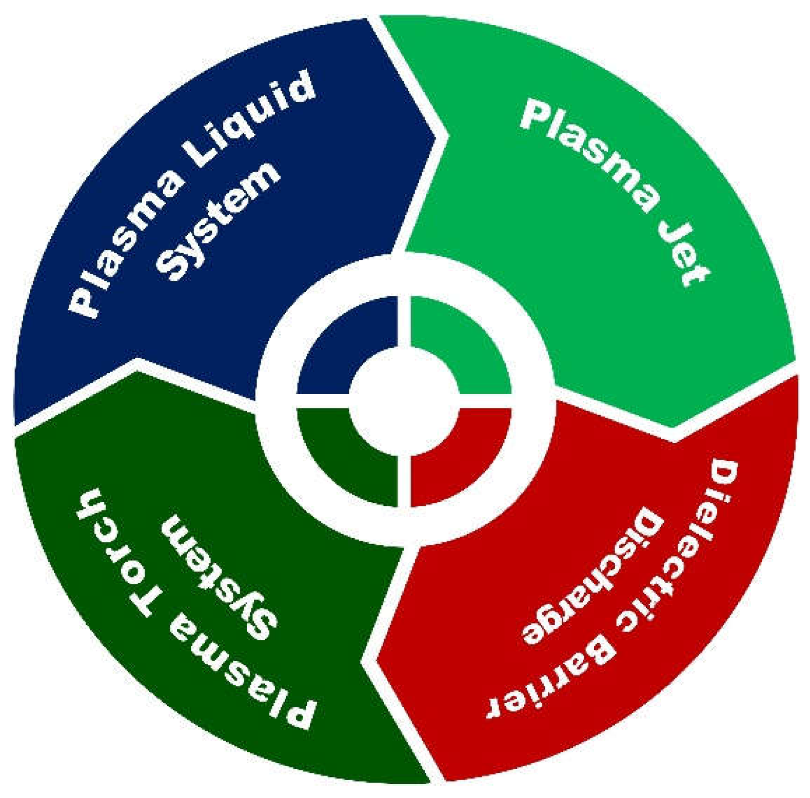
In recent years, various Microplasma systems have been used for nanosynthesis, as shown in Figure 2. These systems illustrate the versatility and flexibility of Microplasma's processing conditions. Here, we classify the system into four categories based on electrode geometry, way of injecting precursors, power coupling method, and plasma power source, as described in the following sub-sections [49,50,51].

2.2.1. Plasma Jet System

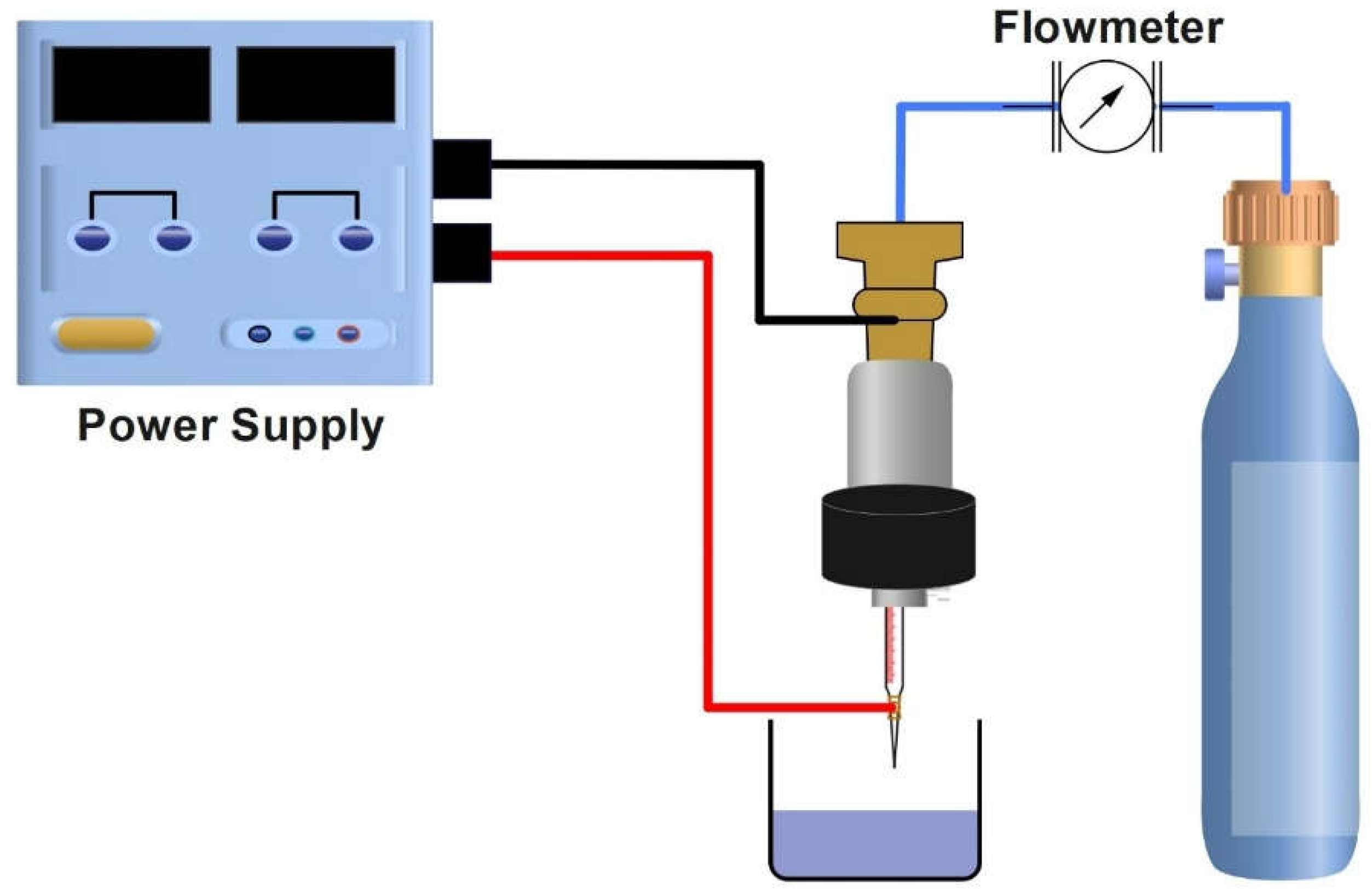
The atmospheric pressure plasma jet system synthesizes nanoparticles by generating inert gas plasma. Habib et al. [55] demonstrated the synthesis of silver nanoparticles by atmospheric pressure plasma jet using silver nitrate as a precursor and trisodium citrate dihydrate. The Microplasma jet system is shown in Figure 3, which consists of a gas flow system and power supply. A high-voltage electrode was coiled around the asymmetric source and was composed of a tube with 3.77 mm. This system has a large reservoir with a diameter of 38.2 mm, connected to a ground electrode. The support was connected to another ground electrode. The discharge was generated by high voltage. The working gas used in this system was helium, and 8A current and 25kHz frequency were applied. The surface of the liquid and plasma jet was kept at a distance of 7 mm. The samples were exposed to plasma for 5 minutes.
In addition, plasma jets have various configurations, such as hollow electrode micro discharge [56], Microplasma jets with external electrodes [57], and Microplasma jets having consumable electrodes [58].

2.2.2. Dielectric Barrier Discharge

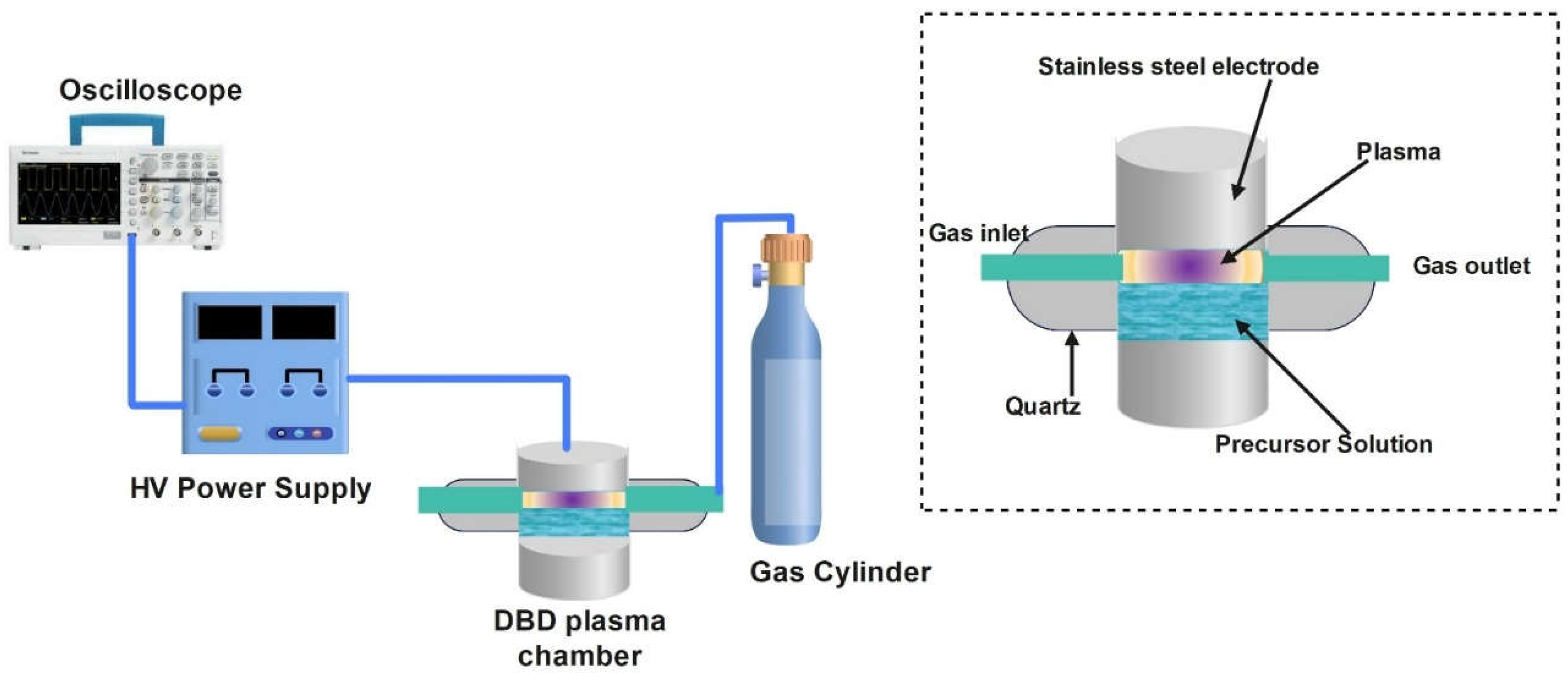
The synthesis of silver nanoparticles by atmospheric pressure dielectric barrier discharge was demonstrated by Janith and co-workers [59], and the schematic diagram of the process is shown in Figure 4. The reactor is a quartz-based cylindrical structure. The sodium citrate solution (34 mM) and aqueous silver nitrate were mixed to prepare a solution of silver precursor. The precursor solution could be injected into the hollow central part of the plasma reactor, which could be sealed by a quartz lid. The central part was linked with the outlet and gas feeding line. The chamber has a total volume of 25 mL; the precursor solution used in this experiment was 4 mL. The bottom and lid of the quartz chamber acted as a dielectric barrier. The dielectric barriers were at a distance of 8 mm from each other. The power supply of 2000 K was used to apply high voltage to the stainless-steel electrodes. The frequency and power supply were 9.1 kHz and 39 kV respectively [59].

2.2.3. Plasma Torch Method

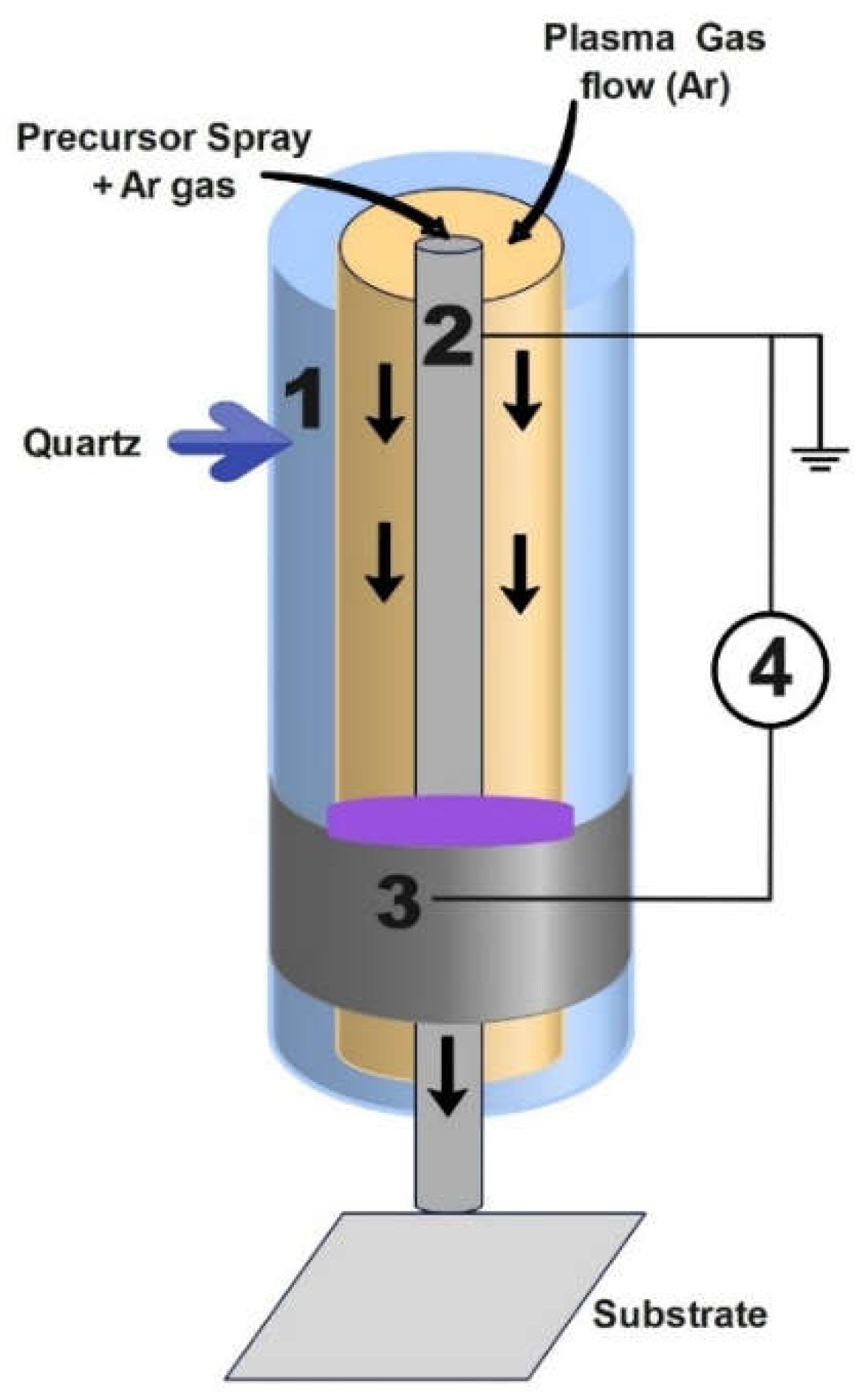
Another technique for nanomaterials synthesis is the atmospheric plasma torch method. Bjelajac et al. [60] reported the synthesis of Au nanomaterials by the atmospheric pressure plasma torch method. Tetra chloroauric acid trihydrate dissolved in ethanol or water was used as a precursor. The atmospheric pressure plasma torch consists of two hollow quartz tubes. The plasma was generated between the outer plasma tube (7mm inner diameter and 9mm outer diameter) and the inner tube (4mm inner diameter and 6mm outer diameter). The inner tube was grounded, and its outer surface was coated with a thick Pt film of 300 nm. Physical vapor deposition was used to deposit Pt film. The aluminum foil 5cm long covered the external quartz tube, and the aluminum foil was connected to the high voltage (H.V) generator. The plasma was produced in the space between the two tubes by applying sinusoidal high voltage to the external electrodes. The internal hollow electrode carried the nebulized gold precursor solution close to the discharge. The schematic diagram of the plasma torch setup is given in Figure 5.

2.2.4. Plasma-Liquid System

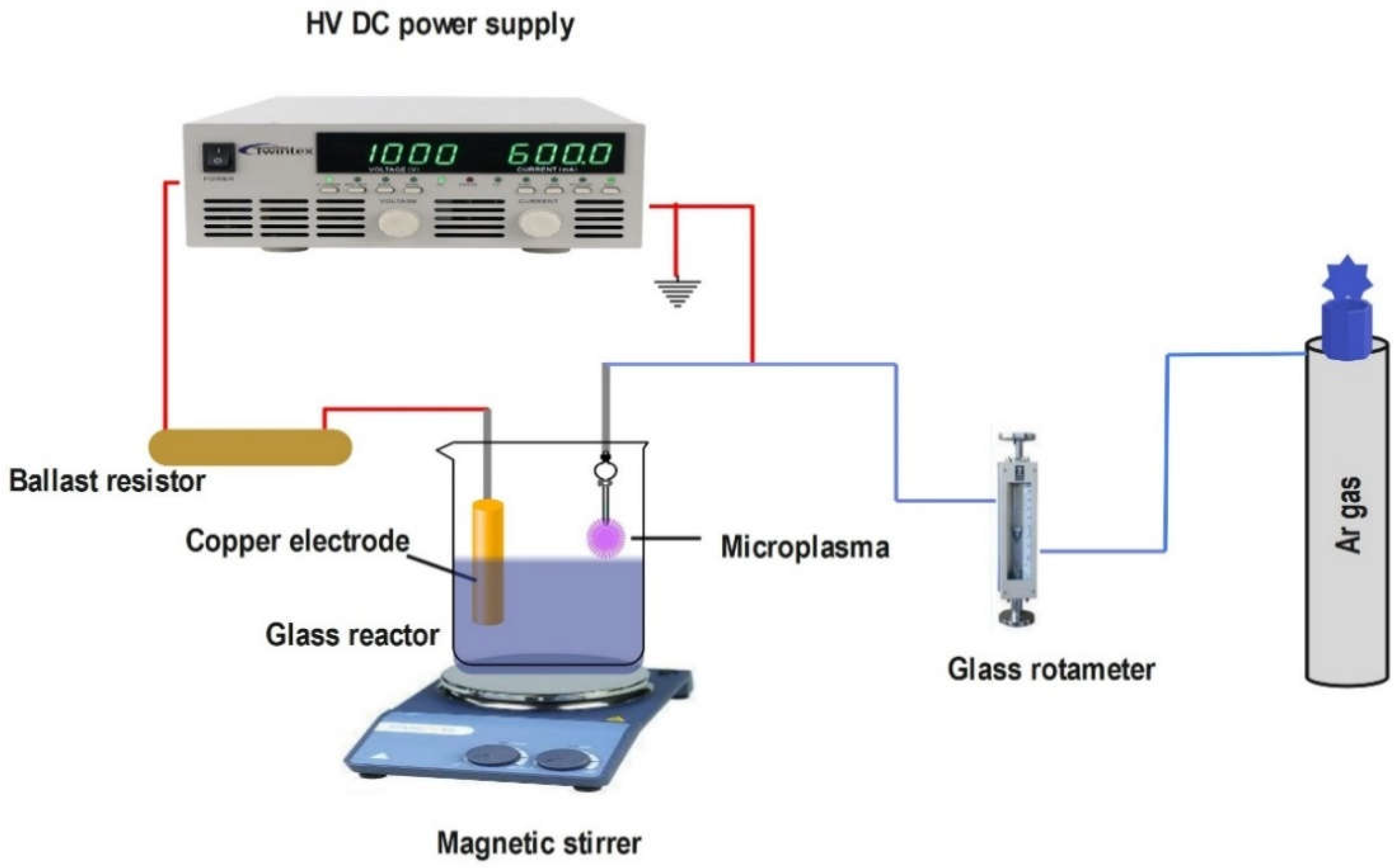
A plasma liquid system is extensively used for nanomaterial synthesis and is also called an atmospheric pressure Microplasma or Microplasma electrochemical synthesis, as shown in Figure 6. Ming and colleagues [61] reported the synthesis of cuprous oxide nanoparticles by microplasma electrochemical synthesis. It consisted of a stainless-steel tube (0.7 mm inside diameter, 8 cm length) placed 3 cm away from the copper electrode. A 2 mm space was maintained between the liquid surface and the capillary tube end. The argon gas flow was connected to the tube, and the glass rotameter was used to control the flow rate at 60 ml/min. The discharge was ignited by applying a high voltage, keeping the current constant. The precursors used in this synthesis were NaOH, NaCl, and NaNO3 with H2O ethylene glycol or DI water as solvents. The plasma was generated at the interface of the gas solution. A Ballast resistor stabilized the current and voltage. A magnetic stirrer was used to stir the solution gently to avoid agglomeration. The sediments collected were washed and centrifuged many times.

3. Literature Review

Thong and colleagues [62] synthesized silver nanoparticles using a D.C. helium Microplasma jet and examined the stabilizing effect of sucrose at various molar concentrations added to the AgNO3 solution. In this setup, the gap between the surface of the solution and the capillary was kept small to lower the voltage required to cause the Microplasma to ignite. Bisht et al. [63] reported the synthesis of silver nanoparticles using atmospheric pressure microplasma. Silver nitrate solution in DI water was used as a precursor. In order to avoid agglomeration, sucrose was used as a stabilizing agent. They synthesized silver nanoparticles with uniform radii ranging from 7-13nm. The Microplasma caused the reduction and nucleation of aqueous metal ions into nanoparticles without using any chemical reducing agents. The obtained nanoparticles were analyzed by dynamic light scattering (DLS), SEM, and UV visible absorption. Kondeti et al. [64] described the surfactant-free synthesis of silver nanoparticles using Argon and Ar +0.64% H2 plasma. A sinusoidal voltage wave modulated at 20 kHz with a 20% duty cycle and 13.4 MHz radio frequency to generate the plasma. It was observed that Ar+0.64%H2 synthesized nanoparticles of small size with a maximum 2-3nm diameter, whereas Ar gas plasma synthesized nanoparticles of broad size distribution. Shepida et al. [65] demonstrated the formation of silver nanoparticles in a solution of AgNO3 and sodium polyacrylate, a non-toxic surfactant. This setup used tungsten wire as a cathode, and the voltage was kept constant at 250V. The silver nanoparticles in the range 2-20 nm were formed at a concentration of 0.05- 0.2mMol L-1 of AgNO3 with 0.5 gL-1 of NaPA. The synthesized silver nanoparticles had established antimicrobial activity against staphylococcus aureus, Escherichia coli, and candida albicans. Huang et al. [66] declared the synthesis of silver nanoparticles by plasma-assisted electrochemical technique. They demonstrated that the interparticle spacing and the size of nanoparticles in the solution could be adjusted by altering the synthesis parameters so that the plasmonic response could be tuned. It was evident that larger-sized, highly dispersive silver nanoparticles were produced at higher solution concentrations and higher temperatures. Additionally, they revealed that silver nanoparticle synthesis can be accomplished without using a stabilizer, allowing control of nanoparticles dispersion. Shuaib et al. [67] synthesized AgNPs using the Microplasma technique. They investigated the role of variation in the molar concentration of fructose on the size of nanoparticles. They concluded that AgNPs with better efficiency against fungi and bacteria can be obtained by using 2mM fructose sample, due to the production of A g + ions. Lin et al. [68] demonstrated the combination of Gemini surfactant with AgNPs to attain a stable nano-surfactant system with strong anti-bacterial activities. Plasma-aided technique prepared high-quality crystalline nanostructures, where electrons acted as reductants, replacing conventional chemical reducing agents. The surfactants stabilized the silver nanoparticles by preventing AgNPs from aggregating. Antibacterial studies were conducted against S. aureus and E. coli, demonstrating the synergetic effects of the compounding systems [69]. Habib et al. [55] reported the silver nanoparticle synthesis in a quick and environment-friendly way using an atmospheric pressure plasma jet. They examined the role of variations in AgNo3 (precursor) and citrate concentration and determined the optimal conditions for synthesizing silver nanoparticles. They found their effective applications in the bio-medical field (antibacterial activities), photonic, and catalytic activities. Saleem et al. [70] demonstrated that by changing the type or concentration of capping agents in optimized Microplasma parameters, the size of AgNPs could be modified, and therefore, it influenced the stability of AgNPs. The results from DLS indicated that Polyvinyl Alcohol (PVA) capped AgNPs were the most stable over 15 days compared to Polyvinyl Pyrrolidone PVP capping agents and sucrose. The AgNP's size variations were within the range of less than 5nm limit. They suggested that these stability results had practical applications in cancer therapy. Skiba et al. [71] examined the catalytic effect of silver nanoparticles synthesized by a non-equilibrium low-temperature plasma technique. The characteristics and formation of nanoparticles were analyzed by DLS, ultraviolet-visible spectroscopy, and scanning electron microscopy. Then, silver nanoparticles were effectively used in the catalytic reduction of 4-NP, and they demonstrated outstanding catalytic performance with a quick reaction time. Iqbal et al. [72] reported that the atmospheric Microplasma approach was successfully employed to generate two- dimensional stumbled silver nanosheets. SEM analysis was used to verify the surface morphology of the synthesized nanosheets, and it showed that their lateral dimensions increased as the precursor concentration increased. The antibacterial activity of silver nanosheets was found to be highly effective against various types of bacteria and to be correlated with the size of nanosheets.
Iqbal and co-workers [73] reported the synthesis of ZnO nanostructure using different ionic surfactants and non-ionic fructose using the Microplasma technique. This study investigated the modification in the surface of hexagonal ZnO with surfactants. The structural study demonstrated the crystalline structure with a hexagonal phase of synthesized ZnO nanostructures. Significant antibacterial activity against the tested pathogens was found in the antibacterial study. Schwan et al. [74] successfully synthesized morphology-controlled ZnO nanoparticles using zinc powder and oxygen with an atmospheric pressure plasma jet. It was discovered that the rate of oxygen in carrier gas and plasma, the energy within the reactor, and the discharge current all affected the particle's morphology. Jain et al. [75] described the synthesis and deposition of ZnO nanocrystalline materials by atmospheric pressure plasma synthesis. Radiofrequency power generated plasma and the precursor was metallic zinc wire. The aggregation of synthesized nanostructures formed a porous film at the substrate. The synthesized nanostructures were thoroughly studied and characterized by UV-visible absorption, transmission electron microscopy, and X-ray diffraction. Abdullah et al. [76] demonstrated the capability of atmospheric pressure plasma jets to prepare high-purity, nanometer-sized ZnO in the gas or liquid phase. The obtained ZnO nanocrystals were characterized by transmission electron microscopy, Fourier transformation infrared (FTIR), and X-ray powder diffraction. The findings revealed that electrolytic media, current density, and reaction temperature influenced the morphology of ZnO nanocrystals.
Rawi and co-workers [42] reported the synthesis of core-shell nanoparticles of Ag-ZnO by an atmospheric pressure plasma jet technique and described their antibacterial and antifungal properties. The characterization of these Ag-ZnO core-shell nanoparticles was done by different techniques such as ultraviolet-visible spectroscopy (UV-vis), transmission electron microscopy (TEM), XRD, energy dispersive X-ray spectroscopy (EDX), and field emission scanning electron microscopy (FE-SEM). The pureness of synthesized Ag-ZnO core-shell N.P.s was proved by XRD and EDX analysis. The antibacterial activity of these core-shell nanoparticles was evaluated on two different types of gram-positive (Staphylococcus aureus and Staphylococcus epidermidis) and gram-negative bacteria (pneumonia and Escherichia coli). Furthermore, the antifungal activity of these core-shell N.P.s was evaluated against two distinct types of yeast. Khalid et al. [77] observed the formation of gold-silver core-shell nanoparticles using cold atmospheric pressure Microplasma. They revealed that the precursor concentration affected the average size of particles; the average size of particles increased with the increase in concentration. The review of plasma configuration and their use for synthesis of silver, zinc-oxide and core-shall nanoparticles is also summarized in Table 1.

4. Summary of Review

Some important factors influencing nanoparticle size, morphology, and properties are discussed here and are summarized in Table 2. In nanoparticle synthesis, solution/ precursor concentration affects nanoparticles' size and anti-microbial properties. Early studies revealed that increasing precursor concentration enhanced nanoparticles' size and anti-microbial properties. Another factor that is significantly important in nanoparticle size and morphology is processing time. The increase in processing time (5-45 mins) causes an increase in the size of nanoparticles and improves the crystallinity of nanoparticles. The concentration of the stabilizing agent or surfactants added to the precursor to avoid agglomeration is important. (Usually, fructose and sucrose are used as stabilizers in the synthesis of nanoparticles. The increase in concentration or molar ratio of surfactants or stabilizing agents increases the average size of nanoparticles. The gas flow rate also affects nanoparticle size; when the flow rate rises, the nanoparticle size decreases. All these factors are of great importance in nanoparticle synthesis of silver and zinc oxide, as they affect the structural properties and their biomedical applications. By altering these factors, the properties of nanoparticles can be varied.

5. Conclusion and final remarks

The atmospheric pressure plasma is an advanced and novel technique that can produce nanoparticles at ambient conditions. It has low thermal temperatures, faster processes, simplified equipment, and a cheap, eco-friendly system with various dimensions and configurations. Recent studies are presented here for nanomaterial synthesis by different configurations of atmospheric pressure Microplasma depending on the slight difference in their setups. This brief review revealed that silver and zinc oxide and their core-shell nanomaterials have great biological properties such as antibacterial, anticancer, and antifungal. The contribution of numerous control parameters like electrode dimension, flow of gas, type of gas, precursors concentration, operating voltage and current, distance between electrodes, and configuration of plasma system on nanoparticles characteristics (size, shape, and applications) is summarized. Nowadays, wide research is being conducted on atmospheric pressure microplasma nano synthesis. Still, there is a broad scope for the development of research, and a research gap is provided in this article. Although silver and zinc oxide nanoparticles by this technique are widely reported, the literature on their core-shell nanoparticles is very limited and needs to be investigated in the future, specifically the pros and cons of such core-shell nanoparticles for biomedical applications.

Author Contributions

Conceptualization, M.N., A.K., and M.R.M.; validation, B.A., U.R., M.N. and A.K.; resources, M.N.; writing—original draft preparation, A.K. and M.N.; writing—review and editing, U.R., B.A., O.A., F.A. and E.T.; supervision, M.N. and M.R.M.; project administration, M.N. All authors have read and agreed to the published version of the manuscript.”.

Funding

This manuscript received no fundings.

Institutional Review Board Statement

Not applicable.

Informed Consent Statement

Not applicable.

Data Availability Statement

The data is provided in this article.

Conflicts of Interest

The authors declare no conflicts of interest.

References

  1. Kumar, A.; Jayeoye, T.J.; Mohite, P.; Singh, S.; Rajput, T.; Munde, S.; Eze, F.N.; Chidrawar, V.R.; Puri, A.; Prajapati, B.G.; et al. Sustainable and consumer-centric nanotechnology-based materials: An update on the multifaceted applications, risks and tremendous opportunities. Nano-Structures Nano-Objects 2024, 38, 101148. [Google Scholar] [CrossRef]
  2. Zhou, A.F.; Feng, P.X. One-Dimensional and Two-Dimensional Nanomaterials for Sensor Applications. 2024, MDPI. p. 622.
  3. Yamaguchi, T.; Kim, H.-J.; Park, H.J.; Kim, T.; Khalid, Z.; Park, J.K.; Oh, J.-M. Controlling the Surface Morphology of Two-Dimensional Nano-Materials upon Molecule-Mediated Crystal Growth. Nanomaterials 2023, 13, 2363. [Google Scholar] [CrossRef] [PubMed]
  4. Lee, Y.-Y.; Sriram, B.; Wang, S.-F.; Kogularasu, S.; Chang-Chien, G.-P. Advanced Nanomaterial-Based Biosensors for N-Terminal Pro-Brain Natriuretic Peptide Biomarker Detection: Progress and Future Challenges in Cardiovascular Disease Diagnostics. Nanomaterials 2024, 14, 153. [Google Scholar] [CrossRef] [PubMed]
  5. Gatou, M.-A.; Vagena, I.-A.; Pippa, N.; Gazouli, M.; Pavlatou, E.A.; Lagopati, N. The Use of Crystalline Carbon-Based Nanomaterials (CBNs) in Various Biomedical Applications. Crystals 2023, 13, 1236. [Google Scholar] [CrossRef]
  6. Cao, M. Recent Development of Nanomaterials for Chemical Engineering. Nanomaterials 2024, 14, 456. [Google Scholar] [CrossRef] [PubMed]
  7. Wang, N.; Chen, L.; Huang, W.; Gao, Z.; Jin, M. Current Advances of Nanomaterial-Based Oral Drug Delivery for Colorectal Cancer Treatment. Nanomaterials 2024, 14, 557. [Google Scholar] [CrossRef] [PubMed]
  8. Gaci, Y.; Guittoum, A.; Hemmous, M.; Nez-Blanco, D.M.; Gorria, P.; Blanco, J.A.; Aouaroun, T. EFFECT OF Fe CONTENT ON THE STRUCTURAL AND MAGNETIC PROPERTIES OF TERNARY (Ni60Co40) 100−xFex NANOMATERIALS SYNTHESIZED BY HYDROTHERMAL ROUTE. Surface Review and Letters (SRL) 2024, 31, 1–9. [Google Scholar]
  9. Gatou, M.-A.; Skylla, E.; Dourou, P.; Pippa, N.; Gazouli, M.; Lagopati, N.; Pavlatou, E.A. Magnesium Oxide (MgO) Nanoparticles: Synthetic Strategies and Biomedical Applications. Crystals 2024, 14, 215. [Google Scholar] [CrossRef]
  10. Hadi, A.J.; Nayef, U.M.; Jabir, M.S.; Mutlak, F.A.-H. Titanium dioxide nanoparticles prepared via laser ablation: evaluation of their antibacterial and anticancer activityI. Surf. Rev. Lett. 2023, 30, 1–10. [Google Scholar] [CrossRef]
  11. Kayed, K.H.; The structural effects on silver nanoparticles plasma edges in optical reflectance spectra of Ag/Ag2O composites synthesized by oxygen plasma treatment of silver thin films. 2021.
  12. Casiano-Muñiz, I.M.; Ortiz-Román, M.I.; Lorenzana-Vázquez, G.; Román-Velázquez, F.R. Synthesis, Characterization, and Ecotoxicology Assessment of Zinc Oxide Nanoparticles by In Vivo Models. Nanomaterials 2024, 14, 255. [Google Scholar] [CrossRef]
  13. Alhoqail, W.A.; Alothaim, A.S.; Suhail, M.; Iqbal, D.; Kamal, M.; Asmari, M.M.; Jamal, A. Husk-like Zinc Oxide Nanoparticles Induce Apoptosis through ROS Generation in Epidermoid Carcinoma Cells: Effect of Incubation Period on Sol-Gel Synthesis and Anti-Cancerous Properties. Biomedicines 2023, 11, 320. [Google Scholar] [CrossRef] [PubMed]
  14. Zenkin, K.; Durmus, S.; Demir, A. Investigating Magnetic, Dielectric, Optic and Morphologic Properties of Nano-Nickel Oxide-Doped NdFeO3. Surface Review and Letters (SRL) 2024, 31, 1–16. [Google Scholar] [CrossRef]
  15. Kumar, P.; Ramesh, M.R.; Doddamani, M.; Suresh, J. Green synthesis of Fe/Ni/Cr oxide nanoparticles using Costus pictus plant extract: Microstructure and biological properties. Surf. Rev. Lett. 2024, 2450065. [Google Scholar] [CrossRef]
  16. Ounis, T.-D.; Rahmouni, K.; Aouar, L.Zaabat, M.; Optical properties and antibacterial activity of Ni, Mg and Fe-doped ZnO. Surface Review and Letters, 2024.
  17. Cho, N.-H.; Cheong, T.-C.; Min, J.H.; Wu, J.H.; Lee, S.J.; Kim, D.; Yang, J.-S.; Kim, S.; Kim, Y.K.; Seong, S.-Y. A multifunctional core–shell nanoparticle for dendritic cell-based cancer immunotherapy. Nat. Nanotechnol. 2011, 6, 675–682. [Google Scholar] [CrossRef] [PubMed]
  18. Prakash, K.M.K.; Prakash, V.; Gopinath, P.G. Influence of Precursor Molarity and Leaf Extract Concentration on Physical Properties of Zinc Oxide (ZnO) Nanoparticles Synthesized Using Moringa Oleifera Leaf Extract. Surf. Rev. Lett. 2024. [Google Scholar] [CrossRef]
  19. Abdulgafour, H.I.; Zainulabdeen, F.S.; Karam, G.S.; Magid, H.C.; Najim, A.A.; Hassan, F.M. Synthesis and characterization of Al-doped ZnO thin films as anti-reflection coatings for solar cell applications. Surf. Rev. Lett. 2024, 31, 1–7. [Google Scholar] [CrossRef]
  20. Wiesmann, N.; Mendler, S.; Buhr, C.R.; Ritz, U.; Kämmerer, P.W.; Brieger, J. Zinc Oxide Nanoparticles Exhibit Favorable Properties to Promote Tissue Integration of Biomaterials. Biomedicines 2021, 9, 1462. [Google Scholar] [CrossRef]
  21. Sugihartono, I.; Tan, S.T.; Arkundato, A.; Fahdiran, R.; Isnaeni, I.; Handoko, E.; Budi, S.; Budi, A.S. The Effect of Al-Cu Co-Dopants on Morphology, Structure, and Optical Properties of ZnO Nanostructures. Materials Research 2023, 26, e20220499. [Google Scholar] [CrossRef]
  22. Khan, S.; Zahoor, M.; Khan, R.S.; Ikram, M.; Islam, N.U. The impact of silver nanoparticles on the growth of plants: The agriculture applications. Heliyon 2023, 9, e16928. [Google Scholar] [CrossRef]
  23. Song, Y.; Yang, F.; Mu, B.; Kang, Y.; Hui, A.; Wang, A. Phyto-mediated synthesis of Ag nanoparticles/attapulgite nanocomposites using olive leaf extract: Characterization, antibacterial activities and cytotoxicity. Inorganic Chemistry Communications 2023, 151, 110543. [Google Scholar] [CrossRef]
  24. Hu, J.; Chen, F.; Mao, J.; Ni, L.; Lu, J. Direction regulation of interface carrier transfer and enhanced photocatalytic oxygen activation over Z-scheme Bi4V2O11/Ag/AgCl for water purification. Journal of Colloid and Interface Science 2023, 641, 695–706. [Google Scholar] [CrossRef] [PubMed]
  25. Yu, S.-j.; Yin, Y.-g.; Liu, J.-f.J.E.S.P.Impacts; Silver nanoparticles in the environment. 2013. 15, 78-92.
  26. Jaswal, T.; Gupta, J. A review on the toxicity of silver nanoparticles on human health. Materials Today Proceedings 2023, 81, 859–863. [Google Scholar] [CrossRef]
  27. Neciosup-Puican, A.A.; Pérez-Tulich, L.; Trujillo, W.; Parada-Quinayá, C. Green Synthesis of Silver Nanoparticles from Anthocyanin Extracts of Peruvian Purple Potato INIA 328—Kulli papa. Nanomaterials 2024, 14, 1147. [Google Scholar] [CrossRef]
  28. Francisco, P.; Amaral, M.N.; Neves, A.; Ferreira-Gonçalves, T.; Viana, A.S.; Catarino, J.; Faísca, P.; Simões, S.; Perdigão, J.; Charmier, A.J.; et al. Pluronic® F127 Hydrogel Containing Silver Nanoparticles in Skin Burn Regeneration: An Experimental Approach from Fundamental to Translational Research. Gels 2023, 9, 200. [Google Scholar] [CrossRef]
  29. Holubnycha, V.; Husak, Y.; Korniienko, V.; Bolshanina, S.; Tveresovska, O.; Myronov, P.; Holubnycha, M.; Butsyk, A.; Borén, T.; Banasiuk, R.; et al. Antimicrobial Activity of Two Different Types of Silver Nanoparticles against Wide Range of Pathogenic Bacteria. Nanomaterials 2024, 14, 137. [Google Scholar] [CrossRef]
  30. Gong, P.; Li, H.; He, X.; Wang, K.; Hu, J.; Tan, W.; Zhang, S.Yang, X.J.N.; Preparation and antibacterial activity of Fe3O4@ Ag nanoparticles. 2007, 18, 285604.
  31. Naysmith, A.; Mian, N.S.; Rana, S. Development of conductive textile fabric using Plackett–Burman optimized green synthesized silver nanoparticles and in situ polymerized polypyrrole. Green Chemistry Letters and Reviews 2023, 16, 2158690. [Google Scholar] [CrossRef]
  32. Ghazwani, M.; Hani, U.; Alqarni, M.H.; Alam, A. Development and Characterization of Methyl-Anthranilate-Loaded Silver Nanoparticles: A Phytocosmetic Sunscreen Gel for UV Protection. Pharmaceutics 2023, 15, 1434. [Google Scholar] [CrossRef] [PubMed]
  33. Patel, R.R.; Singh, S.K.; Singh, M. Green synthesis of silver nanoparticles: methods, biological applications, delivery and toxicity. Materials Advances 2023, 4, 1831–1849. [Google Scholar]
  34. Al-Serwi, R.H.; Eladl, M.A.; El-Sherbiny, M.; Saleh, M.A.; Othman, G.; Alshahrani, S.M.; Alnefaie, R.; Jan, A.M.; Alnasser, S.M.; Albalawi, A.E.; et al. Targeted Drug Administration onto Cancer Cells Using Hyaluronic Acid–Quercetin-Conjugated Silver Nanoparticles. Molecules 2023, 28, 4146. [Google Scholar] [CrossRef]
  35. Adam, A.; Mertz, D. Iron oxide@ mesoporous silica core-shell nanoparticles as multimodal platforms for magnetic resonance imaging, magnetic hyperthermia, near-infrared light photothermia, and drug delivery. Nanomaterials 2023, 13, 1342. [Google Scholar] [CrossRef]
  36. Kar, N.; McCoy, M.; Wolfe, J.; Bueno, S.L.A.; Shafei, I.H.; Skrabalak, S.E. Retrosynthetic design of core–shell nanoparticles for thermal conversion to monodisperse high-entropy alloy nanoparticles. Nat. Synth. 2023, 3, 175–184. [Google Scholar] [CrossRef]
  37. Rani, P.; Varma, R.S.; Singh, K.; Acevedo, R.; Singh, J. Catalytic and antimicrobial potential of green synthesized Au and Au@Ag core-shell nanoparticles. Chemosphere 2023, 317, 137841. [Google Scholar] [CrossRef] [PubMed]
  38. Hu, J.; Liu, X.; Zhang, J.; Gu, X.; Zhang, Y. Plasmon-activated NO2 sensor based on Au@MoS2 core-shell nanoparticles with heightened sensitivity and full recoverability. Sensors Actuators B Chem. 2023, 382. [Google Scholar] [CrossRef]
  39. Subbotina, J.; Rouse, I.; Lobaskin, V. In silico prediction of protein binding affinities onto core–shell PEGylated noble metal nanoparticles for rational design of drug nanocarriers. Nanoscale 2023, 15, 13371–13383. [Google Scholar] [CrossRef]
  40. Awiaz, G.; Lin, J.Wu, A. Recent advances of Au@ Ag core–shell SERS-based biosensors. Wiley Online Library.
  41. Liu, X.; Liang, X.; Yu, J.; Xu, K.; Shen, J.-W.; Duan, W.; Zeng, J. Recent development of noble metal-based bimetallic nanoparticles for colorimetric sensing. TrAC Trends Anal. Chem. 2023, 169, 117386. [Google Scholar] [CrossRef]
  42. Al-Rawi, B.K.Mazhir, S.N.J.I.J.o.N.; Evaluation of Antimicrobial Agents of Ag-ZnO Core-Shell Prepared by Micro-Jet Plasma Technique. 2023, 22, 2350044-16.
  43. Abuzeid, H.M.; Julien, C.M.; Zhu, L.; Hashem, A.M. Green synthesis of nanoparticles and their energy storage, environmental, and biomedical applications. Crystals 2023, 13, 1576. [Google Scholar] [CrossRef]
  44. Miao, W.; Hao, R.; Wang, J.; Wang, Z.; Lin, W.; Liu, H.; Feng, Z.; Lyu, Y.; Li, Q.; Jia, D. Architecture Design and Catalytic Activity: Non-Noble Bimetallic CoFe/fe3O4 Core–Shell Structures for CO2 Hydrogenation. Advanced Science 2023, 10, 2205087. [Google Scholar] [CrossRef]
  45. Chatterjee, K.; Sarkar, S.; Rao, K.J.; Paria, S. Core/shell nanoparticles in biomedical applications. Adv. Colloid Interface Sci. 2014, 209, 8–39. [Google Scholar] [CrossRef]
  46. Xinghao, L.I.U.; Cheng, C.; Zimu, X.U.; Shuheng, H.U.; Jie, S.; Yan, L.A.N.; Paul, K.C. Degradation of tetracycline in water by gas–liquid plasma in conjunction with rGO-TiO2 nanocomposite. Plasma Science and Technology 2021, 23, 115503. [Google Scholar]
  47. Iqbal, T.; Zahra, S.K.; Khan, M.A.R.; Shafique, M.; Raza, S.R.A.; Andleeb, S. Microplasma-assisted electrochemical synthesis of ZnO nanostructures for photocatalytic and antibacterial applications. Phys. Scr. 2021, 96, 125801. [Google Scholar] [CrossRef]
  48. Thai, V.-P.; Furuno, H.; Saito, N.; Takahashi, K.; Sasaki, T.; Kikuchi, T. The essential role of redox potential/equilibrium constant in the ability of non-equilibrium plasma for nano-synthesis in liquids. J. Appl. Phys. 2020, 128, 043305. [Google Scholar] [CrossRef]
  49. Ingsel, T.; Gupta, R.K., Plasma at the nanoscale: An introduction, in Plasma at the Nanoscale. 2022, Elsevier. p. 1-20.
  50. Abou El-Nour, K.M.; Eftaiha, A.a.; Al-Warthan, A.Ammar, R.A.J.A.j.o.c.; Synthesis and applications of silver nanoparticles. 2010. 3, 135-140.
  51. Skіba, M.; Pivovarov, A.; Vorobyova, V.; Derkach, T.Kurmakova, I.; Plasma-chemical formation of silver nanoparticles: The silver ions concentration effect on the particle size and their antimicrobial properties. 2019.
  52. Verma, S.; Rao, B.; Srivastava, A.; Srivastava, D.; Kaul, R.; Singh, B. A facile synthesis of broad plasmon wavelength tunable silver nanoparticles in citrate aqueous solutions by laser ablation and light irradiation. Colloids Surfaces A: Physicochem. Eng. Asp. 2017, 527, 23–33. [Google Scholar] [CrossRef]
  53. Iravani, S.; Korbekandi, H.; Mirmohammadi, S.V.Zolfaghari, B.J.R.i.p.s.; Synthesis of silver nanoparticles: chemical, physical and biological methods. 2014. 9, 385.
  54. Francis, S.; Joseph, S.; Koshy, E.P.; Mathew, B.J.A.c., nanomedicine,biotechnology; Microwave assisted green synthesis of silver nanoparticles using leaf extract of elephantopus scaber and its environmental and biological applications. 2018. 46, 795-804.
  55. Habib, T.; Caiut, J.M.A.Caillier, B.J.N.; Synthesis of silver nanoparticles by atmospheric pressure plasma jet. 2022. 33, 325603.
  56. Lin, L.; Li, S.; Hessel, V.; Starostin, S.A.; Lavrijsen, R.; Zhang, W. Synthesis of Ni nanoparticles with controllable magnetic properties by atmospheric pressure microplasma assisted process. AIChE J. 2018, 64, 1540–1549. [Google Scholar] [CrossRef]
  57. Mariotti, D.; Bose, A.C.Ostrikov, K.J.I.T.o.P.S.; Atmospheric-microplasma-assisted nanofabrication: Metal and metal–oxide nanostructures and nanoarchitectures. 2009, 37, 1027-1033.
  58. Shimizu, Y.; Kawaguchi, K.; Sasaki, T.Koshizaki, N.J.A.P.L.; Generation of room-temperature atmospheric H2/Ar microplasma jet driven with pulse-modulated ultrahigh frequency and its application to gold nanoparticle preparation. 2009, 94(19).
  59. Weerasinghe, J.; Li, W.; Zhou, R.; Zhou, R.; Gissibl, A.; Sonar, P.; Speight, R.; Vasilev, K.; Ostrikov, K. Bactericidal Silver Nanoparticles by Atmospheric Pressure Solution Plasma Processing. Nanomaterials 2020, 10, 874. [Google Scholar] [CrossRef] [PubMed]
  60. Bjelajac, A.; Phillipe, A.-M.; Guillot, J.; Fleming, Y.; Chemin, J.-B.; Choquet, P.; Bulou, S. Gold nanoparticles synthesis and immobilization by atmospheric pressure DBD plasma torch method. Nanoscale Adv. 2023, 5, 2573–2582. [Google Scholar] [CrossRef] [PubMed]
  61. Du, C.Xiao, M.J.S.r.; Cu2O nanoparticles synthesis by microplasma. 2014. 4, 7339.
  62. Thong, Y.L.; Chin, O.H.; Ong, B.H.; Huang, N.M. Synthesis of silver nanoparticles prepared in aqueous solutions using helium dc microplasma jet. Jpn. J. Appl. Phys. 2015, 55, 01AE19. [Google Scholar] [CrossRef]
  63. Bisht, A.; Roshan Deen, G.Ilyas, U.; Synthesis of nanoparticles using atmospheric microplasma discharge. 2013.
  64. Kondeti, V.S.S.K.; Gangal, U.; Yatom, S.; Bruggeman, P.J. Ag+ reduction and silver nanoparticle synthesis at the plasma–liquid interface by an RF driven atmospheric pressure plasma jet: Mechanisms and the effect of surfactant. J. Vac. Sci. Technol. A 2017, 35, 061302. [Google Scholar] [CrossRef]
  65. Shepida, M.; Kuntyi, O.; Sukhatskiy, Y.; Mazur, A.; Sozanskyi, M.J.B.C.Applications; Microplasma synthesis of antibacterial active silver nanoparticles in sodium polyacrylate solutions. 2021. 2021.
  66. Huang, X.Z.; Zhong, X.X.; Lu, Y.; Li, Y.S.; E Rider, A.; A Furman, S.; Ostrikov, K. Plasmonic Ag nanoparticles via environment-benign atmospheric microplasma electrochemistry. Nanotechnology 2013, 24, 095604. [Google Scholar] [CrossRef] [PubMed]
  67. Shuaib, U.; Hussain, T.; Ahmad, R.; Zakaullah, M.; Mubarik, F.E.; Muntaha, S.T.; Ashraf, S. Plasma-liquid synthesis of silver nanoparticles and their antibacterial and antifungal applications. Mater. Res. Express 2020, 7, 035015. [Google Scholar] [CrossRef]
  68. Lin, L.; Li, X.; Zhou, J.; Zou, J.; Lai, J.; Chen, Z.; Shen, J.; Xu, H. Plasma-aided green and controllable synthesis of silver nanoparticles and their compounding with gemini surfactant. J. Taiwan Inst. Chem. Eng. 2021, 122, 311–319. [Google Scholar] [CrossRef]
  69. Vashistha, V.K.; Bala, R.; Das, D.K.; Mittal, A.; Pullabhotla, R.V. Transition metal Nanoparticles as Promising Antimicrobial Agents. Surf. Rev. Lett. 2023. [Google Scholar] [CrossRef]
  70. Saleem, M.T.; Bashir, S.Bashir, M.J.N.E.; Microplasma assisted synthesis of silver nanoparticles capped with PVA, PVP and Sucrose. 2021. 2, 020026.
  71. Skiba, M.; Vorobyova, V.; Pivovarov, A.; Trus, I. Preparation of silver nanoparticles using atmospheric discharge plasma for catalytic reduction of p-nitrophenol: the influence of pressure in the reactor. Pigment. Resin Technol. 2020, 49, 449–456. [Google Scholar] [CrossRef]
  72. Iqbal, T.; Mukhtar, M.; A Khan, M.; Khan, R.; Zaman, R.; Mahmood, H.; Zaka-Ul-Islam, M. Atmospheric pressure microplasma assisted growth of silver nanosheets and their inhibitory action against bacteria of clinical interest. Mater. Res. Express 2016, 3, 125019. [Google Scholar] [CrossRef]
  73. Iqbal, T.; Aziz, A.; Khan, M.; Andleeb, S.; Mahmood, H.; Khan, A.A.; Khan, R.; Shafique, M. Surfactant assisted synthesis of ZnO nanostructures using atmospheric pressure microplasma electrochemical process with antibacterial applications. Mater. Sci. Eng. B 2018, 228, 153–159. [Google Scholar] [CrossRef]
  74. Schwan, A.M.; Chwatal, S.; Hendler, C.; Kopp, D.; Lackner, J.M.; Kaindl, R.; Tscherner, M.; Zirkl, M.; Angerer, P.; Friessnegger, B.; et al. Morphology-controlled atmospheric pressure plasma synthesis of zinc oxide nanoparticles for piezoelectric sensors. Appl. Nanosci. 2023, 13, 6421–6432. [Google Scholar] [CrossRef]
  75. Jain, G.; Macias-Montero, M.; Velusamy, T.; Maguire, P.; Mariotti, D.J.P.P.Polymers; Porous zinc oxide nanocrystalline film deposition by atmospheric pressure plasma: Fabrication and energy band estimation. 2017. 14, 1700052.
  76. Abdullah, E.A.; Anber, A.A.; Edan, F.F.; Fraih, A.J. Synthesis of ZnO Nanoparticles by Using an Atmospheric-Pressure Plasma Jet. OALib 2018, 05, 1–7. [Google Scholar] [CrossRef]
  77. Khalid, A.; Murbat, H.H.; Shanan, Z.J.J.B.Archives, C.; SYNTHESIS AND CHARACTERIZATION OF GOLD–SILVER AU/AG-CORE-SHELL NANOPARTICLES BY COLD ATMOSPHERIC PRESSURE PLASMA. 2019. 19(2).
  78. Sun, D.; Turner, J.; Jiang, N.; Zhu, S.; Zhang, L.; Falzon, B.G.; McCoy, C.P.; Maguire, P.; Mariotti, D.; Sun, D.J.C.S.Technology; Atmospheric pressure microplasma for antibacterial silver nanoparticle/chitosan nanocomposites with tailored properties. 2020. 186: 107911.
  79. Aadim, K.A.Abbas, I.K.J.I.J.o.S.; Synthesis and Investigation of the Structural Characteristics of Zinc Oxide Nanoparticles Produced by an Atmospheric Plasma Jet. 2023: 1743-1752.
  80. Bose, A.C.; Shimizu, Y.; Mariotti, D.; Sasaki, T.; Terashima, K.; Koshizaki, N. Flow rate effect on the structure and morphology of molybdenum oxide nanoparticles deposited by atmospheric-pressure microplasma processing. Nanotechnology 2006, 17, 5976–5982. [Google Scholar] [CrossRef]
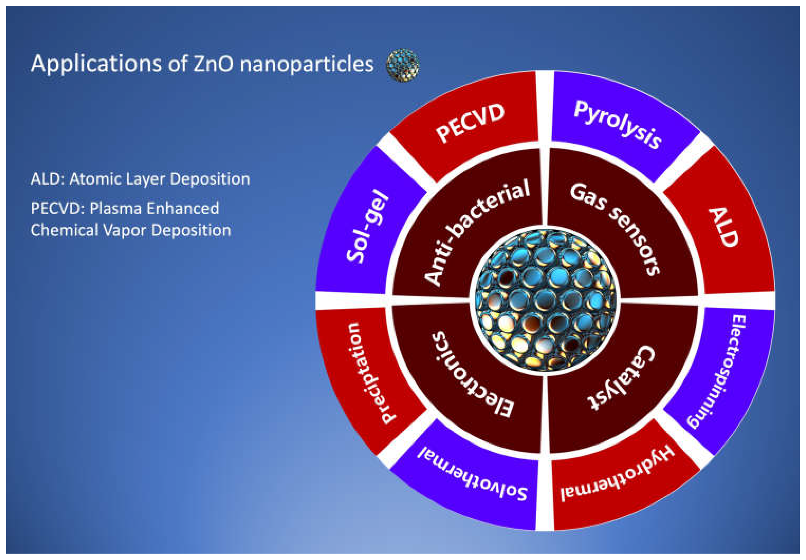
Figure 1. Applications of Zinc Oxide nanoparticles in diverse fields and conventional synthesis techniques.
Preprints 113434 g001
Figure 2. Atmospheric pressure plasma-based approaches for nano synthesis.
Preprints 113434 g002
Figure 3. Schematic diagram of atmospheric pressure plasma jet system for nanomaterials synthesis (redrawn from Reference [55]).
Preprints 113434 g003
Figure 4. Schematic diagram of atmospheric dielectric barrier discharge for nanomaterials synthesis (redrawn from Reference [59]).
Preprints 113434 g004
Figure 5. Schematic diagram of atmospheric pressure plasma torch method for nanomaterials synthesis (redrawn from Reference [60]).
Preprints 113434 g005
Figure 6. Schematic diagram of atmospheric pressure plasma method for nanomaterials synthesis (redrawn from Reference [61]).
Preprints 113434 g006
Table 1. A literature review of atmospheric plasma configurations for synthesizing silver, zinc oxide, and silver-zinc oxide core-shell nanomaterial.
No. Plasma
Configuration
Nanomaterials Applications Capillary
Diameter
Precursor Gas Flow Rate Voltage &
Current
Ref.
01 Atmospheric Pressure Microplasma jet Silver Optoelectronics, sensing, biomedical applications Internal diameter 0.26mm
AgNO3 + sucrose 26sccm 2mA [62]
02 Atmospheric Pressure Microplasma Silver Nanosensors Internal diameter
0.7mm
AgNO3 + sucrose 25sccm 0-15kV [63]
03 R.F. atmospheric pressure Microplasma jet Silver Photovoltaic Internal diameter 5.25mm
AgNO3 1.5slm [64]
04 Microplasma Synthesis Silver Antibacterial activity Internal diameter 0.1mm AgNO3+ NaPA 250V [65]
05 Atmospheric Microplasma electrochemistry Silver Plasmonic applications as sensing Internal diameter 0.175mm AgNO3 + fructose 25sccm 3mA and 2kV [66]
06 Plasma liquid synthesis Silver Anti-bacterial and antifungal activities Internal diameter 0.34mm AgNO3 + fructose 100 sccm 15mA and 600V [67]
07 Plasma-aided green and controllable synthesis Silver Antibacterial activity Internal diameter 0.5 mm AgNO3 + Acetone 30 sccm [68]
08 Atmospheric pressure Plasma jet Silver Bioactivity, catalysis Internal diameter
3.7mm
AgNO3+ trisodium citrate 3 L/min 8A [55]
09 Microplasma assisted synthesis Silver Cancer therapy Internal diameter < 1mm AgNO3+ PVA, PVP & sucrclose 600 sccm 3-5 kV [70]
10 Atmospheric discharge plasma Silver Catalytic properties Internal diameter 2.4mm AgNO3+ AlgNa 500-1000V [71]
11 Atmospheric pressure Microplasma Silver Anti-bacterial activity Internal diameter 0.2 mm AgNO3 + fructose 150 sccm 1000V [72]
12 Atmospheric pressure Microplasma electrochemical process Zinc oxide Antibacterial applications Internal diameter 0.2 mm Zn (NO3)2 + surfactant 150 sccm 1000V [73]
13 Atmospheric pressure plasma jet technique Zinc oxide Piezoelectric sensors Zinc powder 10L/min 200-400A [74]
14 Atmospheric pressure plasma (R.F. Power) Zinc oxide Light-emitting diodes Internal diameter
0.7mm
Zinc wire 150 sccm [75]
15 Atmospheric pressure plasma jet Zinc oxide Solar cells, Gas sensors Internal diameter
0.6mm
Zinc anode + NaOH+ HNO3+ sucrose 60ml/min 3kV
5-10 mA
[76]
16 Atmospheric pressure Microplasma Jet Ag-ZnO core shells Antimicrobial activity AgNO3 + Zn (NO3)2 13kV [42]
17 Atmospheric pressure Microplasma Au-Ag core shells Optical and biological properties Internal diameter
1mm
AgNO3 + HAuCl4. 3H2O 2 l/min 10kV [77]
Table 2. Summary of influence of control parameters on the properties of silver, zinc oxide, and core-shell nanoparticles.
Material Control parameters Effect of Parameters Ref.
Silver Solution concentration
  • The size of nanosheets increases with increase in solution concentration,
  • Antibacterial activity of silver nanosheets enhanced with an increase in solution concentration
[72]
  • The average size of nanoparticles increases with an increase in precursor concentration
  • An increase in precursor concentration causes a significant increase in the inhibition zone against bacteria and fungi.
[78]
  • The large size and highly dispersive nanoparticles formed by increasing solution concentration
[66]
ZnO


Processing time
  • An increase in processing time improves the crystallinity
[76]
  • The average diameter and size of nanoparticles increase with the increase in processing time.
[79]
Silver
  • Increasing the exposure time to plasma increases the average size of nanoparticles.
[63]
Silver Stabilizing agent concentration
  • The greater the concentration of fructose as a stabilizing agent, the more dispersed and relatively smaller nanoparticles are formed, and it reduces agglomeration. At greater fructose concentrations, the smaller nanoparticles have enhanced properties against bacteria.
[67]
Silver Stabilizing agent concentration
  • The average size of nanoparticles decreases by increasing the molar ratio of sucrose in the precursor.

[62]
Molybdenum oxide Gas flow rate
  • The size of nanoparticles reduces as the flow rate of gases rises.
[80]
Disclaimer/Publisher’s Note: The statements, opinions and data contained in all publications are solely those of the individual author(s) and contributor(s) and not of MDPI and/or the editor(s). MDPI and/or the editor(s) disclaim responsibility for any injury to people or property resulting from any ideas, methods, instructions or products referred to in the content.
Copyright: This open access article is published under a Creative Commons CC BY 4.0 license, which permit the free download, distribution, and reuse, provided that the author and preprint are cited in any reuse.
Prerpints.org logo

Preprints.org is a free preprint server supported by MDPI in Basel, Switzerland.

Subscribe

© 2024 MDPI (Basel, Switzerland) unless otherwise stated