Preprint
Article

Ultrasonic Synthesis of Zein Protein Microcapsules: Stability, Morphology and Curcumin Inclusion Studies by Optical Spectroscopy and Microscopy Experiments

Altmetrics

Downloads

80

Views

41

Comments

0

Submitted:

30 July 2024

Posted:

31 July 2024

You are already at the latest version

Alerts
Abstract
Zein protein (ZP) is the major storage protein of corn (maize). It is safe, biodegradable, edible, and characterized by unique amphiphilic and self-assembly properties. We exploited these properties to prepare stable ZP microcapsules by oil-in-water ultrasound-assisted emulsification. The opti-mal experimental conditions were determined in terms of the sonication frequency, applied acoustic power, sonication time, protein concentration and temperature. The morphology and stability of ZP microcapsules were characterized by optical spectroscopy and microscopy tech-niques. We found that a thick ZP outer layer protects the inner oily phase from internal/external diffusion, and enables the microencapsulation of Curcumin, a very active anti-inflammatory and nutraceutical agent.
Keywords: 
Subject: Chemistry and Materials Science  -   Physical Chemistry

1. Introduction

Encapsulation of materials in microparticles dispersed in solution may find application in several fields, such as food chemistry, imaging, energy production and therapeutic/diagnostic medicine [1]. Examples of microsystems able to encapsulate active ingredients, microsystems including functional polymeric micro- and nanoparticles, core–shell structures, microsponges and microspheres have been developed [2]. In particular, oil-in-water (O/W) emulsification is a simple, low-cost and effective method for preparing aqueous suspensions of microcapsules filled with oil-soluble active ingredients [3].
The microcapsule shell prevents the diffusion of the confined compounds out of the core, providing a thick barrier that can also protect sensitive compounds from acidic, alkaline or enzymatic degradation. Moreover, microcapsules may respond to external stimuli such as changes in pH, temperature, light, magnetic and acoustic fields, to enable the timely and spatially controlled release of the encapsulated components [4]. Microcapsules made from proteins have the advantage of being biocompatible and biodegradable and, for these properties, they have been extensively studied for pharmaceutical and food targeted applications [5].
Zein (ZP) is the major storage protein of corn (maize), recognized as safe by the Food and Drug Administration [6]. It has unique amphiphilic and self-assembly properties, it is biodegradable, edible, and it is sourced in large scale from the surpluses of corn and byproducts of the milling and bioethanol industries [7,8,9,10,11]. Applications of ZP include use in fibers, adhesives, coating, ceramics, inks, cosmetics, textiles, and biodegradable plastics [6,12,13,14,15]. However, ZP insolubility in water and its poor nutritional quality (negative nitrogen balance) limit its use in human food products, while it is primarily used in food packaging and coating [16]. For instance, ZP is known to form a robust, lustrous, hydrophobic grease-proof coating that can be used as a biodegradable film and plastics, providing resistance to microbial attack [17].
From the molecular point of view, ZP is encoded in a large multigene family and is constituted by a heterogenous mixture of polypeptides of different molecular sizes, solubility and charge [18]. Several ZP fractions have been identified, that can be divided into four main fraction, i.e., α-(70-80%), β- (10-20%), γ- and δ-ZP. Presently, more than 60 structures are deposited on the ZP UniProt KB database [19], and more than 40 for the α-ZP [20]. α-ZP is divided in α-ZP19 and α-ZP22, of approximate weights of 19 and 22 kDa, respectively; β-ZP contains two 14-15 kDa polypeptides; γ-ZP include two polypeptides of 27 and 16 kDa, respectively; δ-ZP is a minor fraction of the total ZP and has an approximate weight of 10 kDa. [21,22] ZP is particularly rich in hydrophobic amino acids (more than 50%), deficient in acidic and basic amino acids, but rich in glutamic acid residues (21-26%), most of which, however, are amidated as glutamines and asparagines.
The high proportion of non-polar amino acids causes the low ZP solubility in water and its prevalent hydrophobic character. ZP is soluble in binary solutions consisting of lower aliphatic alcohols and water, such as 50-90% aqueous ethanol [23]. It should be stressed, however, that different ZP fractions show different solubility properties. Interestingly, enzymatic modification with alcalase was used to make ZP water soluble [24].
The secondary structure of ZP was investigated by circular dichroism (CD), and FTIR spectroscopies. It is reported that ZP is characterized by a ~40-60% helical content, complemented by β-sheets (30%) and β-turns (20%) structures [25,26,27]. On the other hand, so far, there is no consensus on the tertiary structure of ZP [10]. Argos et al. [28] proposed a ZP 3D model comprising nine adjacent, anti-parallel helices joined by glutamine-rich “turns” or “loops” clustered within a distorted cylinder. The polar and hydrophobic residues distributed along the helical surfaces developed intra- and intermolecular hydrogen bonding, so that ZP could be arranged in planes. It has also been shown that ZP is able to self-assemble into mesophases of different morphology depending on the specific experimental conditions, that can be exploited to modulate the hydrophilic-hydrophobic balance of the system [29,30]. In particular, ZP led to the formation of stable nanostructures, such as fibers, nanoparticles, microspheres and thin films [17].
Emulsification is commonly used as a tool for the confinement of solid, liquid, or gaseous materials in micrometer-sized capsules. Among the methods usually applied for emulsion preparation, ultrasounds have attracted much attention because it is considered as an eco-friendly and cost-effective technique [2]. Acoustic cavitation is the primary mechanism for the synthesis of microcapsules, achieved by the formation and collapse of microbubbles under ultrasound pressure through multiple in-phase expansion and contraction steps [3]. In addition, acoustic cavitation can also boost the disruption of oil droplets, facilitating the formation of stable emulsions [1].
Cavitation effects are highly dependent on the applied ultrasound frequency. The amount of energy released by the bubble collapse and the maximum bubble size before collapse (resonance size) are correlated, and approximately inversely proportional to the applied frequency. Ultrasound parameters such as frequency and acoustic power can be tuned to control the number of cavitation events, the microcapsules size, and their surface roughness and structure [1]. While the low-frequency ultrasound region (16-100 kHz) gives rise to the formation of larger bubbles followed by intense bubble collapse, resulting in localized shear stress and high local temperatures, the intermediate ultrasound region (100 and 1000 kHz) results in a moderately intense bubble collapse with an efficient radical production in the range between 200 and 500 kHz. Low frequency ultrasounds have already been used to prepare protein microcapsules and microbubbles, stabilized by crosslinked protein shells which can be dispersed in aqueous media [31]. During the emulsification stage, the bubbles or liquid droplets (the core) act as ‘templates’ for the formation of the protein shells. Reversible protein coatings are stabilized through hydrophobic interactions and/or hydrogen bonding, while irreversible coatings require that covalent linkages, like disulfide bonds, are formed. In the latter case, covalent links can be induced by reactions involving free radicals generated through ultrasonic cavitation [2,3,4].
Herein, the amphiphilic character of ZP was exploited to stabilize the interface of core droplets dispersed in the medium, thus forming a tight and effective protective coating for the encapsulated compounds. The most common methods applied for obtaining ZP nano- and micro-structures are antisolvent precipitation, coacervation/phase separation, evaporation-induced self-assembly, electrospraying and electrospinning, Pickering emulsion [32,33]. Although several studies focused on the ZP properties as stabilizer in emulsion formulations, only few studies have reported the production of ZP microcapsules by ultrasound assisted emulsification (UAE).

2. Results

2.1. Spectroscopic Characterization of Zein Protein (ZP)

2.1.1. UV-Vis Absorption

It is reported that αZP has a globular structure, characterized by a dominant content of α-helices and virtually no β-structures [10]. The ZP UV-Vis absorption spectrum shows a strong peak at λ=195 nm and a weaker absorption at λ=220 nm, associated to the π→π* and the n→π* transitions of the peptide bond, respectively, together with two less intense absorption bands peaked at 280 nm and 320 nm, ascribable to the Tyr [34] and the dityrosine groups [35] (Supplementary Materials, Figure SM1). Dityrosine is a natural component of ZP, as a product of environmental stress and protein modification, [36] and it has been detected and quantified in previous studies [37].

2.1.2. Electronic Circular Dichroism

Far-UV CD measurements (Figure 1) were carried out to analyze the secondary structure of ZP. Deconvolution of the experimental curves through BestSel software analysis, [38], shows the prevalence of α-helix and random coil conformations, while β-turns and antiparallel β-sheets appear to be less populated, consistently with PDB and literature data on the αZP fraction (Table 1) [21]. This conformational landscape remains stable over 10 days since the solution preparation. CD experiments from 15 to 75°C have also been carried out to analyze the ZP conformational changes in this range of temperatures (Figure 1).
As the temperature increase, the α-helix contribution to the conformational landscape of ZP decreases by about 30%, favoring the population of random coil, β-turn and parallel β-sheet conformations, in agreement with the literature [39]. CD spectra carried out at 25°C, before and after the heating/cooling cycle, indicate that the ZP temperature-dependent behavior is fully reversible (Figure SM2, Table SM1). The reversibility of this process was also confirmed by UV-Vis absorption and fluorescence emission experiments (Figures SM3Figures SM4). Interestingly, in previous studies on prolamines, it was shown that the formation of ZP aggregates at 40°C is favored by an increment of β-sheet structures [40]. It was also reported that proteins with an α-helical content higher than 40%, formed β-structures during the aggregation process, passing through coiled-coil intermediate structures [41]. Direct transition from α-helix to β-strands, was also suggested in studies focused on the formation of microstructures during maize and ZP processing [42].
* ref. [21]

2.1.3. Steady-State Fluorescence

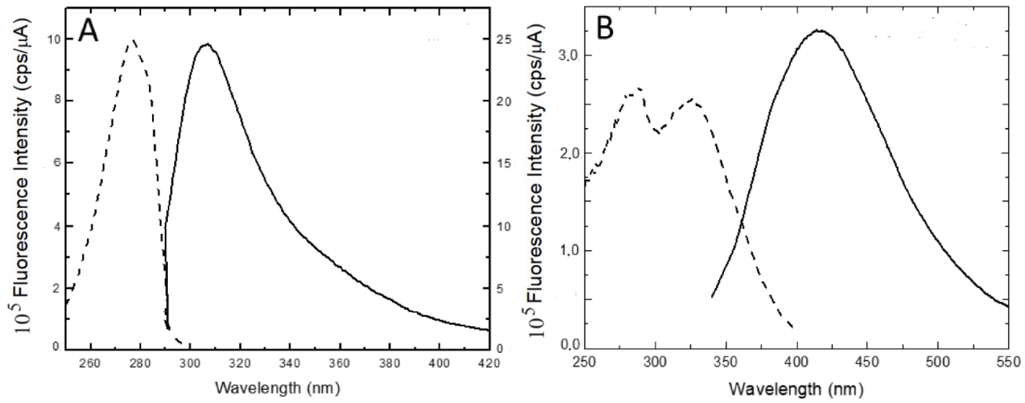
The emission and excitation spectra of ZP are shown in Figure 2. The emission spectrum reported in Figure 2A is typical of the phenol side-chain of tyrosine, as confirmed by the excitation spectrum at λem =308 nm, while the long-wavelength emission measured from 360 to 500 nm (Figure 2B) can be ascribed to the dityrosine group. The double-horned peak clearly revealed in the excitation spectrum reported in Figure 2B was assigned to the ionized and neutral forms of dityrosine, characterized by absorption maxima at 315 nm and 283 nm, respectively [35]. Interestingly, the anisotropy coefficient of dityrosine at 25°C (r=0.180±0.001) is twice larger than the tyrosine anisotropy coefficient (r=0.074±0.001) measured at the same temperature, and this difference is also maintained at 75°C (r=0.12±0.01 and r=±0.04±0.01, respectively), as a consequence of the restrained rotational motion of the former. Temperature-dependent fluorescence anisotropy measurements confirmed the reversibility of the process, the anisotropy coefficients of tyrosine and dityrosine groups being 0.084±0.005 and 0.17±0.01, respectively, at 25°C for solutions submitted to heating/cooling cycles.

2.2. Zein Protein Microcapsules

2.2.1. Zein Microcapsules Synthesis: Procedure and Optimization of the Experimental Conditions

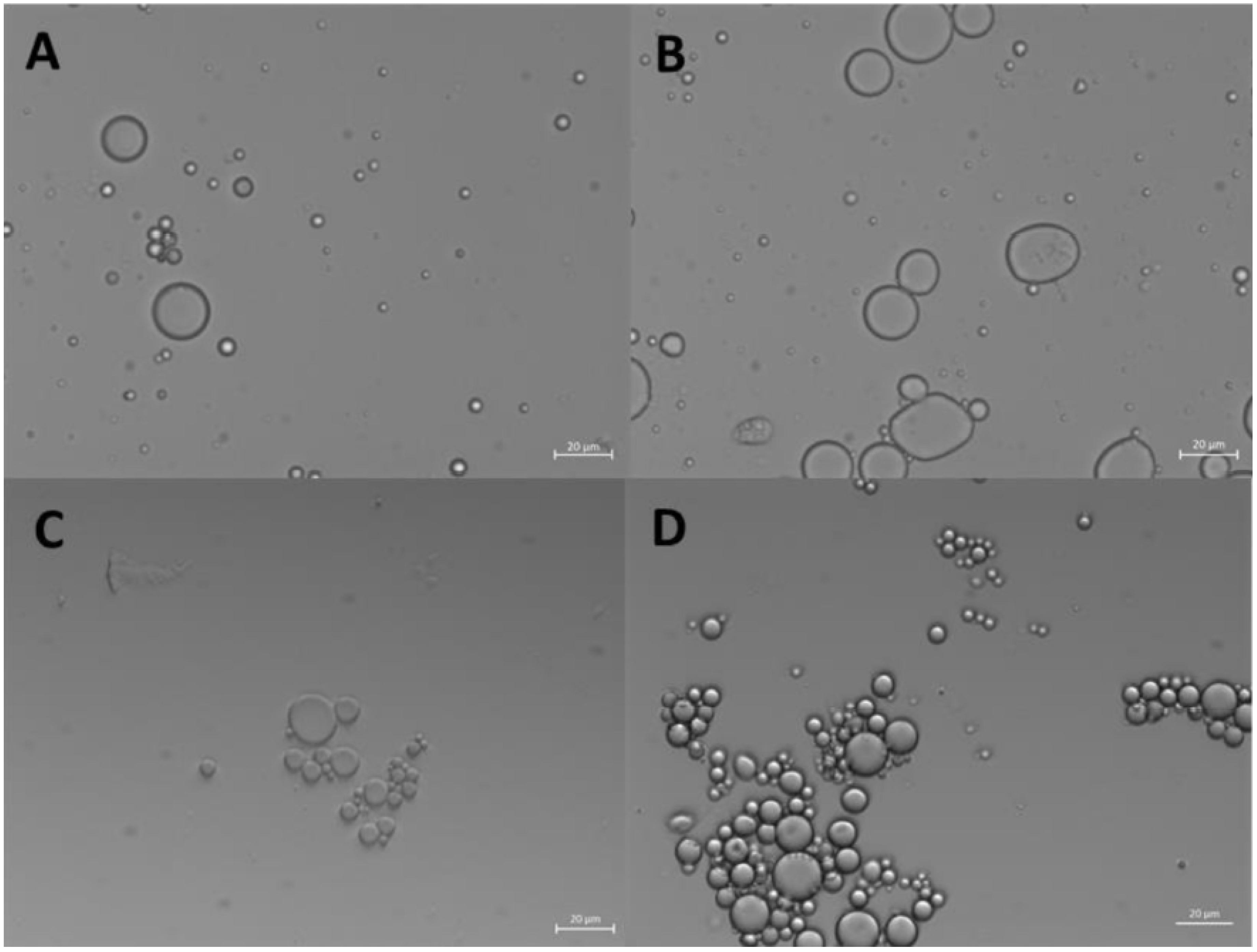
ZP microcapsules were obtained by dissolving ZP in EtOH/H2O 70/30 (v/v) solutions at increasing concentrations from 2.5 mg/mL to 10.0 mg/mL and upon ultrasonic emulsification of 50 μL soybean oil (SO) in these solutions for 60 s at a frequency of 20 kHz and a power of 220 W. Stable oil-filled microcapsules were observed by optical microscopy only for ZP concentrations higher than 2 mg/ml. The formation of ZP microcapsules could be clearly detected by optical microscopy, right after the ultrasound treatment and up to 4 days of storage at room temperature (Figure 3).
ZP microstructures have been compared to control samples, consisting of EtOH/H2O 70/30 (v/v) and SO solutions, that were emulsified using the same ultrasound setting parameters. Blank samples formed O/W emulsions, that over the time displayed a very different behavior, as shown in Figure SM5. In blank samples, the droplets undergo coalescence and progressive phase separation. Instead, ZP solutions, showed the formation of a precipitate with a different shape and texture with the oil layer progressively depositing to the bottom. In addition, ZP precipitates can be re-dispersed in the solution by gently shaking the test tube, restoring the emulsion status, that was not possible for the control samples. Optical microscopy images of ZP samples (2.5 and 5 mg/ml) with different SO amounts (50, 25 and 10 µL) showed spherical shape, displaying well-defined protein coating borders (Figure SM6A), some coalescence of the microcapsules (Figures SM6BFigures SM6C), and concentration-dependent microcapsule number density (Figure SM6D).
The experiments carried out for the optimization of the experimental conditions are reported as SM in the Design of Experiment (DOE) section (Table SM2, Figures SM7-SM10). Very briefly, a set of experiments was designed to evaluate the system response, while tuning several synthetic parameters (acoustic power, sonication time, ZP concentration, storage conditions). The list of the performed experiments is reported in Table SM2. Each system was closely monitored for 10 days. The optimal experimental conditions were therefore fixed at: 25 s sonication time, 20 kHz frequency, 220 W electric power, 10 µl of SO and 10 mg/ml ZP solutions in 1 ml EtOH/H2O 70/30 (v/v). Under these experimental conditions, minimal oil/water phase separation was detected.

2.2.2. Morphological Characterization of ZP Microcapsules

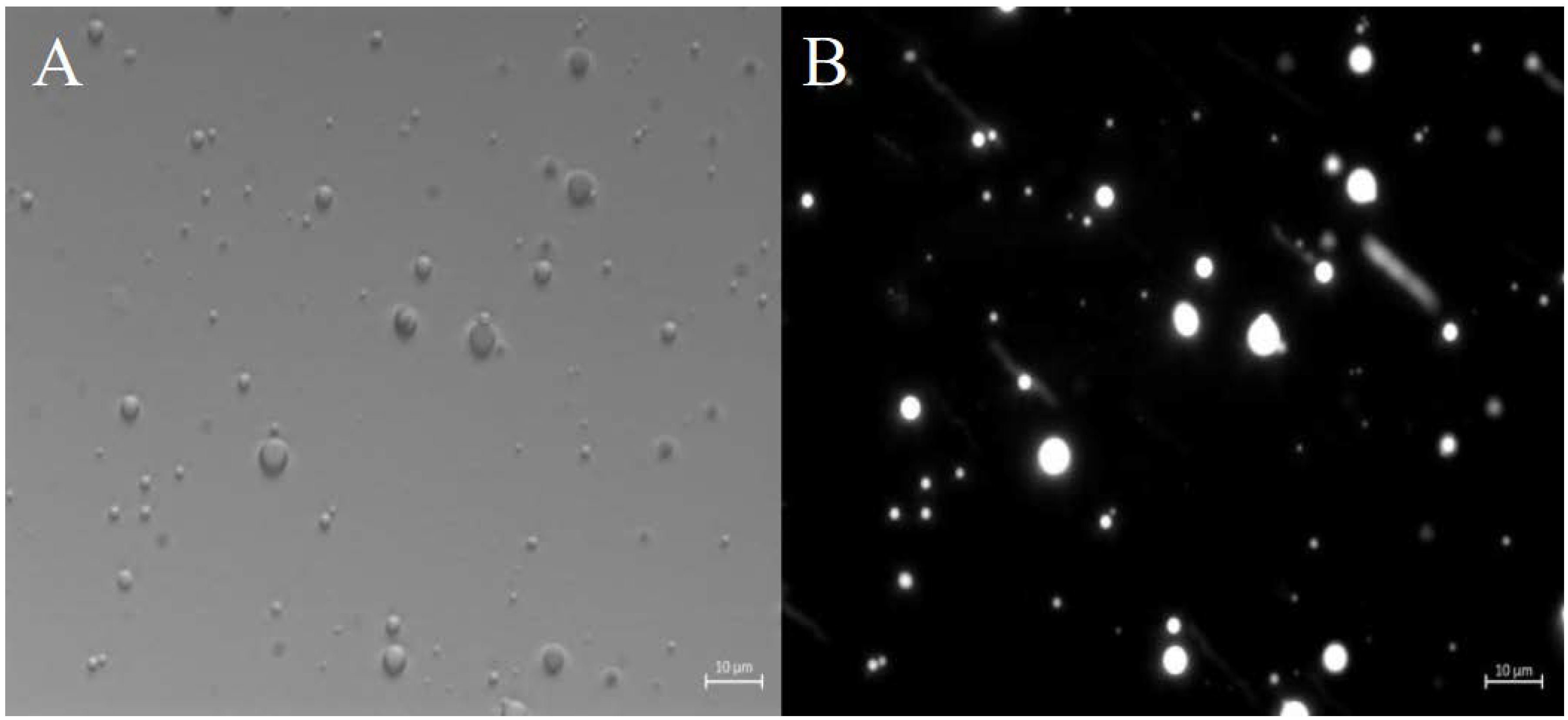
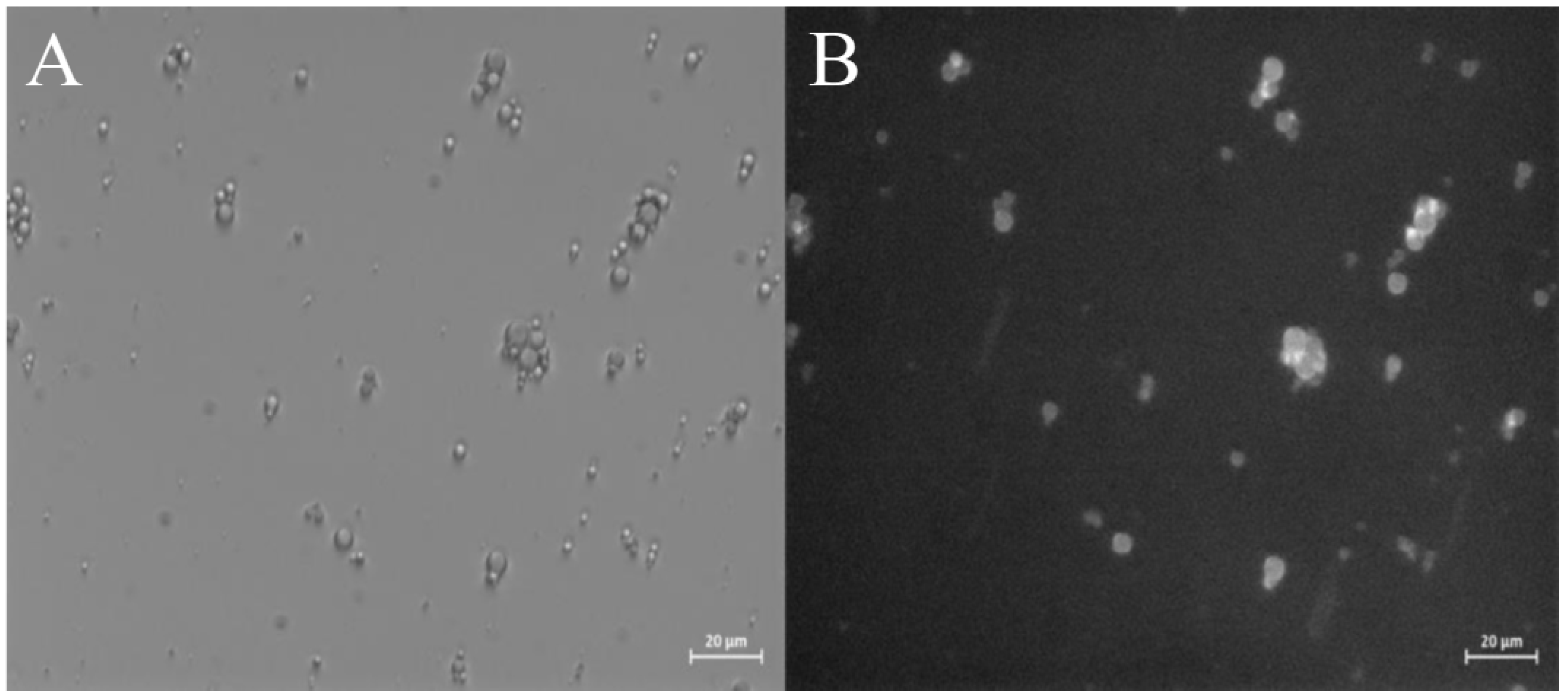
The fluorescent hydrophobic dye Nile Red (NLR, 9-diethylamino-5H-benzo[a]phenoxazine-5-one) was included in the oily phase to monitor the oil partition in the microcapsules prepared by UAE. NLR is an excellent dye for the detection of intracellular lipid droplets by fluorescence microscopy, because its fluorescence is fully quenched in water, while it is strongly enhanced in a hydrophobic environment [42]. Recently, Weissmueller et al. successfully encapsulated NLR in the hydrophobic environment of ZP nanocarriers, obtained by flash nanoprecipitation [14]. The dyed samples were therefore observed under an optical microscope, collecting both bright-field and fluorescence images. The microstructures revealed by the two imaging techniques strongly overlapped, suggesting that the ZP microcapsules are filled with oil, with the protein coating the oil droplets (Figure 4).
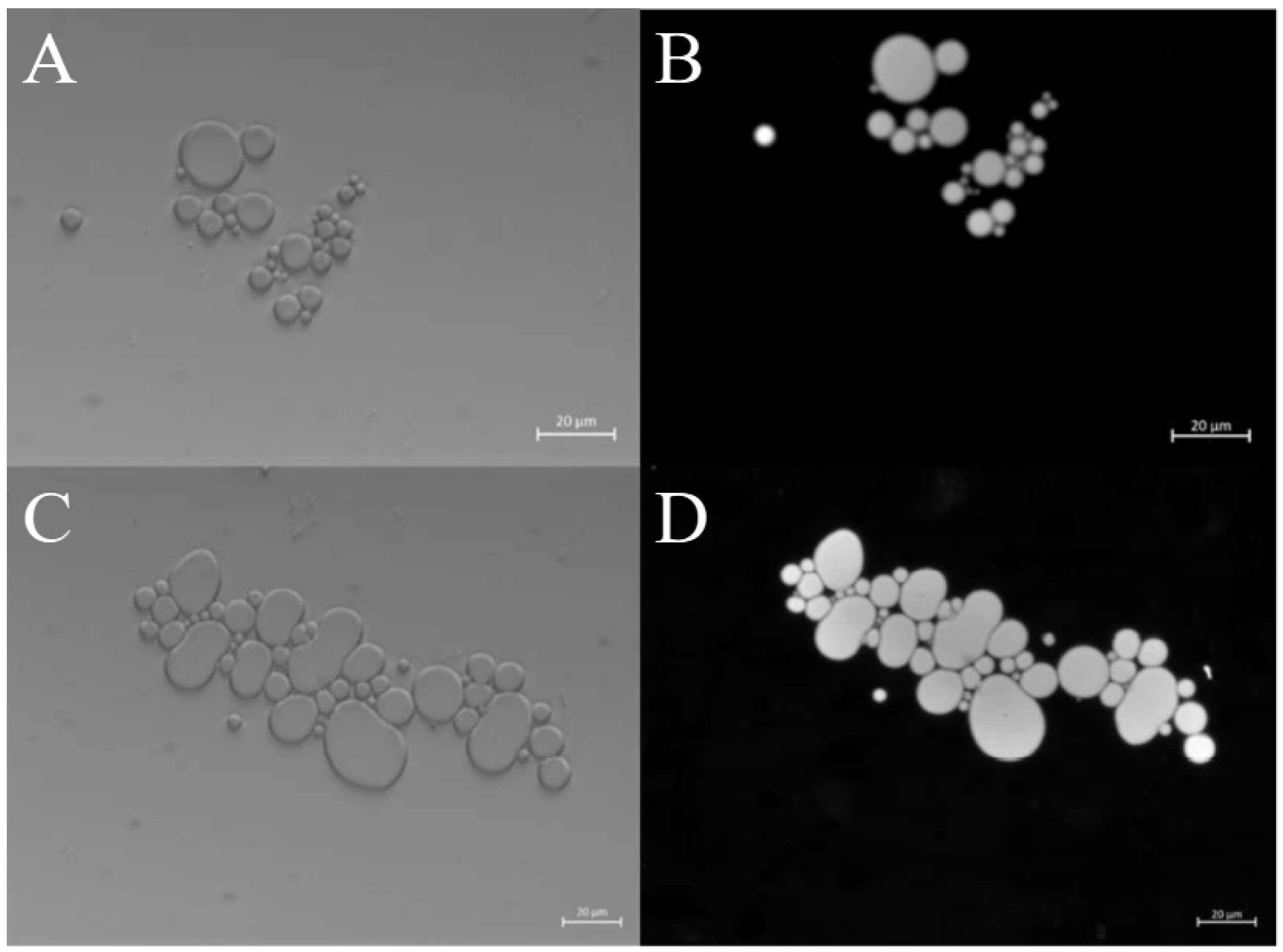
Interestingly, in the case of clustered microcapsules produced by coalescence (Figure 5), the fluorescence signals highlighted how the oil content is not dispersed, and the larger microstructures are oil-filled. This is favored by the merging of the microcapsule outer layer and the pooling of the oil phases. Each structure remains delimited by recognizable borders, supporting the idea of ZP acting as an outer coating that stabilize the oil-filled globular microstructures.
To get further evidence on their morphology and structuration, ZP microcapsules were stained with the fluorescent dye Rhodamine B [RhB, 9-(2-Carboxyphenyl)-6-(diethylamino)-N,N-diethyl-3H-xanthen-3-iminium chloride]. Recently, it has been shown that RhB can be easily conjugated to ZP exploiting electrostatic, hydrogen bonds and van der Waals interactions [44].
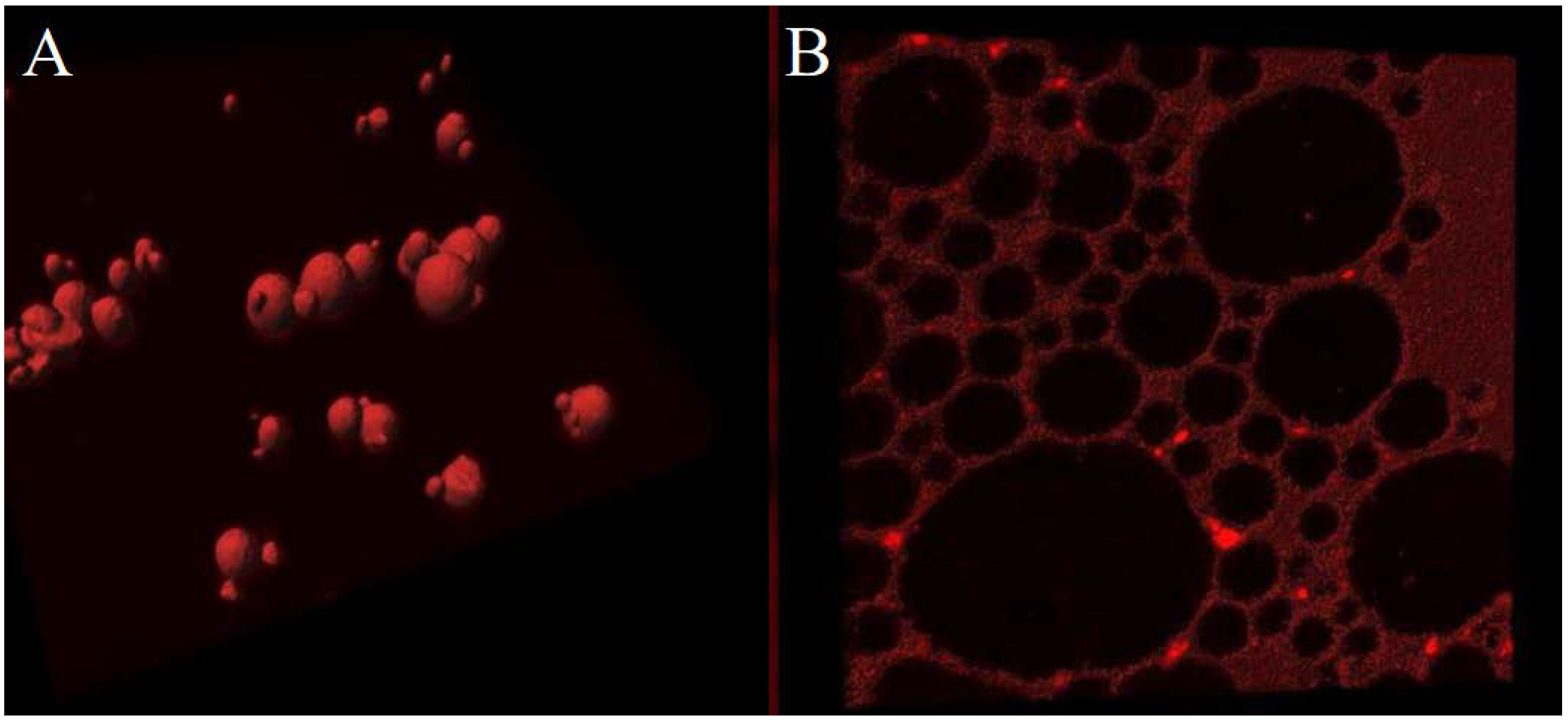
ZP microcapsules including NLR or stained with RhB were imaged by CLSM (Figure 6A and Figure 6B, respectively). The sample including NLR (Figure 6A) confirms the globular morphology of ZP microcapsules, characterized by an oil-filled inner core. A protein outer layer can be observed in the RhB stained microparticles (Fig. 6B), despite the background noise due to residual RhB in solution. Overall these results positively indicate that ZP microcapsules are oil-filled structures, stabilized by a ZP outer shell.
ESEM produces images of good quality and resolution in the case of wet samples. This greatly facilitates the imaging of biological samples that are unstable under the high vacuum conditions required by conventional electron scanning microscopes, making ESEM most suitable for imaging of non-metallic, uncoated and biological materials.
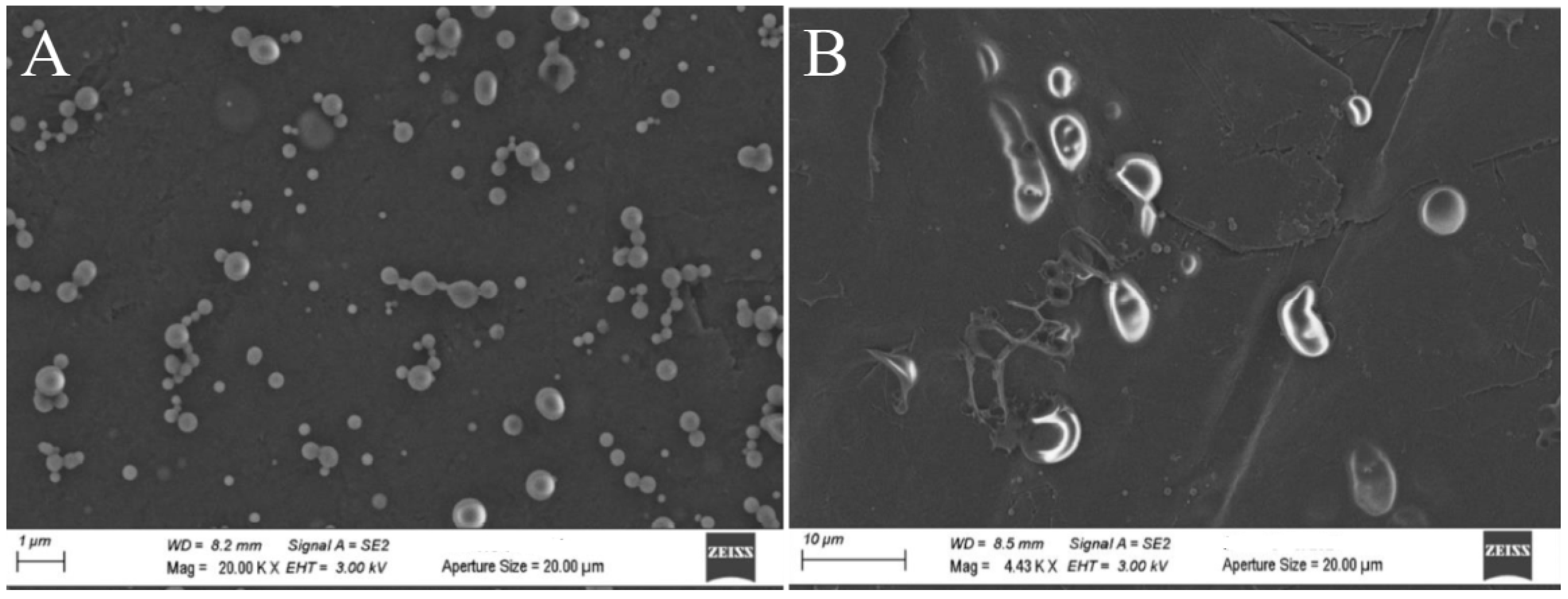
FE-ESEM imaging was carried out comparing freshly synthesized ZP microcapsules with the same samples after 18 days of storage at room temperature (Figure 7). Many microspheres were observed in the protein-containing samples, confirming the role of ZP in stabilizing the oil droplets by a protein coating shell. Remarkably, no significant structures could be detected in the control sample (O/W emulsion in EtOH/H2O 70/30 (v/v) solution, Figure SM11) [45]. Freshly synthesized and 18 days-old samples also showed interesting differences. In particular, the 18-days-old protein microstructures collapsed and appear rather elongated (Figure 7B), suggesting that perturbation of the protein shell integrity may promote the coalescence of ZP microcapsules over time.
Important results were obtained when ZP microcapsules were re-dispersed in EtOH/H2O 70/30 (v/v) after drying. The protein microcapsules could be successfully recovered, although a general increase of their average size and heterogeneity, major coalescence and decreased particle density were observed in comparison with the analogous wet sample. A quite stable condition was finally achieved after 6 days from the ZP microcapsules re-solubilization (Figure SM10).
The stability of ZP microcapsules was also assessed at different temperatures with the aim to test if temperature shock could be a suitable stimulus to trigger the controlled release of encapsulated materials. ZP microcapsules in EtOH/H2O 70/30 (v/v) solution were therefore heated to 75°C, and the change in size and number density of microstructures was monitored. Interestingly, the light scattering intensity of ZP microcapsules decreased as the temperature increased, showing a major decrease for temperatures higher than 50°C (Figure SM12A). In particular, from 25°C to 75°C, a 34% drop in the particle number density was observed, which parallels the light scattering decrease. In contrast, ZP microcapsules in EtOH/H2O 10/90 (v/v) showed a quite different behavior when heated at 75°C, giving rise to increasing light scattering intensities as the temperature increased (Fig. SM12B). The latter results indicate that, in a highly hydrophobic environment, ZP microcapsules are stabilized at higher temperatures. Interestingly, in both cases, heating/cooling cycles demonstrated that such effects are irreversible.

2.2.3. Encapsulation of Curcumin into ZP microcaspules

Curcumin [bis(4-hydroxy-3-methoxyphenyl)-1,6-heptadiene-3,5-dione] is a natural yellow-orange dye derived from the rhizome of Curcuma Longa, a member of the ginger family Zingiberaceae, showing interesting pharmaceutical and nutraceutical properties, due to its antioxidant, antibacterial and anti-inflammatory activities, and the recognized safe (GRAS) properties [46]. It was chosen for inclusion studies because it lacks solubility in water, it is soluble in oil up to 2.90 ± 0.15 mg per oil gram [47], and it is strongly fluorescent [48]. Those properties make Curcumin a proper candidate for successfully encapsulation in O/W droplets, and a suitable probe for imaging. In particular, the inclusion of Curcumin in ZP nanoparticles and ZP-polymer hybrid assemblies has widely been studied for delivery of food ingredients or therapeutics [49,50,51,52,53,54,55,56,57,58].
The UV-Vis absorption and the fluorescence emission and excitation spectra of Curcumin in SO and ethanol are reported in Figures SM13 and Figures SM14, respectively. As it can be seen in the reported figures, the absorption and fluorescence spectra of Curcumin in SO are more structured, while in ethanol the absorption and emission spectral profiles are definitely smoother, as a consequence of spectral relaxation in a polar solvent [59].
Curcumin has been used as a probe to test the ability of ZP microcapsules to encapsulate an active ingredient, using UAE O/W formulation. Bright field optical and fluorescence microscopy measurements revealed that Curcumin was successfully encapsulated in ZP microcapsules, and it was also retained therein after drying and re-dispersing the microcapsules in water, as shown in Figure 8 and Figure SM15. It should be noted that in both the fluorescence microscopy images reported in the figures, the Curcumin signal is overlapped to the ZP microcapsules imaged by bright field optical microscopy.
Noteworthy, the fluorescence emission of the Curcumin in the ZP microcapsules (λem,max = 498 nm) is red-shifted by 12 nm with respect to its fluorescence emission spectrum in SO (λem,max = 486 nm) (Figure SM16). This finding suggests that Curcumin is encapsulated in the ZP microcapsules, but, at least, partially exposed to water molecules. This could attributed to a preferential interaction of Curcumin with the ZP shell coating the microcapsules [58]. Interestingly, Hu et al. synthesized core-shell ZP nanoparticles coated by a Pectin hydrophilic outer layer and fortified in the hydrophobic inner phase by Curcumin. The latter was found to interact with ZP through its aromatic rings and inter-ring chains. [49]

3. Materials and Methods

3.1. Materials

ZP was purchased by Sigma Aldrich [maize z1C2(541924] and UniProt access numbers (Q41896 and P04700). According to the product information, the sample consists of two αZP polypeptide chains of 22 and 24 KDa, respectively, the amino acid composition of which are reported in Table SM3. All the solutions for spectroscopy and microscopy experiments were prepared from this same batch, sealed in a dry, cool, and dark environment. Curcumin (powder, min. 95% p/p) was purchased from Galeno srl (Italy), and used as such without further purification. SO, NLR, and RhB were purchased by Sigma Aldrich. Spectroscopic absolute ethanol (anhydrous, ≥ 99.9 %) was purchased from Carlo Erba. Milli-Q filtered water was obtained by a Millipore system (18.2 MΩ cm at 25°C).

3.2. Methods and Instrumentation

3.2.1. Protein Solubilization

ZP sample was supplied as dry powder and used as such without further purification or pre-treatments. ZP was solubilized in EtOH/H2O 70/30 (v/v) solution and kept under stirring. Unless differently stated, ZP solutions were freshly prepared before each experiment.

3.2.2. UV–Vis Absorption Spectrophotometry

UV-Vis absorption experiments were carried out with a Cary 100 Scan spectrophotometer (Varian, Middelburg, Netherlands) equipped with a Peltier thermostat, using quartz cuvettes (Hellma) with optical lengths of 10 and 4 mm.

3.2.3. Circular Dichroism

Circular dichroism (CD) experiments were carried out using a Jasco J-1500 CD spectropolarimeter (Jasco International Co.) using a cell holder equipped with a Peltier thermostat (PTC-510). The setup was purged with ultra-pure nitrogen gas. Spectra were recorded in the far-UV wavelength range, i.e., from 190 to 250 nm (amide region), and from 250 to 350 nm for the aromatic region (tyrosine), using a scan speed of 20 nm/min, a bandwidth of 2 nm, and a sensitivity of 20 mdeg. For each sample, the spectra were accumulated 10 times to maximize the signal-to-noise ratio. The solvent spectrum was subtracted for background correction. Quartz cuvettes (Hellma) with optical lengths of 1 mm were used. Temperature-dependent experiments were carried out applying a ramp of 10°C/min, a waiting time of 120 s, until a maximum oscillation of ±0.1 °C to the set temperature was achieved. Data analysis for secondary structure determination was performed relying on the BestSel algorithm [38].

3.2.4. Steady-State Fluorescence

Steady-state fluorescence experiments were carried out applying single photon counting (SPC) detection using a Fluoromax-4 spectrofluorometer (Horiba, Jobin Yvon) equipped with automatically controlled Glan-Thomson polarizers. The temperature was set through an external thermostat, allowing for a maximum oscillation of ±0.1 °C. Emission and excitation spectra were acquired using a 1 cm quartz cell (Hellma) and 3/3 nm excitation (ex) and emission (em) slits opening. 4/10 mm asymmetric quartz cell (Hellma), 1/1 nm ex/em slits were used for Curcumin in SO, while a 5 mm quartz cell (Hellma) and 1.5/1.5 nm ex/em slits were used for spectral measurements of Curcumin embedded in ZP microcapsules. For this experiment, due to the presence of microcapsules, a cut-off emission filter was used to minimize light scattering contamination. Unless otherwise stated, fluorescence intensities are reported as signal-to-reference [count per second (cps)/microamperes (µA)] units. All the measurements were carried with absorbances lower than 0.1 at the excitation wavelength to minimize inner filter effects. In any case, inner filter effect correction was applied to all the measured fluorescence spectra.
Fluorescence anisotropy measurements for tyrosine and dityrosine in ZP were carried out using Glan-Thomson polarizers, a 1 cm quartz cell (Hellma), 10 s/nm integration time, and 3/3 nm ex/em slits. Fluorescence emission anisotropy was accounted for through the fluorescence anisotropy coefficient r [60]:
r = G I V V I V H I V V + 2 I V H
where IVV and IVH are the fluorescence emission intensities measured with the excitation and emission polarizers set in the vertical/vertical (V/V) and vertical/horizontal (V/H) arrangements, respectively. The instrumental correction factor G was evaluated measuring IHV and IHH, i.e., the fluorescence emission intensities measured with the excitation and emission polarizers set in horizontal/vertical (H/V) and horizontal/horizontal (H/H) positions, respectively.

3.2.5. Optical Microscopy

Widefield microscopy images were obtained with a Zeiss microscope, Scope A1 (Carl Zeiss, Oberkochen, Germany) equipped by a Mercury Lamp HBO 50 and CCD AxioCam ICm1 (Zeiss). The images were acquired with the software Zen-Blue (Zeiss) using magnification in air (10x, 20x, 40x, 63x) and in oil immersion (100x) objectives. Fluorescence imaging was performed in single channel mode of excitation (red channel for NLR and RhB, blue channel for Curcumin). Images and particles diameters were analyzed with the software ImageJ [61,62]. Mean diameters and standard deviations of each sample are based on a gaussian fit of the size distribution. Data analysis was performed using Origin Pro 2016 software (by OriginLab Corporation, USA).
Confocal Laser Scanning Microscopy (CLSM) experiments were carried out through an FV1000 (Olympus) laser scanning microscope equipped with a He-Ne laser. The images were captured with a 40x oil immersion objective adding 4x and 5x zoom (160-200x total magnification). 3D image reconstruction was developed using the software Imaris 6.2.1 (Bitplane).

3.2.6. Field Emission - Environmental Scanning Electron Microscope (FE-ESEM)

ZP microcapsules were imaged by a FE-ESEM LEO 1530 (Zeiss) microscope, depositing a 5 μl ZP microcapsules solution on a graphite substrate, maintained under vacuum conditions for 30 minute before recording topography information [63].

3.2.7. Light Scattering

Rayleigh Light Scattering (RLS) intensities of ZP microcapsule solutions were measured using the same fluorescence equipment described above, reporting the RLS intensities as signal-to-reference [count per second (cps)/microamperes (µA)]. Both excitation and emission monochromators were set at λexem=600 nm, well outside the sample absorption or fluorescence emission regions. A 5x5 mm quartz cell (Hellma) was used, with 0.5/0.5 nm ex/em slits opening.

3.2.8. Synthesis of Zein Microcapsules

ZP microcapsules were produced from UAE of O/W emulsions. Specifically, for the O/W system, 50 μL SO were added to 0.5 to 10 mg ZP dissolved in 1 mL EtOH/H2O 70/30 (v/v). Disposable test tubes were used for each emulsification. A 20 kHz ultrasound horn (Branson Digital Sonifier) with a 3 mm diameter was placed at the O/W interface and the sonication was performed for different times (i.e., 25, 30, 45 s) and acoustic power (i.e., 120 W, 160 W, 220 W). To reduce the temperature shock at the O/W interface due to the UAE process, the system was immersed in an ice bath during sonication. In the standard protocol, the obtained microcapsules were stored in the test tubes at room temperature, and not separated nor washed from the solution.

3.2.9. Inclusion of Dyes and Active Compounds

NLR has been included in ZP microcapsules, by using SO stained with NLR during the microcapsules synthesis. The stained SO was stored under dark conditions at 4 °C. The NLR UV-Vis absorption spectrum in SO is peaked at around 510 nm, while its fluorescence emission spectrum in SO is peaked at 570 nm.
ZP microcapsules were stained by dissolving RhB in ZP EtOH/H2O 70/30 (v/v), and following the standard microcapsules synthesis protocol. The UV-Vis absorption spectrum of RhB is located between 500 and 580 nm, while its fluorescence emission spectrum is located between 550 nm and 680 nm.
Curcumin has been included in ZP microcapsules by using Curcumin-containing SO in the standard microcapsule synthesis protocol. 2.66 mg/g Curcumin was added to SO and dissolved under agitation, storing the SO/Curcumin solution under dark conditions at 4 °C.

4. Conclusions

In this work, ZP material was first characterized with the aim to develop an affordable and low-cost method for the synthesis and preparation of protein microcapsules. Next, the properties of the ZP microcapsules synthesized and stored under optimal experimental conditions, were characterized focusing on their stability over time and after drying and thermal shocking. Finally, as a proof-of-principle experiment, the ability of ZP microcapsules to encapsulate Curcumin, was tested and confirmed.
Due to the ZP solubility properties, a binary mixture of EtOH/H2O 70/30 (v/v) was chosen as solvent and used for the spectroscopic measurements. The ZP secondary structure was extrapolated by deconvolution of the far-UV CD signal, providing results in agreement with literature data and consistent with PDB previsions. It has been shown that proteins with an α-helical content higher than 40%, tend to form β-structures during the aggregation process, passing through coiled-coil intermediate structures, and that the formation of ZP aggregates is favored by an increment of β-sheet structures [39]. It should be noted that the reversibility of the ZP thermal-induced modifications is controversial in literature, as earlier studies support the formation of ZP oligomers and protein ordered arrangements during the thermal treatment [64].
CD data support this scenario, as they displayed a partial structural reorganization from α to β structure, together with the increase of random coil conformations at increasing temperatures. Moreover, quenching of the fluorescence emission intensity is typical of aggregation phenomena. However, aggregation should have also caused increased contribution of scattered light, which has not been detected.
ZP microcapsules were formed by UAE of O/W solutions, producing oil droplets coated by a protein shell, as the result of the emulsification process and the ZP amphiphilic properties. ZP microcapsule size and density were determined by the concomitant effect of the applied ultrasound acoustic power, sonication times, protein concentration and temperature. In particular, the protein concentration turned out to be the most influential parameter, as regard the particle density and size, while the applied acoustic power and sonication time mainly affect the microcapsule structure and integrity over time. This is because the acoustic power is related to shear forces, that are fundamental in the assembly of the protein units at the interface of the oil droplets [2].
Combining all the available information from the DOE analysis, the following experimental conditions should be considered as the parameters of choice: O/W emulsion with 1:100 oil/solution (v/v) ratio, 20 kHz sonication frequency, 10 mg/mL ZP concentration in EtOH/H2O 70/30 (v/v) solution, 220 W acoustic power, 25 s sonication time in an ice bath. Under these experimental conditions ZP microcapsules with diameters of 0.9 ± 0.3 μm, showing minimum coalescence and particle density drop over long times, were obtained.
The ZP microcapsule morphology was characterized by FE-ESEM and CLSM imaging, providing evidence of an oil-filled inner phase stabilized by a ZP outer shell coating. Noteworthy, the oil core is preserved when coalescence occurs, resulting in larger oil-filled microcapsules. Dispersibility and stability in water of dried ZP microcapsules have also been studied, finding that, although ZP is not soluble in water, ZP microcapsules can be successfully transferred and stored in an aqueous environment, as H2O/EtOH 90/10 (v/v) or even pure water, a crucial result in view of future applications.
ZP microcapsules in EtOH/H2O 70/30 (v/v), H2O/EtOH 90/10 (v/v) and H2O were exposed to different temperatures (25 – 75°C) to study their thermal stability. Responses to temperature varied between the different samples, but ZP microcapsules were not disrupted by the thermal treatment.
Proof-of-principle studies on the inclusion of an active ingredient into ZP microcapsules were also carried out using Curcumin, a fluorescent molecule showing unique pharmaceutical and nutraceutical properties. Unfortunately, its utilization in food and supplement products is still limited because of its extremely low (3×10⁻⁸ M) water solubility, poor chemical stability, and low oral bioavailability. These drawbacks make Curcumin difficult to incorporate into many products and to be solubilized in the aqueous fluids within the gastrointestinal tract. Moreover, a major factor that limits Curcumin bioavailability is its rapid degradation by hydrolysis and chemical instability under physiological conditions. It has already been shown that solubility, stability and bioavailability of Curcumin can be strongly enhanced by encapsulation [65].
The established microencapsulation protocol proved to be successful, and Curcumin, was efficiently included and retained in the ZP microcapsules. These results set a promising ground for future studies on the subject, considering the existing barriers to ZP transfer and delivery, passing through the stomach and the gastrointestinal tract [66]. In vitro and in vivo studies concerning the capacity of ZP microcapsules to preserve the encapsulated Curcumin, and allow for its controlled spatial and temporal release will be the object of future investigation.

Supplementary Materials

The following supporting information can be downloaded at the website of this paper posted on Preprints.org. Spectroscopic characterization of Zein protein: UV-Vis, CD, Fluorescence; Figures SM1-SM4, Table SM1. Optical microscopy of ZP microcapsules: Figures SM5-SM6. DOE (determination of optimal conditions for microcapsules synthesis): Figures SI7-SI9, Table SM2. Re-dispersibility of ZP microcapsules: Figure SM10. Temperature stability of ZP microcapsules: Figure SM12. Coumarin spectroscopic characterization in solution and in ZP microcapsules: Figures SM13-SM16). Composition of ZP (Table SM3).

Author Contributions

Conceptualization, M.V. and F.C..; methodology, F.C.; software, A.Q.; validation, M.V. and F.C..; formal analysis, M.V.; investigation, A.Q.; resources, F.C.; data curation, A,Q. and M.V.; writing—original draft preparation, M.V. and A.Q..; writing—review and editing, M.V. and F.C.; visualization, A.Q..; supervision, M.V.; project administration, F.C.; funding acquisition, F.C. All authors have read and agreed to the published version of the manuscript.”

Funding

This project received funding from the European Union Horizon 2020 Research and Innovation Program under the Marie Skłodowska- Curie grant agreement no. 872233 (“PEPSA-MATE”).

Data Availability Statement

Data is contained within the article or supplementary material

Acknowledgments

Dr. Antonio Rinaldi (ENEA, Rome, Italy) is acknowledged for hospitality (A.Q.), and having FE-ESEM experiments made possible.

Conflicts of Interest

The authors declare no conflict of interest.

Sample Availability

Zein protein is available at low cost from many producers.

References

  1. Bah, M.G.; Bilal, H.M.; Wang, J. Fabrication and application of complex microcapsules: A review. Soft Matter 2020, 16, 570–590. [Google Scholar] [CrossRef] [PubMed]
  2. Taha, A.; Emam, A.; Ismaiel, A.; Ashokkumar, M.; Xu, X.; Pan, S.; Hu, H. Ultrasonic emulsification: An overview on the preparation of different emulsifiers-stabilized emulsions. Trends Food Sci. Technol. 2020, 105, 363–377. [Google Scholar] [CrossRef]
  3. Leong, T.S.H.; Martin,G. J.O.; Ashokkumar M. Ultrasonic encapsulation – A review. Ultrason. Sonochem. 2017, 35, 605–614. [Google Scholar] [CrossRef] [PubMed]
  4. Tortora, M.; Cavalieri, F.; Mosesso, P.; Ciaffardini, F.; Melone, F.; Crestini, C. Ultrasound driven assembly of lignin into microcapsules for storage and delivery of hydrophobic molecules. Biomacromolecules 2014, 15, 1634–1643. [Google Scholar] [CrossRef] [PubMed]
  5. Ramos, R.; Bernard, J.; Ganachaud, F.; Miserez, A. Protein-based encapsulation strategies: Toward micro- and nanoscale carriers with increased functionality. Small Sci. 2022, 2, 2100095. [Google Scholar] [CrossRef]
  6. Lawton, J.W. Zein: A history of processing and use. Cereal Chem. 2002, 79, 1–18. [Google Scholar] [CrossRef]
  7. Kim, S.; Xu, J. Aggregate formation of Zein and its structural inversion in aqueous ethanol. J. Cereal Sci. 2008, 47, 1–5. [Google Scholar] [CrossRef]
  8. Wang, Y.; Padua, G.W. Nanoscale characterization of Zein self-assembly. Langmuir 2012, 28, 2429–2435. [Google Scholar] [CrossRef]
  9. Wang, Q.; Xian, W.; Li, S.; Liu, C.; Padua, G.W. Topography and biocompatibility of patterned hydrophobic/hydrophilic Zein layers. Acta Biomater. 2008, 4, 844–851. [Google Scholar] [CrossRef]
  10. Turasan, H.; Kokini, J.L. Advances in understanding the molecular structures and functionalities of biodegradable Zein-based materials using spectroscopic techniques: A review. Biomacromolecules 2017, 18, 331–354. [Google Scholar] [CrossRef]
  11. Wang, Y.; Su, C.-P.; Schulmerich, M.; Padua, G. W. Characterization of core–shell structures formed by Zein. Food Hydrocoll. 2013, 30, 487–494. [Google Scholar] [CrossRef]
  12. 1rache, J.M.; González-Navarro, C.J. Zein nanoparticles as vehicles for oral delivery purposes. Nanomed. 2017, 12, 1209–1211. [Google Scholar] [CrossRef]
  13. Girija Aswathy, R.; Sivakumar, B.; Brahatheeswarani, D.; Fukuda, T.; Yoshida, Y.; Maekawa, T.; Sakthi Kumar, D. Biocompatible fluorescent Zein nanoparticles for simultaneous bioimaging and drug delivery application. Adv. Nat. Sci. Nanosci. Nanotechnol. 2012, 3, 025006. [Google Scholar] [CrossRef]
  14. Weissmueller, N.T.; Lu, H.D.; Hurley, A.; Prud’homme, R.K. Nanocarriers from GRAS Zein proteins to encapsulate hydrophobic actives. Biomacromolecules 2016, 17, 3828–3837. [Google Scholar] [CrossRef] [PubMed]
  15. Hurtado-López, P.; Murdan, S. Zein microspheres as drug/antigen carriers: A study of their degradation and erosion in the presence and absence of enzymes. J. Microencapsul. 2006, 23, 303–314. [Google Scholar] [CrossRef]
  16. Sharif, N.; Fabra, M.J.; López-Rubio, A. Nanostructures of Zein for encapsulation of food ingredients, in Biopolymer Nanostructures for Food Encapsulation Purposes, Elsevier, 2019, 217–245. [CrossRef]
  17. Beck, M.I.; Tomka, I.; Waysek, E. Physico-chemical characterization of Zein as a film coating polymer: A direct comparison with ethyl cellulose. Int. J. Pharm. 1996, 141, 137–150. [Google Scholar] [CrossRef]
  18. Song, R.; Llaca, V.; Linton, E.; Messing, J. Sequence, regulation, and evolution of the maize 22-kD α-Zein gene family. Genome Res. 2001, 11, 1817–1825. [Google Scholar] [CrossRef]
  19. “zein AND reviewed:yes in UniProtKB.” https://www.uniprot.org/uniprot/?query=zein&fil=reviewed%3Ayes&sort=score (accessed , 2024). 17 July.
  20. “alpha zein AND reviewed:yes in UniProtKB.” https://www.uniprot.org/uniprot/?query=alpha%20zein&fil=reviewed%3Ayes&sort =score (accessed , 2024). 17 July.
  21. Cabra, V.; Arreguin, R.; Galvez, A.; Quirasco, M.; Vazquez-Duhalt, R.; Farres, A. Characterization of a 19 kDa α-Zein of high purity. J. Agric. Food Chem., 2005, 53, 725–729. [Google Scholar] [CrossRef]
  22. Zhang, X.; Dong, C.; Hu, Y.; Gao, M.; Luan, G. Zein as a structural protein in Gluten-free systems: An overview. Food Sci. Hum. Wellness, 2021, 10, 270–277. [Google Scholar] [CrossRef]
  23. Wang, Y.; Padua, G.W. Formation of Zein microphases in ethanol−water. Langmuir 2010, 26, 12897–12901. [Google Scholar] [CrossRef]
  24. Mannheim, A.; Cheryan, M. Water-soluble Zein by enzymatic modification in organic solvents. Cereal Chem. 1993, 70, 115–121. [Google Scholar]
  25. Forato, L.A.; Bicudo, T.D.C.; Colnago, L.A. Conformation of alpha Zeins in solid state by Fourier Transform IR. Biopolymers 2003, 72, 421–426. [Google Scholar] [CrossRef]
  26. Tatham, A. S.; Field, J.M; Morris, V.J.; l’Anson, K.J.; Cardle, L.; Dufton, M.J.; Shewry, P.R. Solution conformational analysis of the alpha-Zein proteins of maize. J. Biol. Chem. 1993, 268, 26253–26259. [Google Scholar] [CrossRef] [PubMed]
  27. Forato, L.A.; Doriguetto, A.C.; Fischer, H.; Mascarenhas, Y.P.; Craievich, A.F.; Colnago,L. A. Conformation of the Z19 Prolamin by FTIR, NMR, and SAXS. J. Agric. Food Chem. 2004, 52, 2382–2385. [Google Scholar] [CrossRef] [PubMed]
  28. Argos, P.; Pedersen, K.; Marks, M.D.; Larkins, B.A. A Structural model for maize Zein proteins. J. Biol. Chem. 1982, 257, 9984–9990. [Google Scholar] [CrossRef] [PubMed]
  29. Matsushima, N.; Danno, G.; Takezawa, H.; Izumi, Y. Three-dimensional structure of maize alpha-Zein proteins studied by small-sngle X-ray scattering. Biochim. Biophys. Acta. 1997, 1339, 14–22. [Google Scholar] [CrossRef] [PubMed]
  30. Momany, F.A.; Sessa, D.J.; Lawton, J.W.; Selling, G.W.; Hamaker, S.A.H. Willett, J.L. Structural characterization of alpha-Zein. J. Agric. Food Chem. 2006, 54, 543–547. [Google Scholar] [CrossRef]
  31. Padua, G.W.; Guardiola, L.V. Microcapsules produced from Zein, in Microencapsulation and Microspheres for Food Applications, Elsevier, 2015, pp. 3– 20. [CrossRef]
  32. Oh, Y.K.; Flanagan, D.R. Swelling and permeability characteristics of Zein membranes. PDA J. Pharm. Sci. Technol. 2003, 57, 208–217. [Google Scholar]
  33. Wu, Y.; Luo, Y.; Wang, Q. Antioxidant and antimicrobial properties of essential oils encapsulated in Zein nanoparticles prepared by liquid–liquid dispersion method. LWT - Food Sci. Technol. 2012, 48, 283–290. [Google Scholar] [CrossRef]
  34. Beaven, G.H.; Holiday, E.R. Ultraviolet absorption spectra of proteins and amino acids. Advances in Protein Chemistry. [CrossRef]
  35. Malencik, D.A.; Anderson, S.R. Dityrosine as a product of oxidative stress and fluorescent probe. Amino Acids 2003, 25, 233–247. [Google Scholar] [CrossRef]
  36. Heinecke, J.W.; Li, W.; Daehnke, H.L.; Goldstein, J. A. Dityrosine, a specific marker of oxidation, is synthesized by the Myeloperoxidase-Hydrogen Peroxide system of human neutrophils and macrophages. J. Biol. Chem. 1993, 268, 4069–4077. [Google Scholar] [CrossRef] [PubMed]
  37. Nguyen, D.D.; Johnson, S.K.; Clarke, M.W. Identification and quantification of dityrosine in grain proteins by isotope dilution liquid chromatography-tandem mass spectrometry. Food Anal. Methods, 2017, 10, 3321–3328. [Google Scholar] [CrossRef]
  38. BeStSel: a web server for accurate protein secondary structure prediction and fold recognition from the circular dichroism spectra. Nucleic Acids Research, Oxford Academic. https://academic.oup.com/nar/article/46/W1/W315/5035652?login=false (accessed , 2024). 23 July.
  39. Cabra, V.; Arreguin, R.; Vazquez-Duhalt, R.; Farres, A. Effect of temperature and pH on the secondary structure and processes of oligomerization of 19 kDa alpha-Zein. Biochim. Biophys. Acta BBA - Proteins Proteomics 2006, 1764, 1110–1118. [Google Scholar] [CrossRef] [PubMed]
  40. Gao, C.; Taylor, J.; Wellner, N.; Byaruhanga, Y.B.; Parker, M.L.; Mills, E.N.C.; Belton, P.S. Effect of preparation conditions on protein secondary structure and bofilm formation of Kafirin. J. Agric. Food Chem. [CrossRef]
  41. Richardson, J.S.; Richardson, D.C. Natural β-sheet proteins use negative design to avoid edge-to-edge aggregation. Proc. Natl. Acad. Sci. 2002, 99, 2754–2759. [Google Scholar] [CrossRef] [PubMed]
  42. Batterman-Azcona, S.J.; Hamaker, B.R. Changes occurring in protein body structure and α-Zein during cornflake processing. Cereal Chem. J. 1998, 75, 217–221. [Google Scholar] [CrossRef]
  43. Klymchenko, A.S. Fluorescent probes for lipid membranes: From the cell surface to organelles. Acc. Chem. Res. 2023, 56, 1–12. [Google Scholar] [CrossRef] [PubMed]
  44. Feng, Y.; Liu, W.; Mercadé-Prieto, R.; Chen X., D. Dye-protein interactions between Rhodamine B and Whey proteins that affect the photoproperties of the dye. J. Photochem. Photobiol. Chem. 2021, 408, 113092. [Google Scholar] [CrossRef]
  45. Stokes, D.J.; Thiel, B.L.; Donald, A.M. Direct observation of water−oil emulsion systems in the liquid state by Environmental Scanning Electron Microscopy. Langmuir 1998, 14, 4402–4408. [Google Scholar] [CrossRef]
  46. Hewlings, S.J.; Kalman, D.S. Curcumin: A review of its’ effects on human health, Foods 2017, 10, 92. 10. [CrossRef]
  47. Kharat, M.; Du, Z.; Zhang, G.; McClements, D.J. Physical and chemical stability of Curcumin in aqueous solutions and emulsions: Impact of pH, temperature, and molecular environment. J. Agric. Food Chem. 2017, 65, 1525–1532. [Google Scholar] [CrossRef]
  48. Chignell, C.F.; Bilski, P.; Reszka, K.J.; Motten, A. G.; Sik, R. H.; Dahl, T. A. Spectral and photochemical properties of Curcumin. Photochem. Photobiol. 1994, 59, 295–302. [Google Scholar] [CrossRef]
  49. Hu, K.; Huang, X.; Gao, Y.; Huang, X.; Xiao, H.; McClements, D.J. Core-shell biopolymer nanoparticle delivery systems: Synthesis and characterization of Curcumin fortified Zein-Pectin nanoparticles. Food Chem. 2015, 182, 275–281. [Google Scholar] [CrossRef] [PubMed]
  50. Dai, L.; Sun, C.; Li, R.; Mao, L.; Liu, F.; Gao, Y. Structural characterization, formation mechanism and stability of Curcumin, in Zein-Lecithin composite nanoparticles fabricated by antisolvent co-precipitation. Food Chem. 2017, 237, 1163–1171. [Google Scholar] [CrossRef] [PubMed]
  51. Liu, Q.; Jing, Y.; Han, C.; Zhang, H.; Tian, Y. Encapsulation of Curcumin in Zein/Caseinate/sodium Alginate nanoparticles with improved physicochemical and controlled release properties. Food Hydrocoll. 2019, 93, 432–442. [Google Scholar] [CrossRef]
  52. Chen, S.; Han, Y.H.; Huang, J.Y.; Dai, L.; Du, J. , McClements, D. J.; Mao, L.; Liu, J.; Gao, Y.X. Fabrication and characterization of layer-by-layer composite nanoparticles based on Zein and Hyaluronic acid for co-delivery of Curcumin and Queercetagetin, ACS Appl. Mater. Interf. 2019, 11, 16922–16933. [Google Scholar] [CrossRef]
  53. Feng, S.; Sun, Y.; Wang, D.; Sun, P.; Shao, P. Effect of adjusting pH and Chondroitin sulfate in the formation of Curcumin Zein nanoparticles: Synthesis, characterization and morphology. Carbohydr. Polym. 2020, 250, 116970. [Google Scholar] [CrossRef] [PubMed]
  54. Chen, S.; Li, Q.; McClements, D.J.; Han, Y.H.; Dai, L.; Mao, L.; Gao, Y.X. Co-delivery of Curcumin and Piperine in Zein-Carrageenan core-shell nanoparticles: Formation, structure, stability and in vitro gastrointestinal digestion. Food Hydrocoll. 2020, 99, 105334. [Google Scholar] [CrossRef]
  55. Liu, J.; Li, Y.; Zhang, H.; Liu, S.; Yang, M.; Cui, M.; Zhang, T.; Yu, Y.; Xiao, H.; Du, Z. Fabrication, characterization and functional attributes of Zein-Egg White derived peptides (EWDP)-chitosan tertiary nanoparticles for encapsulation of Curcumin: Role of EWDP. Food Chem. 2022, 372, 131266. [Google Scholar] [CrossRef] [PubMed]
  56. Peng, Y.; Li, X.; Gu, P.; Cheng, W.; Zhang, R.; Hu, K. Curcumin-loaded Zein/Pectin nanoparticles: Caco-2 cellular uptake and the effects on cell cycle arrest and apoptosis of human hepatoma cells (HepG2). J. Drug Deliv. Sci. Technol. 2022, 74, 103497. [Google Scholar] [CrossRef]
  57. Ding, R.; Zhang, M.; Zhu, Q.; Qu, Y.; Jia, X.; Yin, L. Curcumin loaded Zein-Alginate nanogels with “core-shell” structure: Formation, characterization and simulated digestion. Int. J. Biol. Macromol. 2023, 251, 126201. [Google Scholar] [CrossRef]
  58. Zhang, H.; Shi, X.; Li, Y.; Li, S.; Zhang, L. Huang, X.; Yang, M.; Du, X.; Liu, J.; Zhang, T. Enhancing the stability and biological activity of Curcumin through incorporating Zein-sodium Alginate-egg white peptides hybrid assemblies. Food Bioscience 2024, 59, 103868. [Google Scholar] [CrossRef]
  59. Mondal, S.; Ghosh, S.; Moulik, S. P. Stability of Curcumin in different solvent and solution media: UV–Visible and steady-state fluorescence spectral study. J. Photochem. Photobiol. B 2016, 158, 212–218. [Google Scholar] [CrossRef] [PubMed]
  60. Lakowicz, J.R. Principles of Fluorescence Spectroscopy, 3rd ed. New York: Springer, 2006.
  61. Abràmoff, M.D.; Magalhães, P.J.; Ram, S.J. Image Processing with ImageJ. Biophotonics Int., 2004, 11, 36–42. [Google Scholar]
  62. Schneider, C.A.; Rasband, W.S.; Eliceiri, K. W. NIH image to ImageJ: 25 years of image analysis. Nat. Methods. [CrossRef]
  63. Kirk, S.E.; Skepper, J.N.; Donald, A.M. Application of Environmental Scanning Electron Microscopy to determine biological surface structure. J. Microsc. 2009, 233, 205–224. [Google Scholar] [CrossRef] [PubMed]
  64. Selling, G.W.; Hamaker, S.A.H.; Sessa, D.J. Effect of solvent and temperature on secondary and tertiary structure of Zein by Circular Dichroism. Cereal Chem. J. 2007, 84, 265–270. [Google Scholar] [CrossRef]
  65. Brotons-Canto, A.; González-Navarro, C.J.; Gil, A.G.; Asin-Prieto, E.; Saiz, M.J.; Llabrés, J.M. Zein nanoparticles improve the oral bioavailability of Curcumin in Wistar rats. Pharmaceutics 2021, 13, 361. [Google Scholar] [CrossRef]
  66. Pan, Y.; Tikekar, R.V.; Wang, M.S.; Avena-Bustillos, R.J.; Nitin, N. Effect of barrier properties of Zein colloidal particles and oil-in-water emulsions on oxidative stability of encapsulated bioactive compounds. Food Hydrocoll. 2015, 43, 82–90. [Google Scholar] [CrossRef]
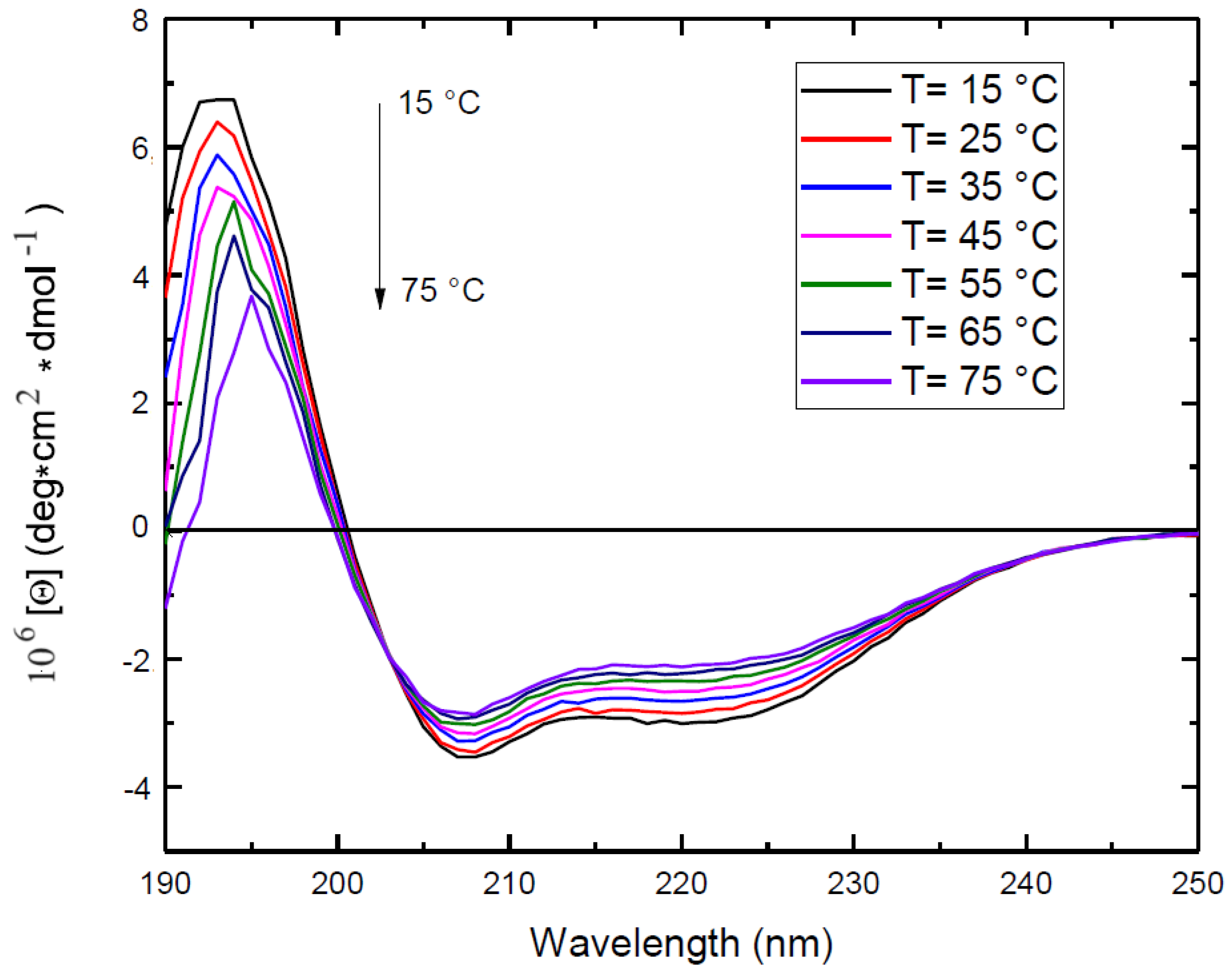
Figure 1. Far-UV circular dichroism spectrum of 0.3 mg/mL ZP in EtOH/H2O 70/30 (v/v) at different temperatures. Black: 15°C, red: 25°C, blue: 35°C, magenta: 45°C, green: 55 °C, dark blue: 65°C, purple: 75°C.
Preprints 113790 g001
Figure 2. Fluorescence emission and excitation spectra of 0.3 mg/mL Zein in EtOH/H2O 70/30 (v/v) at 25 °C. A) Dashed line: excitation spectrum of tyrosine (λem=308 nm); solid line: emission spectrum of tyrosine(λexc=278 nm). B) Dashed line: excitation spectrum of dityrosine (λem=420 nm); solid line: emission spectrum of dityrosine, (λexc=320 nm).
Preprints 113790 g002
Figure 3. Optical microscopy images of O/W emulsions from EtOH/H2O 70/30 (v/v) ZP solutions and 50 µL SO. A: 2.5 mg/mL ZP, t=0; B: 5 mg/mL ZP, t=0; C: 2.5 mg/mL ZP, t = 4 days; D: 5 mg/mL ZP, t = 4 days. Scale bar: 20 μm.
Preprints 113790 g003
Figure 4. Bright-field (A) and fluorescence (B) microscopy images of O/W emulsion prepared by UAE of 5.0 mg/mL ZP EtOH/H2O 70/30 (v/v) solution and 10 µL SO. NLR has been included in SO before the microcapsule preparation. Sample volume: 1mL; frequency: 20 kHz, power: 220 W, UAE in continuous mode. Scale bar: 10 μm.
Preprints 113790 g004
Figure 5. Bright-field (A, C) and fluorescence (B, D) microscopy images of O/W emulsions formed by 5.0 mg/mL ZP in EtOH/H2O 70/30 (v/v) and 10 µL SO solution. NLR has been included in SO before the synthesis. Particle clusters are imaged after +4 days from the synthesis. Sample volume: 1mL; frequency: 20 kHz, power: 220 W, UAE in continuous mode. Scale bar: 20 μm.
Preprints 113790 g005
Figure 6. 3D confocal microscopy image of O/W emulsions from 10 mg/mL ZP EtOH/H2O 70/30 (v/v) and 10 mL SO solution. A: NLR added to the oil phase before the synthesis. B: RhB added to the aqueous solution before the synthesis. Sample volume: 1mL. Synthesis conditions: power= 220 W, time = 25 s; frequency:20 kHz, UAE in continuous mode, test tube in ice bath.
Preprints 113790 g006
Figure 7. SEM images of O/W emulsions from 10 mg/ml Zein EtOH/H2O 70/30 (v/v) and 10 ml SO solutions, A: day of synthesis. B: after 18 days. Synthesis conditions: power= 220 W, time= 25 s, frequency= 20 kHz, UAE in continuos mode.
Preprints 113790 g007
Figure 8. Bright field optical (A) and fluorescence (B) microscopy images of Zein microcapsule encapsulating Curcumin. Curcumin has been included in SO before the microcapsule synthesis. Scale bar: 20 μm.
Preprints 113790 g008
Table 1. Secondary structure for αZP-22KDa, αZP-22KDa and ZP from CD experiments (T=25°C).
Secondary 刘structure ZP (fresh)刘(%) ZP (1 day)刘(%) ZP (10 days)刘(%) ZP*刘(%)
α-helix 48.6 45.0 45.8 56.7
antiparallel β-sheet 6.1 0.8 1.2 7.1
turns 13.9 13.3 13.4 8.2
random coil 31.3 40.9 39.7 28.0
Disclaimer/Publisher’s Note: The statements, opinions and data contained in all publications are solely those of the individual author(s) and contributor(s) and not of MDPI and/or the editor(s). MDPI and/or the editor(s) disclaim responsibility for any injury to people or property resulting from any ideas, methods, instructions or products referred to in the content.
Copyright: This open access article is published under a Creative Commons CC BY 4.0 license, which permit the free download, distribution, and reuse, provided that the author and preprint are cited in any reuse.
Prerpints.org logo

Preprints.org is a free preprint server supported by MDPI in Basel, Switzerland.

Subscribe

© 2024 MDPI (Basel, Switzerland) unless otherwise stated