Preprint
Article

Essential Oils as an Antifungal Alternative to Control Cladosporium spp., Lasiodiplodia spp., Colletotrichum spp., Fusarium spp. and Aspergillus spp. Isolated of Musa paradisiaca

Altmetrics

Downloads

66

Views

61

Comments

0

This version is not peer-reviewed

Submitted:

28 August 2024

Posted:

29 August 2024

You are already at the latest version

Alerts
Abstract
Antifungal properties are found in essential oil extracts of oregano (Origanum vulgare), rosemary (Salvia rosmarinus), clove (Syzygium aromaticum), thyme (Thymus), cinnamon (Cinnamomum verum), basil (Ocimum basilicum), therefore, the ability to prevent or inhibit the growth of Cladosporium spp., Lasiodiplodia spp., Colletotrichum spp., Fusarium spp., and Aspergillus spp. was studied. Cladosporium spp., Lasiodiplodia spp. Colletotrichum spp. Fusarium spp. y Aspergillus spp. fungi that were isolated and purified from the rot found on the banana peels (Musa paradisiaca). To perform a visual (macro-scopic) analysis of the Petri dish containing the pure fungus and a more detailed (microscopic) analysis of the fungal spores to classify it by genus, then, dilutions are prepared to inoculate onto amended PDA medium with chloramphenicol. Weekly re-inoculations are performed until iso-lated and purified fungi are obtained, which are then evaluated for in vitro inhibition and in vivo growth, according to the results of the in vivo analysis, the fungi were classified from higher to lower severity in: Colletotrichum spp., Lasiodiplodia spp., Aspergillus spp., Fusarium spp. and Cladosporium spp and based on fungal growth observations, it was found that basil and rosemary do not inhibit growth at the maximum concentration. Cinnamon inhibits at 400 ppm, rosemary halts fungal growth at 600 ppm, clove at 1000 ppm, and oregano at 800 ppm.
Keywords: 
Subject: Biology and Life Sciences  -   Agricultural Science and Agronomy

1. Introduction

The origin of the banana is from India, Asia, it is an exotic and climatic fruit known as cambur, gualele, guineo, musacea, platano. In Ecuador, in El Oro province, Machala, has led exports since 1910 to the United States, Peru, and Chile, highlighting its quality, which is unmatched by fruits offered at lower prices from other countries [1]. Currently it is the largest exporter of banana with internationally recognized quality product with a productivity of 1500 boxes per hectare per year, it is consumed when the peel is yellow, being an important source of vitamins, potassium, carbohydrates, and minerals [2]. The rot in the banana increases in transport and the fungal growth decreases the quality of the fruit after harvest and produces the rot of the crown, known as “Crown rot” caused mainly by Colletotrichum spp. [3].
Oregano has bioactive compounds such as carvacrol, thymol, phenolic acids that have antimicrobial properties, antioxidants, cinnamon has cinnamaldehyde, eugenol, flavonoids that has anti-inflammatory, antioxidant, glucose regulation potential and the clove has Eugenol, acetyl eugenol, flavonoids acting as analgesic, antimicrobial, antioxidant [4,5]. Cinnamon essential oil was used as a bioactive compound to extend the lifetime of the strawberry as a post-harvest treatment [6].
Basil has bioactive compounds such as eugenol, Rosmarinus acid, flavonoids that generates antioxidant, anti-inflammatory properties, thyme has thymol, carvacrol, flavonoids that generates antibacterial properties, antioxidant, enhances digestion and rosemary acid Rosmarinus, carnosol, flavonoids with antioxidative, anti-inflammatory, potential neuroprotective effect [7,8,9].
The Ascomycetes family includes the genus of fungi Cladosporium spp. and are commonly found in indoor and outdoor environments, are microscopically distinguished by having septate hyphae and conidia in groups or chains, the conidia are black, olive green, or sometimes brown, and frequently form in chains, are globose or ellipsoidal in shape. the hyphae are dark, branched, septate, and have a rough, cottony, or velvety texture. They may be plant pathogens, sometimes affect humans causing respiratory problems and allergies, are used in biodegradation, although they do not have significant industrial applications [10,11].
Lasiodiplodia spp. causes rot in fruit crops and is another type of fungus that is part of the Ascomycetes, the conidia are spherical in shape and occasionally form in chains, the hyphae are septate and frequently contain conidia in their spores, the texture may be cottony or velvety, with colors ranging from brown to black, and microscopically, conidia in spores and septate hyphae can be observed. The main impact of this fungus is on fruits and crops, causing diseases such as fruit rot. Its impact on agriculture is significant, although it is not frequently used in biotechnology [12,13].
Anthracnose is a common disease in many plants and is caused by various fungal pathogens, including those in the genera Colletotrichum spp., is classified within the Ascomycetes, microscopically the conidia appear in clusters or groups and have a cylindrical or ellipsoidal shape, the hyphae usually have conidia in their cluster and are septate, the texture may be velvety or cottony, and the color may be pink, red or orange. It is used to control pests in agriculture and is used in biopesticides [3,9]. Studies are being conducted on antifungal activity (in vitro) of plant extracts for the control of anthracnosis [14].
Plant diseases such as wilt and root rot are caused by Fusarium spp., some species produce mycotoxins that are harmful to animals and humans. It is a fungus of the Ascomycetes or Basidiomycetes families, the conidia may be white, pink, red, or purple, are elongated, and frequently appear in chains. Macroconidia and microconidia can be observed in the septate hyphae, and the texture may be cottony, velvety, or powdery [15,16].
Aspergillus spp. is found in food production, are used in the production of organic acids, enzymes and antibiotics, produce aflatoxins, which are toxic and carcinogenic, may also be opportunistic pathogens that infect people with weakened immune systems, are of the Ascomycetes family, microscopically, the conidia may be in chains or clusters, septate hyphae and the mycelium may be dense and abundant, the colonies may be green, black, white and yellow and the texture may be smooth, velvety, or cottony [17,18].

2. Materials and Methods

2.1. Isolation and Purification of Microorganisms

It is based on the scheme of Figure 1., to prepare the amended medium, PDA medium is made according to the supplier's specifications, and then amended with 0.5 g/L of chloramphenicol, which prevents bacterial growth, allowing for the study of fungi only [19].
Figure 2 shows the process for obtaining the initial dilution, which begins by decomposing the fruit to take a piece of the peel, rinsing it with sterile water twice, and discarding the used water [3]. The pieces of fruit peel affected by the presence of pathogens are placed in an Erlenmeyer flask with 200 mL of a 0.05% (v/v) Tween 80 solution and stirred in the vortex for approximately 2 minutes.
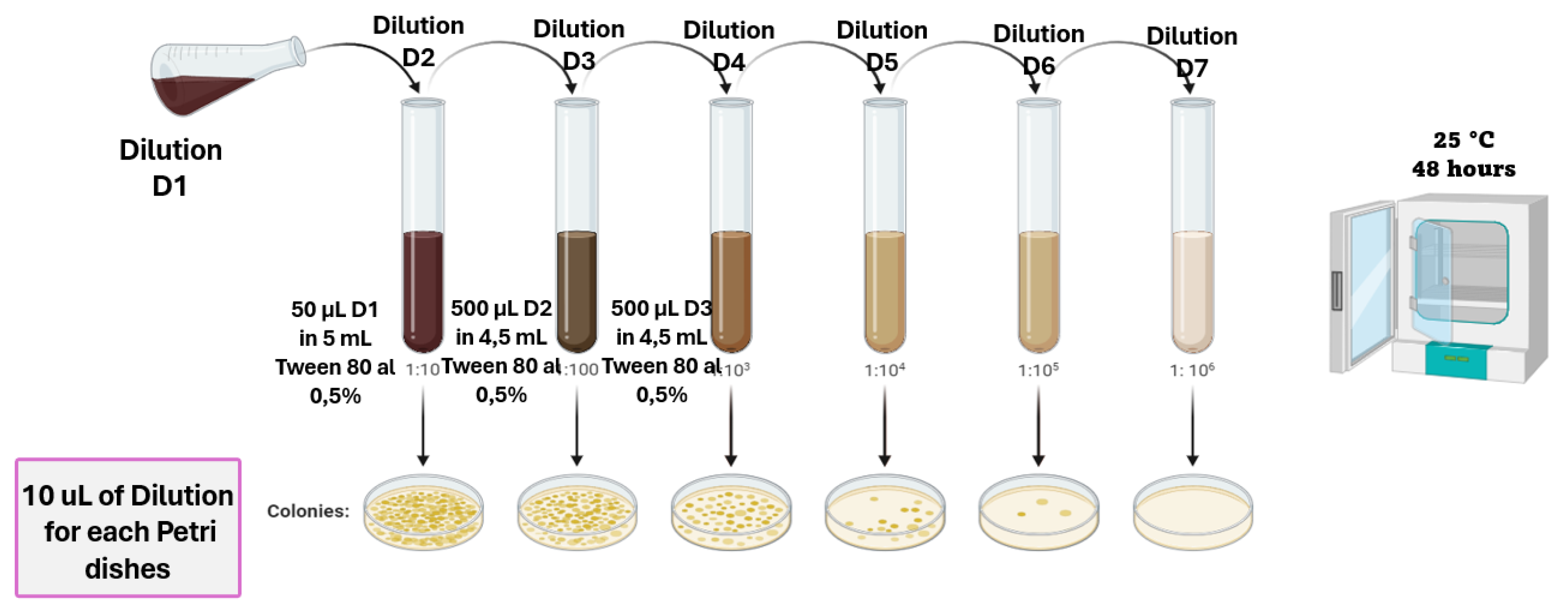
From dilution 1, four dilution series are prepared, using a 0.1% preparation of the initial solution to stir each dilution. Then, 0.1 mL of each dilution is plated onto Petri dishes with PDA to observe the macroscopic and microscopic morphology [3]. Figure 3 provides the diagram for preparing the dilutions used to analyze the growth.
The pathogen was visibly isolated on a Petri dish with PDA medium, which was incubated at 25°C for 7 days. A colony was picked with a sterile loop and subculture weekly until the strains were purified.

2.2. Macroscopic Identification

The purified fungi, three replicates were evaluated each week after plating the isolated pathogens. Their macroscopic characteristics were recorded and compared with bibliographic information from books and guides on fungal morphology to identify the pathogen’s genus. During the analysis, aspects such as colony shape, elevation, edges, and appearance were considered.

2.3. Microscopic Identification

The fungus obtained from the culture was examined in triplicate under the microscope using adhesive tape to collect the aerial mycelium and adhere it to a slide. The slide was observed under a microscope with 40X and 60X magnification lenses [5]. The evaluation focused on the hyphae, mycelium, spores, and the microscopically observed structures.

2.4. In Vivo Fungal Activity

After the morphological characterization, the pathogens Cladosporium spp., Lasiodiplodia spp. Colletotrichum spp. Fusarium spp. and Aspergillus spp. which were isolated from the banana (Musa paradisiaca), were evaluated.
The inhibition index was calculated in quadruplicate based on the measurement corresponding to the diameter of fungal growth to assess the severity of the identified pathogens. In the Figure 4 provides the diagram for obtaining a desired concentration of 106 conidia/mL of each inoculum.
The fungal growth diameter on the days following inoculation was the response variable studied, based on each fungal genus, the most severe pathogen was determined. Figure 5 provides the diagram for the inoculation of the fungi on the banana.

2.5. In Vitro Antifungal Activity with Essential Oils

Antifungal activity was analyzed under controlled laboratory conditions using essential oils of oregano (Origanum vulgare), rosemary (Salvia rosmarinus), clove (Syzygium aromaticum), thyme (Thymus), cinnamon (Cinnamomum verum), and basil (Ocimum basilicum). PDA media were prepared with concentrations of 200, 400, 600, 800, and 1000 ppm of each essential oil, and a control without essential oil was included (Figure 6).
The solutions with the specific concentrations of essential oil were solidified for the inoculation of the pathogens maintained at 25°C in the incubator to identify the most effective concentration. The prepared Petri dishes were visually inspected in triplicate every 48 hours to determine the percentage of inhibition, and the effectiveness of the essential oil used.

3. Results

3.1. Macroscopic Identification

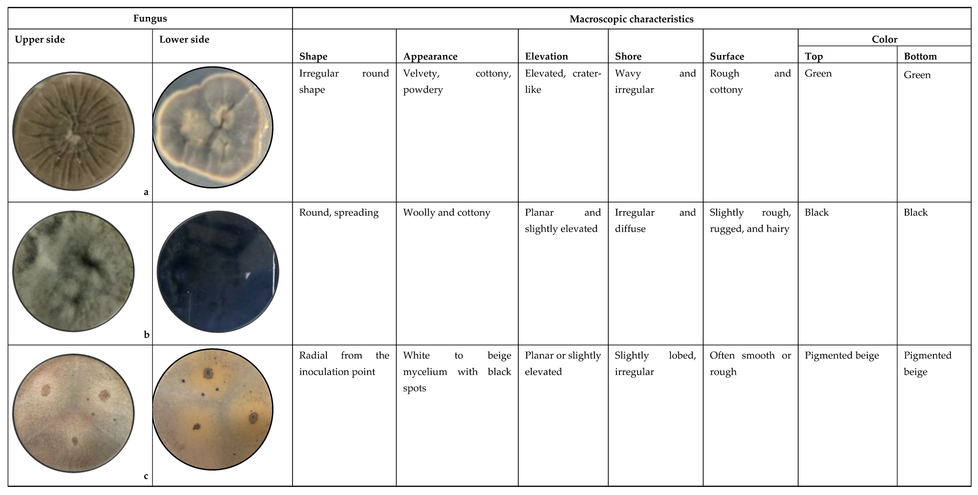
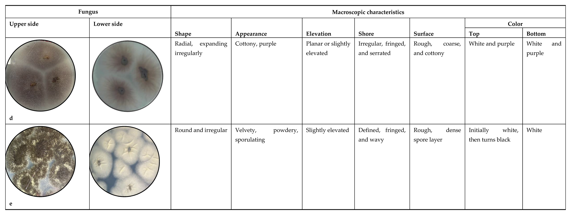
Table 1 shows the macroscopic comparison of purified fungi by characteristics such as colony shape, appearance, elevation, shore, surface and color of both the front and reverse.

3.2. Microscopic Identification

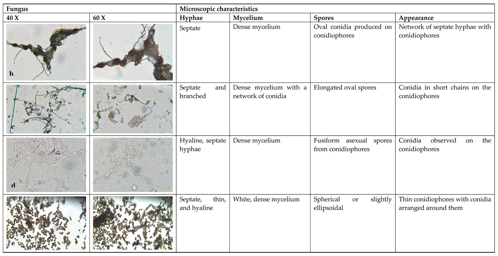
Table 2 presents the aerial mycelium of Cladosporium spp., Lasiodiplodia spp., Colletotrichum spp., Fusarium spp., and Aspergillus spp. The observations were made using 40X and 60X microscope lenses. The table evaluated the hyphae, mycelium, spores, and appearance in an analysis of their morphological characteristics.

3.3. In vivo fungal activity

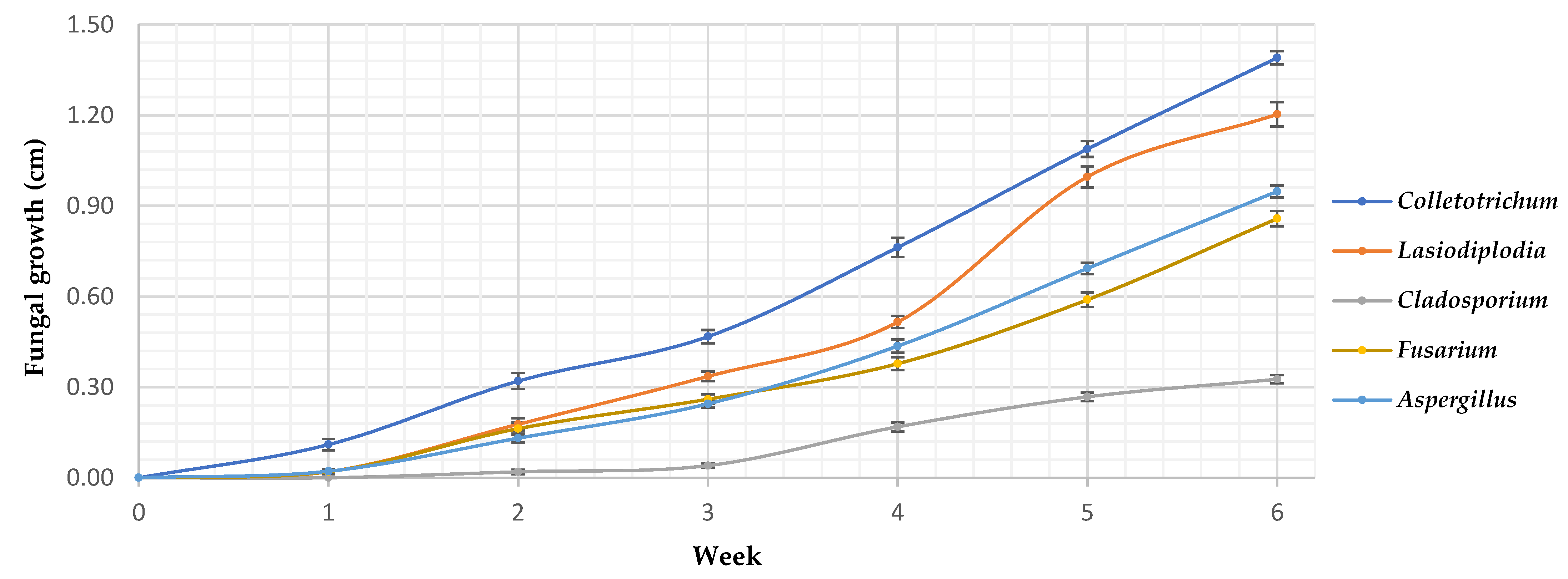
The severity of fungal infection was assessed through an in vivo analysis. Figure 7 illustrates the evaluation of fungal growth across 20 banana samples, which were monitored over a period of 6 weeks. The analysis focused on identifying and assessing the growth of various fungal strains, including Cladosporium spp., Lasiodiplodia spp., Colletotrichum spp., Fusarium spp., and Aspergillus spp.
The results provide insights into the impact of these fungal strains on the bananas over the study period, highlighting differences in growth and infection severity of the different fungal species.

3.4. In Vitro Antifungal Activity with Essential Oils

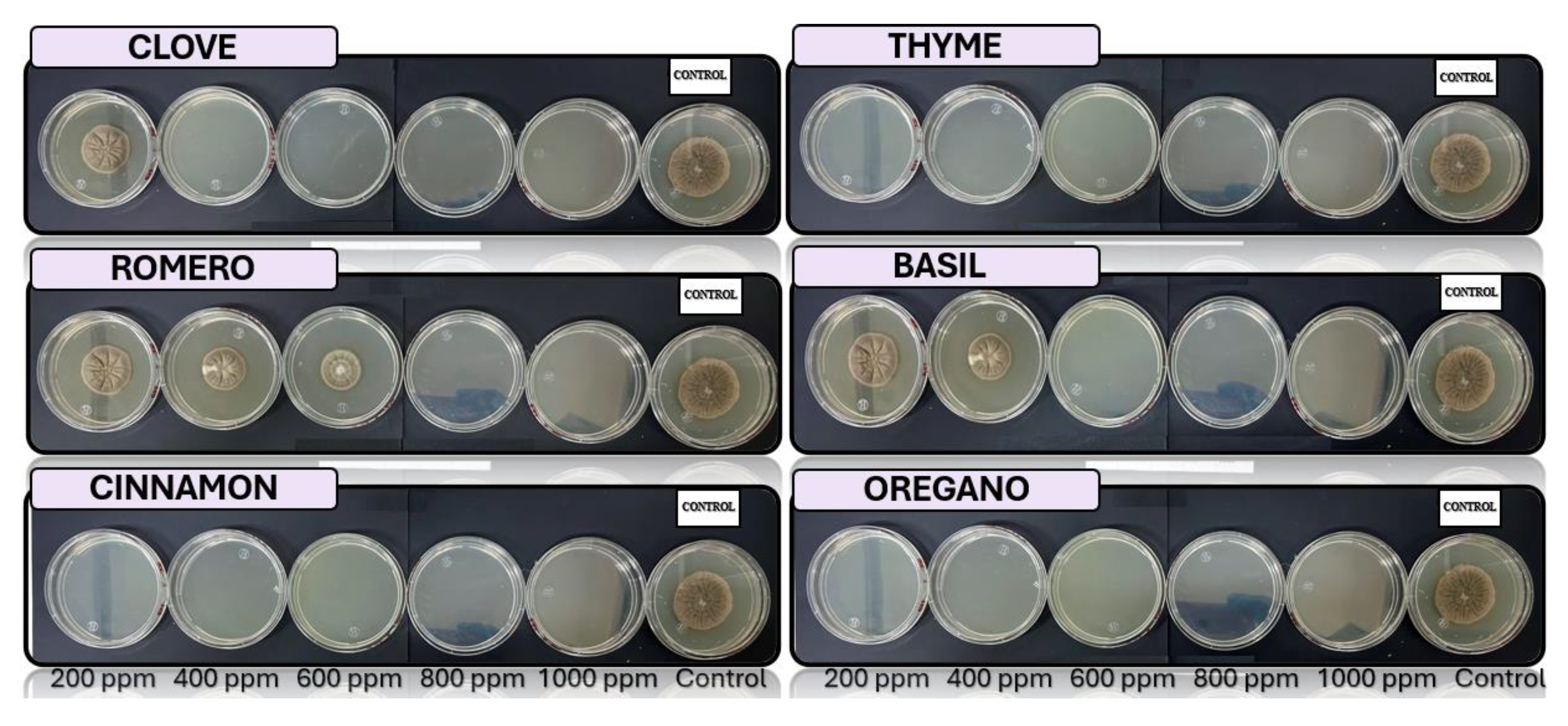
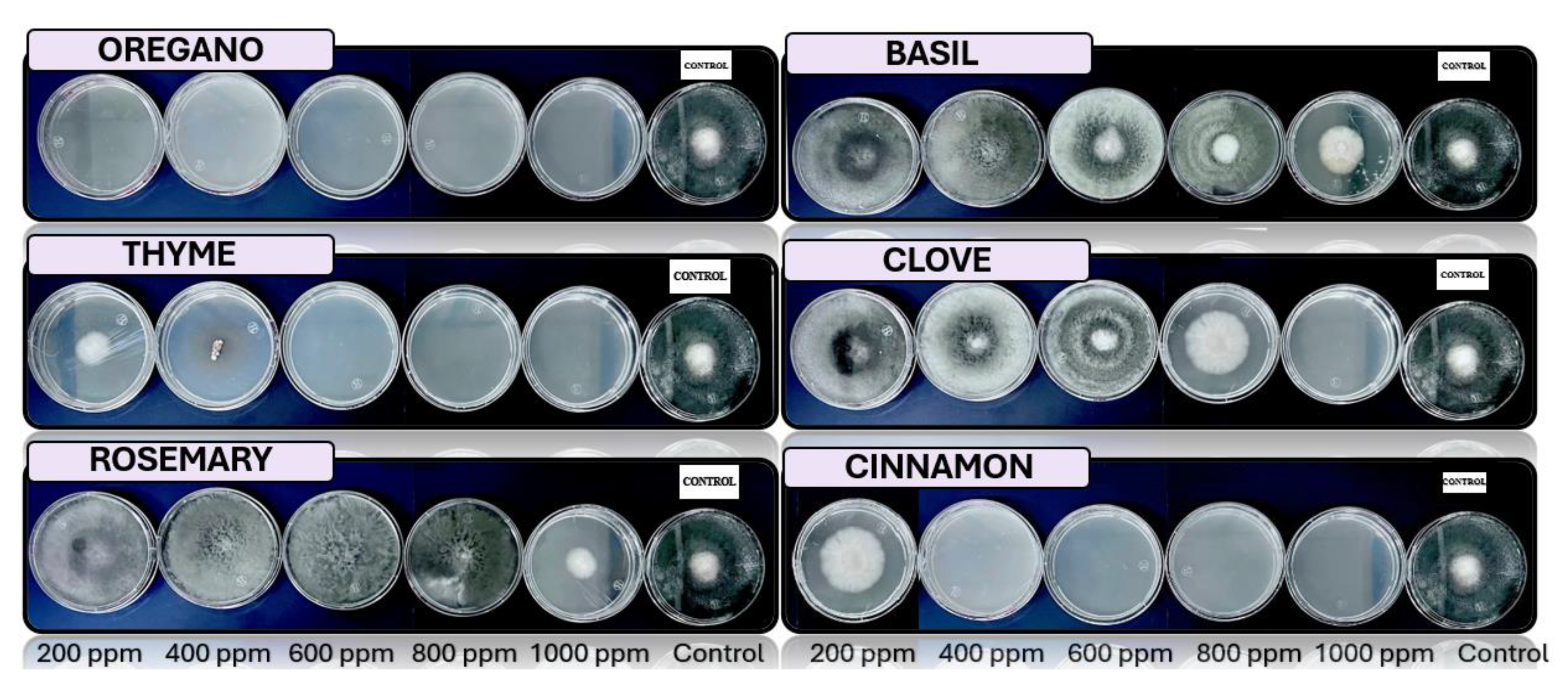
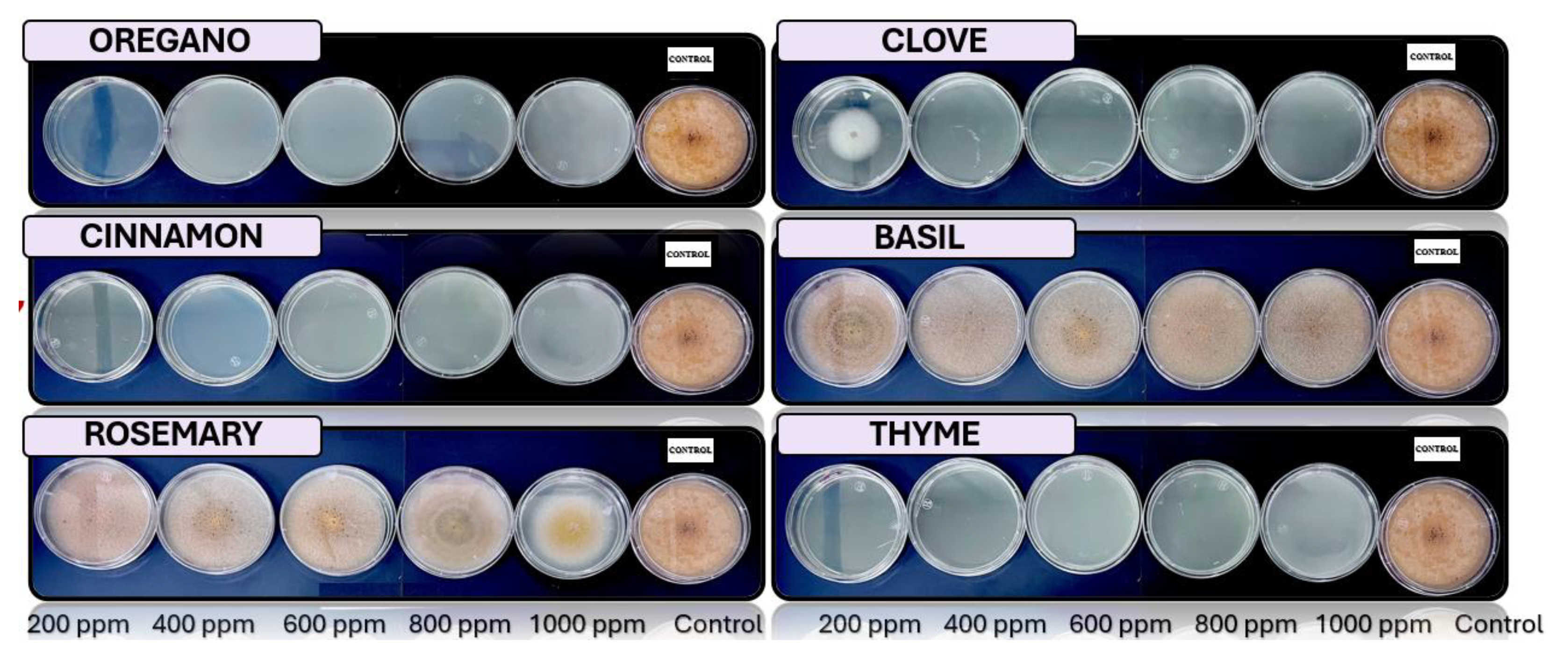
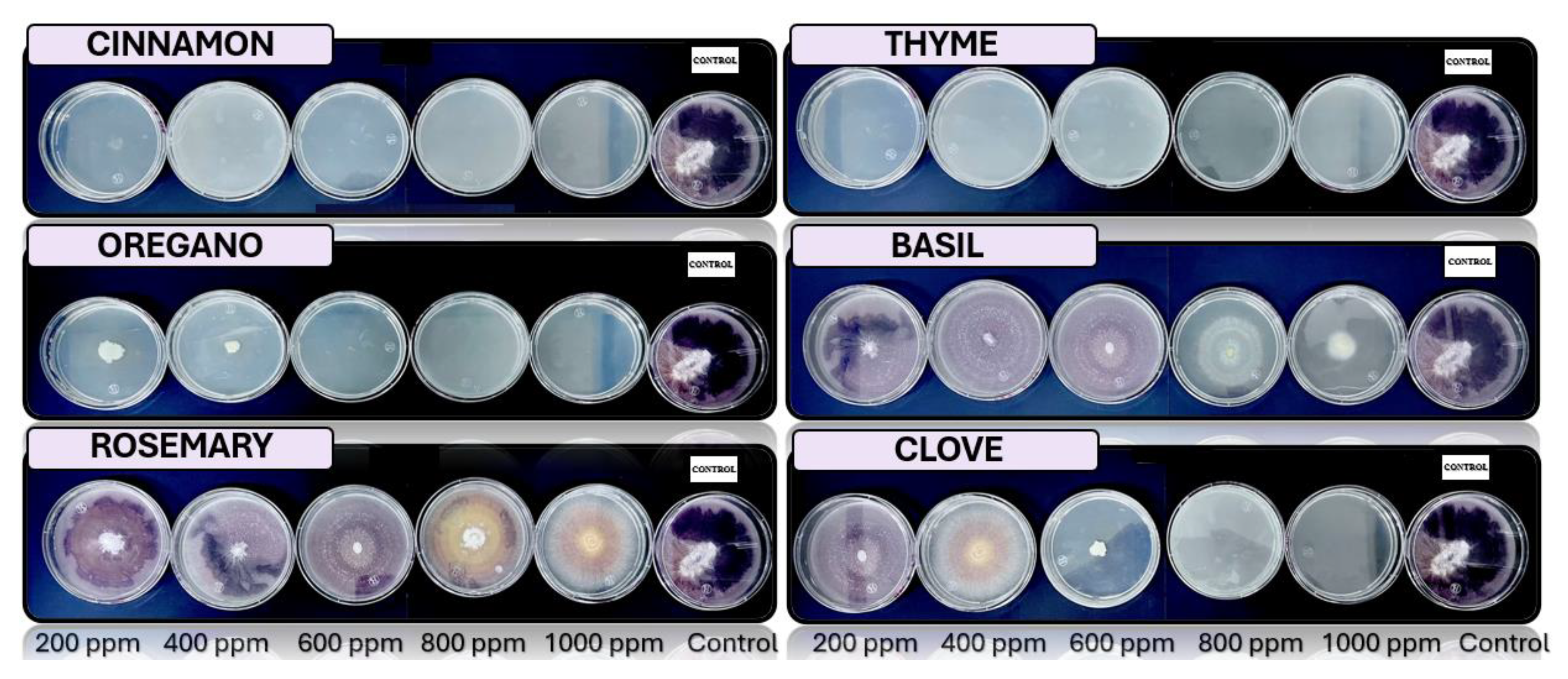
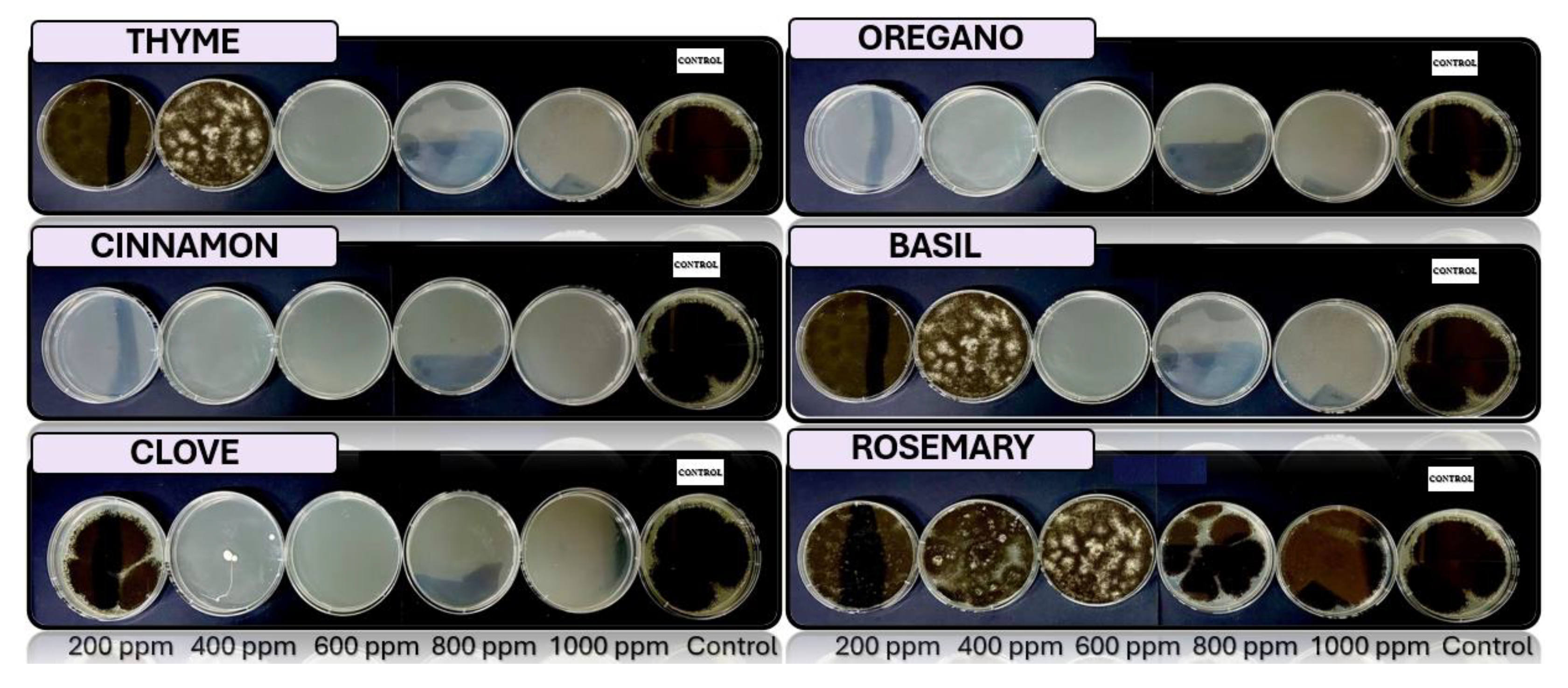
Figure 8, Figure 9, Figure 10, Figure 11 and Figure 12 illustrate the growth of Cladosporium spp., Lasiodiplodia spp., Colletotrichum spp., Fusarium spp., and Aspergillus spp. on PDA medium with essential oil concentrations of 200, 400, 600, 800, and 1000 ppm. The essential oils evaluated include oregano, basil, cinnamon, rosemary, thyme, and clove.

4. Discussion

4.1. Macroscopic Identification

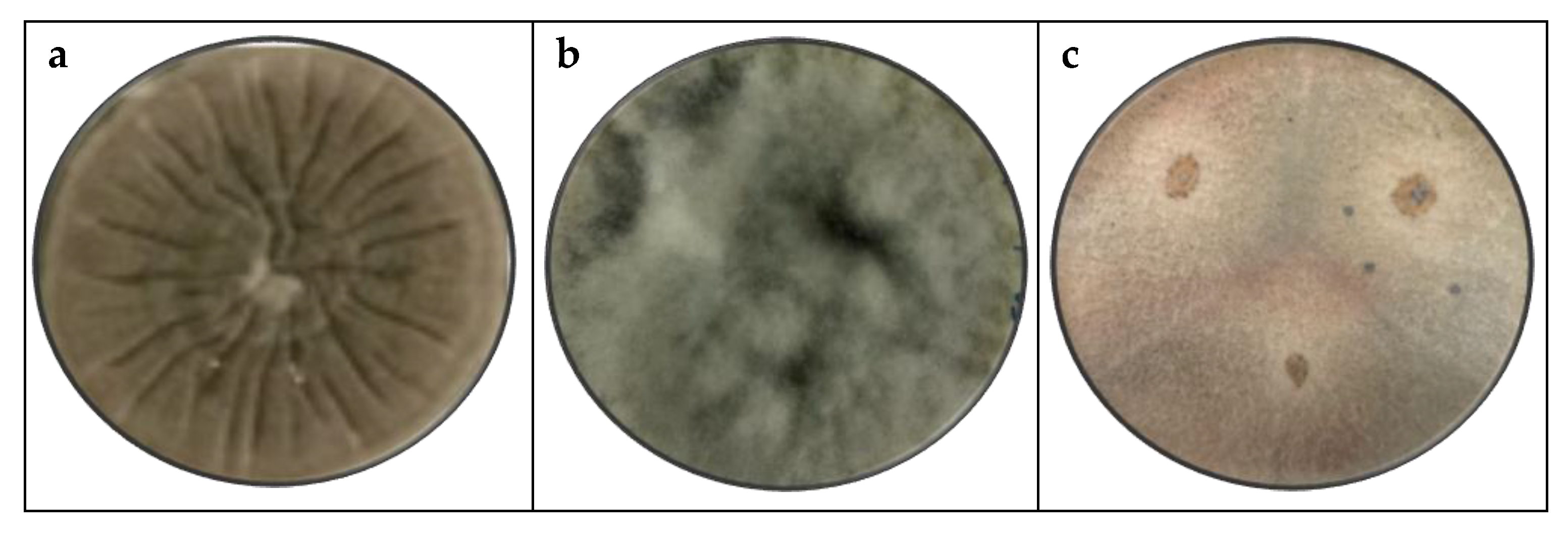
Figure 13 shows the initial isolation of pathogens obtained from perceptibly affected banana peels, which were plated on PDA and incubated at 25°C. The figure displays the purified fungi that are implicated in banana rot.
This macroscopic analysis suggests that the fungus is of the genus Cladosporium spp. due to its powdery appearance, green color on both the front and reverse sides, and its velvety, cottony, and cratiform (crater-like) appearance [20], Lasiodiplodia spp. based on the macroscopic characteristics, which report a woolly appearance with a black to gray color, rapid growth, and a rough surface. These features are similar to those reported when evaluating banana crown rot [21], Colletotrichum spp. have mycelium that is pigmented beige with a slightly raised, planar elevation, and has been identified as the most significant fungal agent in banana [22].
Postharvest deterioration of bananas is due to the presence of Fusarium spp. of various species that have been studied previously [15,23], this species exhibits a white and purple coloration with irregular edges and a cottony appearance, Aspergillus spp. have various applications in biotechnology, including the secretion of organic acids, proteins, enzymes, and secondary metabolites due to their prolific nature the genus is suspected based on its appearance on PDA, where it presents as round black spores with a rough texture, black coloration on the front, and white on the reverse [24].

4.2. Microscopic Identification

Based on the results of the macroscopic analysis and the comparison of microscopic characteristics with bibliographic references, the identification is Cladosporium spp. for septate and branched hyphae, and conidia that are generally ellipsoidal and are found in long chains [20], Lasiodiplodia spp. the identification is based on the black coloration, the ability to form multicellular oval conidia, and the septate hyphae, with conidia located at the apex of the conidiophore [21].
Colletotrichum spp. With high pathogenic potential, they have oval and elongated conidia arranged in short chains along the conidiophore, with septate and branched hyphae [25], Fusarium spp. is characterized by conidia that can be unicellular or multicellular, with a varied shape, including fusiform and ellipsoidal. Due to the diversity of species, sequencing is necessary to ensure accurate analysis and identification, and Aspergillus spp. is distinguished by having septate, thin, and hyaline hyphae, with dense white and black mycelium, and spores that are spherical or slightly ellipsoidal.

4.3. In Vivo Fungal Activity

After the macroscopic and microscopic characterization, the fungal strain was evaluated of Cladosporium spp., Lasiodiplodia spp. Colletotrichum spp. Fusarium spp. and Aspergillus spp. To establish the calculated inhibition index for 20 samples based on the corresponding measurement of the circular growth diameter of the fungi, in order to assess the severity of the fungus [26].
The response variable studied was fungal growth over the 6 weeks following inoculation. To further analyze the fungal genera, the most severe agent was determined by statistically analyzing the obtained results using Stat graphics Centurion XV.
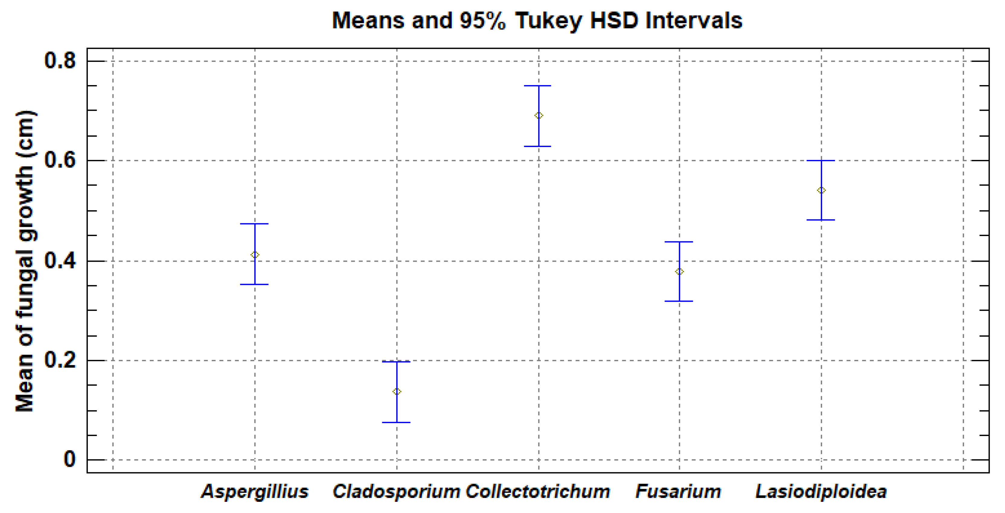
Figure 14 presents the analysis of means. The p-value of the F-test is less than 0.05, indicating a statistically significant difference between the means of the 5 variables with a 95.0% confidence level. It is concluded that the pathogens were assessed in the following order of severity, from highest to lowest: Colletotrichum spp., Lasiodiplodia spp., Aspergillus spp., Fusarium spp. and Cladosporium spp..

4.4. In Vitro Antifungal Activity with Essential Oils

The potential of essential oils as antifungal agents was evaluated in vitro to oregano (Origanum vulgare), rosemary (Salvia rosmarinus), clove (Syzygium aromaticum), thyme (Thymus), cinnamon (Cinnamomum verum) and basil (Ocimum basilicum), from commercial stores and extracted through steam distillation, were evaluated for their antifungal potential. Solutions were prepared using PDA (Potato Dextrose Agar) medium with varying concentrations of essential oils 200, 400, 600, 800 y 1000 ppm.
The different concentrations were meticulously formulated to assess their antifungal efficacy against the targeted fungal strains. Each concentration was mixed into the PDA medium to ensure uniform distribution before solidifying in petri dishes for subsequent testing. A control without essential oil was included for comparison.
The most effective concentration was identified through the analysis conducted in four replicates, and the Petri dishes were examined every 48 hours to determine the percentage of inhibition and effectiveness of the essential oil at the established concentration [27].
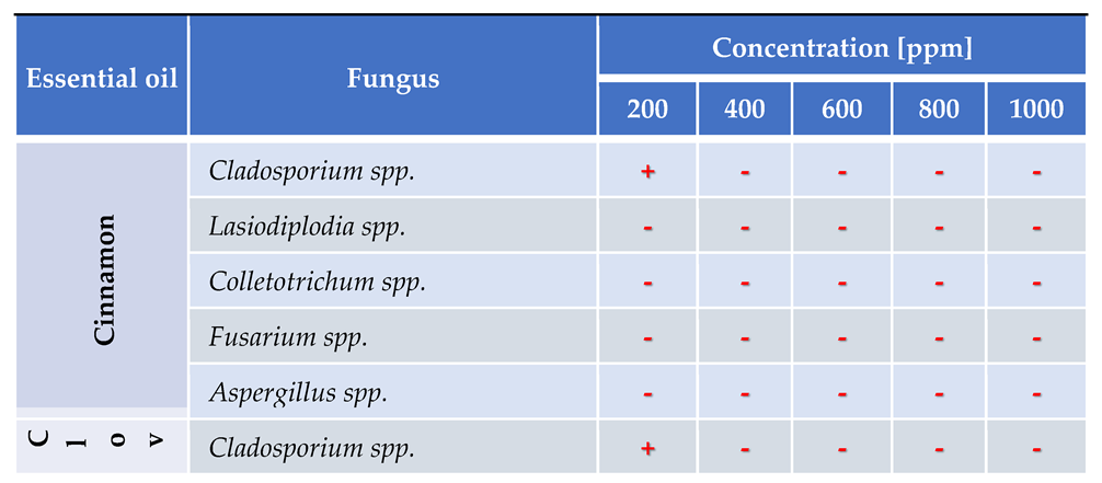
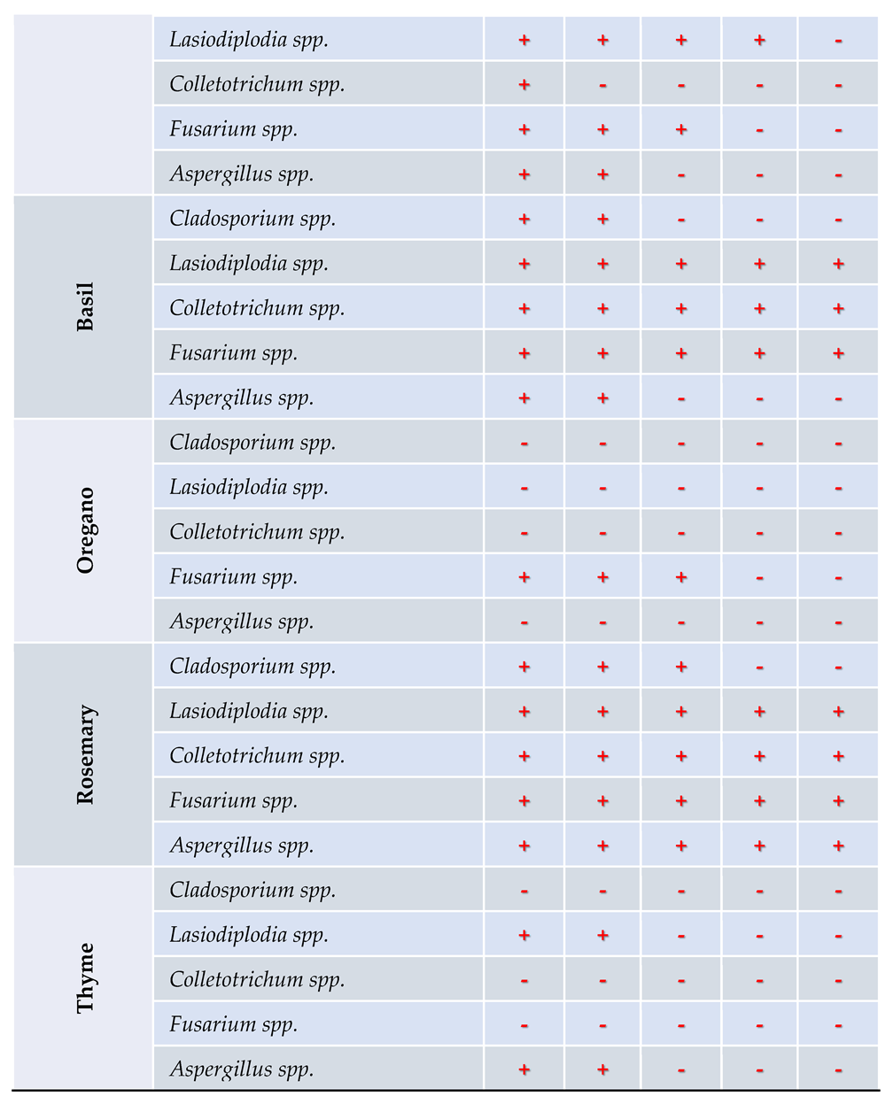
Table 3 presents the in vivo analysis of antifungal activity, demonstrating that a concentration of 400 ppm of cinnamon essential oil effectively inhibits the growth of all five fungal species tested.

5. Conclusions

During the characterization process, the fungal strains were assessed and ranked based on their severity, from the most severe to the least severe. This ranking was determined through of an evaluation of the analysis in vivo report: Colletotrichum spp., Lasiodiplodia spp., Aspergillus spp., Fusarium spp. y Cladosporium spp. The in vitro antifungal activity analysis reveals that a concentration of 400 ppm of cinnamon essential oil is effective in inhibiting the growth of all five fungal species examined.
In the in vitro study to evaluate the efficacy of essential oils, cinnamon was effective at 400 ppm, thyme at 600 ppm, and clove at 1000 ppm, while basil and rosemary did not inhibit the growth of the analyzed pathogens. Specifically: Colletotrichum spp. was controlled with 200 ppm of oregano, thyme, 400 ppm of cinnamon, and 1000 ppm of clove. Lasiodiplodia spp. was controlled with 200 ppm of cinnamon, oregano, 600 ppm of thyme, and 1000 ppm of clove. Aspergillus spp. was controlled with 200 ppm of cinnamon, oregano, 600 ppm of clove, basil, and thyme. Fusarium spp. was controlled with 200 ppm of cinnamon, thyme, 800 ppm of clove, and oregano. Cladosporium spp. was controlled with 200 ppm of oregano, thyme, 400 ppm of cinnamon, clove, 600 ppm of basil, and 800 ppm of rosemary.

Contributions

All authors listed have significantly contributed to the development and writing of this article. All authors have read and agreed to the published version of the manuscript.”.

Acknowledgments

The authors acknowledge the support from Departamento de Ciencias Ambientales y Biotecnología – Escuela Politécnica Nacional y FICAYA – Universidad Técnica del Norte.

Conflicts of Interest

The funders had no role in the design of the study, in the collection, analysis, or interpretation of data, in the writing of the manuscript, or in the decision to publish the results.

References

  1. Mata Anchundia D, Suatunce Cunuhay P, Poveda Morán R. Análisis económico del banano orgánico y convencional en la provincia Los Ríos, Ecuador. Avances 2021;23:419–30.
  2. Ruiz Medina MD, Ruales J. Postharvest Alternatives in Banana Cultivation. 2024. [CrossRef]
  3. Vilaplana R, Pazmiño L, Valencia-Chamorro S. Control of anthracnose, caused by Colletotrichum musae, on postharvest organic banana by thyme oil. Postharvest Biology and Technology 2018;138:56–63. [CrossRef]
  4. Arcila-Lozano CC, Loarca-Piña G, Lecona-Uribe S, González de Mejía E. El orégano: propiedades, composición y actividad biológica de sus componentes. Archivos Latinoamericanos de Nutrición 2004;54:100–11.
  5. Villanueva C, Vilca R, Ítalo A, Cotrina G. Aplicación de Aceite Esencial de Canela (cinnamomum verum) y Clavo de Olor (syzygium aromaticum) en la cobertura comestible y tiempo de vida útil de la Fresa (fragaria ananassa). Ciencia Latina Revista Científica Multidisciplinar, vol. 5, Perú: 2021, p. 1504–26. [CrossRef]
  6. Ruiz M, Ávila J, Ruales J. Diseño de un recubrimiento comestible bioactivo para aplicarlo en la frutilla (Fragaria vesca) como proceso de postcosecha 2016;17:276–87.
  7. López Luengo MT. El romero. Planta aromática con efectos antioxidantes. Offarm 2008;27:60–3.
  8. Farías C, Cisternas C, Morales G, Muñoz L, Valenzuela Rodrigo. Albahaca: Composición química y sus beneficios en salud. Revista Chilena de nutrición 2022;49. [CrossRef]
  9. Alzate D, Mier G, Afanador L, Durango D, García C. Evaluación de la fitotoxicidad y la actividad antifúngica contra Colletotrichum acutatum de los aceites esenciales de tomillo (Thymus vulgaris), limoncillo (Cymbopogon citratus), y sus componentes mayoritarios 2009;16.
  10. [10] Bensch K, Braun U, Groenewald JZ, Crous PW. The genus Cladosporium. Studies in Mycology 2012;72:1–401. [CrossRef]
  11. Crous PW, Braun U, Schubert K, Groenewald JZ. Delimiting Cladosporium from morphologically similar genera. Studies in Mycology 2007;58:33–56. [CrossRef]
  12. Picos-Muñoz PA, García-Estrada RS, León-Félix J, Sañudo-Barajas A, Allende-Molar R, Picos-Muñoz PA, et al. Lasiodiplodia theobromae en Cultivos Agrícolas de México: Taxonomía, Hospedantes, Diversidad y Control. Revista mexicana de fitopatología 2015;33:54–74.
  13. Sangeetha G, Anandan A, Rani SU. Morphological and molecular characterisation of Lasiodiplodia theobromae from various banana cultivars causing crown rot disease in fruits. Archives Of Phytopathology And Plant Protection 2012. [CrossRef]
  14. Villacís-Aldaz LA, León-Gordon O, Santana-Mayorga R, Mangui-Tobar J, Carranza G, Pazmiño-Miranda P. Actividad anti fúngica (in vitro) de extractos vegetales para el control de antracnosis (Colletotrichum acutatum). Journal of the Selva Andina Biosphere 2017;5:59–64. [CrossRef]
  15. Randy P. Marchites por fusarium del plátano 2015. [CrossRef]
  16. Ortiz E, Riascos D, Angarita M, Castro O, Rivera C, Romero D, et al. Tópicos taxonómicos para el estudio del género Fusarium 2020;33:61–6.
  17. Chang PK, Horn BW, Abe K, Gomi K. Aspergillus: Introduction. Encyclopedia of Food Microbiology: Second Edition, Elsevier Inc.; 2014, p. 77–82. [CrossRef]
  18. Rokas A. Aspergillus. Current Biology 2013;23:R187–8. [CrossRef]
  19. Suárez L, Rangel A. Aislamiento de microorganismos para control biológico de Moniliophthora roreri 2013;62.
  20. Ogórek R, Pusz W, Lejman A. Characteristic and taxonomy of Cladosporium fungi. Prace Pogladowe 2012;19:80–5.
  21. Santos JEA, Silva DEM, Vieira RFBS, Cordeiro MVM, Almeida MMM, Lima M a. S, et al. First Report of Lasiodiplodia brasiliensis Causing Crown Rot on Banana in Brazil. Plant Disease 2023;107:2538. [CrossRef]
  22. Camargo Piñeres Y, Zambrano Montenegro G, Ortega-Cuadros M, Gutierrez Montero DJ, Yepes JA, Camargo Piñeres Y, et al. Actividad antifúngica in vitro del aceite esencial de Swinglea glutinosa Merr sobre Colletotrichum sp. patógeno de mango (Mangifera indica L.). Revista Colombiana de Biotecnología 2021;23:62–71. [CrossRef]
  23. Jiménez M, Logrieco A, Bottalico A. Occurrence and Pathogenicity of Fusarium Species in Banana Fruits. Journal of Phytopathology 1993;137:214–20. [CrossRef]
  24. Pascal A, Simon T. Aspergillus fumigatus in Poultry 2011;20.
  25. Aguilar-Anccota R, Apaza-Apaza S, Maldonado E, Calle-Cheje Y, Rafael-Rutte R, Montalvo K, et al. Control in vitro e in vivo de Thielavipsis paradoxa y Collettrichum musae cn biofungicidas en frutos de banano orgánico. Manglar 2024;21:57–63. [CrossRef]
  26. Angarita Merchán M, Torres Caicedo MI, Díaz Torres AK. Técnicas de Biología Molecular en el desarrollo de la investigación. Revisión de la literatura. Revista Habanera de Ciencias Médicas 2017;16:796–807.
  27. Pérez-Rodríguez LR, Pérez-Moreno L, Guzmán-Mendoza R, Sanzón-Gómez D, Belmonte-Vargas JR, Pérez-Rodríguez LR, et al. Sensibilidad in vitro de hongos fitopatógenos causantes de enfermedades en fresa a controladores biológicos y fungicidas, en el estado de Guanajuato, México. Acta universitaria 2019;29. [CrossRef]
Figure 1. Preparation of the amended medium with chloramphenicol (0.5 g/L), to obtain pure fungi responsible for the rot present in banana peels.
Preprints 116561 g001
Figure 2. Preparation of the initial dilution containing the microorganisms causing banana rot.
Preprints 116561 g002
Figure 3. Preparation of serial dilutions for plating 10µL on amended PDA medium.
Preprints 116561 g003
Figure 4. Diagram for obtaining a concentration of 106 conidia/mL of each inoculum.
Preprints 116561 g004
Figure 5. Inoculation of the fungi with a concentration of 106 conidia/mL.
Preprints 116561 g005
Figure 6. Preparation scheme of the medium with established essential oil concentrations.
Preprints 116561 g006
Figure 7. Fungal growth (cm) for 6 weeks in 20 banana samples inoculated with Cladosporium spp., Lasiodiplodia spp. Colletotrichum spp., Fusarium spp. and Aspergillus spp., stored at 13°C and 95% HR.
Preprints 116561 g007
Figure 8. In vitro growth analysis of Cladosporium spp. on PDA medium with basil, cinnamon, clove, oregano, rosemary, and thyme essential oils at 200, 400, 600, 800, and 1000 ppm, stored at 25°C (n=4).
Preprints 116561 g008
Figure 9. In vitro growth analysis of Lasiodiplodia spp. on PDA medium with basil, cinnamon, clove, oregano, rosemary, and thyme essential oils at 200, 400, 600, 800, and 1000 ppm, stored at 25°C (n=4).
Preprints 116561 g009
Figure 10. In vitro growth analysis of Colletotrichum spp. on PDA medium with basil, cinnamon, clove, oregano, rosemary, and thyme essential oils at 200, 400, 600, 800, and 1000 ppm, stored at 25°C (n=4).
Preprints 116561 g010
Figure 11. In vitro growth analysis of Fusarium spp. on PDA medium with basil, cinnamon, clove, oregano, rosemary, and thyme essential oils at 200, 400, 600, 800, and 1000 ppm, stored at 25°C (n=4).
Preprints 116561 g011
Figure 12. In vitro growth analysis of Aspergillus spp. on PDA medium with basil, cinnamon, clove, oregano, rosemary, and thyme essential oils at 200, 400, 600, 800, and 1000 ppm, stored at 25°C (n=4).
Preprints 116561 g012
Figure 13. Fungi isolated and purified from banana peel rot, inoculated on selective medium (PDA with chloramphenicol), and stored on PDA at a 25°C in the incubator, (a) strain of Cladosporium spp., (b) Lasiodiplodia spp., (c) Colletotrichum spp., (d) Fusarium spp. spp. and (e) Aspergillus spp..
Preprints 116561 g013aPreprints 116561 g013b
Figure 14. Mean diameter (cm) of fungal decay circles in banana samples inoculated with Cladosporium spp., Lasiodiplodia spp. Colletotrichum spp., Fusarium spp. and Aspergillus spp. for 6 weeks, stored at 13°C and 95% HR (n=20).
Preprints 116561 g014
Table 1. Macroscopic characterization of (a) Cladosporium spp., (b) Lasiodiplodia spp., (c) Colletotrichum spp., (d) Fusarium spp. and (e) Aspergillus spp. considering appearance of upper side and lower side.
Preprints 116561 i001Preprints 116561 i002
Table 2. Microscopic characterization of (a) Cladosporium spp., (b) Lasiodiplodia spp., (c) Colletotrichum spp., (d) Fusarium spp. spp. and (e) Aspergillus spp. observed under the microscope.
Preprints 116561 i003Preprints 116561 i004
Table 3. Evaluation of antifungal activity in vitro of essential oil against Cladosporium spp., Lasiodiplodia spp., Colletotrichum spp., Fusarium spp. and Aspergillus spp. strain, using oregano, rosemary, clove, thyme, cinnamon and basil essential oils.
Preprints 116561 i005Preprints 116561 i006
Disclaimer/Publisher’s Note: The statements, opinions and data contained in all publications are solely those of the individual author(s) and contributor(s) and not of MDPI and/or the editor(s). MDPI and/or the editor(s) disclaim responsibility for any injury to people or property resulting from any ideas, methods, instructions or products referred to in the content.
Copyright: This open access article is published under a Creative Commons CC BY 4.0 license, which permit the free download, distribution, and reuse, provided that the author and preprint are cited in any reuse.
Prerpints.org logo

Preprints.org is a free preprint server supported by MDPI in Basel, Switzerland.

Subscribe

© 2024 MDPI (Basel, Switzerland) unless otherwise stated