Preprint
Article

Phytochemical Analysis, Antimicrobial, Antibiofilm and Antioxidant Effects of a New Arctic Lichen Placidium Doesaiense Growing in Pakistan

Altmetrics

Downloads

112

Views

74

Comments

0

A peer-reviewed article of this preprint also exists.

Submitted:

29 August 2024

Posted:

30 August 2024

You are already at the latest version

Alerts
Abstract
Lichenochemical composition and antimicrobial, antibiofilm and antioxidant effects of a newly described arctic lichen Placidium doesaiense growing in Pakistan were investigated. HPLC–DAD methods were used for identification of secondary metabolites in acetone and methanol extracts. The total polyphenol and the total flavonoid content was determined spectrophotometrically. The study examined the antioxidant (DPPH scavenging activity and reducing power), antibacterial (minimum inhibitory concentration (MIC) and minimum bactericidal concentration (MBC)) and antibiofilm (inhibition of biofilm formation and reduction of mature biofilm) activities of extracts of the lichen P. doesaiense and isolated parietin. The chemical constituents olivetol, olivetolic acid, haematommic acid, fallacinol and parietin, were identified as major compounds in the tested extracts of the lichen. Parietin was isolated from the acetone extract on a separation column. The methanol extract had higher values of TPC (21.67 mg GA/g) and TFC (377.40 mg RE/g) than the acetone extract. Although the extracts showed the best antibacterial activity (especially against Proteus mirabilis ATCC 12453), parietin demonstrated superior antibiofilm activity (especially against Staphylococcus aureus ATCC 25923). Isolated parietin showed the best antioxidant activity measures according to DPPH scavenging activity (IC50= 51.616 μg/mL) and reducing power. This is the first report on the phytochemical composition of the lichen Placidium doesaiense and the first description of the chemical composition of some of the 28 species of the genus Placidia. This research will pave the way for further exploration of new activities of this lichen and its metabolites, which are important for medicine and pharmacy.
Keywords: 
Subject: Medicine and Pharmacology  -   Pharmacy

1. Introduction

A lichen represents a stable symbiotic relationship between a fungus and algae (typically green) and/or cyanobacteria. Lichens have the remarkable ability to produce over 1000 distinct secondary metabolites, which include monoaromatic compounds, anthraquinones, xanthones, dibenzofurans, depsones, depsides, and depsidones [1]. Many of these compounds are unique to lichens and exhibit various pharmaceutical activities. These include antioxidant, antiviral, antimicrobial, anti-inflammatory, and antiproliferative properties, as well as additional effects such as antipyretic, antiherbivore, allelopathic, and photoprotective activities [1,2,3,4,5]. Placidium is a small genus and only 28 species have been reported worldwide [6,7,8]. Placidium deosaiense Usman & Khalid was described in 2021 from Deosai Plains and its adjacent areas, Gilgit Baltistan, Pakistan [8]. It grows on soil and falls in the group of Pyrenocarpous lichens of Ascomycota. After the Tibetan plateau, Deosai Plains is the second highest alpine plateau in the world, and it comprises 2240 km2 of alpine tundra with an altitudinal range up to 5200 meters above sea level. It is located in Northern part of Pakistan between the Himalayas and Karakorum i.e. the world-famous mountain ranges [9,10]. Annual precipitation varies from 350 to 550 mm mostly received during winter as snow [11]. The plateau’s ecosystem is characterized by extreme cold with low atmospheric pressure, coupled with relatively low oxygen and carbon dioxide levels, aridity, intense and rapid solar ultraviolet radiation [12].
According to our knowledge, previous investigations of the chemistry of lichens from the genus Placidium have not confirmed the presence of any chemical compounds in them. Chemical analysis of the lichen Placidium squamulosum var. argentinum belonging to the Placidium genus showed that all tests for the presence of secondary metabolites have been negative [13]. Chemical analysis for the species Placidium nitidulum, Placidium nigrum and Placidium varium found in China also showed that all the spot tests have been negative, and that no substances have been detected by TLC [14].
The aim of this study was to determine, for the first time, the total phenolic and flavonoid contents in the acetone and methanol extracts of Placidium deosaiense, and to investigate their antimicrobial, antibiofilm, and antioxidant activities, highlighting their potential applications in medicine and pharmacy. Additionally, the phenolic compounds in P. deosaiense extracts were identified using high-performance liquid chromatography coupled with a photodiode array (HPLC-DAD).

2. Results

2.1. Phytochemical Analyses

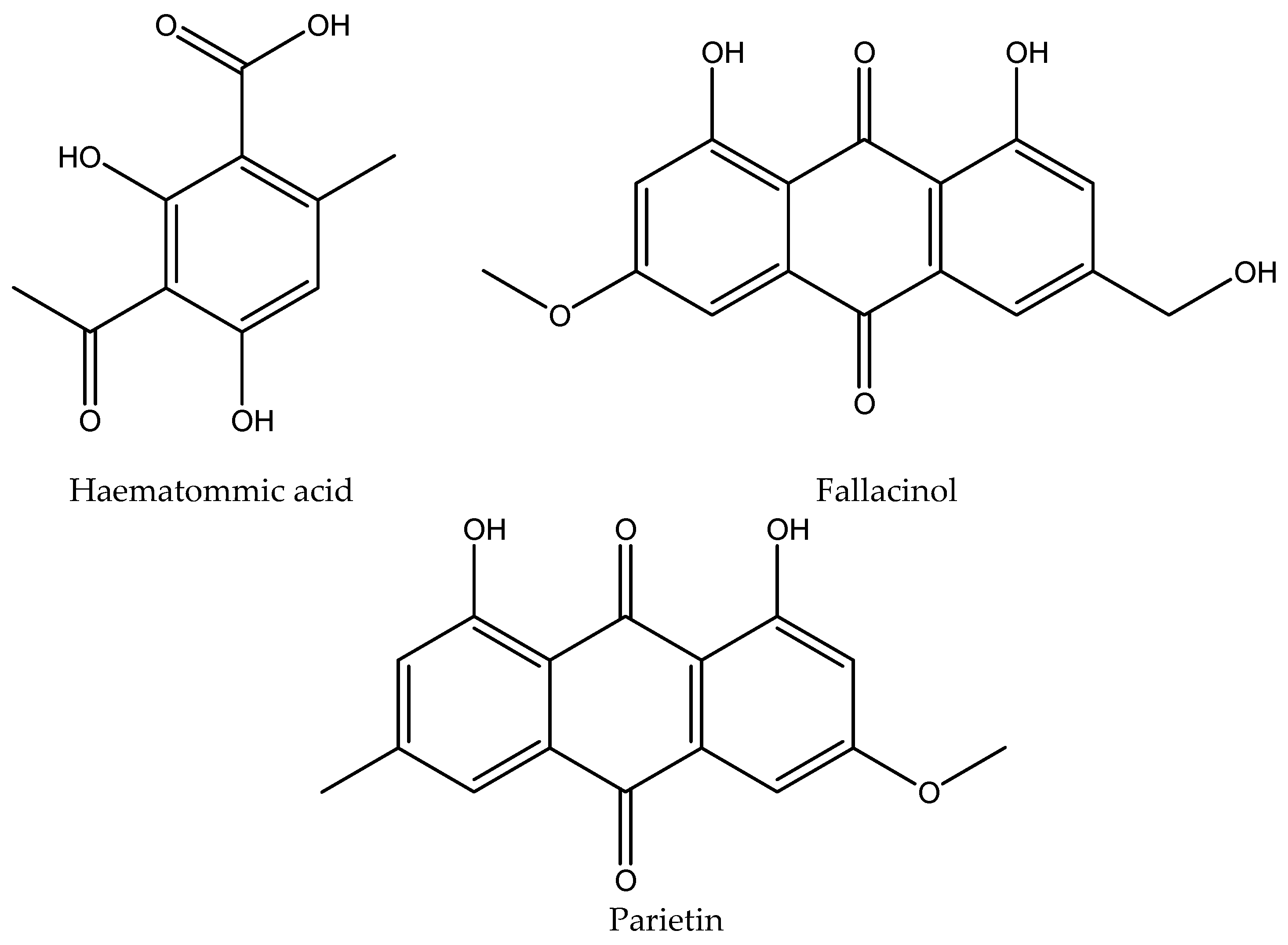
HPLC chromatograms of the acetone and methanol extracts of P. doesaiense, lichen collected in the Himalayas in Pakistan, as well as secondary metabolite parietin, are presented in Figure 1. A total of five different compounds (olivetol, olivetolic acid, haematommic acid, fallacinol and parietin) were identified in extracts, with olivetol being the most abundant compound (Table 1). Structures of all of the detected compounds are shown in Figure 2. In addition, parietin was isolated from the acetone extract on a separation column using different separation systems and used for further tests.

2.2. Yield of the Extraction, Total Polyphenols and Flavonoids Content and the Amount of Parietin

The data in Table 2 shows that higher yields were obtained by extracting P. doesaiense lichen with methanol than with acetone. Quantitative analysis performed using the HPLC method showed that the amount of parietin in the methanol extract was 0.310 mg/mL, while the amount of this compound in the acetone extract was about ten times less and amounted to 0.028 mg/mL.
Table 2 shows the results of the total phenolic content (TPC) and total flavonoid content (TFC) in the acetone and methanol extracts of the tested lichen. The methanol extract demonstrated significantly higher TPC (21.67 mg GA/g) and TFC (377.40 mg RE/g) values compared to the acetone extract.

2.3. Antibacterial Activity

Table 3 displays the in vitro antibacterial activity results for the acetone extract, methanol extract, parietin, and the positive control (tetracycline). In this experiment, the minimum inhibitory concentration (MIC) and the minimum bactericidal concentration (MBC) values ranged from <1.25 to >10 mg/mL for the acetone extract, methanol extract, and parietin, and from <0.25 to >128 µg/mL for tetracycline. The results of testing the antimicrobial activity of the acetone and methanol extracts of P. deosaiense lichen and parietin showed that the best antibacterial activity was recorded for the species Proteus mirabilis ATCC 12453 and Bacillus cereus ATCC 11778. The analyzed extracts of lichen P. deosaiense and parietin showed weak antibacterial activity on the other tested bacterial species. Based on the results, it can be concluded that the methanol extract of P. doesaiense exhibited superior antibacterial activity (MIC=1.25 mg/mL) against Proteus mirabilis ATCC 12453 compared to the acetone extract (MIC=2.5 mg/ml) and parietin (MIC=>10 mg/mL). The acetone extract showed better antibacterial activity (MIC=5 mg/mL; MBC=5 mg/mL) against Bacillus cereus ATCC 11778 than the methanol extract (MIC=10 mg/mL; MBC=10 mg/mL) and parietin (MIC and MBC= <10 mg/mL). Parietin showed very weak antibacterial activity against all tested bacterial species (MIC and MBC= <10 mg/mL). When compared to the positive control (tetracycline), the tested extracts and parietin exhibited limited (parietin) to moderate (the methanol and acetone extracts) effects against certain bacterial species.

2.4. Antibiofilm Activity

To evaluate the in vitro antibiofilm activity of the acetone and methanol extracts of P. doesainse and parietin, inhibition of biofilm formation and reduction of mature biofilm was investigated. The antibiofilm effect of the extracts and parietin on the P. mirabilis ATCC 12453, P. aeruginosa ATCC 10145, and S. aureus ATCC 25923 biofilm formation were examined. The results (Table 4 and Table 5) show the influence of different concentrations on the percentage of biofilm inhibition and reduction. The solvent control (5% DMSO) did not affect bacterial growth. The examined extracts of P. doesainse and parietin showed the greatest inhibition of biofilm formation S. aureus ATCC 25923. Parietin exhibited the best inhibition of biofilm formation S. aureus ATCC 25923, mostly at all concentrations (range: from 99.6 % to 63.7 %). Parietin has demonstrable strong inhibition of biofilm formation of P. mirabilis ATCC 12453 (10 mg/mL-92.5%) and P. aeruginosa ATCC 10145 (10 mg/mL-99.8 %), but only at higher concentrations. The acetone extract was more effective than the methanol extract. In the highest concentration (10 mg/mL) the acetone extract inhibited 92.1% of P. aeruginosa ATCC 10145 and 92% of S. aureus ATCC 25923. The acetone extract showed a strong inhibition of the formation of S. aureus ATCC 25923 biofilm and in lower concentrations (1.25 mg/mL, 2.5 mg/mL and 5 mg/mL) over 90%. The methanol extract also inhibited the biofilm formation of S. aureus ATCC 25923 at all tested concentrations. At concentrations of 5 mg/mL and 10 mg/mL, the methanol extract also inhibited the biofilm of P. aeruginosa ATCC 10145 and at the concentration of 10 mg/mL, it had an inhibitory effect on the formation of P. mirabilis ATCC 12453 biofilm.
Analysis of the effect of the acetone and methanol extracts of P. doesainse and parietin on the reduction of mature biofilms (Table 5) was more moderate. The maximum reduction was achieved in relation to the mature biofilm S. aureus ATCC 25923 (up to 85.3% reduction by parietin; 75.5% reduction by the methanol extract; and 69.8% reduction by the acetone extract). The reduction of the mature biofilm P. mirabilis ATCC 12453 is up to 20.4% by the methanol extract, but the same extract has no effect on inhibition of mature biofilm of P. aeruginosa ATCC 10145. The mature biofilm of P. aeruginosa ATCC 10145 was inhibited by the acetone extract up to 28.2% and parietin up to 59.8%.

2.5. Antioxidant Activity

The antioxidant activity of two P. doesaiense extracts and the isolated compound parietin was assessed using two methods: DPPH scavenging activity and reducing power (Table 6). The assessment of the antioxidant activity showed that all tested extracts were able to scavenge the DPPH radical. The isolated compound parietin showed higher DPPH free radical scavenging activity (IC50= 51.616 µg/mL) than the tested extracts of the lichen P. doesaiense. The acetone and methanol extracts of the lichen P. doesaiense showed similar activity in the scavenging of DPPH radicals. The results of the reducing power of the tested lichen extracts and parietin are shown in Table 6, where the examined extracts and parietin had less activity compared to ascorbic acid, at higher concentrations, but at lower concentrations, the difference in the activity was not so pronounced. Parietin shows the highest reducing power, while the acetone extract shows the weakest at higher concentrations, but at lower concentrations, the difference was smaller compared to the other tested extract and parietin.

3. Discussion

The present paper for the first time deals with the chemical analyses of the acetone and methanol extracts of a novel arctic alpine lichen, Placidium doesaiense, as well as the investigation of in vitro antimicrobial, antibiofilm and antioxidant effects of above-mentioned extracts and isolated parietin.
As can be seen in the HPLC chromatogram of the acetone extract (Figure 1), five different compounds were identified for the first time. The signal at retention time 2.42 min originated from olivetol. Olivetol belongs to the monoaromatic compound, as well as olivetolic acid whose signal is located at 2.91 min. Olivetolic acid (olivetol carboxylic acid) is a carboxylated derivative of olivetol and these two compounds do not occur so often in lichens and are not widely distributed. These two compounds have been reported from lichens Cetrelia monachorum and Ramalina conduplicans and their biological activity, including anti-inflammatory, antioxidant (DPPH and ABTS scavenging, protection against hydroxyl radical-induced DNA damage), antihyperglycemic, antimicrobial, antitumor, antiviral activities and others, has been extensively investigated [15,16,17,18]. Olivetol is a compound known to occur in some lichen species [19] but is best known as a precursor of tetrahydrocannabinol, found in Cannabis sativa [20]. The small signal that occurs at 4.56 min comes from haematommic acid, which is a common metabolite in lichens [21]. The signal with weak-medium intensity comes from the anthraquinone parietin and it appears at 17.54 min. Its structure was confirmed on the basis of the retention time values and the UV spectrum of a standard substance previously isolated from the lichen Xanthoria parietina [22]. It is mostly characteristic of lichen genera Xanthoria, Teloschistes, and Caloplaca [23] and this is the first time it was found in the genus Placidum. In contrast to the chromatogram of the acetone extract, the signal originating from parietin was more intense. Given the photoprotective and protective role of parietin, which is very important for the survival of this lichen under extreme external influences, its amount in the extracts was determined.
Methanol often provides a higher yield of extraction compared to acetone in lichens due to several factors related to its chemical properties and the nature of the compounds present in lichens. Many bioactive compounds in lichens, such as phenolics, are polar and dissolve better in polar solvents like methanol, which enhances the extraction yield of these compounds [24]. Parietin is a polar compound, and methanol, being a highly polar solvent, is more effective in dissolving and extracting polar compounds. This higher polarity of the methanol allows it to solubilize parietin more efficiently than the acetone. Methanol has a smaller molecular size and better penetration capabilities, which allows it to break down the cell walls of lichens more effectively. This results in a higher release of intracellular compounds, including parietin [25].
In vitro evaluations of various lichens against human pathogenic bacteria have been conducted [26]. Recent research has shown that the methanol extract of P. squamulosum showed high antibacterial activity against all seven tested bacterial strains (E. coli ATCC1652, S. typhi ATCC1679, P. mirabilis ATCC2601, S. aureus ATCC1885, E. faecalis ATCC2321, S. epidermidis ATCC2405, and B. cereus ATCC13061), with amount of MIC value from of 250 to 500 mg/mL [27]. However, our study is the first to investigate the antimicrobial activity of P. doesaiense extracts. The best antibacterial activity of the tested extracts in our work was shown for the species P. mirabilis ATCC 12453 and B. cereus ATCC 11778, and much weaker results for the other tested bacterial lines. The relative resistance of E. coli, S. aureus, P. aeruginosa, E. faecalis compared to P. mirabilis and B. cereus can be attributed to several factors related to their intrinsic and acquired resistance mechanisms: genetic adaptability, biofilm formation, efflux pumps, enzymatic degradation and cell wall structure [28]. The acetone and methanol extracts of P. doesaiense demonstrated superior antibacterial activity compared to isolated parietin. This finding aligns with other studies indicating that acetone and methanol extracts of lichens generally exhibit better antimicrobial activity than isolated compounds. This enhanced activity is attributed to the complex mixture of bioactive compounds in the extracts, which can work synergistically to produce a more potent antimicrobial effect. Methanol, being a polar solvent, is particularly effective at extracting phenolic compounds and flavonoids, which are known for their antimicrobial properties. This complex mixture in extracts can target multiple microbial pathways, enhancing overall antimicrobial efficacy compared to a single isolated compound [29,30,31]. Isolated parietin showed weak antibacterial activity against the tested bacterial lines. Basile et al. investigated the antimicrobial activity of acetone extract of Xanthoria parietina and parietin. The extract and parietin were tested for antimicrobial activity against nine American Type Culture Collection (ATCC) standard bacterial strains and clinically isolated bacterial strains. Both samples demonstrated strong antibacterial activity against all tested bacterial strains and clinical isolates, particularly against S. aureus from both standard and clinical sources [32]. In relation to our testing of the antibacterial activity, they were performed on different bacterial lines and ATCC strains.
To the best of the authors' knowledge, the antibiofilm activity of P. doesaiense extracts has not been investigated previously, although there is an existing study on the antibiofilm activity of parietin. In that study the antibiofilm activity of parietin has been demonstrated against S. aureus and E. faecalis [33], which is particularly significant since biofilms are involved in 80% of human microbial infections. Anthraquinone derivatives, commonly found in plants, are recognized for their antimicrobial properties, with several mechanisms identified. However, they are also linked to toxic and laxative effects, which could cause undesirable side effects if used in drug development [34]. In a study by Mitrovic et al. antibiofilm potentials (against S. aureus ATCC 25923 and P. mirabilis ATCC 12453) of acetone, ethyl acetate and methanol extracts of lichen species Platismatia glauca and Pseudevernia furfuracea were evaluated. GC-MS analyses of the extracts led to the identification of olivetol (which is also in high abundance in our examined extracts). The acetone extracts of Platismatia glauca and Pseudevernia furfuracea showed better antibiofilm activity than their methanol extract (as well as our examined extracts, but they had a weaker ability of inhibition) [35].
The biofilm, an assemblage of bacteria on extracellular polymer matrices of biotic or abiotic surfaces, confers greater resistance to varying environmental conditions and biocides compared to the planktonic form of bacteria [36].
In the present study, the acetone and methanol extracts and parietin from P. doesaiense exhibit dual actions by both preventing biofilm formation and eradicating mature biofilms. Although the extracts showed the best antibacterial activity, parietin demonstrated a superior antibiofilm effect. This suggests that lichen metabolites are the primary contributors to the antibiofilm properties of the tested extracts. It should be noted that the tested extracts and parietin exhibited better antibiofilm effects against the Gram-positive strain S. aureus at all concentrations, while their effects were weaker against the Gram-negative strains P. aeruginosa and P. mirabilis. The exception was at higher concentrations, where there was significant biofilm inhibition against P. aeruginosa. The antibiofilm activity of the tested extracts and parietin is particularly effective against Gram-positive bacteria such as S. aureus, likely due to their cell wall structure. Gram-positive bacteria have a thick peptidoglycan layer, which is a primary target for many antimicrobial agents found in lichen extracts. These extracts can effectively disrupt the synthesis and integrity of the peptidoglycan layer, impairing the bacteria's ability to form and maintain biofilms [30,31]. The biofilm eradication ability of P. doesaiense extracts and parietin was reduced but remained significant against S. aureus biofilm. The literature data does not provide an accurate explanation of the mechanism of antibiofilm activity of lichens and their extracts, but secondary lichen metabolites are generally thought to be responsible for antibiofilm effect as well as antimicrobial activity [35,37]. Several lichen compounds, including usnic acid, atranorin, evernic acid, psoromic acid, and butyrolactone analogs have demonstrated antibiofilm activity [37,38,39].
Several studies have examined the antioxidant potential of various lichen species [40,41], but this is the first study about the antioxidant activity of the new lichen P. doesaiense. Other authors examined lichen extracts that contained metabolites that were also identified in our samples. Taslimi and Gulçin evaluated the antioxidant properties of olivetol using various methods. The IC50 values of olivetol in the DPPH, ABTS•+, DMPD•+,O2•-, and metal chelating assays were 17.77, 1.94, 19.25, 53.30, and 2.83 μg/mL, respectively [42]. The tested extracts of P. doesaiense lichen as well as isolated parietin showed the ability to scavenge DPPH radicals as well as reducing power, whereby isolated parietin showed the best antioxidant activity. The examination of parietin as an antioxidant agent has already been examined in other studies, as well as the other metabolites present in the examined extracts [43,44]. In tests by other authors, the antioxidant activity mostly depended on the total phenols present and the lichen components themselves (depside, depsidone, tridepside and other phenolic components). In most studies, total phenolic content was positively correlated with antioxidant activity [45,46].

4. Materials and Methods

4.1. The Collection and Identification of Lichen Sample

Lichen was collected from Deosai National Park in the Gilgit-Baltistan region of the Islamic Republic of Pakistan (35°01'32.5"N; 75°22'22.5"E; 4,057 m; on soil). Specimens of the types of lichen Placidium deosaiense Usman & Khalid were determined at the Institute of Botany, University of the Punjab, Lahore (LAH Herbarium; voucher number: LAH36819) using the relevant key and monographs [8,9,10,47].

4.2. Extract Preparation

The lichen material was cleaned from impurities (soil, pebbles, other lichens, etc.), dried at 20–25°C and then ground (pulverised). Twenty grams of the lichen were ground and half of this amount (10 g each) was placed in two 250 mL Erlenmeyer flasks. 150 mL of the acetone and methanol were poured into each Erlenmeyer flask. The Erlenmeyer flasks were closed and left to stand for 48 hours at room temperature with constant stirring. After that, both contents were filtered. The liquid part was evaporated on a rotary vacuum evaporator until dry. The dry extracts were measured, and the percentage yield of all extracts was determined gravimetrically using the dry weight of concentrate (a) and the plant dry weight (b) as follows: %yield = a/b.

4.3. Isolation of Parietin

A measured quantity of 10 g of the lichen was immersed in 150 mL of acetone and macerated for 48 hours with constant stirring. After that, the entire amount was filtered through filter paper and the liquid part evaporated on a vacuum evaporator under reduced pressure. The solid residue was applied to a chromatographic column and eluted with the following solvent systems: toluene, toluene-acetone (80:20, 60:40, 40:60) and acetone. The first eluted compound from the column was parietin. After collection of fractions and evaporation, the residue was recrystallized in the acetone to give orange crystals. Its structure was confirmed on the basis of spectral values by comparison with literature data [48]. Parietin was used for the antioxidant, antimicrobial and antibiofilm testing.

4.4. High-Performance Liquid Chromatography (HPLC) Analysis

HPLC was used to expand and identify individual constituents of extracts. Analyzes were performed on the Agilent 1200 Series (Agilent Technologies, Santa Clara, CA, USA) using the C18 column (ZORBAX Eclipse XDB-C18; 25cm×4.6mm; 5 μm). Separate dot detection was performed using a Diode Array Detector (DAD) detector at 280, 330, and 350 nm, and the absorption spectra of the components were recorded in the range of 200 to 400 nm. Dissolved solubilized samples were filtered through using a pore size of 0.45 μm. Chromatographic separation was carried out using methanol–water–phosphoric acid solvent system (85:15:0.9, v/v/v). The mobile phase flow rate was 1 mL/min, and the injected sample amount was 10 μL. The column was thermostated at a temperature of 30 °C. This procedure was previously explained and used [49,50]. Experimental water was generated using a Milli-Q water purification system (Milford, MA, USA), methanol was HPLC grade (Merck, Darmstadt, Germany), and phosphoric acid was the analytical reagent grade (Sigma Aldrich). Chromatograms and UV spectral data were obtained at a wavelength of 254 nm. The identification of the secondary metabolites of the acetone and methanolic extracts of the lichen U. crustulosa was made by comparing the retention times (tR) and UV spectra of the metabolites with standards (λ=200-400 nm). The standards used for HPLC identification were obtained from the following sources: olivetol and olivetolic acid from the lichen Cladonia macaronesica, haematommic acid from Umbilicaria crustulosa, parietin and fallacinol from the lichen Xanthoria parietina. The standard compounds have been previously isolated in our laboratory and their structures were confirmed by mass spectrometry, 1H and 13C-NMR.

4.5. Determination of Total Phenolic Content (TPC)

To determine the content of total phenolic compounds in tested extracts the method described by Singleton et al. was employed [51]. Briefly, 1 mL of the extracts at a concentration of 0.25 mL was mixed with 5 mL of 10-times diluted Folin–Ciocalteu reagent and 4 mL of 7.5% NaHCO3. After 15 min of incubation at room temperature, the absorbance of mixtures was read at 765 nm using UV–Vis double beam spectrophotometer Halo DB-20S (Dynamica GmbH, Dietikon, Switzerland). All measurements were done in triplicate. The TPC values were expressed in equivalents of gallic acid (mg GA/ g dry extract).

4.6. Determination of Total Flavonoid Content (TFC)

The evaluation of total flavonoid content in examined extracts was done according to the AlCl3 method by Brighente et al. [52]. The same volume of extracts solutions at a concentration of 0.25 mL and 2% solution of aluminum trichloride (AlCl3) were mixed and incubated at room temperature for an hour. Thereafter, the absorbance of mixtures was read at 415 nm. All measurements were done in triplicate. The TFC values were expressed in equivalents of quercetin (mg QU/ g dry extract).

4.7. Antibacterial Activity

Minimal inhibitory concentrations (MICs) and minimal bactericidal concentrations (MBCs) of tested extracts were determined according to the broth microdilution method established by the Clinical and Laboratory Standards Institute (CLSI, 2012) [53]. Gram-negative bacteria Escherichia coli ATCC 25922, Proteus mirabilis ATCC 12453, Pseudomonas aeruginosa ATCC 10145, and Gram-positive bacteria Staphylococcus aureus ATCC 25923, methicillin-resistant Staphylococcus aureus ATCC 43300, Enterococcus faecalis ATCC 29212, and Bacillus cereus ATCC 11778 were used. Stock solutions of the different extracts at a concentration of 20 mg/mL were prepared in DMSO and then diluted in Mueller-Hinton broth (Torlak, Belgrade, Serbia) to achieve a 10% DMSO. Bacterial suspensions were prepared by a direct colony suspension method and adjusted to 0.5 McFarland turbidity standard using a densitometer (DEN-1, BIOSAN, Riga, Latvia). A total of 10 µL of diluted bacterial suspension of approximately 5 × 10⁶ colony-forming units (CFU)/mL were added to 100 µL of Mueller-Hinton broth supplemented with two-fold serially diluted stock concentrations (10 - 0.04 mg/mL) of tested extracts. The prepared 96-well microtiter plates were incubated at 37°C for 20 hours and then re-incubated for 2 h after adding 5 µL of resazurin solution (Alfa Aesar GmbH & Co., Karlsruhe, Germany), the indicator of microbial growth. MIC values were defined as the lowest concentration of tested extracts that prevented resazurin color change from blue to pink. MBC values were determined by subculturing 10 µL aliquots from wells with no color change on Nutrient agar plates (Torlak, Belgrade, Serbia). The lowest extract concentration at which no bacterial growth was observed after overnight incubation was considered MBC. Each experiment included growth control (broth + bacterium), sterility control (broth + extract) and solvent control (5% DMSO and lower). The antibiotic tetracycline (Sigma-Aldrich Co., St. Louis, USA), dissolved in Mueller-Hinton broth, was used as a positive control.

4.8. Determination of Antibiofilm Activity

4.8.1. Inhibition of Biofilm Formation

Three biofilm-positive bacterial strains Proteus mirabilis ATCC 12453, Pseudomonas aeruginosa ATCC 10145, and Staphylococcus aureus ATCC 25923 were chosen. Their ability of in vitro biofilm formation was confirmed according to Stepanović et al. [54]. Following a crystal violet assay, the effect of extracts on biofilm formation was evaluated in 96-well flat-bottomed polystyrene tissue culture (TC) treated microtiter plates. Tryptic soy broth (TSB) (Torlak, Belgrade, Serbia), supplemented with additional glucose to a final concentration of 1%, was used as a nutrient broth. Two-fold serial dilutions of the extracts were prepared resulting in a decreasing concentration range (10 - 0.312 mg/mL). Optical densities (OD) of samples were measured at 550 nm using an ELISA plate reader (RT-2100C, Rayto, Shenzhen, China). Each experiment included growth control (broth + bacterium), extract control (broth + extract), broth control (broth only) and solvent control (5% DMSO and lower). The percentage of biofilm inhibition was calculated using the following formula presented in Ali et al. [55]:
Inhibition   ( % ) = OD GC - OD B - OD S - OD EC OD GC - OD B x 100
where ODGC is the OD value of the growth control, ODB is the OD value of the broth control, ODS is the OD value of the sample and ODEC is the OD value of the extract control.

4.8.2. Inhibition of Formed Biofilm

In order to examine the potential effect of the extracts on formed biofilm, 20 μL of bacterial suspension (0.5 McFarland turbidity standard) of each strain was first inoculated in 180 μL of TSB with 1% glucose in 96-well flat-bottomed TC-treated microtiter plates and incubated at 37°C for 20 hours without adding extracts. After incubation, free-floating bacteria were removed and wells were rinsed. Then, formed biofilms were treated with 100 μL of plant extracts at varying concentrations (10 - 0.312 mg/mL) and incubated further for 24 hours. The biofilms were stained according to the method previously described. The OD values of samples were measured at 550 nm using an ELISA plate reader. The percentage of reduction of biofilm biomass was calculated using the formula (1).

4.9. Antioxidant Activity

4.9.1. Determination of DPPH Free Radical Scavenging Activity

To determine the ability to neutralize DPPH· radicals, we used the method described by Takao et al. [56]. The method is based on the principle of a hydrogen donor as an antioxidant, whereby the ability to neutralize free radicals is measured. In the neutralization reaction, after taking over a hydrogen atom from the antioxidant, the stable compound 2,2-diph-enyl-1-picrylhydrazyl is obtained, and the color changes from purple to yellow. Antioxidant activity is proportional to the decrease in absorbance measured spectrophotometrically at 517 nm. A series of 10 double dilutions with a volume of 2 mL was made from the sample solutions (extracts and standards). 2 mL of 40 mM DPPH solution is added to 2 mL of the sample. In comparison, a control is prepared with methanol instead of the sample. Then the reaction mixture is left in the dark for 30 min and the ability to neutralize DPPH radicals is determined by measuring the absorbance at 517 nm. All working trials and controls were tested in triplicate. Ascorbic acid was used as positive controls. The free radical neutralization capacity was calculated according to the following Equation 4:
Inhibition capacity of the DPPH radical (%) = Ac-As/ Ac × 100,
The Ac-absorbance of the control solution (negative control), As is the absorbance of the sample solution or standard. The dependence curve of the percentage of neutralization on the concentration of the samples was used to calculate the IC50 value, which is defined as the concentration of the tested sample that inhibits the action of free radicals by 50%.

4.9.2. Reduction Capacity

To determine the reductive activity (reducing capacity or power) of lichen extracts, we used the ferricyanide/Prussian blue method described by Oyaizu [57]. The method is based on the reduction of Fe3+ to Fe2+ in the presence of antioxidants in acidic conditions, whereby the complex known as Prussian blue with λmax = 700 nm is formed. A series of 10 double dilutions with a volume of 1 mL was made from the sample solution (extract and standard). At the same time, a control with methanol is prepared instead of the sample. 2.5 mL of phosphate buffer (0.2 M, pH 6.6) and 2.5 mL of 1% potassium ferricyanide were added to all samples (extract, standard, control). The mixture is incubated for 20 minutes at a temperature of 50 0C. Then 2.5 mL of 10 % TCA was added to the mixture. Centrifugation follows. After centrifugation, two layers separated. Take 2.5 mL of the supernatant and add 2.5 mL of distilled water and 0.5 mL of FeCl3. The absorbance of the solution was measured at 700 nm on a spectrophotometer. As a standard (positive control), ascorbic acid was used to compare the activity. The increase in the absorbance of the mixture solution shows how much the reducing power is increased. All measurements were repeated three times, and the results are shown as mean value ± standard deviation. A higher absorbance value indicates a higher reductive capacity of the sample.

4.10. Statistical Analysis

The data are expressed as the mean ± standard deviation (SD) from three different measurements. ANOVA analysis was used to compare the different groups. The results were considered statistically significant if p < 0.05. Microsoft Excel (Microsoft Excel® version 2013, Microsoft Co., Ltd., Redmond, WA, USA) was used for generating graphs and calibration curves. Commercial IBM SPSS version 20.0 for Windows was used for all other statistical analysis.

5. Conclusions

According to our knowledge, previous scientific research has not dealt with the chemical analysis of species from the genus Placidium. Five secondary metabolites, olivetol, olivetolic acid, haematommic acid, fallacinol and parietin as major compounds were identified in Placidium deosaiense Usman & Khalid using the HPLC-DAD method. The special chemotaxonomic importance of this work lies in the fact that for the first time the chemical composition of some Placidium species was analyzed.
This study also showed that the tested lichen has an important quantities of phenolic and flavonoid compounds. Parietin was isolated from the acetone extract on a separation column. It is an orange anthraquinone pigment which are characteristic for sun-exposed habitats. Previous studies have shown that parietin is a photoprotective secondary product of the lichen Xanthoria parietina [58]. Other studies have confirmed that parietin absorbs light and can help protect the photosynthesis apparatus of the photobiont against damage by high light levels [59]. This is the most likely explanation for its unexpected presence in this lichen that grows at 5200 meters above sea level and is exposed to a large amount of sunlight. It was of particular importance to show the antimicrobial, antibiofilm and antioxidant activities of this specific and new species given that it grows under specific conditions at high altitude (5200 meters above sea level). Testing the activity of the acetone and methanol extract of the lichen P. deosaiense showed its antibacterial and antioxidant properties, which were tested for the first time. The isolated compound parietin demonstrated the strongest DPPH free radical scavenging activity (IC50 = 51.616 µg/mL) and the highest reducing capacity compared to the tested extracts of the lichen P. doesaiense. Although the extracts showed the best antibacterial activity (especially against Proteus mirabilis ATCC 12453), parietin demonstrated superior antibiofilm activity (especially against Staphylococcus aureus ATCC 25923). Based on the obtained results, it can be assumed that the specific secondary metabolites identified in the new species probably play an important protective role against the extreme factors of the external environment in which this lichen grows. This research will serve for further examination of new activities of this lichen and its metabolites of importance for medicine and pharmacy.

Author Contributions

Conceptualization, A.M., A.N.K., N.M. and J.T.; methodology, A.M., M.U., O.S., N.Đ. and N.M.; software, A.M., N.Đ. and J.T.; validation, A.M., A.N.K., O.S., N.M. and J.T.; formal analysis, A.M., M.U., O.S., N.Đ., and N.M.; investigation, A.M., A.N.K., M.U., O.S., N.Đ., N.M., and J.T.; resources, O.S., A.N.K., N.M. and J.T; data curation, A.M., N.Đ., O.S. and J.T; writing—original draft preparation, A.M., A.N.K., O.S., N.M. and J.T.; writing—review and editing, A.M., M.U., N.Đ. and J.T.; visualization, A.M., M.U., O.S., and N.M.; supervision, O.S., A.N.K., and N.M.; project administration, O.S., N.M., and J.T.; funding acquisition, O.S and N.M. All authors have read and agreed to the published version of the manuscript.

Funding

This work was supported by the Ministry of Science, Technological Development and Innovation of the Republic of Serbia (Agreement No: 451-03-65/2024-03/200111 and 451-03-65/2024-03/200124), and by the Science Fund of the Republic of Serbia, The Program Ideas, Grant No. 7743504, NES.

Institutional Review Board Statement

Not applicable.

Informed Consent Statement

Not applicable.

Data Availability Statement

The data presented in this study are available upon request from the corresponding author.

Conflicts of Interest

The authors declare no conflicts of interest.

References

  1. Ranković, B.; Kosanić, M.; Stanojković, T.; Vasiljević, P.; Manojlović, N. Biological activities of Toninia candida and Usnea barbata together with their norstictic acid and usnic acid constituents. Int. J. Mol. Sci. 2012, 13, 14707–14722. [Google Scholar] [CrossRef]
  2. Manojlović, N.; Ranković, B.; Kosanić, M.; Vasiljević, P.; Stanojković, T. Chemical composition of three Parmelia lichens and antioxidant, antimicrobial and cytotoxic activities of some their major metabolites. Phytomedicine. 2012, 19, 1166–1172. [Google Scholar] [CrossRef] [PubMed]
  3. Kosanic, M.; Rankovic, B.; Stanojkovic, T.; Vasiljevic, P.; Manojlovic, N. Biological activities and chemical composition of lichens from Serbia. Excli J. 2014, 13, 1226–1238. [Google Scholar] [CrossRef] [PubMed]
  4. Kocovic, A.; Jeremic, J.; Bradic, J.; Sovrlic, M.; Tomovic, J.; Vasiljevic, P.; Andjic, M.; Draginic, N.; Grujovic, M.; Mladenovic, K.; Baskic, D.; Popovic, S.; Matic, S.; Zivkovic, V.; Jeremic, N.; Jakovljevic, V.; Manojlovic, N. Phytochemical Analysis, Antioxidant, Antimicrobial, and Cytotoxic Activity of Different Extracts of Xanthoparmelia stenophylla Lichen from Stara Planina, Serbia. Plants. 2022, 11, 1624. [Google Scholar] [CrossRef]
  5. Solárová, Z.; Liskova, A.; Samec, M.; Kubatka, P.; Büsselberg, D.; Solár, P. Anticancer Potential of Lichens' Secondary Metabolites. Biomol. 2020, 10, 87. [Google Scholar] [CrossRef]
  6. Breuss, O. An updated world-wide key to the catapyrenioid lichens (Verrucariaceae). Herzogia. 2010, 23, 205–216. [Google Scholar] [CrossRef]
  7. Prieto, M.; Martınez, I.; Aragon, G.; Gueidan, C.; Lutzoni, F. Molecular phylogeny of Heteroplacidium, Placidium, and related catapyrenioid genera (Verrucariaceae, lichen-forming Ascomycota). Am. J. Bot. 2012, 99, 23–35. [Google Scholar] [CrossRef] [PubMed]
  8. Usman, M.; Dyer, P.S.; Khalid, A.N. A novel arctic-alpine lichen from Deosai National Park, Gilgit Baltistan, Pakistan. Bryologist. 2021, 124, 484–493. [Google Scholar] [CrossRef]
  9. Hussain, Z. Vegetation analysis, grassland productivity and carrying capacity of Deosai National Park, Gilgit-Baltistan. Doctoral dissertation, Arid Agriculture University Rawalpindi Pakistan, 2014. [Google Scholar]
  10. Khan, K.S.; Jan, S.A. Deosai National Park: conservation, control and conflicts. PUTAJ-HSS. 2018, 25, 59–66. [Google Scholar]
  11. WAPDA. Meteorological Data on Deosai Plains (Watersheds of Sadpara Dam Skardu). Unpublished Data recorded from Water & Power Development Authority, Government of Pakistan, Lahore. 2012.
  12. Bellemain, E.; Nawaz, M.A.; Valentini, A.; Swenson, J.E.; Taberlet, P. Genetic tracking of the brown bear in northern Pakistan and implications for conservation. Biol. Conserv. 2007, 134, 537–547. [Google Scholar] [CrossRef]
  13. Halıcı, M.; Güllü, M.; Kahraman, Y.M.; Barták, M. Three new records of lichenised fungi for Antarctica. Polar Rec. 2022, 58, E22. [Google Scholar] [CrossRef]
  14. Zhang, T.; Zhang, X.; Yang, Q.; Wei, X. Hidden Species Diversity was Explored in Two Genera of Catapyrenioid Lichens (Verrucariaceae, Ascomycota) from the Deserts of China. J. Fungi. 2022, 8, 729. [Google Scholar] [CrossRef] [PubMed]
  15. Oettl, S.K.; Gerstmeier, J.; Khan, S.Y.; Wiechmann, K.; Bauer, J.; Atanasov, A.G.; Rollinger, J.M. Imbricaric acid and perlatolic acid: Multi-targeting anti-inflammatory depsides from Cetrelia monachorum. PLoS One. 2013, 8, e76929. [Google Scholar] [CrossRef] [PubMed]
  16. Kumar, T.K.; Siva, B.; Anand, A.; Anusha, K.; Mohabe, S.; Reddy, A.M.; Le Devehat, F.; Tiwari, A.K.; Boustie, J.; Babu, K.S. Comprehensive Lichenometabolomic Exploration of Ramalina conduplicans Vain Using UPLC-Q-ToF-MS/MS: An Identification of Free Radical Scavenging and Anti-Hyperglycemic Constituents. Molecules. 2022, 27, 6720. [Google Scholar] [CrossRef]
  17. Lee, Y. E.; Kodama, T.; Morita, H. Novel insights into the antibacterial activities of cannabinoid biosynthetic intermediate, olivetolic acid, and its alkyl-chain derivatives. J. Nat. Med. 2023, 77, 298–305. [Google Scholar] [CrossRef]
  18. Biskup, I.; Zaczynska, E.; Krauze-Baranowska, M.; Fecka, I. Evaluation of cytotoxicity of 5-n-alkylresorcinol homologs and fraction on mouse fibroblast cell line L929. Eur. Food Res. Technol. 2017, 243, 1137–1148. [Google Scholar] [CrossRef]
  19. Johnson, C.J. Imbricaric Acid and Perlatolic Acid: Multi-Targeting Anti-Inflammatory Depsides from Cetreliamonachorum. PLoS One. 2013, 8, e76929. [Google Scholar] [CrossRef]
  20. Kearsey, L.J.; Prandi, N.; Karuppiah, V.; Yan, C.; Leys, D.; Toogood, H.; Takano, E.; Scrutton, N.S. Structure of the Cannabis sativa olivetol-producing enzyme reveals cyclization plasticity in type III polyketide synthases. FEBS J. 2020, 287, 1511–1524. [Google Scholar] [CrossRef]
  21. Đukić, V.; Usman, M.; Khalid, A.N.; Manojlović, A.; Zarić, M.; Čanović, P.; Živković-Zarić, R.; Manojlović, N. Phytochemical composition and antitumor activity of a new arctic lichen Anamylopsora pakistanica. Nat. Prod. Res. 2024, 1–8. [Google Scholar] [CrossRef]
  22. Manojlović, T.N.; Solujić, S.; Sukdolak, S.; Krstić, L.J. Anthraquinones from the lichen Xanthoria parietina. J. Serb. Chem. Soc. 1998, 63, 7–11. [Google Scholar]
  23. Tripathi, A.H.; Negi, N.; Gahtori, R.; Kumari, A.; Joshi, P.; Tewari, L.M.; Joshi, Y.; Rajesh, B.; Upreti, D.K.; Upadhyay, S. K. A review of anti-cancer and related properties of lichen-extracts and metabolites. Anti-Cancer Agent. ME. 2022, 22, 115–142. [Google Scholar] [CrossRef]
  24. Stanly, C.; Hag Ali, D.M.; Keng, C.L.; Boey, P.L.; Bhatt, A. Comparative evaluation of antioxidant activity and total phenolic content of selected lichen species from Malaysia. J Pharm Res 2011, 4, 2824–2827. [Google Scholar]
  25. Díaz-Reinoso, B.; Rodríguez-González, I.; Domínguez, H. Towards greener approaches in the extraction of bioactives from lichens. Rev. Environ. Sci. Biotechnol. 2021, 20, 917–942. [Google Scholar] [CrossRef]
  26. Srivastava, P.; Logesh, A.; Upreti, D.; Dhole, T.; Srivastava, A. In-vitro evaluation of some Indian lichens against human pathogenic bacteria. Mycosphere 2013, 4, 734–743. [Google Scholar] [CrossRef]
  27. Valadbeigi, T.; Shaddel, M. Antibacterial and Antifungal Activities of Gelatinose and non-Gelatinose Lichen Species. J. Arch. Mil. Med. 2015, 3, e31610. [Google Scholar] [CrossRef]
  28. Chakraborty, B.; Nath, A.; Saikia, H.; Sengupta, M. Bactericidal activity of selected medicinal plants against multidrug resistant bacterial strains from clinical isolates. Asian Pac. J. Trop. Med. 2014, 7, S435–S441. [Google Scholar] [CrossRef] [PubMed]
  29. Ahmed, M.Z.; Rao, T.; Khan, N.A.; Aslam, M.; Pane, Y.S. Antimicrobial Activities of Lichens. In Chemistry, Biology and Pharmacology of Lichen, 1st ed.; John Wiley & Sons Ltd.: Hoboken, NJ, USA, 2024; Volume 13, pp. 169–191. [Google Scholar] [CrossRef]
  30. Kumar, P.; Nayaka, S.; Verma, T.; Niranjan, A.; Upreti, D.K. Comparative analysis of antimicrobial, antioxidant activities and phytochemicals of Himalayan lichens. Biomass Conv. Bioref. 2024, 1–21. [Google Scholar] [CrossRef]
  31. Sargsyan, R.; Gasparyan, A.; Tadevosyan, G.; Panosyan, H. Antimicrobial and antioxidant potentials of non-cytotoxic extracts of corticolous lichens sampled in Armenia. AMB Expr. 2021, 11, 1–11. [Google Scholar] [CrossRef]
  32. Basile, A.; Rigano, D.; Loppi, S.; Di Santi, A.; Nebbioso, A.; Sorbo, S.; Conte, B.; Paoli, L.; De Ruberto, F.; Molinari, A.M.; Altucci, L.; Bontempo, P. Antiproliferative, Antibacterial and Antifungal Activity of the Lichen Xanthoria parietina and Its Secondary Metabolite Parietin. Int. J. Mol. Sci. 2015, 16, 7861–7875. [Google Scholar] [CrossRef] [PubMed]
  33. May Zin, W.W.; Buttachon, S.; Dethoup, T.; Pereira, J.A.; Gales, L.; Inácio, A.; Costa, P.M.; Lee, M.; Sekeroglu, N.; Silva, A.M.S.; Pinto, M.M.M.; Kijjoa, A. Antibacterial and antibiofilm activities of the metabolites isolated from the culture of the mangrove-derived endophytic fungus Eurotium chevalieri KUFA 0006. Phytochem. 2017, 141, 86–97. [Google Scholar] [CrossRef] [PubMed]
  34. Malmir, M.; Serrano, R.; Silva, O. Anthraquinones as potential antimicrobial Agents–a Review. In Antimicrobial Research: Novel Bioknowledge and educational Programs, 1st ed.; Méndez Vilas, A., Ed.; Formatex Research Center S.L.: Badajoz, Spain, 2017; Volume 20, pp. 55–61. [Google Scholar] [CrossRef]
  35. Mitrovic, T.; Stamenkovic, S.; Cvetkovic, V.; Radulovic, N.; Mladenovic, M.; Stankovic, M.; Topuzovic, M.; Radojevic, I.; Stefanovic, O.; Vasic, S.; Comic, L. Platismatia glaucia and Pseudevernia furfuracea lichens as sources of antioxidant, antimicrobial and antibiofilm agents. EXCLI J. 2014, 13, 938–953. [Google Scholar]
  36. Nomura, H.; Isshiki, Y.; Sakuda, K.; Sakuma, K.; Kondo, S. Effects of oakmoss and its components on biofilm formation of Legionella pneumophila. Biol. Pharm. Bull. 2013, 36, 833–837. [Google Scholar] [CrossRef]
  37. Pompilio, A.; Pomponio, S.; Di Vincenzo, V.; Crocetta, V.; Nicoletti, M.; Piovano, M.; Garbarino, J.A.; Di Bonaventura, G. Antimicrobial and antibiofilm activity of secondary metabolites of lichens against methicillin-resistant Staphylococcus aureus strains from cystic fibrosis patients. Future Microbiol. 2013, 8, 281–292. [Google Scholar] [CrossRef]
  38. Gökalsın, B.; Sesal, N.C. Lichen Secondary Metabolite Evernic Acid as Potential Quorum Sensing Inhibitor Against Pseudomonas Aeruginosa. World J. Microbiol. Biotechnol. 2016, 32, 1–7. [Google Scholar] [CrossRef]
  39. Sweidan, A. Antibiofilm activity of lichen secondary metabolites. Doctoral dissertation, Human health and pathology. Institut des Sciences Chimiques de Rennes. Université de Rennes, 2017. [Google Scholar]
  40. Tomović, J.; Kosanić, M.; Ranković, B.; Vasiljević, P.; Najman, S.; Manojlović, N. Phytochemical analysis and biological ac-tivity of extracts of lichen Physcia Semipinnata: As a new source of pharmacologically active compounds. Farmacia. 2019, 67, 346–353. [Google Scholar] [CrossRef]
  41. Aslan, A.; Güllüce, M.; Sökmen, M.; Adιgüzel, A.; Sahin, F.; Özkan, H. Antioxidant and Antimicrobial Properties of the Lichens Cladonia foliacea., Dermatocarpon miniatum., Everinia divaricata., Evernia prunastri., and Neofuscella pulla. Pharm. Biol. 2006, 44, 247–252. [Google Scholar] [CrossRef]
  42. Taslimi, P.; Gulçin, İ. Antioxidant and anticholinergic properties of olivetol. J. Food Biochem 2018, 42, e12516. [Google Scholar] [CrossRef]
  43. Yenigun, S.; Ipek, Y.; Marah, S.; Demirtas, I.; Ozen, T. DNA protection, molecular docking, antioxidant, antibacterial, enzyme inhibition, and enzyme kinetic studies for parietin, isolated from Xanthoria parietina (L.) Th. Fr. J. Biomol. Struct. Dyn. 2024, 42, 848–862. [Google Scholar] [CrossRef]
  44. Boulebd, H.; Spiegel, M. Computational assessment of the primary and secondary antioxidant potential of alkylresorcinols in physiological media. RSC Adv. 2023, 13, 29463–29476. [Google Scholar] [CrossRef]
  45. Ranković, B.; Ranković, D.; Kosanić, M.; Marić, D. Antioxidant and antimicrobial properties of the lichen Anaptychia ciliaris, Nephroma parile, Ochrolechia tartarea and Parmelia centrifuga. Cent. Eur. J. Biol. 2010, 5, 649–655. [Google Scholar] [CrossRef]
  46. White, P. A.; Oliveira, R.C.; Oliveira, A.P.; Serafini, M.R.; Araújo, A.A.; Gelain, D.P.; Moreira, J.C.F.; Almeida, J.R.G.S.; Quintans, J.S.S.; Quintans-Junior, L.J.; Santos, M.R. Antioxidant activity and mechanisms of action of natural compounds isolated from lichens: A systematic review. Molecules 2014, 19, 14496–14527. [Google Scholar] [CrossRef]
  47. Dobson, F.S. Lichens. In An illustrated guide to the British and Irish species, 6th ed.; Richmond Publishing Co.: London, UK, 2011. [Google Scholar]
  48. Huneck, S.; Yoshimura, I. Identification of Lichen Substances, 1st ed.; Springer: Berlin/Heidelberg, Germany, 1996; pp. 125–446. [Google Scholar] [CrossRef]
  49. Manojlović, N.T.; Rančić, A.B.; Décor, R.; Vasiljević, P.; Tomović, J. Determination of chemical composition and antimicrobial, antioxidant and cytotoxic activities of lichens Parmelia conspersa and Parmelia perlata. J. Food Meas. Charact. 2020, 15, 686–696. [Google Scholar] [CrossRef]
  50. Aoussar, N.; Achmit, M.; Es-Sadeqy, Y.; Vasiljević, P.; Rhallabi, N.; Ait Mhand, R.; Zerouali, K.; Manojlović, N.; Mellouki, F. Phytochemical constituents, antioxidant and antistaphylococcal activities of Evernia prunastri (L.) Ach., Pseudevernia furfuracea (L.) Zopf. and Ramalina farinacea (L.) Ach. from Morocco. Arch. Microbiol. 2021, 203, 2887–2894. [Google Scholar] [CrossRef]
  51. Singleton, V.L.; Orthofer, R.; Lamuela-Raventós, R.M. Analysis of total phenols and other oxidation substrates and antioxidants by means of folin-ciocalteu reagent. Methods. Enzymol. 1999, 299, 152–178. [Google Scholar] [CrossRef]
  52. Brighente, I.M.C.; Dias, M.; Verdi, L.G.; Pizzolatti, M.G. 2007. Antioxidant activity and total phenolic content of some brazilian species. Pharm. Biol. 2007, 45, 156–161. [Google Scholar] [CrossRef]
  53. Clinical and Laboratory Standards Institute (CLSI). Methods for Dilution Antimicrobial Susceptibility Tests for Bacteria That Grow Aerobically; Approved Standard – Ninth Edition. CLSI document M07-A9, Wayne, Pennsylvania, USA, 2012.
  54. Stepanović, S.; Vuković, D.; Hola, V.; Bonaventura, G.D.; Djukić, S.; Ćirković, I.; Ruzicka, F. Quantification of biofilm in microtiter plates: overview of testing conditions and practical recommendations for assessment of biofilm production by staphylococci. APMIS. 2007, 115, 891–899. [Google Scholar] [CrossRef] [PubMed]
  55. Ali, I.A.; Matinlinna, J.P.; Lévesque, C.M.; Neelakantan, P. Trans-cinnamaldehyde attenuates Enterococcus faecalis virulence and inhibits biofilm formation. Antibiotics 2021, 10, 702. [Google Scholar] [CrossRef]
  56. Takao, T.; Kitatani, F.; Watanabe, N.; Yagi, A.; Sakata, K. A simple screening method for antioxidants and isolation of several antioxidants produced by marine bacteria from fish and shellfish. Biosci. Biotechnol. Biochem. 1994, 58, 1780–1783. [Google Scholar] [CrossRef]
  57. Oyaizu, M. Studies on products of browning reaction prepared from glu-coseamine. Jpn. J. Nutr. 1986, 44, 307–314. [Google Scholar] [CrossRef]
  58. Solhaug, K.A.; Gauslaa, Y. Parietin, a photoprotective secondary product of the lichen Xanthoria parietina. Oecologia. 1996, 108, 412–418. [Google Scholar] [CrossRef] [PubMed]
  59. Fahselt, D. Secondary biochemistry of lichens. Symbiosis. 1994, 16, 117–165. [Google Scholar]
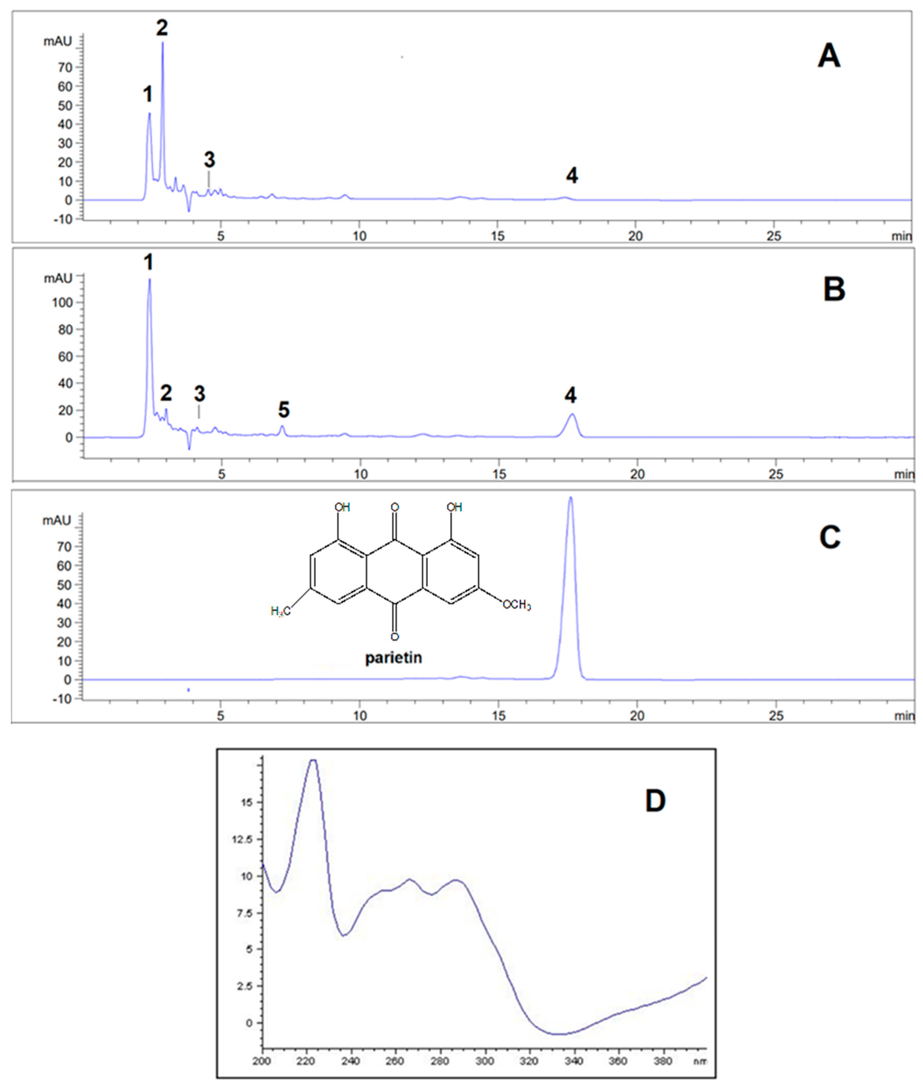
Figure 1. HPLC chromatograms of the acetone and methanol extracts of the lichen P. deosaiense and isolated parietin obtained at 254 nm as well as UV spectrum of parietin. 1—olivetol; 2—olivetolic acid (olivetil carboxylic acid); 3—haematommic acid. 4—parietin; 5— fallacinol. (A) —chromatogram of Placidium deosaiense acetone extract obtained at 254 nm. (B) —chromatogram of Placidium deosaiense methanol extract obtained at 254 nm. (C) — chromatogram of the isolated parietin. D – UV spectrum of parietin from 200-400 nm.
Preprints 116687 g001
Figure 2. The chemical structures of compounds identified in extracts.
Preprints 116687 g002aPreprints 116687 g002b
Table 1. Retention time, absorbance maxima and relative abundance of the examined lichen substances.
Sr. no Compound
Retention time (tR±SD)*
(min)
Absorbance maxima (nm) Relative abundance %
(254 nm)
Extracts**
Acetone Methanol
1 Olivetol 2.42±0.01 278 20.3995 34.0514
2 Olivetolic acid 2.91±0.02 218, 263, 301 25.8901 4.0678
3 Haematommic acid 4.56±0.01 202, 237, 259 5.3064 2.2663
4 Parietin 17.54±0.02 222, 266, 286, 438 1.0588 10.7476
5 Fallacinol 7.20±0.01 223, 325, 435 / 1.6712
* Values are the means of three determinations ± SD; **/-not detected.
Table 2. The yield of the extraction, total polyphenol and flavonoid content of the extracts of the lichen P. doesaiense and the amount of parietin in the extracts.
Lichen extracts Yield
(%)
Phenolics content
(mg GA/g)
Flavonoids content
(mg RE/g)
Amount of parietin
(mg/mL)
Acetone
Methanol
0.676 19.46 ± 0.75 44.29 ± 2.79 0.028
1.063 21.67 ± 0.41 377.40 ± 10.38 0.310
*Values are expressed as mean ± SD of triplicate measurements; GA – gallic acid equivalents; RE - rutin equivalents.
Table 3. Antibacterial activity (MIC and MBC values) of the acetone and methanol extracts of the lichen P. doesaiense as well as parietin.
Bacterial species Acetone Methanol Parietin Tetracycline
MIC MBC MIC MBC MIC MBC MIC MBC
mg/mL µg/mL
Escherichia coli ATCC 25922 >10 >10 >10 >10 >10 >10 4 6
Proteus mirabilis ATCC 12453 2.5 >10 1.25 >10 >10 >10 64 >128
Pseudomonas aeruginosa ATCC 10145 >10 >10 >10 >10 >10 >10 32 >128
Staphylococcus aureus ATCC 25923 >10 >10 >10 >10 >10 >10 2 3
Staphylococcus aureus MRSA ATCC 43300 >10 >10 >10 >10 >10 >10 <0.25 3
Enterococcus faecalis ATCC 29212 >10 >10 >10 >10 >10 >10 8 12
Bacillus cereus ATCC 11778 5 5 10 10 >10 >10 0.25 0.5
Table 4. Percent (%) of the biofilm formation inhibition for tested extracts of the lichen P. doesaiense and parietin.
Acetone extract
Bacterial species 10 mg/mL 5 mg/mL 2.5 mg/mL 1.25 mg/mL 0.625 mg/mL 0.3125 mg/mL
Proteus mirabilis ATCC 12453 / / / / / /
Pseudomonas aeruginosa ATCC 10145 92.1 / / / / /
Staphylococcus aureus ATCC 25923 92.0 91.1 87.9 90.6 66.8 54.2
Methanol extract
Proteus mirabilis ATCC 12453 36.0 / / / / /
Pseudomonas aeruginosa ATCC 10145 81.7 4.7 / / / /
Staphylococcus aureus ATCC 25923 90.3 89.7 85.2 73.4 68.9 63.7
Parietin
Proteus mirabilis ATCC 12453 92.5 25.6 / / / /
Pseudomonas aeruginosa ATCC 10145 99.8 48.1 / / / /
Staphylococcus aureus ATCC 25923 99.6 91.1 89.8 92.3 93.5 63.6
* "/" - there was no inhibition of biofilm formation at the tested concentrations;.
Table 5. Percent (%) of the reduction of mature biofilm for tested extracts of the lichen P. doesaiense and parietin.
Acetone extract
Bacterial species 10 mg/mL 5 mg/mL 2.5 mg/mL 1.25 mg/mL 0.625 mg/mL 0.3125 mg/mL
Proteus mirabilis ATCC 12453 10.2 8.3 5.1 / / /
Pseudomonas aeruginosa ATCC 10145 28.8 23.7 20.2 / / /
Staphylococcus aureus ATCC 25923 69.8 63.6 59.8 62.0 52.0 /
Methanol extract
Proteus mirabilis ATCC 12453 20.4 11.9 / / / /
Pseudomonas aeruginosa ATCC 10145 / / / / / /
Staphylococcus aureus ATCC 25923 75.5 72.3 68.9 66.8 59.9 48.4
Parietin
Proteus mirabilis ATCC 12453 3.4 3.0 / / / /
Pseudomonas aeruginosa ATCC 10145 59.58 / / / / /
Staphylococcus aureus ATCC 25923 85.3 73.7 56.4 51.85 47.3 39.4
* "/" - no reduction of the formed biofilm occurred at the tested concentrations.
Table 6. The antioxidant activity (DPPH scavenging and reducing power) of the extracts of the lichen P. doesaiense and parietin.
Lichen extract/
compound
DPPH scavenging IC50 (μg/mL) Reducing power
Absorbance (700 nm)
1000 μg/mL 500 μg/mL 250 μg/mL 125 μg/mL 62.5 μg/mL 31.25 μg/mL
Acetone 270.22±4.80 0.074±0.001 0.059±0.001 0.060±0.001 0.056±0.001 0.055±0.001 0.053±0.002
Methanol 275.124±9.713 0.0853±0.001 0.066±0.004 0.066±0.001 0.062±0.002 0.062±0.002 0.061±0.002
Parietin 51.616±0.490 0.1036±0.001 0.1026±0.001 0.097±0.001 0.095±0.001 0.093±0.001 0.091±0.001
Ascorbic acid 4.451±0.202 1.541±0.054 0.853±0.005 0.406±0.017 0.215±0.004 0.110±0.015 0.105±0.003
Disclaimer/Publisher’s Note: The statements, opinions and data contained in all publications are solely those of the individual author(s) and contributor(s) and not of MDPI and/or the editor(s). MDPI and/or the editor(s) disclaim responsibility for any injury to people or property resulting from any ideas, methods, instructions or products referred to in the content.
Copyright: This open access article is published under a Creative Commons CC BY 4.0 license, which permit the free download, distribution, and reuse, provided that the author and preprint are cited in any reuse.
Prerpints.org logo

Preprints.org is a free preprint server supported by MDPI in Basel, Switzerland.

Subscribe

© 2024 MDPI (Basel, Switzerland) unless otherwise stated