Preprint
Article

Artificial Intelligence and Innovative Surveillance Methods—An Additional Value in Livestock Farming?

Altmetrics

Downloads

99

Views

59

Comments

0

A peer-reviewed article of this preprint also exists.

Submitted:

02 September 2024

Posted:

03 September 2024

You are already at the latest version

Alerts
Abstract
Artificial intelligence (A.I.) is the key to process big amounts of data to useful information for operators. In the present study the suitability of a 24/7 A.I. sound based coughing monitoring system for early detection of respiratory distress caused by porcine respiratory disease complex (PRDC) associated pathogens was assessed in the nursery of a conventional pig farm. Screening for PRDC associated pathogens was conducted by PCR examination of oral fluids (OFs) and bioaerosol samples (AS). A significant correlation between A.I. and human gained health data was observed in both batches. An increase in coughing episodes, either measured by A.I. or human, was significantly correlated with decreasing Ct-values of swIAV. The Odds to detect nucleic acids of PRRSV or Actinobacillus (A.) pleuropneumonia was significantly higher for OFs compared to AS. Moreover, PCR examinations of OFs revealed significantly lower Ct-values for swIAV and A. pleuropneumonia compared to AS. The quantity of swIAV RNA in OFs and AS was significantly associated with the A.I. calculated respiratory health score and thus, with clinical disease. Due to its reliable data A.I. based health monitoring is beneficial for early recognition of respiratory distress and thus can assist farmer’s daily work.
Keywords: 
Subject: Medicine and Pharmacology  -   Veterinary Medicine

1. Introduction

Health monitoring is a crucial element in the production of livestock. Next to animal welfare issues and economical losses [1,2,3,4,5], animal health also depicts a major column for the public health sector. A major challenge for pigs’ health depicts the porcine respiratory disease complex (PRDC) [6]. This disease complex describes a clinical condition associated with treatment-resistant respiratory disease of growing-finishing pigs of multifactorial etiology including infectious and noninfectious factors [7]. Porcine circovirus type 2 (PCV2) [8,9], porcine reproductive and respiratory syndrome virus (PRRSV) [6,10], swine influenza A virus (swIAV) [11,12] and Mesomycoplasma (M.) hyopneumoniae [6,13] are considered as the major pathogens involved in the PRDC [6]. Continuous monitoring of animal behavior or evaluation of clinical signs related to pathological changes of the animal’s health is required to reach and maintain a high health status in swine operations. In recent years several approaches concerning automated analysis of changes in animal behavior or appearance of clinical signs were published [14,15,16,17,18,19,20,21]. Within this group, wearable devices or sensor technology are assumed to increase animal health and economy in terms of precision livestock farming [20] and thus, might even be beneficial concerning human health in terms of the one health strategy of the WHO. In a more concrete way artificial intelligence (A.I.) is considered to be helpful to get insight in farm infection dynamics [22], detection of clinical signs [14] and decision making [23]. Although automated recognition of animal health related behavior or clinical signs is thought to be an increasing market [20], the clinical observation of animal health by humans depicts the counterpart and daily work of farmers and veterinarians in the field of livestock farming. A herd inspection often reveals subjectively perceived results unless standardized examinations are carried out. In addition, due to increasing herd sizes the time for inspection of the animal population or individuals is limited. From that perspective, 24 h automated animal health monitoring seems to be a promising approach that can support the daily work of farmers and covers the time when no human-based inspection is possible. Moreover, it accommodates the possible role of circadian rhythm concerning the inflammatory response on the clinical outcome of respiratory disease as reviewed for humans elsewhere [24].
Next to clinical investigations, laboratory diagnostics are needed to gain sufficient information for the implementation of both short-term actions such as antimicrobial and antiphlogistic treatment, and long-term measures as vaccinations or management changes. Proper diagnostics and monitoring resemble an important limitation in the control of the porcine respiratory disease complex (PRDC) as the current methods are often labor-intensive, expensive and based on invasive techniques such as collecting blood or sampling the respiratory tract (i.e., nasal swabs, tonsil scratches, tracheobronchial swabs). In the recent decade aggregate samples such as oral fluids (OFs) or environmental samples have gained increasing interest as cost-effective, non-invasive sampling procedures. Particularly OFs are meanwhile well established for surveillance or monitoring of viral [25,26] or bacterial pathogens [27,28,29]. Next to OFs, bioaerosol samples (AS) have been evaluated for surveillance or monitoring of viral pathogens of humans [30] or viral and bacterial pathogens of animals [31,32,33]. Concerning porcine pathogens, PCV2, swIAV [34,35], PRRSV and M. hyopneumoniae [36,37] could be detected in bioaerosols under experimental or field conditions. At least for influenza A virus bioaerosol sampling revealed comparable results compared to oral fluids in a farrow-to-feeder facility [35].
The aim of this longitudinal study was to evaluate an A.I. system for monitoring of respiratory distress in a conventional nursery facility combined with the qualitative and quantitative detection of PRDC associated pathogens in OFs and bioaerosol samples.

2. Materials and Methods

The Farm Description

The blinded prospective follow up study was conducted in a conventional one-site piglet producing farm in Germany with a known history of recurring respiratory distress. The farm was known to be positive for PRRSV, swIAV, A. pleuropneumoniae and M. hyopneumoniae by PCR analysis based on diagnostic investigations performed by the herd attending veterinarian. Piglets were vaccinated against PRRSV (UNISTRAIN® PRRS, HIPRA, Spain, i.m.), PCV2 (Ingelvac CircoFLEX®, Boehringer Ingelheim, Germany), M. hyopneumoniae (Hyogen® (Ceva, Germany) at 21 days of age and Lawsonia (L.) intracellularis (Porcilis® Lawsonia, MSD, Germany) at weaning with commercially available vaccines. Three weeks prior to farrowing, the sows of the corresponding farm were vaccinated with a combined A. pleuropneumoniae + Pasteurella (P.) multocida and Streptococcus (S.) suis autogenous vaccine. In addition, the sows were vaccinated twice a year against swIAV (Respiporc® FLU3 and Respiporc® FLUpan H1N1, Ceva, Germany) and quarterly against PRRSV (UNISTRAIN® PRRS, HIPRA, Spain, i.d.) with commercially available vaccines.
The sow farm was producing piglets in a one-week batch farrowing interval. After weaning at 28 days of age, the pigs were transferred to the corresponding nursery facilities. The nursery unit was managed batch wise all- in all- out. The nursery unit was designed to house 600 pigs per airspace and consisted of 20 pens with 25-30 pigs per pen on plastic slatted floors. The animals received a commercial diet via a sensor-controlled liquid feeding trough. Water was available ad libitum via bowl- and nipple-drinkers. The ventilation system consisted of a diffused ceiling for supply air and underfloor extraction for exhaust air. Cleaning and disinfection were conducted after each batch.

Study Design and Data Collection

For the present study, two consecutive batches (Batch 1 (B1), 520 pigs and batch 2 (B2), 519 pigs) in the nursery were enrolled. The day of placement into the nursery unit was considered as study day 0. Piglets in both batches were monitored for 39 days. All procedures were approved by the internal ethical commission of the Veterinary Faculty of the Ludwig-Maximilians-University München (reference 269-24-04-2021).

Automated Coughing Monitoring

The commercially available A.I. device (SoundTalks® NV, Leuven, Belgium) performs continuous and automated measurements of coughs and is able to distinguish between a pigs’ cough and other environmental noises in the barn. It compares the previous day’s coughing level with the current one and calculates a numerical score (respiratory health status; ReHS) with values ranging from 0 (worst case) to 100 (best case). The alerts can be recognized through LED lights on the monitor and through the website. A normal ReHS, which ranges from 60 to 100 is represented by a green signal. Yellow and red alerts indicate a potential and a high risk of a respiratory problem and are equivalent to 40–59 and 0–39 ReHS, respectively. Two SoundTalks® boxes (M1, M2) were installed in the alleyway of the barn (Figure 1). Blinding of the investigator was ensured by setting the LEDs of the monitors to “science mode” (blue LED) over the entire study period. The ReHS was obtained daily (24/7) over the entire study period. The total ReHS displays the lowest score obtained from one of the two monitors.

Clinical Coughing Monitoring

In addition to the continuously automated A.I. based cough monitoring, a clinical coughing score (CCS) was conducted as published elsewhere [38]. Briefly, the coughing index was calculated as follows:
Coughing index (CI) [%] = Total number of coughing bouts (CC) / [number of examined pigs (n) * total time of observation (min)] *100
The score was gathered daily within the first five days after weaning. Subsequently the clinical monitoring was conducted three times a week (Monday, Wednesday, Friday) in each batch until the end of the observational period at day 39. The coughing score was always determined by the same person (the investigator) at the same time in the morning hours.
Laboratory Diagnostic Sampling
Four OFs and two AS were collected (Figure 1) on the same days as the clinical coughing score. For the collection of the oral fluids a cotton rope (BASKO Aleksander Skoracki, Posen, Poland; length 100cm, 3 single cords à Ø1cm, in total ca. Ø3cm) was placed for approximately 20 minutes in each pen next to one of the SoundTalks® monitors. The overall sampling procedure and further processing was done as published elsewhere [39].
Airsamples (AS) were obtained using two battery-powered AirPrep™ Cub samplers (AirPrep Model ACD210; InnovaPrep, Missouri, USA) according to the sampling scheme of the clinical coughing score. Prior to sampling, a sterile single use filter (Ø52mm) made of dielectric polymer fiber was inserted into each sampler. In the compartment both samplers were programmed by means of an intuitive control panel to a flow rate of 200 l/min with a collection time of 60 min. At the end of the 60 min. and thus a filtration of 12 m³, the sampler stopped automatically. One sampler was placed in the front and one in the back of the study compartment (Figure 1, red diamonds (AS5 and AS6)). To ensure that constant measurement points could be used for both Airprep cubs, chains were attached to the existing steel beams and a link was marked. The height of the air sampler was adjusted to the size of the piglets during nursery. In B1, the air inlet opening was located at a height of 77cm (measured from the floor) from study day 1-26, and at a height of 87cm from study day 27-39. In B2, the air inlet opening was at a height of 77cm from study day 1-33 and at a height of 92cm above the floor from study day 34-39. At the end of the sampling period, the filter was rinsed using the AirPrep Filter & Elution Kit supplied. Shortly, the top of the filter was pressed onto the sample cup and the elutor adapter was placed on top. Subsequently the filter was washed out with the elution fluid canister containing TRIS medium using a specially developed procedure via wet foam. This foam breaks the electrical tension on the filter surface and disintegrates within a few seconds in the sample cup resulting in 6ml of wash solution.
The samples were shipped cooled and promptly processed upon arrival at the laboratory. All assays were performed in a commercial laboratory using accredited assay methods (Table 1). To extract the genomes the laboratory used KingFisher™ Duo Prime (Thermo Fisher Scientific; Waltham. Massachusetts, USA) with ID Gene™ Mag Universal Extraction Kit (Innovative Diagnostics, Grabels, France). Subsequently, eluates were frozen and stored in KingFisher™ Elution Strips (Thermo Fisher Scientific; Vantaa, Finland) at -20°C. After completion of one run, the selected OFS and AS were analyzed collectively. The “BALF” profile provided by the laboratory was used for this purpose, which includes the PCR assays listed in Table 1.. The real-time PCR was rated as positive if Ct-value was ≤40.

Statistical Analysis

The statistical program IBM SPSS Statistics® (version 29.0, IBM® SPSS Inc., Chicago, Illinois; USA) and Microsoft EXCEL® (version 2019, Microsoft Office; USA) were used for the statistical analysis of the data and to create figures and tables. The significance level was p < 0.05. The confidence interval was 0.95.
Continuous data were tested for normal distribution using the Kolmogorov-Smirnov test. Due to non-normal distribution of the data, correlations between CCS, ReHS, Ct-values of OF and Ct-values of AS were determined using Spearman’s rho. Associations between the non-parametric continuous data Ct-value in dependency of the sample material (OF and AS) were calculated using Mann-Whitney U test. Frequency data (detection of pathogens by OF / AS) were analyzed by cross tables and fishers exact test was used to evaluate association between the frequency of detection of any pathogen DNA and the sample material. Odds ratios were calculated when appropriate additionally. The level of agreement between OFS and AS for each sampling time was calculated using Cohen’s kappa coefficient (κ). Agreement was considered poor if κ ≤0.2, fair if 0.21 ≤ κ ≤ 0.4, moderate if 0.41 ≤ κ ≤ 0.6, substantial if 0.61 ≤ κ ≤ 0.8 and good if κ >0.8 [40].

3. Results

3.1. Coughing Monitoring

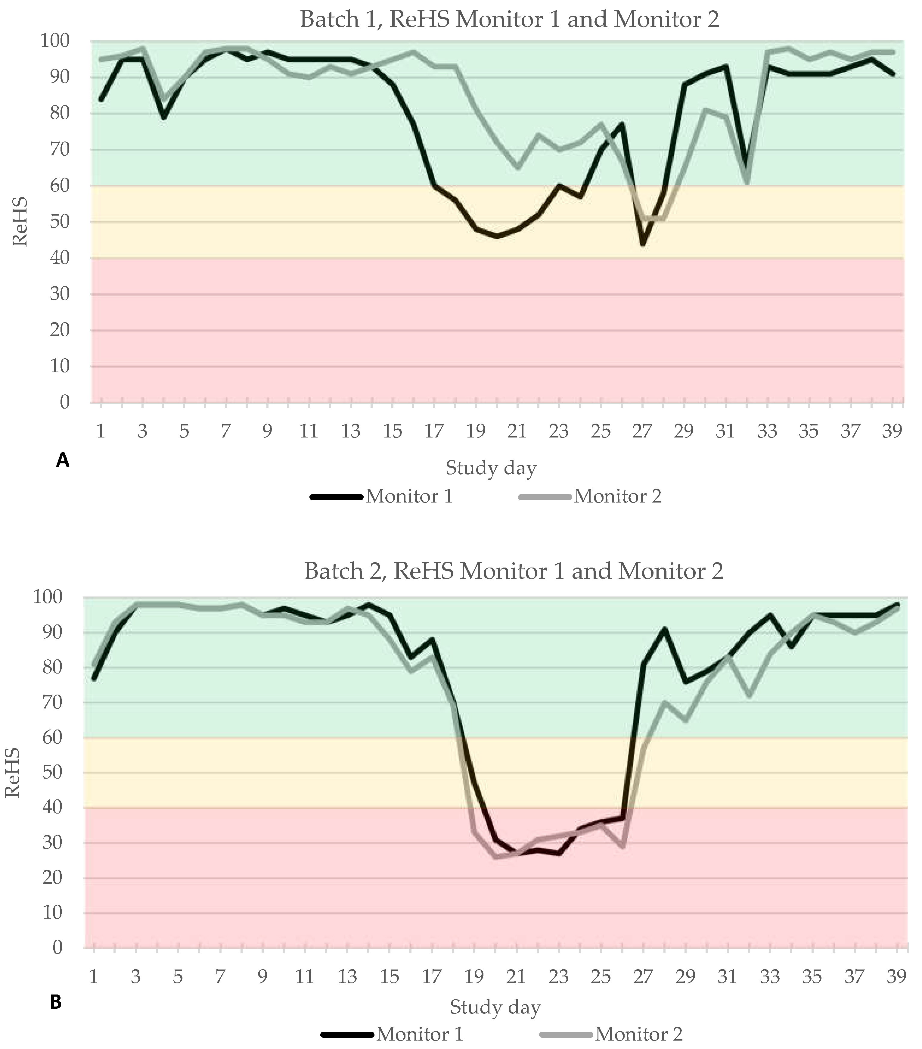
The automated A.I. based coughing monitoring system worked without error and ReHS could be calculated for every study day. In B1 first ReHS alert (yellow) was observed on study day 17 only by SoundTalks® monitor 1, located near the entrance, whereas both devices recorded a second alert (yellow) on study day 27 (Figure 2A). In B2, both monitors documented an alert on study day 19, however, monitor 2 already recorded a red alert whereas monitor 1 showed a yellow alert on day 19 and changed to red on day 20 (Figure 2B).
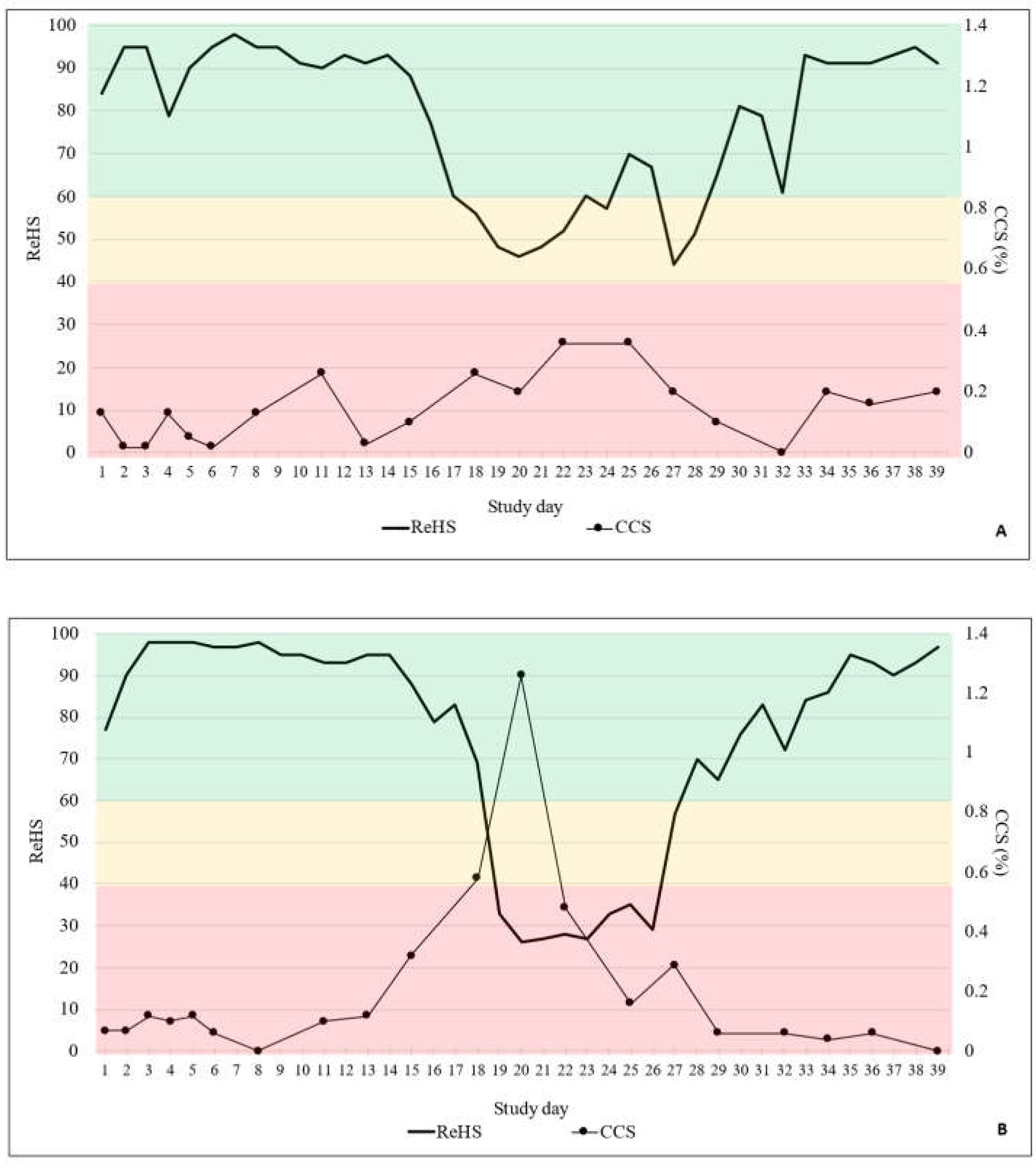
The total ReHS from both SoundTalks® monitors and the CCS are displayed in Figure 3A for B1 and in Figure 3B for B2, respectively. Briefly, in B1, 29/39 days were classified with a good ReHS (green) and 10/39 days with a moderate ReHS (yellow). In B2, 30/39 were assigned to a good ReHS (green), 2/39 days to a moderate ReHS (yellow) and 7/39 days to a poor ReHS (red).
Twenty human conducted clinical coughing scores were obtained in each batch. In B1 only mild clinical signs were recorded by the observer, which did not exceed a coughing index of 0.4%. In B2 more severe coughing was obvious with a maximum coughing index of 1,26% on study day 20.
Bivariate correlations (Spearman’s rho) revealed a significant moderate to medium negative correlation between the ReHS and the CCS for B1 (Spearman´s rho -.478; p ≤ 0.001) and B2 (Spearman´s rho: -.468; p ≤ 0.001) accordingly, indicating that an increase in the CCS is associated with a decrease in the ReHS.

Molecular Biological Examinations

In total 240 OFs (120 per batch) and 80 AS (40 per batch) were available for PCR analysis. A detailed overview on the detection rate of each pathogen in the two different batches is presented in Table 2.
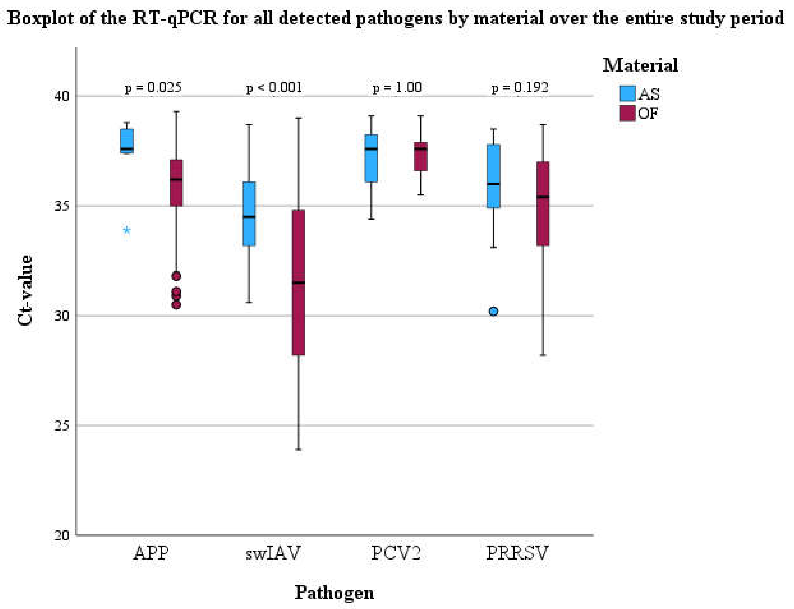
Briefly, all targeted pathogens with the exception of M. hyopneumoniae could be detected by PCR in both batches. Significant differences concerning the frequency of the detection between OFs and AS could be observed for PRRSV and A. pleuropneumoniae in both batches (Table 2). In detail, the odds to detect PRRSV in OFs was 3.72 times higher (95% CI: 1.96-7.08, p < 0.001) compared to AS. Moreover, there was 24.89 higher odds (95% CI: 10.17-60.92, p < 0.001) to find A. pleuropneumoniae in OFs by PCR compared to AS. In addition, Cohen´s Kappa was calculated to evaluate the agreement between OFs and AS to assign the study population as pathogen positive or negative for each sample day. Only for swIAV a significant agreement (p < 0.001) between OFs and AS with a moderate kappa (0.578) was observed. For all other pathogens, no significant agreement could be detected. Medians of PCR Ct-values for swIAV-, PRRSV-, PCV2, and A. pleuropneumoniae positive OFs and AS are shown using boxplots (Figure 4). RNA or DNA loads of A. pleuropneumoniae and swIAV were significantly higher in OFs compared to AS (Figure 4), respectively.
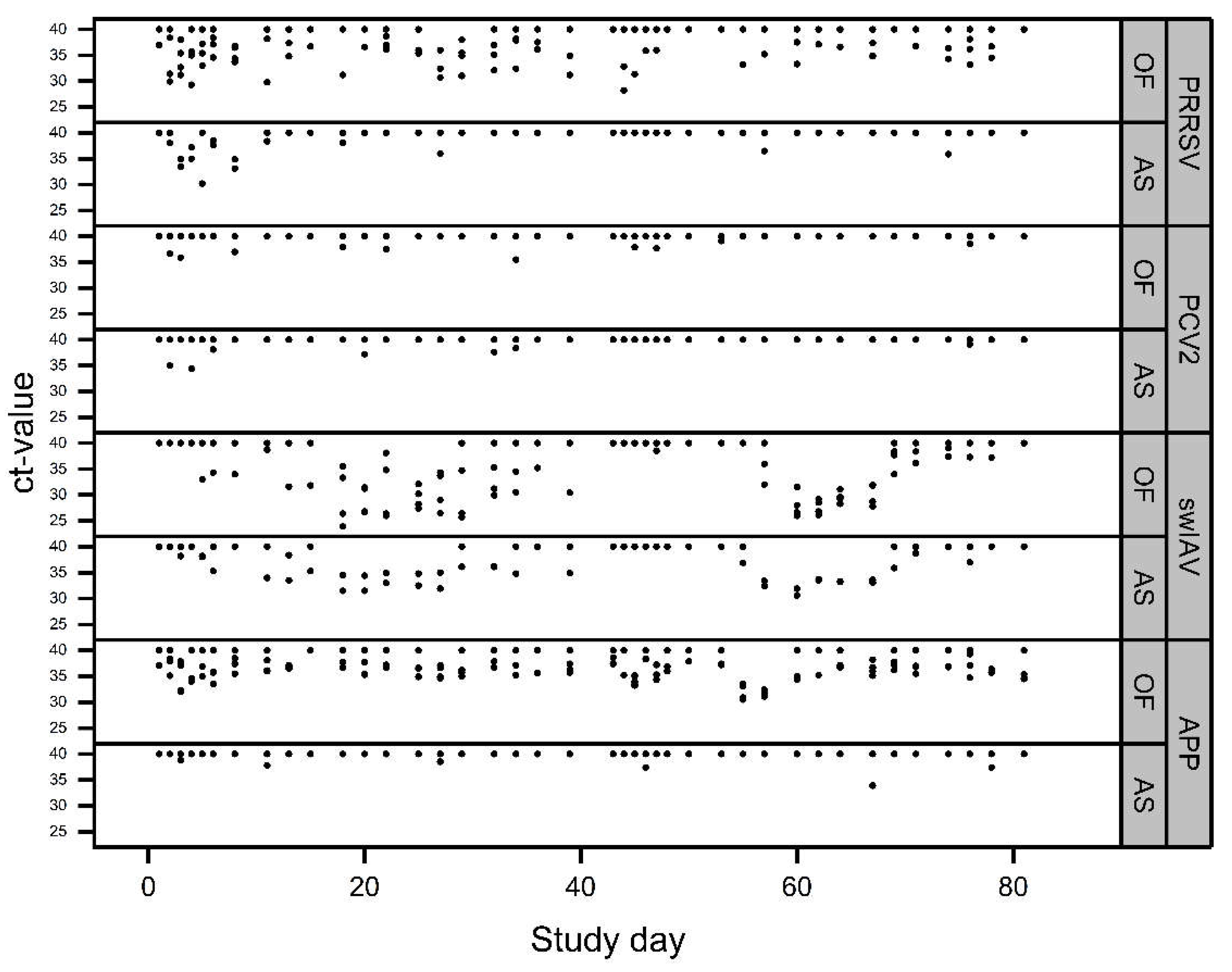
The Ct-values of positive OFs and AS over the entire study period are shown in Figure 5.
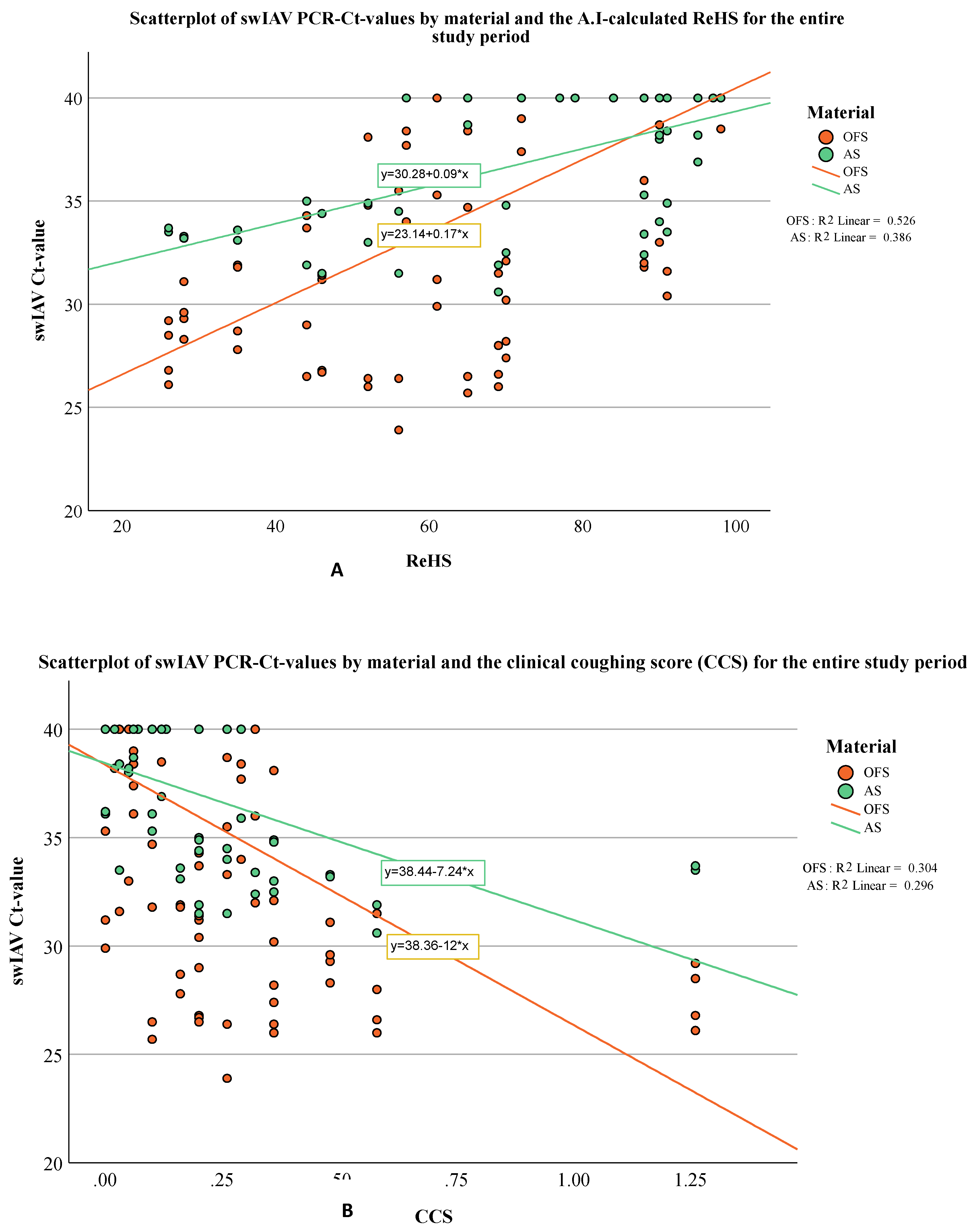
To evaluate potential relationships between the extend of coughing (expressed by ReHS or CCS) and the viral or bacterial loads (expressed by Ct-values) in OFs or AS bivariate correlations (Spearman´s rho) were calculated. A significant correlation was observed between the Ct-values of the swIAV PCR-results and the ReHS (Spearman´s rho: AS: 0.630, p < 0.001; OFs: 0.693, p < 0.001) and CCS (Spearmans rho: AS: -0.542, p < 0.001, OFs: -0.535, p < 0.001) for the entire study period. Figure 6a and Figure 6b depict the Ct-values of swIAV for OFs and AS in correlation to the ReHS (Figure 6a) or CCS (Figure 6b) including the regression equation for swIAV Ct-values depending on the used sampling material.

4. Discussion

Early detection of respiratory distress is essential to preserve animal health and to combat economic losses due to infections with respiratory pathogens. Within the present study we combined a sound based A.I. respiratory health monitoring system with a molecular biological laboratory screening based on OFs and AS for the detection of pathogens associated with PRDC. Within this scope, we also compared OFs and AS as matrices for the detection of nucleic acids of PRRSV, PCV2, APP and swIAV. As observed by others [41] the A.I. based monitoring system proved to be technically reliable and respiratory health data could be obtained for each study day. However, limitations concerning the use of such a monitoring might display the lack of internet within the range of the stables, which should be checked prior to the installation of such a monitoring system. The comparison between the 24/7 A.I. and manual coughing monitoring correlated on a moderate to medium level, as also reported by others [16]. In principle this correlation was expected as coughing is needed to have a corresponding response by the A.I. monitoring system. However, the ReHS gave more detailed information, e.g., in the first batch, when coughing was present particularly in the front part of nursery unit. Thus, next to the recognition of clinical signs, the A.I-based long-time monitoring on farms might also demonstrate localized pattern of disease that indicate issues concerning the farm environment or internal biosecurity. The advantage of continuous A.I based monitoring becomes clearly recognizable as different times of activity of pigs over a day period can be addressed, whereas a clinical examination is limited in time and only covers the time of physical presence in the corresponding pig population. This might explain the only moderate correlation between ReHS and CCS in the present examination. The circadian rhythm of the pig in its surrounding environment influences the times of activity of the pigs [42] and thus, the appearance of clinical signs due to the circadian oscillation of the immune system. In terms of respiratory diseases in humans, the severity of clinical signs also shows circadian variability across the 24-h cycle. An increased inflammation and disease severity at night is described for obstructive airways diseases and allergic rhinitis with the consequence of greater effects to exposure of inflammatory insults at night [24]. Based on a previous investigation the optimal number of devices for the 600 head barn was determined to be two [43]. However, to ensure optimal sound coverage and to account for variations in infection dynamics the number of required monitors and the placement of the devices must be thoroughly considered.
The diagnostic screening by PCR revealed the presence of multiple respiratory pathogens in the nursery of the study farm. Thus, the principal preconditions for PRDC were present in the study population. Nevertheless, in order to definitively determine whether a certain combination of pathogens contributed to the clinical signs, pathomorphological examinations would have been required. Interestingly, a significant correlation was observed between decreasing Ct-values of the swIAV PCR and increasing respiratory distress measured by the A.I. or the investigator. Thus, an increase of the swIAV viral load in OFs or AS coincided with the extend of the clinical signs in terms of coughing expressed by reduced ReHS or increased CCS, respectively. Comparable observations were made by Neira, et al. [44] who reported a correlation between the quantitative detection of swIAV in oral fluids and the coughing score. Besides the link between swIAV viral load in OFs or As and the extend of coughing, this observation also gives evidence to suggest that swIAV might have been the driving force for respiratory distress in this nursery unit. Moreover, based on the comparable pattern of swIAV RNA detection in both batches an endemic swIAV infection can be assumed. As already postulated by Prost, et al. [35] and Anderson, et al. [34], no significant differences concerning the qualitative detection of swIAV by PCR were evident between OFs and bioaerosol samples. However, in contrast to Prost, et al. [35] swIAV RNA loads were significantly higher in OFs compared to bioaerosol samples. The detection of PRRSV might be a result of the MLV vaccination of the piglets as no significant correlation between clinical observations and PRRSV detection was observed in the present study. However, no further diagnostics to discriminate between field or vaccine strains could be conducted due to the low viral loads in both sampling materials. Shedding of MLVs is a well-known observation after vaccination and viral RNA could be detected in nasal swabs by PCR under experimental conditions up to 42 days after vaccination with an MLV [45]. Although airborne transmission of PRRSV over more than four kilometers was reported [36], the detection rate of viral nucleic acids in bioaerosol samples compared to OFs in our study was rather low. This observation indicates less suitability of bioaerosols for monitoring or surveillance purposes of PRRSV compared to OFs whose sensitivity and suitability to monitor PRRSV was already shown elsewhere [46,47,48]. Still, these results should be interpreted with caution because airborne transmission of PRRSV might be strain depended as reported by others [37] and the attenuation of the vaccine strain that was putatively detected here might bias our findings. Significant advantages of OFs over AS could also be observed for the detection of A. pleuropneumoniae. The high sensitivity of OFs concerning the detection of A. pleuropneumoniae by PCR was already shown in a previous study under field conditions [27]. The sporadic detection of A. pleuropneumoniae DNA in the bioaerosols is in line with the limited capability of A. pleuropneumoniae aerosol transmission reported elsewhere [49] and the higher within pen transmission rate compared to the transmission between different pens [50]. However, the sporadic detection of APP in the bioaerosols indicates the possibility of airborne infections with A. pleuropneumoniae within a pig population as shown by others [49,50]. Moreover, differences in the ventilation rate, air humidity, particle size and the overall bacterial load in the pig population might influence the detection of bacteria in bioaerosol samples. Particularly the underfloor extraction for the exhaust air of the stable in the present study might contribute to a reduced the number of bacteria in the air, which could also in a certain extend apply to A. pleuropneumoniae. Concerning PCV2, Harms, et al. [8] demonstrated the relevance of the combination of PCV2 and swIAV in a PRDC case series. However, in the present study the qualitative and quantitative detection rate of PCV2 was low in both sample types. Although Nielsen, et al. [51] postulated that PCV2 detection in OF are not necessarily correlated with clinical signs, PCV2 does not seem to play a major role concerning the clinical outcome in the present nursery, as evident from Figure 5. Moreover, the pigs of the present examination were vaccinated against PCV2. The positive effects of PCV2 vaccination concerning PRDC or PMWS affected herds [52,53] on clinical outcomes and co-infections are widely known.

5. Conclusions

The A.I. based coughing monitoring (SoundTalks®) used in the present examination reliably delivered detailed data on pig health and showed localized disease pattern in the nursery. The extend of the respiratory distress, expressed by ReHS, correlated with the quantity of swIAV RNA in OFs and AS. Under the present study conditions, bioaerosol sampling was less sensitive to detect A. pleuropneumoniae and PRRSV by PCR compared to oral fluids. Moreover, A. pleuropneumoniae bacterial and swIAV viral loads were significantly higher in oral fluids compared to AS. The application of a long term A.I. based coughing monitoring system in combination with laboratory examinations can be a useful tool to identify relevant pathogens and recurring disease patterns on farms and to early recognize respiratory distress under field conditions.

Supplementary Materials

The manuscript does not contain supplementary materials.

Author Contributions

“Conceptualization, M.E., J.B., M.R., J.S.; methodology, C.A., M.G., J.B.; software, F.F., M.E., J.S.; validation, M.R., M.E.; formal analysis, F.F., M.E., J.S, S.R..; investigation, F.F., M.E., J.S.; resources, M.G., C.A., J.B.; data curation, F.F., M.E., J.S ; writing—original draft preparation, F.F., M.E., J.S; writing—review and editing, M.R, M.G., J.B., S.R.; C.A.; visualization, F.F., M.E., J.S., S.R.; supervision, M.R., M.E., J.S.; project administration, F.F., M.E., J.S.; funding acquisition, J.B., M.G., C.A. All authors have read and agreed to the published version of the manuscript

Funding

This research was funded by Boehringer Ingelheim Vetmedica GmbH, Ingelheim, Germany.

Institutional Review Board Statement

The study was conducted in accordance with the German Animal Welfare Legislation and approved by the internal ethical commission of the Veterinary Faculty of the Ludwig-Maximilians-Universität München (reference 269-24-04-2021).

Data Availability Statement

The dataset is available from the corresponding author upon request.

Acknowledgments

We gratefully thank the participating farmer and veterinarian for their valuable support. We would like to thank Dale Polson for providing air samplers and the filtration systems.

Conflicts of Interest

The authors declare no conflicts of interest. JB, MG and CA are employees of Boehringer Ingelheim Animal Health, Ingelheim, Germany who provided funding for the study. The funders were not involved in collection, analyses, or interpretation of data.

References

  1. McOrist, S. “Defining the Full Costs of Endemic Porcine Proliferative Enteropathy.” Veterinary Journal 170, no. 1 (2005): 8-9. [CrossRef]
  2. McOrist, S., S. H. Smith, and L. E. Green. “Estimate of Direct Financial Losses Due to Porcine Proliferative Enteropathy.” The Veterinary record 140, no. 22 (1997): 579-81. [CrossRef]
  3. Alarcon, P., J. Rushton, and B. Wieland. “Cost of Post-Weaning Multi-Systemic Wasting Syndrome and Porcine Circovirus Type-2 Subclinical Infection in England - an Economic Disease Model.” Preventive veterinary medicine 110, no. 2 (2013): 88-102. [CrossRef]
  4. Renken, C., C. Nathues, H. Swam, K. Fiebig, C. Weiss, M. Eddicks, M. Ritzmann, and H. Nathues. “Application of an Economic Calculator to Determine the Cost of Porcine Reproductive and Respiratory Syndrome at Farm-Level in 21 Pig Herds in Germany.” Porcine health management 7, no. 1 (2021): 3. [CrossRef]
  5. Boeters, M., B. Garcia-Morante, G. van Schaik, J. Segales, J. Rushton, and W. Steeneveld. “The Economic Impact of Endemic Respiratory Disease in Pigs and Related Interventions - a Systematic Review.” Porcine health management 9, no. 1 (2023): 45. [CrossRef]
  6. Thacker, E. L. “Immunology of the Porcine Respiratory Disease Complex.” Vet Clin North Am Food Anim Pract 17, no. 3 (2001): 551-65. [CrossRef]
  7. Eddicks, Matthias, Lina Eddicks, Julia Stadler, Walter Hermanns, and Mathias Ritzmann. “Der Porcine Respiratory Disease Complex (Prdc)–Eine Klinische Übersicht.” Tierärztliche Praxis Ausgabe G: Großtiere/Nutztiere 49, no. 02 (2021): 120-32.
  8. Harms, P. A., P. G. Halbur, and S. D. Sorden. “Three Cases of Porcine Respiratory Disease Complex Associated with Porcine Circovirus Type 2 Infection.” Journal of Swine Health and Production 10, no. 1 (2002): 27-30. [CrossRef]
  9. Kim, J., H. K. Chung, and C. Chae. “Association of Porcine Circovirus 2 with Porcine Respiratory Disease Complex.” Veterinary Journal 166, no. 3 (2003): 251-56. [CrossRef]
  10. Opriessnig, T., L. G. Gimenez-Lirola, and P. G. Halbur. “Polymicrobial Respiratory Disease in Pigs.” Animal health research reviews / Conference of Research Workers in Animal Diseases 12, no. 2 (2011): 133-48.
  11. Deblanc, C., and G. Simon. “Involvement of Swine Influenza a Viruses in the Porcine Respiratory Disease Complex.” Virologie (Montrouge) 21, no. 5 (2017): 225-38.
  12. Schmidt, C., S. P. Cibulski, C. P. Andrade, T. F. Teixeira, A. P. Varela, C. M. Scheffer, A. C. Franco, L. L. de Almeida, and P. M. Roehe. “Swine Influenza Virus and Association with the Porcine Respiratory Disease Complex in Pig Farms in Southern Brazil.” Zoonoses and public health 63, no. 3 (2016): 234-40. [CrossRef]
  13. Thacker, E. L., P. G. Halbur, R. F. Ross, R. Thanawongnuwech, and B. J. Thacker. “Mycoplasma Hyopneumoniae Potentiation of Porcine Reproductive and Respiratory Syndrome Virus-Induced Pneumonia.” Journal of clinical microbiology 37, no. 3 (1999): 620-7. [CrossRef]
  14. Chung, Y., S. Oh, J. Lee, D. Park, H. H. Chang, and S. Kim. “Automatic Detection and Recognition of Pig Wasting Diseases Using Sound Data in Audio Surveillance Systems.” Sensors (Basel) 13, no. 10 (2013): 12929-42. [CrossRef]
  15. Pessoa, J., M. Rodrigues da Costa, E. García Manzanilla, T. Norton, C. McAloon, and L. Boyle. “Managing Respiratory Disease in Finisher Pigs: Combining Quantitative Assessments of Clinical Signs and the Prevalence of Lung Lesions at Slaughter.” Prev Vet Med 186 (2021): 105208. [CrossRef]
  16. Silva, Apsp, G. Y. Storino, F. S. M. Ferreyra, M. Zhang, E. Fano, D. Polson, C. Wang, R. J. Derscheid, J. J. Zimmerman, M. J. Clavijo, and B. L. Arruda. “Cough Associated with the Detection of Mycoplasma Hyopneumoniae DNA in Clinical and Environmental Specimens under Controlled Conditions.” Porcine Health Manag 8, no. 1 (2022): 6. [CrossRef]
  17. Wang, S., H. Jiang, Y. Qiao, S. Jiang, H. Lin, and Q. Sun. “The Research Progress of Vision-Based Artificial Intelligence in Smart Pig Farming.” Sensors (Basel) 22, no. 17 (2022). [CrossRef]
  18. Jorquera-Chavez, M., S. Fuentes, F. R. Dunshea, R. D. Warner, T. Poblete, R. S. Morrison, and E. C. Jongman. “Remotely Sensed Imagery for Early Detection of Respiratory Disease in Pigs: A Pilot Study.” Animals (Basel) 10, no. 3 (2020). [CrossRef]
  19. Neethirajan, S. “Affective State Recognition in Livestock-Artificial Intelligence Approaches.” Animals (Basel) 12, no. 6 (2022). [CrossRef]
  20. Neethirajan, Suresh. “Recent Advances in Wearable Sensors for Animal Health Management.” Sensing and Bio-Sensing Research 12 (2017): 15-29. [CrossRef]
  21. Arulmozhi, E., A. Bhujel, B. E. Moon, and H. T. Kim. “The Application of Cameras in Precision Pig Farming: An Overview for Swine-Keeping Professionals.” Animals (Basel) 11, no. 8 (2021). [CrossRef]
  22. Esener, N., M. J. Green, R. D. Emes, B. Jowett, P. L. Davies, A. J. Bradley, and T. Dottorini. “Discrimination of Contagious and Environmental Strains of Streptococcus Uberis in Dairy Herds by Means of Mass Spectrometry and Machine-Learning.” Scientific reports 8, no. 1 (2018): 17517. [CrossRef]
  23. Ezanno, P., S. Picault, G. Beaunee, X. Bailly, F. Munoz, R. Duboz, H. Monod, and J. F. Guegan. “Research Perspectives on Animal Health in the Era of Artificial Intelligence.” Veterinary research 52, no. 1 (2021): 40. [CrossRef]
  24. Comas, Maria, Christopher J. Gordon, Brian G. Oliver, Nicholas W. Stow, Gregory King, Pawan Sharma, Alaina J. Ammit, Ronald R. Grunstein, and Craig L. Phillips. “A Circadian Based Inflammatory Response – Implications for Respiratory Disease and Treatment.” Sleep Science and Practice 1, no. 1 (2017): 18. [CrossRef]
  25. Hernandez-Garcia, J., N. Robben, D. Magnee, T. Eley, I. Dennis, S. M. Kayes, J. R. Thomson, and A. W. Tucker. “The Use of Oral Fluids to Monitor Key Pathogens in Porcine Respiratory Disease Complex.” Porcine health management 3 (2017): 7. [CrossRef]
  26. Ramirez, A., C. Wang, J. R. Prickett, R. Pogranichniy, K. J. Yoon, R. Main, J. K. Johnson, C. Rademacher, M. Hoogland, P. Hoffmann, A. Kurtz, E. Kurtz, and J. Zimmerman. “Efficient Surveillance of Pig Populations Using Oral Fluids.” Preventive veterinary medicine 104, no. 3-4 (2012): 292-300. [CrossRef]
  27. Kleinmans, M., K. Fiebig, R. Tabeling, H. Swam, A. Duivelshof-Crienen, M. Ritzmann, and M. Eddicks. “Explorative Field Study on the Use of Oral Fluids for the Surveillance of Actinobacillus Pleuropneumoniae Infections in Fattening Farms by an Apx-Real-Time Pcr.” Vet Sci 9, no. 10 (2022). [CrossRef]
  28. Schott, F., K. Hoffmann, E. Sarno, P. D. Bangerter, R. Stephan, G. Overesch, M. Haessig, X. Sidler, and R. Graage. “Evaluation of Oral Fluids for Surveillance of Foodborne and Zoonotic Pathogens in Pig Farms.” J Vet Diagn Invest 33, no. 4 (2021): 655-63.
  29. Deffner, P., R. Maurer, V. Cvjetkovic, W. Sipos, R. Krejci, M. Ritzmann, and M. Eddicks. “Cross-Sectional Study on the in-Herd Prevalence of Mycoplasma Hyopneumoniae at Different Stages of Pig Production.” The Veterinary record (2022): e1317.
  30. Tedeschini, E., S. Pasqualini, C. Emiliani, E. Marini, A. Valecchi, C. Laoreti, S. Ministrini, B. Camilloni, R. Castronari, L. Patoia, F. Merante, S. Baglioni, E. De Robertis, M. Pirro, A. Mencacci, and L. Pasqualini. “Monitoring of Indoor Bioaerosol for the Detection of Sars-Cov-2 in Different Hospital Settings.” Front Public Health 11 (2023): 1169073.
  31. Song, L., C. Wang, G. Jiang, J. Ma, Y. Li, H. Chen, and J. Guo. “Bioaerosol Is an Important Transmission Route of Antibiotic Resistance Genes in Pig Farms.” Environ Int 154 (2021): 106559. [CrossRef]
  32. Nguyen, N. T. T., Y. T. H. Luu, T. D. Hoang, H. X. Nguyen, T. D. Dao, V. N. Bui, and G. C. Gray. “An Epidemiological Study of Streptococcus Suis Prevalence among Swine at Industrial Swine Farms in Northern Vietnam.” One Health 13 (2021): 100254. [CrossRef]
  33. Anderson, B. D., J. A. Lednicky, M. Torremorell, and G. C. Gray. “The Use of Bioaerosol Sampling for Airborne Virus Surveillance in Swine Production Facilities: A Mini Review.” Front Vet Sci 4 (2017): 121. [CrossRef]
  34. Anderson, B. D., M. Yondon, E. S. Bailey, E. K. Duman, R. A. Simmons, A. G. Greer, and G. C. Gray. “Environmental Bioaerosol Surveillance as an Early Warning System for Pathogen Detection in North Carolina Swine Farms: A Pilot Study.” Transboundary and emerging diseases 68, no. 2 (2021): 361-67. [CrossRef]
  35. Prost, K., H. Kloeze, S. Mukhi, K. Bozek, Z. Poljak, and S. Mubareka. “Bioaerosol and Surface Sampling for the Surveillance of Influenza a Virus in Swine.” Transboundary and emerging diseases 66, no. 3 (2019): 1210-17. [CrossRef]
  36. Dee, S., S. Otake, S. Oliveira, and J. Deen. “Evidence of Long Distance Airborne Transport of Porcine Reproductive and Respiratory Syndrome Virus and Mycoplasma Hyopneumoniae.” Veterinary research 40, no. 4 (2009): 39. [CrossRef]
  37. Torremorell, M., C. Pijoan, K. Janni, R. Walker, and H. S. Joo. “Airborne Transmission of Actinobacillus Pleuropneumoniae and Porcine Reproductive and Respiratory Syndrome Virus in Nursery Pigs.” American journal of veterinary research 58, no. 8 (1997): 828-32. [CrossRef]
  38. Nathues, H., J. Spergser, R. Rosengarten, L. Kreienbrock, and E. Grosse Beilage. “Value of the Clinical Examination in Diagnosing Enzootic Pneumonia in Fattening Pigs.” Veterinary Journal 193, no. 2 (2012): 443-7. [CrossRef]
  39. Prickett, J., R. Simer, J. Christopher-Hennings, K. J. Yoon, R. B. Evans, and J. J. Zimmerman. “Detection of Porcine Reproductive and Respiratory Syndrome Virus Infection in Porcine Oral Fluid Samples: A Longitudinal Study under Experimental Conditions.” J Vet Diagn Invest 20, no. 2 (2008): 156-63. [CrossRef]
  40. Landis, J. Richard, and Gary G. Koch. “The Measurement of Observer Agreement for Categorical Data.” Biometrics 33, no. 1 (1977): 159-74. [CrossRef]
  41. Duerlinger, S., C. Knecht, S. Sawyer, G. Balka, M. Zaruba, T. Ruemenapf, C. Kraft, P. H. Rathkjen, and A. Ladinig. “Efficacy of a Modified Live Porcine Reproductive and Respiratory Syndrome Virus 1 (Prrsv-1) Vaccine against Experimental Infection with Prrsv Aut15-33 in Weaned Piglets.” Vaccines (Basel) 10, no. 6 (2022). [CrossRef]
  42. Ingram, D. L., and M. J. Dauncey. “Circadian Rhythms in the Pig.” Comparative Biochemistry and Physiology Part A: Physiology 82, no. 1 (1985): 1-5.
  43. Polson D., Playter S., Berckmans D., Cui Z., Quinn B. “Determining the Optimal Placement and Configuration of an Audio-Based Sensor Platform to Enable Improved Detection and Characterisation of Clinical Respiratory Episodes in Large Growing Pig Airspaces and Sites.” In Proceedings of the European Conference on Precision Livestock Farming, pp. 490-95. Cork, Ireland, 26–29 August 2019.
  44. Neira, V., M. Allerson, C. Corzo, M. Culhane, A. Rendahl, and M. Torremorell. “Detection of Influenza a Virus in Aerosols of Vaccinated and Non-Vaccinated Pigs in a Warm Environment.” PLoS One 13, no. 5 (2018): e0197600. [CrossRef]
  45. Madapong, A., K. Saeng-Chuto, A. Tantituvanont, and D. Nilubol. “Safety of Prrsv-2 Mlv Vaccines Administrated Via the Intramuscular or Intradermal Route and Evaluation of Prrsv Transmission Upon Needle-Free and Needle Delivery.” Scientific reports 11, no. 1 (2021): 23107. [CrossRef]
  46. Biernacka, K., P. Karbowiak, P. Wrobel, T. Chareza, M. Czopowicz, G. Balka, C. Goodell, R. Rauh, and T. Stadejek. “Detection of Porcine Reproductive and Respiratory Syndrome Virus (Prrsv) and Influenza a Virus (Iav) in Oral Fluid of Pigs.” Research in veterinary science 109 (2016): 74-80. [CrossRef]
  47. Kittawornrat, A., J. Prickett, W. Chittick, C. Wang, M. Engle, J. Johnson, D. Patnayak, T. Schwartz, D. Whitney, C. Olsen, K. Schwartz, and J. Zimmerman. “Porcine Reproductive and Respiratory Syndrome Virus (Prrsv) in Serum and Oral Fluid Samples from Individual Boars: Will Oral Fluid Replace Serum for Prrsv Surveillance?” Virus research 154, no. 1-2 (2010): 170-6.
  48. Kittawornrat, A., Y. Panyasing, C. Goodell, C. Wang, P. Gauger, K. Harmon, R. Rauh, L. Desfresne, I. Levis, and J. Zimmerman. “Porcine Reproductive and Respiratory Syndrome Virus (Prrsv) Surveillance Using Pre-Weaning Oral Fluid Samples Detects Circulation of Wild-Type Prrsv.” Veterinary Microbiology 168, no. 2-4 (2014): 331-9. [CrossRef]
  49. Kristensen, C. S., O. Angen, M. Andreasen, H. Takai, J. P. Nielsen, and S. E. Jorsal. “Demonstration of Airborne Transmission of Actinobacillus Pleuropneumoniae Serotype 2 between Simulated Pig Units Located at Close Range.” Veterinary Microbiology 98, no. 3-4 (2004): 243-9. [CrossRef]
  50. Tobias, T. J., A. Bouma, J. van den Broek, A. van Nes, A. J. Daemen, J. A. Wagenaar, J. A. Stegeman, and D. Klinkenberg. “Transmission of Actinobacillus Pleuropneumoniae among Weaned Piglets on Endemically Infected Farms.” Preventive veterinary medicine 117, no. 1 (2014): 207-14. [CrossRef]
  51. Nielsen, G. B., J. P. Nielsen, J. Haugegaard, S. C. Leth, L. E. Larsen, C. S. Kristensen, K. S. Pedersen, H. Stege, C. K. Hjulsager, and H. Houe. “Comparison of Serum Pools and Oral Fluid Samples for Detection of Porcine Circovirus Type 2 by Quantitative Real-Time Pcr in Finisher Pigs.” Porcine health management 4 (2018): 2. [CrossRef]
  52. Fachinger, V., R. Bischoff, S. B. Jedidia, A. Saalmueller, and K. Elbers. “The Effect of Vaccination against Porcine Circovirus Type 2 in Pigs Suffering from Porcine Respiratory Disease Complex.” Vaccine 26, no. 11 (2008): 1488-99. [CrossRef]
  53. Kixmoeller, M., M. Ritzmann, M. Eddicks, A. Saalmueller, K. Elbers, and V. Fachinger. “Reduction of Pmws-Associated Clinical Signs and Co-Infections by Vaccination against Pcv2.” Vaccine 26, no. 27-28 (2008): 3443-51. [CrossRef]
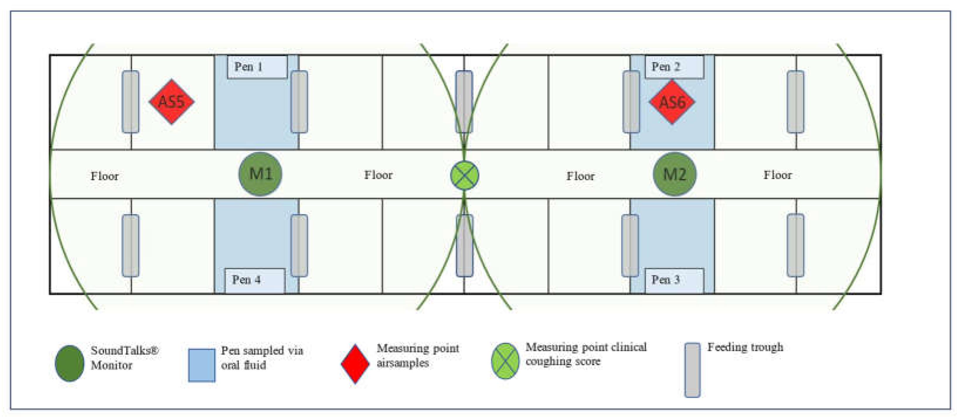
Figure 1. Outline of the study compartment and position of Oral Fluids and Air Sample collection points, clinical coughing score assessment and Sound Talks® monitor (M1 and M2). The radius lines correspond to the coverage range (10 meters) of the respective Sound Talks® monitor (M1 and M2).
Preprints 116986 g001
Figure 2. ReHS of B1 (A) and B2 (B) obtained by monitor 1 and monitor 2 over the entire study period.
Preprints 116986 g002
Figure 3. Total ReHS and CCS of B1 (A) and B2 (B) for the observational period of 39 days.
Preprints 116986 g003
Figure 4. Boxplots (median, lower and upper quartile, minimum and maximum) of the PCR results for all detected pathogens in OFs and AS over the entire study period.
Preprints 116986 g004
Figure 5. Detection patterns of the different pathogens over the entire study period (B1: study days 0-39; B2: study days 40-80) in OFs and AS. Ct-values ≥ 40 represent negative results. Overlaying results are possible.
Preprints 116986 g005
Figure 6. Scatterplot of swIAV PCR Ct-values in correlation with the ReHS (A) or CCS (B) for the entire study period for AS and OFs and the corresponding regression equation.
Preprints 116986 g006
Table 1. Name and manufacturer of the assays used in the present study.
Pathogen Name of the used assay Manufacturer
PRRSV Virotype PRRSV NA/EU INDICAL BIOSCIENCE GmbH; Leipzig, Germany
PCV 2 Virotype PCV 2/PCV 3 INDICAL BIOSCIENCE GmbH; Leipzig, Germany
swIAV Virotype Influenza A RT-PCR INDICAL BIOSCIENCE GmbH; Leipzig, Germany
M. hyopneumoniae EXOone Mycoplasma hyopneumoniae exopol; San Mateo de Gállego, Zaragoza, Spain
A. pleuropneumoniae EXOone Actinobacillus pleuropneumoniae exopol; San Mateo de Gállego, Zaragoza, Spain
Table 2. Percentage (%) and frequency of detection (n) of specific pathogens by PCR in OFs and AS of B1 and B2 and for the entire study period, as well as p-values in case of significant differences in the frequency of detection.
B1 B2 Entire study period
OFs
(n = 64)
AS
(n = 32)
p-value OFs
(n = 60)
AS
(n = 30)
p-value OFs
(n = 124)
AS
(n = 62)
p-value
PRRSV 65.0 %
(52/80)
32.5 %
(13/40)
< 0.001 27.5 %
(22/80)
5.0 %
(2/40)
0.003 46.3 %
(74/160)
18.8 %
(15/80)
< 0.001
PCV2 7.5 %
(6/80)
15.0 %
(6/40)
0.211 5.0 %
(4/80)
2.5 %
(1/40)
0.664 6.3 %
(10/160)
8.8 %
(7/80)
0.594
swIAV 45.0 %
(36/80)
57.5 %
(23/40)
0.246 36.3 %
(29/80)
35.0 %
(14/40)
1.00 40.6 %
(65/160)
46.3 %
(37/80)
0.410
APP1 63.7 %
(51/80)
7.5 %
(3/40)
<0.001 70.0 %
(56/80)
7.5 %
(3/40)
< 0.001 66.9 %
(107/160)
7.5 %
(6/80)
< 0.001
M. hyo2 0.0 %
(0/64)
0.0 %
(0/32)
- 0.0 %
(0/60)
0.0 %
(0/30)
- 0.0 %
(0/124)
0.0 %
(0/62)
n.d.*
* n.d.: not detected, 1A. pleuropneumoniae, 2M. hyopneumoniae.
Disclaimer/Publisher’s Note: The statements, opinions and data contained in all publications are solely those of the individual author(s) and contributor(s) and not of MDPI and/or the editor(s). MDPI and/or the editor(s) disclaim responsibility for any injury to people or property resulting from any ideas, methods, instructions or products referred to in the content.
Copyright: This open access article is published under a Creative Commons CC BY 4.0 license, which permit the free download, distribution, and reuse, provided that the author and preprint are cited in any reuse.
Prerpints.org logo

Preprints.org is a free preprint server supported by MDPI in Basel, Switzerland.

Subscribe

© 2024 MDPI (Basel, Switzerland) unless otherwise stated